Surg Endosc (2001) 15:209-212
DOI: I 0.1007/s004640000265
Surgical Endoscopy
Ultrasound and
Interventional Techniques
9 Springer-Verlag New York Inc. 2001Use of a fluorescent bile acid to enhance visualization of the biliary
tract and bile leaks during laparoscopic surgery in rabbits
F. Holzinger, 1~" L. Kriihenbiihl, 1 C. D. Schteingart, 2 H.-T. Ton-Nu, 2 A. F. Hofmann 2
Department of Visceral and Transplantation Surgery, Inselspital, University of Bern, Murtenstrasse 35, CH-3010 Bern, Switzerland
2 Division of Gastroenterology, Department of Medicine, University of California, San Diego, La Jolla, CA 92093-0813, USA
Received: 6 December 1999/Accepted in final form: 8 February 2000/Online publication: 5 October 2000
Abstract
Background: W e set out to determine whether intravenously
administered cholylglycylaminofluorescein (CGF), a fluo-
rescent bile acid, would enhance the visualization of the
biliary tract and bile leaks in rabbits undergoing laparoscop-
ic cholecystectomy (LC).
Methods: C G F was infused at doses of 1, 5, and 10 mg/kg
b.w. Biliary recovery was determined spectrophotometri-
cally (six rabbits). For LC (seven rabbits), a blue (fluores-
cein) falter was attached to the light source, and a fluores-
cein-emission filter was attached to the charge coupled de-
vice (CCD) camera. The biliary tract and bile leak (made by
incising the gallbladder) was observed under standard and
fluorescent illumination.
Results: Apple-green fluorescence appeared in 2 rain and
persisted for 30-450 rain, enhancing visualization of bile
duct anatomy as well as the bile leak. Biliary recovery o f
CGF at 90 rain was high (86-96% of the infused dose).
Conclusion: In rabbits, CGF is secreted quantitatively in
bile, induces biliary fluorescence, and enhances visualiza-
tion of the bile ducts and bile leaks when viewed with
appropriate filters.
Key words: Fluorescence - - Laparoscopic surgery - -
Fluorescent bile acids - - Cholylglycylaminofluorescein - -
Laparoscopic cholecystectomy - - Rabbits
erative cholangiography can be used to detect an injury that
has already occurred [6]. However, its use has not been
reported to decrease injury rates; and at present, most sur-
geons do not perform intraoperative cholangiography rou-
tinely since the benefit is small in relation to the effort
required.
Attempts have been made to find a method for improved
identification of the biliary tract during LC. In 1992, Araki
et al. [1] described a method for enhanced visualization o f
the biliary tract using indocyanine green (ICG) and reported
its use in 54 patients undergoing LC. ICG was chosen be-
cause of its rapid hepatic uptake, efficient biliary excretion,
lack of toxicity, and prompt fecal elimination [9]. In 1993,
Pertsemlidis et al. [10] reported that ICG enhanced visual-
ization of the extrahepatic bile duct in 14 of 18 patients
undergoing LC.
Herein we report a series of experiments aimed at test-
ing whether an intravenously administered fluorescent bile
acid would enhance visualization of the biliary tract and
experimental bile leaks in rabbits undergoing LC. We chose
cholylglycylaminofluorescein (CGF), because previous
studies [5, 6] have shown that this bile acid is not toxic, it
resembles natural conjugated bile acids in its hepatic trans-
port properties, and it does not undergo an enterohepatic
circulation. The experiments were performed in rabbits be-
cause this species has a gallbladder, and the laparoscopic
equipment available for small animal surgery is of the ap-
propriate size for this species.
Iatrogenic biliary tract injury represents the single greatest
problem in laparoscopic cholecystectomy (LC). Major bile
duct injuries have been reported in 0.3-0.6% of operations.
If all biliary tract injuries and bile leaks are considered, the
total incidence is likely to be still higher [15]. At present,
there is no reliable laparoscopic method for the prevention
or diagnosis of intraoperative biliary tract injury. Intraop-
Correspondence to: F. Holzinger
Materials and methods
Chemicals
Cbolylglycylaminofluorescein
(CGF) was synthesized as by conjugation of
commercially available 5-aminofluorescein (Sigma Chemical Co., St.
Louis, MO, USA) with the carboxylic group of the natural bile acid cholyl-
glycine (glycocholate) (CG), as described previously [13]. The chemical
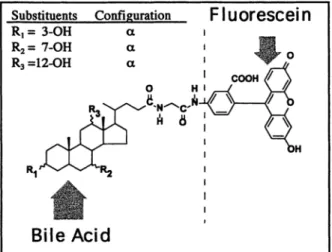
structure of CGF was confirmed by nuclear magnetic resonance and is
given in Fig. 1. The compound was purified by adsorption chromatography
using silica gel column chromatography, as previously described [2], and
210 Substituents C o n f i g u r a t i o n R I = 3 - O H ct R= = 7 - O H ~t R3 = I 2 - O H ct o 14 RI ~" v V - ~ R 2
m
Bile Acid
F luorescein
go
Fig. 1. Chemical structure of the fluorescent bile acid CGF. CGF was synthesized by conjugation of 5-aminofluorescein with the curboxylic group of cholylglycine (also called glycocholic acid or glycocholate).
the final compound was 98% pure by high-pressure liquid chromatography (HPLC) [11]. The fluorescence properties of CGF are essentially those of fluorescein; the wave length for maximum excitation is 492 nm, whereas that for maximum emission is 512 nm [5, 6].
Surgical procedures
S u r g i c a l p r e p a r a t i o n
Animal studies were approved by the Animal Subjects Committee of the University of Bern. Thirteen Burgundian rabbits (Tierstall Inselspital, Bern, Switzerland) weighing 2.8-~--0.4 kg were fasted overnight before the experiment. Anesthesia was induced by intramuscular injection of keta- mine (Nurketan 10, 45 mg/kg b.w.; Chassot AG, Bern, Switzerland) and xylazine-hydrochloride (Xylapan, 6.5 mg/kg b.w.; Chassot AG). Anesthe- sia was maintained by repeated intramusculur doses of 0.5 ml of a 1:1 (by volume) mixture of ketamine and xylazine-hydrochloride every 30-45 min. After the induction of anesthesia, the animals were shaved in the abdominal region, placed on a small animal operating table (Karl Kaps, Asslur, Ger- many), and secured in a supine position. An electric warming pad (Sotis Electronics AG, Glattbrug, Switzerland) had been placed on the operating table to prevent development of hypothermia during the surgical procedure. The jugular vein was cannulated using PE-10 polyethylene tubing (Clay Adams, Parsippany, NJ, USA). A solution of 0.9% sodium chloride (Bich- sel AG, Interlaken, Switzerland) was infused at a rate of 6 ml/h using a Harvard syringe pump (Harvard Apparatus Co., MiUis, MA, USA). Two sets of experimental models were used.
B i l i a r y r e c o v e r y o f C G F in the b i l i a r y f i s t u l a a n i m a l In the fast set of experiments, the abdomen was opened by a midline incision, and an external biliary fistula was constructed by cannulating the common bile duct using a 14-gange intravenous catheter (Abbocath-T; Abbot, Sligo, Ireland) connected to a Combidyn polyethylene tube (B. Braun Melsungen AG, Melsungen, Germany). Bile was collected every 5 min in preweighed plastic vials.
After a 30-min control period, CGF (dissolved in 0.9% sodium chlo- ride, pH adjusted to 8.0 with 0.05 N NaOH) was infused into the Jugular vein over a 5-min period in doses of 0.25, 1.25, or 2.5 i~mol/kg min. These 5-min infusions resulted in a total input dose of I, 5, and 10 mg CGF/kg b.w., respectively. At the end of the infusion period of CGF, saline was infused for a subsequent 90 rain. Biliary output, recovery, and dose-related time curves of CGF fluorescence were measured.
To test for the renal excretion of CGF and its metabolites, the urinary
Fig. 2. Experimental technique for laparoscopic surgery in rabbits.
bladder was cannulated at the end of each experiment, and urine was collected for thin-layer chromatography (TLC). At the end of each experi- ment, the biliary fistula was removed, and the common bile duct was closed with a running suture using Vicryl 6/0 (Ethicon, Norderstedt, Ger- many). The lapurotomy was closed with a two-layer running suture.
L a p a r o s c o p i c c h o l e c y s t e c t o m y
In the second set of experiments, a 5-mm skin incision was made about one-third the distance between the xiphoid and the os pubis. Under visual control, the camera trocar (Aesculap El 562, Tuttlingen, Germany) was introduced into the abdominal cavity using a blunt obturator and attached to a flexible arm. A pursestring suture was applied to prevent gas leakage. COz was insufflated (1 L/min) to establish a pneumoperitoneum with a maximal intraabdominal pressure of 5 mmHg. The 4-ram 30* wide-angle lapuroscope (Aesculap PE 504A, Tuttlingen, Germany) was introduced into the peritoneal cavity. All laparoscopic instruments (micro-scissors, straight and angled forceps) were 2.7 mm in diameter (Wolf, Knittlingen, Germany). No electric coagulation device was needed.
Following a diagnostic overview of the abdominal cavity, additional working 3-mm and 10-mm trocars were introduced into the upper right and left hemi-abdomen (Fig. 2). The gallbladder and extrahepatic biliury tract were exposed and observed under standard and fluorescence conditions before and after intravenous infusion of 1, 5, and 10 mg CGF/kg b.w., respectively. Before completing LC, simulated biliary tract injury was induced by making a small incision with the micro-scissors into the gall- bladder neck.
After we had observed the fluorescence of the bile leak using fluores- cence illumination, a cholecystectomy was performed using conventional illumination. The cystic duct was ligated using a 5-mm Endo Clip Dispos- able Applier (Auto Suture, Elancourt, France). The gallbladder was re- sected using micro-scissors and extracted through the left-side 10-mm trocar. At the end of the laparoscopic experiment, the trocars were re- moved, and the trocur sites were closed with a two-layer technique. The whole operative procedure was recorded simultaneously on videotape with time measurement.
Visualization of biliary fluorescence by selective
optical filters
In order to eliminate autofluorescCnce and to obtain a high signal/noise ratio of CGF fluorescence, optical filters (Curl Storz GmbH, Tuttlingen, Germany) were introduced into the lighting system of the lapuroscopic equipment. A fluorescein-blue filter (maximal transmission at 497 nm) was attached to the light source and used for excitation of CGF. A fluorescein emission filter (transmission peak at 530 nm) was attached in front of the CCD camera and used to detect the fluorescent signal emitted by CGF. The filter system was connected in such a way as to permit instantaneous
211 0.55 " i 0.50 0.45
~_
0.40 O C~0.35 10 20 30 40 50 60 70 80 90 100110120 I I ~ 1 1 , I I I i I a I m C G F I n f u s i o n l o 1 m g / k g b . w . l e 5 m g / k l z b . w . I v I0 m g / K g b . w 0.30 I~l 0.25 9 ~ 0.20 ='~ 0.15 0.10 0.05 v, r~ . o. ~ V . v . .'o.o.o:$:8:8:8:~;~;li';il';ir;iL
o.oo 't-'r-T*, , , , , , , , 0 10 20 30 40 50 60 70 80 90 100110120 T i m e (min)Fig. 3. Time course of biliary recovery of intravenously infused CGF for the three doses used in this study. Data are the mean +-. SD for six rabbits.
0.55 0.50 0.45 0.40 0.35 0.30 0.25 0.20 0.15 0.10 0.05 0.00
switching between standard (cold light) and fluorescent (blue light) illu- mination.
Analytical methods and data analysis
Concentrations of CGF in bile were determined spectrophotometrically (Unicam UV/Vis Spectrometer UV2; ATI Unicam, Cambridge, England) at 492 rim, as previously described [5]. For the determination of hepatic biotransformation and renal excretion of CGF, bile and urine aliquots (2--6 p.l) were examined by TLC using silica gel plates (Silica Gel 60 F-254, 20 x 20 cm, thickness 0.25 mm; E. Merck, Darmstadt, Germany). A solvent system for conjugated bile acids was used (isoamylacetate: propionic acid: N-propanol" water, 4:3:2:1, vol:vol) [4]. CGF and its metabolites were visualized with ultraviolet light (at 366 nm). Biliary volume was deter- mined gravimetrically, assuming a density for bile of 1.00 g/ml. The ap- parent choleretic activity of CGF [ACA = A bile flow/A biliary recovery of CGF] was determined by linear regression (SigmaPIot; Jandel Scientific, San Rafael, CA, USA).
Data are expressed as mean • SD.
Results
Biliary fistula rabbit
Figure 3 shows the time course of the biliary output of C G F after infusion o f 1, 5, and 10 m g CGF/kg b.w. The maximal transport rate occurred at 15 min for 1 and 5 mg C G F / k g b.w. and at 20 min for 10 mg C G F / k g b.w. Biliary recovery of C G F was high. R e c o v e r y at 90 min (in % dose) was 96 _ 1.5 for the 1 mg/kg dose, 89 -+ 2.2 for the 5 mg/kg dose, and 86 ___ 1.9 for the 10 mg/kg dose. The biliary secretion of C G F - i n d u c e d bile flow with the apparent choleretic activity of C G F was 25 /xl/l~mol.
C G F was recovered solely in bile at doses o f 1 and 5 mg/kg b.w. since no fluorescence was detected in urine by TLC. H o w e v e r , in experiments using 10 mg C G F / k g b.w., a small fluorescent spot (<1%) with identical Re value to C G F was detectable in urine. Based on T L C o f bile, C G F
Fig. 4. Enhanced visualization of the common bile duct by CGF. The duct
exhibits an apple-green fluorescence, permitting it to be readily distin- guished from its overlying blood vessel in the hepatic pedicle.
did not undergo appreciable biotransformation during hepa- tocyte transport.
Biliary fluorescence began to fade after 35 _ 2 min with the 1 mg/kg dose and after 60 -.+ 5 min for the 5 mg/kg dose. With the highest dose (10 mg CGF/kg), biliary fluorescence was still present at the end of the experiment.
Small animal laparoscopy
W h e n visualized using the fluorescein-blue filter system, apple-green fluorescence was detectable in the biliary tract after a mean hepatic transit time of 2.4 _+ 0.3 rain. W h e n compared to the preinfusion state, C G F fluorescence en- hanced the visualization of the c o m m o n bile duct and cystic duct. Indeed, the fluorescent bile duct was clearly distin- guishable from its accompanying blood vessels in the he- patic pedicle, which were not fluorescent (Fig. 4). With standard light for illumination, biliary fluorescence was still discernible but relatively indistinct.
In animals in which an experimental biliary tract injury was performed by incision into the gallbladder neck, the biliary fluorescence allowed immediate identification of bile leakage.
D i s c u s s i o n
T h i s study shows that intravenous infusion of C G F , a fluo- rescent bile acid, enhances the visualization of the biliary tract and experimental bile leaks when appropriate optical filters are used in rabbits undergoing cholecystectomy. The study also extends our previous work using the anesthetized biliary fistula rat [5] and the isolated perfused liver [6]. These studies, like the present one, show that C G F is rapidly and efficiently excreted into bile without undergoing bio-
212
transformation during hepatocyte transport. Another fluo-
rescent bile acid, cholyllysylfluorescein, has been shown to
have transport properties similar to those of CGF [6]; there-
fore, this fluorescent bile acid should, at least in principle,
also be suitable for the application described here [8]. For
visualizing the bifiary tree, fluorescent bile acids appear to
be superior to fluorescein because fluorescent bile acids will
enter only those cells that have a suitable anion transporter,
such as the hepatocyte, whereas fluorescein enters all cells
by passive diffusion [12].
Our method for exciting CGF emission and detecting
CGF fluorescence uses a fluorescein-blue filter system and
a fluorescein emission filter. These are inexpensive filters
that are easily attached to standard laparoscopic equipment.
We added a switch that permitted rapid alternation between
fluorescent illumination and conventional (cold light) illu-
mination. The ability to switch rapidly between cold fight
and fluorescent light makes the procedure a simple one.
Switching from the emission filter to no filter was relatively
unimportant, since visualization of the operative field under
cold light was not influenced by the presence of the emis-
sion filter. A dose of 5 mg CGF/kg induced fluorescence for
60 rain, an interval sufficient for cholecystectomy in the
rabbit.
Other investigators have also used induced fluorescence
to enhance the visualization of surgical fields or to detect
ischemic tissues, but the optical methods employed in these
studies seem less convenient than the one described here.
Oddi et al. [8] induced biliary tract fluorescence using an-
other fluorescent bile acid, cholyl-lysyl-fluorescein, during
open laparotomy in rabbits; fluorescence was induced by a
manually held Wood light. To detect mesenteric ischemia
during laparoscopy, Galandiuk et al. [3] used an argon laser
as the excitation source after an intravenous infusion of
fluorescein. Kam and Scheeres [7] modified the method of
Galandiuk et al. by adding an argon laser to the conven-
tional halogen light source. A 1-mm quartz fiber was intro-
duced through the operating channel of the laparoscope and
used to illuminate the abdominal cavity with a 488-nm blue
argon laser light. The video system was equipped with a
488-514-nm amber Plexiglas filter for fluorescein emission.
Although excellent in principle, this technique is expensive
and complex.
In LC, correct identification of biliary tract a n a t o m y - -
that is, isolation of the cystic duct, common bile duct, and
cystic artery in the triangle of Calot--is considered to be the
most important part of the entire procedure. Nevertheless,
aberrant ductal anatomy of the right hepatic duct and minor
ducts in the fiver bed may not be recognized, so that these
structures are liable to be injured during subsequent removal
of the gallbladder [14]. There are also cases in which the
cystic duct is not ligated properly, and continuous bile leak-
age leads to postoperative biloma formation. Our method
can clearly enhance intraoperative visualization of the bili-
ary tract in rabbits, but additional experiments are required
to define whether CGF can induce enhanced visualization of
the bifiary tract during biliary surgery in patients. This en-
hanced visualization might prevent and/or facilitate recog-
nition of bifiary tract injury or bile leaks.
In summary, in the rabbit, CGF, a fluorescent bile acid,
is secreted quantitatively in bile, inducing biliary fluores-
cence. By employing optical filters for excitation and emis-
sion, autofluorescence is eliminated, and the biliary tract
and bile leaks are visualized more readily than with con-
ventional illumination. Clinical studies are needed to deter-
mine whether this technique can be used in patients, and if
so, whether its application will result in decreased bile duct
injury.
Acknowledgments.
We also acknowledge the help of Takanobu Goto, vis- iting postdoctoral fellow from Numazn College of Technology, Japan, in purifying CGF by silica gel column chromatography, as well as the video- technical assistance of Reto Hiinni, "IV studio Inselspital, Bern, Switzer- land. This work was supported in part by a grant-in-aid from the Depart- ment of Clinical Investigation (DKF), University of Bern, CH-3010 Bern, Switzerland.References
1. Araki K, Namakiwa K, Mizutani J, Doiguchi M, Yamamoto H, Arai H, Yamaguchi T, Ido Y, Uno K, Hayashi N (1992) Indocyanine green staining for visualization of the biliary system during laparoscopic cholecystectomy. Endoscopy 24:803
2. Del Vecchio S, Ostrow JD, Mukerjee P, Ton-Nu I-IT, Schteingart CD, Hofmann AF, Cerr~ C, Roda A (1995) A method for the removal of surface active impurities and calcium from conjugated bile salt prepa- rations: comparison with silicic acid chromatography. J Lipid Res 36: 2639-2654
3. Galandiuk S, Fazio VW, Petras RE (1988) Fluorescein endoscopy: a technique for non-invasive assessment of mesenteric ischemia. Dis Colon Rectum 31:848-853
4. Hofmann AF (1962) Thin-layer adsorption chromatography of free and conjugated bile acids on silicic acid. J Lipid Res 3:127-128 5. Holzinger F, Schteingart CD, Ton-Nu HT, Eming SA, Monte M J,
Hagey LR, Hofmann AF (1997) Fluorescent bile acid derivatives: relationship between chemical structure and hepatic and intestinal transport in rat. Hepatology 26:1263-1271
6. Holzinger F, Schteingart CD, Ton-Nu H-T, Cerr~ C, Steinbacb H, Yeh H-Z, Hofmann AF (1998) Transport of fluorescent bile acids by the isolated peffused rat liver: kinetics, sequestration, and mobilization. Hepatology 28:510---520
7. Kam DM, Scheeres DE (1993) Fluorescein-assisted laparoscopy in the identification of arterial mesenteric ischemia. Surg Endosc 7:75-78 8. Oddi A, Mills CO, Custureri F, Di Nicola V, Elias E, Di Matteo G
(1995) Intraoperative biliary tree imaging with cholyl-lysyl- fluorescein: an experimental study in the rabbit. Surg Laparosc Endosc 6:198-200
9. Paumgartner G (1975) The handling of indocyanine green by the liver. Schweiz Med Wochenschr 105:5-30
10. Pertsemlidis D, Barzilai A, Pertsemlidis DSS, Zinberg J, Aufses AH (1993) Enhanced laparoscopic visualization of the extrahepatic bile duct with intravenous indocyanine green. Am J Gastroenterol 88: A230
11. Rossi SS, Converse JL, Hofmann AF (1987) High pressure liquid chromatographic analysis of conjugated bile acids in human bile: si- multaneous resolution of sulfated and unsulfated lithocholyl amidates and the common conjugated bile acids. J Lipid Res 28:589-595 12. Schmidt R, Buscher H-P (1991) Hepatic uptake of fluorescein inves-
tigated by video fluorescence microscopy and digital image analysis. J Hepatol 13:208-212
13. Schteingart CD, Eming S, Ton-Nu HT, Crombie DL, Hofmann AF (1992) Synthesis, structure, and transport properties of fluorescent derivatives of conjugated bile acids. In: Paumgartner G, Gerok W, Stiehl A (eds) Bile acids and hepatobiliary system. Kluwer Academic, London, pp 177-183
14. Strasberg S (1999) Laparoscopic biliary surgery. In: Cooper A (ed) Gastroenterology clinics of North America. WB Saunders, Philadel- phia, pp 117-132
15. Woods MS, Traverso LW, Kozarek R.A, Tsao J, Rossi RL, Gough D, Donohue JH (1994) Characteristics of biliary tract complications dur- ing laparoscopic cholecystectomy: a multi-institutional study. Am J Surg 167:27-34