HAL Id: hal-03142058
https://hal-amu.archives-ouvertes.fr/hal-03142058
Submitted on 15 Feb 2021
HAL is a multi-disciplinary open access
archive for the deposit and dissemination of
sci-entific research documents, whether they are
pub-lished or not. The documents may come from
teaching and research institutions in France or
abroad, or from public or private research centers.
L’archive ouverte pluridisciplinaire HAL, est
destinée au dépôt et à la diffusion de documents
scientifiques de niveau recherche, publiés ou non,
émanant des établissements d’enseignement et de
recherche français ou étrangers, des laboratoires
publics ou privés.
Distributed under a Creative Commons Attribution| 4.0 International License
Description of Prevotella rectalis sp. nov., a new
bacterium isolated from human rectum
S. Belkacemi, C.I. Lo, S. Khelaifia, D. Raoult, P.-E. Fournier, F. Fenollar
To cite this version:
S. Belkacemi, C.I. Lo, S. Khelaifia, D. Raoult, P.-E. Fournier, et al.. Description of Prevotella rectalis
sp. nov., a new bacterium isolated from human rectum. New Microbes and New Infections, Wiley
Online Library 2020, 36, pp.100703. �10.1016/j.nmni.2020.100703�. �hal-03142058�
Description of Prevotella rectalis sp. nov., a new bacterium isolated from
human rectum
S. Belkacemi1,2, C. I. Lo2,3, S. Khelaifia1,2, D. Raoult1,2, P.-E. Fournier2,3and F. Fenollar2,3
1) Aix Marseille Université, IRD, AP-HM, MEPHI, 2) IHU-Méditerranée Infection and 3) Aix Marseille Université, IRD, AP-HM, SSA, VITROME, Marseille, France
Abstract
Using a taxonogenomics method, we describe here a Gram-negative bacterium named Prevotella rectalis sp. nov., strain Marseille-P4334T(= CSUR P4334) isolated from the rectum. Strain Marseille-P4334Thas a genome that measure 3.03 Mbp with 43.3 mol% G + C content. © 2020 The Authors. Published by Elsevier Ltd.
Keywords: Culturomics, Human gut, Prevotella rectalissp. nov., Rectum
Original Submission: 20 December 2019; Revised Submission: 19 May 2020; Accepted: 22 May 2020 Article published online: 28 May 2020
Corresponding author. F. Fenollar, Institut Hospitalo-Universitaire Méditerranée-Infection, 19–21 Boulevard Jean Moulin, 13385, Mar-seille, Cedex 05. France.
E-mail:florence.fenollar@univ-amu.fr
Introduction
Studying the diversity of bacteria is fundamental to under-standing their involvement in human diseases and normal physiological functions [1]. To unveil human microbial diversity, the culturomics approach, based on diversified culture condi-tions, was designed to isolate as yet uncultured species and to complement 16S rRNA metagenomics [2–4]. Furthermore, a new taxonomic strategy, named taxonogenomics, was devel-oped to include the analysis of complete genome sequences in combination with phenotypic characteristics [5]. Herein, we report a detailed description of Prevotella rectalis sp. nov., for which the type strain Marseille-P4334Twas isolated from the human rectum.
Isolation and growth conditions
In 2017, we isolated an unidentified bacterium strain from a human rectum. The study was approved by the Institut
Federatif de Recherche 48 (agreement number 09-022, Mar-seille, France), and the patient’s consent was obtained. After three tests using matrix-assisted laser desorption/ionization time offlight mass spectrometry (MALDI-TOF MS), the strain was not identified. Screening was carried out on a Microflex LT spectrometer (Bruker Daltonics, Bremen, Germany) as previ-ously described [6]. Spectra obtained from the strain (Fig. 1) were imported and analysed using BIOTYPER3.0 software against
the Bruker database that was permanently updated with the local MEPHI database (https://www.mediterranee-infection. com/urms-data-base).
The growth of strain Marseille-P4334Twas possible on 5% sheep’s blood-agar (bioMérieux, Marcy l’Étoile, France) under anaerobic atmosphere (anaeroGEN; Oxoid, Dardilly, France) at 37°C after 3 days of pre-incubation and 5 days in incubation. Strain identification
The 16S rRNA gene was sequenced to classify this bacterium. Amplification was performed using the primer pair fD1 and rP2 (Eurogentec, Angers, France) and sequenced using the Big Dye® Terminator v1.1 Cycle Sequencing Kit and 3500xLGe-netic Analyzer capillary sequencer (Thermofisher, Saint-Aubin, France), as previously described [7]. The 16S rRNA nucleotide sequence was assembled and corrected using CODONCODE
ALIGNER software (http://www.codoncode.com). Strain
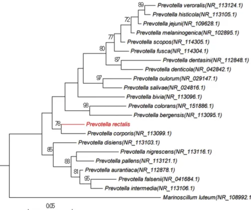
Prevotella corporis strain JCM 8529 (GenBank accession number: NR_113099.1), its phylogenetically closest species with stand-ing in nomenclature (Fig. 2). Considering this value less than a threshold value established previously [8], we consequently proposed to classify strain Marseille-P4334Tas a new species within the genus Prevotella in the phylum Bacteroidetes. Phenotypic characteristics
Colonies of strain Marseille-P4334T were black and smooth with a mean diameter of 0.5–1 mm. Bacterial cells were Gram-negative bacilli ranging from 1 to 3μm in length and 0.4 to 0.6 μm in width. Strain Marseille-P4334T
showed catalase-positive
and oxidase-negative activities. Electron micrograph showing the shapes of this new bacterial strain (Fig. 3) was performed with the Hitachi TM4000 instrument (Hitachi Group, Krefeld, Germany). A comparative study of the biochemical character-istics of strain Marseille-P4334T with other closely related species in phylum Bacteroidetes is presented inTable 1. Results of biochemical tests performed using the API ZYM and 50 CH strips (bioMérieux) are shown inTable 2.
Genome sequencing
Genomic DNA was extracted using the EZ1 biorobot with the EZ1 DNA tissue kit (Qiagen, Hilden, Germany) and then
FIG. 1.MALDI-TOF MS reference spectra of Prevotella rectalis sp. nov., strain Marseille-P4334T. Separately the two reference spectra were generated by comparison of spectra from 12 individual colonies from each strain.
2 New Microbes and New Infections, Volume 36 Number C, July 2020
NMNI
© 2020 The Authors. Published by Elsevier Ltd, NMNI, 36, 100703
sequenced on a MiSeq sequencer (Illumina Inc., San Diego, CA, USA) with the Nextera Mate Pair sample prep kit and Nextera XT Paired End (Illumina), as previously described [9]. The
as-softwares (VELVET[10], SPADES[11] and SOAPDENOVO[12]), and
trimmed data (MISEQ and TRIMMOMATIC [13] softwares) or
un-trimmed data (only MISEQ software). GAPCLOSER was used to FIG. 2. Phylogenetic tree
high-lighting the position of Prevotella rec-talis sp. nov. strain Marseille-P4334T with regard to other closely related species. GenBank accession numbers of 16S rRNA are indicated in paren-theses. Sequences were aligned using MUSCLE with default parameters, phylogenetic inferences were ob-tained using the maximum likelihood method and MEGA 7 software. Bootstrap values obtained by repeating the analysis 1000 times to generate a majority consensus tree are indicated at the nodes.
FIG. 3.Scanning electron microscopy (SEM) of the new species. For each strain a colony was collected from agar and immersed into a 2.5% glutaraldehydefixative solution. Then, a drop of the suspension was directly deposited on a poly-L-lysine-coated microscope slide for 5 minutes and treated with 1% phosphotungstic acid aqueous solution (pH 2.0) for 2 minutes to increase the SEM image contrast. The slide was gently washed in water, air-dried and examined in a tabletop SEM (Hitachi TM4000). Scales and acquisition settings are shown on thefigures.
depth value < 25% of the mean depth were removed. The best assembly was selected using different criteria (number of scaf-folds, N50, number of N). Strain Marseille-P4334T had a genome size of 3 039 397 bp with 43.3 mol% G + C content. The degree of genomic identity of this strain with closely related species was calculated using ORTHOANI software [14].
Results of this analysis are displayed inFig. 4. For strain Mar-seille-P4334T, ORTHOANI values among closely related species
ranged from 68.57% between Prevotella corporis and Prevotella bergensis to 68.70% between Prevotella bergensis and Prevotella salivae. When P. rectalis was compared with these closely related species, values ranged from 69.91% with P. salivae to 76.38% with P. corporis.
TABLE 1.Differential characteristics of Prevotella rectalis sp. nov., strain Marseille-P4334Tcompared with related species within
their respective genus
Properties Prevotella rectalis Prevotella multiformis Prevotella bivia Prevotella melaninogenica Prevotella aurantiaca
Gram stain — — — — — Motility — — — — — Endospore formation — — — — — Catalase + — NA NA — Glycerol — + — — — D-Cellobiose — + — — — D-Raffinose — + — + + α-Galactosidase + — — NA — β-Galactosidase — + + + — α-Fucosidase — NA + + + α-Glucosidase — — NA + NA β-Glucosidase + + + + NA Arginine aminopeptidase — — — + NA
Source Human rectal Subgingival plaque Vagina Human respiratory tract Human oral cavity
+, positive result;−, negative result; NA, data not available.
TABLE 2. Phenotypic characterizations of Prevotella rectalis sp. nov., based on API tests
API tests Characteristics P4334T
ZYM Alkaline phosphatase —
Esterase (C4) + Esterase lipase (C8) + Lipase (C14) — Leucine arylamidase — Valine arylamidase + Cystine arylamidase — Trypsin — α-Chymotrypsin — Acid phosphatase — Naphthol-AS-BI-phosphohydrolase + α-Galactosidase + β-Galactosidase — β-Glucuronidase — α-Glucosidase — β-Glucosidase + N-Acetyl-β-glucosaminidase — α-Mannosidase — α-Fucosidase — 2-NitrophenylβD-galactopyranoside — 50 CH Glycerol — Erythritol — D-Arabinose — L-Arabinose — D-Ribose — D-Xylose — L-Xylose — D-Adonitol — MethylβD-Xylopyranoside — D-Galactose + D-Glucose — D-Fructose — D-Mannose — L-Sorbose — L-Rhamnose — Dulcitol — Inositol — D-Mannitol — D-Sorbitol — Methyl-αD-Mannopyranoside — Methyl-αD-Glucopyranoside — N-Acetylglucosamine — Amygdalin — Arbutin —
Esculin ferric citrate —
Salicin —
D-Cellobiose —
D-Maltose —
TABLE 2.Continued
API tests Characteristics P4334T
D-Lactose — D-Melibiose — D-Saccharose — D-Trehalose — Inulin — D-Melezitose — D-Raffinose — Amidon — Glycogen — Xylitol — Gentiobiose — D-Turanose — D-Xylose — D-Tagalose — D-Fucose — L-Fucose — D-Arabitol — L-Arabitol — Potassium gluconate — Potassium 2-ketogluconate — Potassium 5-ketogluconate —
4 New Microbes and New Infections, Volume 36 Number C, July 2020
NMNI
© 2020 The Authors. Published by Elsevier Ltd, NMNI, 36, 100703
Conclusion
On the basis of unique phenotypic features, including the MALDI-TOF spectrum, a 16S rRNA sequence divergence >1.3% and an ORTHOANI value > 95% with the phylogenetically closest species with standing in nomenclature, we formally proposed strain Marseille-P4334Tas the type strain of Prevotella rectalis sp. nov., as a new member within the phylum Bacteriodetes.
Description of Prevotella rectalis sp. nov
Prevotella rectalis (rec.ta’lis. L. gen. n. rectalis pertaining to rectum, the chamber that begins at the end of the large intes-tine from which this bacterium was isolated). It is a Gram-negative bacterium, non-motile and non-spore-forming. Strain Marseille-P4334Tis the type strain of Prevotella rectalis sp. nov.,
potential pathogenicity of the type strain Marseille-P4334T(= CSUR P4334) is unknown. This strain has a genome size of 3.03 Mbp long with a 43.3 mol% G + C content. The 16S rRNA gene sequence and whole-genome shotgun sequence of strain Marseille-P4334 were deposited in GenBank under accession numbers LS488976 and UWTY00000000, respectively. It has been isolated from the human rectum.
Conflicts of interest
None to declare.
Funding sources
This study was supported by the Institut
Hospitalo-FIG. 4. Heatmap generated with ORTHOANI values calculated using the OAT software for Prevotella rec-talis sp. nov., strain Marseille-P4334 compared with other closely related species with standing in nomenclature.
reference ANR-10-IAHU-03, the Région Provence-Alpes-Côte d’Azur and European funding FEDER PRIMI.
Acknowledgements
The authors thank Hitachi Corporation for providing the TM4000Plus Tabletop microscope and Aurelia Caputo for submitting the genomic sequence to GenBank.
References
[1] Lagier J-C, Armougom F, Million M, Hugon P, Pagnier I, Robert C, et al. Microbial culturomics: paradigm shift in the human gut microbiome study. Clin Microbiol Infect 2012;18:1185–93.
[2] Lagier J-C, Hugon P, Khelaifia S, Fournier PE, La Scola B, Raoult D. The rebirth of culture in microbiology through the example of cul-turomics to study human gut microbiota. Clin Microbiol Rev 2015;28:237–64.
[3] Lagier J-C, Khelaifia S, Tidjani Alou M, Ndongo S, Dione N, Hugon P, et al. Culture of previously uncultured members of the human gut microbiota by culturomics. Nat Microbiol 2016;1:16203.
[4] Lagier J-C, Edouard S, Pagnier I, Mediannikov O, Drancourt M, Raoult D. Current and past strategies for bacterial culture in clinical microbiology. Clin Microbiol Rev 2015;28:208–36.
[5] Fournier P-E, Lagier J-C, Dubourg G, Raoult D. From culturomics to taxonomogenomics: a need to change the taxonomy of prokaryotes in clinical microbiology. Anaerobe 2015;36:73–8.
[6] Lo CI, Fall B, Sambe-Ba B, Diawara S, Gueye MW, Mediannikov O, et al. MALDI-TOF mass spectrometry: a powerful tool for clinical microbiology at Hôpital Principal de Dakar, Senegal (West Africa). PLoS One 2015;30:10–2.
[7] Morel AS, Dubourg G, Prudent E, Edouard S, Gouriet F, Casalta JP, et al. Complementarity between targeted real-time specific PCR and conventional broad-range 16S rDNA PCR in the syndrome-driven diagnosis of infectious diseases. Eur J Clin Microbiol Infect Dis 2015;34:561–70.
[8] Stackebrandt E, Ebers J. Taxonomic parameters revisited : tarnished gold standards. Microbiol Today 2006;6:152–5.
[9] Lo CI, Sankar SA, Fall B, Ba B-S, Diawara S, Gueye MW, et al. High-quality draft genome sequence and description of Haemophilus massi-liensis sp. nov. Stand Genomic Sci 2016;11:31.
[10] Zerbino DR, Birney E. Velvet: algorithms for de novo short read as-sembly using de Bruijn graphs. Genome Res 2008;18:821–9. [11] Bankevich A, Nurk S, Antipov D, Gurevich AA, Dvorkin M,
Kulikov AS, et al. SPAdes: a new genome assembly algorithm and its applications to single-cell sequencing. J Comput Biol 2012;19:455–77. [12] Luo R, Liu B, Xie Y, Li Z, Huang W, Yuan J, et al. SOAPdenovo2: an empirically improved memory-efficient short-read de novo assembler. Gigascience 2012;1:18.
[13] Bolger AM, Lohse M, Usadel B. Trimmomatic: aflexible trimmer for Illumina sequence data. Bioinformatics 2014;30:2114–20.
[14] Lee I, Ouk Kim Y, Park SC, Chun J. OrthoANI: an improved algorithm and software for calculating average nucleotide identity. Int J Syst Evol Microbiol 2016;66:1100–3.
6 New Microbes and New Infections, Volume 36 Number C, July 2020
NMNI
© 2020 The Authors. Published by Elsevier Ltd, NMNI, 36, 100703