ABSTRACT: Guava fruit was identified as a particularly rich source of 13-hydroperoxide lyase activity. The enzyme proved stable to chromatographic procedures and was purified to ho-mogeneity. Based on gel filtration and gel electrophoresis, the native enzyme appears to be a homotetramer with subunits of 55 kD. Starting with primers based on the peptide sequence, the enzyme was cloned by polymerase chain reaction with 3′ and 5′ rapid amplification of cDNA ends. The sequence shows approximately 60–70% identity to known 13-hydroperoxide lyases and is classified in cytochrome P450 74B subfamily as CYP74B5. The cDNA was expressed in Escherichia coli (BL21 cells), with optimal enzyme activity obtained in the absence of isopropyl-β-D-thiogalactopyranoside and δ-aminolevulinic acid. The expressed enzyme metabolized linolenic acid over 10-fold faster than 13(S)-hydroperoxy-linoleic acid and the 9-hydroperoxides of 13(S)-hydroperoxy-linoleic and linolenic acids. 13(S)-Hydroperoxylinolenic acid was converted to 12-oxododec-9(Z)-enoic acid and 3(Z)-hexenal, as identified by gas chromatography–mass spectrometry. The turnover number with this substrate, with enzyme concentration estimated from the Soret absorbance, was ≈2000/s, comparable to values reported for the related allene oxide synthases. Distinctive features of the
guava 13-hydroperoxide lyase and related cytochrome P450 are discussed.
Paper no. L8473 in Lipids 35, 709–720 (July 2000).
The metabolism of fatty acid hydroperoxides [lipoxygenase (LOX) products] involves conversion to epoxides, aldehydes, alcohols, and other derivatives, and these reactions are often catalyzed by cytochrome P450 enzymes. In green plant tis-sue, the fatty acid hydroperoxide lyase (HPL), an enzyme in the LOX pathway, catalyzes the cleavage of 13- and 9-hy-droperoxides of linoleic and linolenic acid into volatile C6- or C9-aldehydes and C12- or C9-oxoacids, respectively (1,2). The C6and C9volatile compounds have a commercial value in the production of “natural” flavor in the food industry, and are potentially important in plant defense against pathogens (3) and pests (4).
The green notes [hexanal, hexan-1-ol, 2(E)-hexenal, 3(Z)-hexenal, 2(E)-hexen-1-ol, and 3(Z)-hexen-1-ol (also known as pipol)] are used widely in flavors (particularly fruit) to im-part a fresh green character. The synthesis of these com-pounds starts from free polyunsaturated fatty acids such as linoleic [9(Z),12(Z)-octadecadienoic] and linolenic acids [9(Z),12(Z),15(Z)-octadecatrienoic]. In nature, these acids are released from cell membranes by lipolytic enzymes after cell damage. Fatty acid 13-hydroperoxides are formed by the ac-tion of a specific lipoxygenase (13-LOX), and these are sub-sequently cleaved by a 13-hydroperoxide lyase (13-HPL) into a C6-aldehyde and a C12-ω-oxoacid moiety. The aldehydes can subsequently undergo thermal isomerization and/or be re-duced by dehydrogenase enzymes to give the other C6 prod-ucts mentioned above (2,5).
13-HPL was demonstrated for the first time in banana fruits (6) and subsequently characterized in a number of dif-ferent plant materials (7–10). The enzyme has been purified to apparent homogeneity from tea leaves (11) and more re-cently green bell pepper fruits (12) and sunflower (13). 1Present address: Department of Entomology, Forbes 410, University of
Ari-zona, P.O. Box 210036, Tucson, AZ 85721-0036.
2Present address: Institute of Food Research, Norwich Research Park,
Col-ney, Norwich NR4 7UA, England.
3Present address: RWTH Aachen, Institut für Biologie III (Plant Physiology), Worringerweg, D-52074, Aachen, Germany.
4Present address: Firmenich Asia Pte. Ltd, 10 Tuas West Road, Singapore 638377.
*To whom correspondence should be addressed at Dept. of Pharmacology, Vanderbilt University, 23rd Ave. at Pierce, Nashville, TN 37232-6602. E-mail: [email protected]
Abbreviations: δ-ALA, δ-Aminolevulinic acid; AOS, allene oxide synthase; GC–MS, gas chromatography–mass spectrometry; GPC, gel permeation chromatography; HIC, hydrophobic interaction chromatography; HPL, hy-droperoxide lyase; HPLC, high-pressure liquid chromatography; IFC, iso-electric focusing chromatography; IPTG, isopropyl-β-D -thiogalactopyran-oside; LOX, lipoxygenase; NTA, nitrilotriacetic acid; PAGE, polyacrylamide gel electrophoresis; PCR, polymerase chain reaction; RACE, rapid amplifi-cation of cDNA ends; RP–HPLC, reversed-phase–high pressure liquid chro-matography; SDS, sodium dodecyl sulfate; TFA, trifluoroacetic acid; UV, ultraviolet.
Purification, Molecular Cloning, and Expression
of the Gene Encoding Fatty Acid 13-Hydroperoxide
Lyase from Guava Fruit (Psidium guajava)
Nathalie Tijeta,1, Urs Wäspib, Duncan J.H. Gaskinc, Peter Hunzikerd,Bernard L. Mullere, Evgeny N. Vulfsonc,2, Alan Slusarenkob,3, Alan R. Brasha,*, and Ian M. Whiteheade,4
aDepartment of Pharmacology, Vanderbilt University, Nashville, Tennessee 37232, bDepartment of Plant Biology, University of Zürich, CH-8008, Switzerland, cInstitute of Food Research, Department of Macromolecular Sciences,
Reading RG6 6BZ, Berkshire, England, dDepartment of Biochemistry, University of Zürich, CH-8057, Switzerland, and eFirmenich S.A., Geneva 8, CH-1211, Switzerland
Cloning of the pepper 13-HPL cDNA confirmed that the en-zyme is a member of the cytochrome P450 family of hemo-proteins (14). The P450 family is designated as CYP74B. Subsequently, the 13-HPL of Arabidopsis was cloned from published expressed sequence tag sequences and identified as a related cytochrome P450 (15).
We began the present work with a survey of HPL activi-ties in various commercial fruits and vegetables, and on this basis selected guava fruit as the starting material for further characterization. The 13-HPL activity in guava is substan-tially higher than in several other well-characterized sources such as bean and pepper. Guava is available on an almost year-round basis, making it an attractive source for the com-mercial production of fatty acid aldehydes. In the present paper, we describe purification of the 13-HPL from guava fruit, molecular cloning of the cDNA, and expression of the active protein in Escherichia coli.
EXPERIMENTAL PROCEDURES
Materials. For protein purification, fruits of Psidium guajava
from Thailand (bought at a local market in Zürich) were frozen and stored at −20°C. For the molecular cloning, im-mature guava (≈3 cm in diameter) were collected in Brazil, frozen on dry ice on the same day, and subsequently stored at −80°C.
Measurement of 13-HPL activity in fruits and vegetables.
The lyase reactions were performed in a four-necked glass vessel equipped with a mechanical stirrer, a dropping funnel topped with a nitrogen bubbler, a thermometer, and a pH elec-trode. Guava fruit homogenate (20 g) prepared in a Waring blender was stirred vigorously under nitrogen at the selected temperature and pH. An aqueous solution of hydroperoxyoc-tadecadienoic acid or hydroperoxyoctadecatrienoic acid, 20 g, containing 35 g/kg hydroperoxide as determined by iodomet-ric titration, was then added. This solution of hydroperoxides consisted of either saponified sunflower oil (serving predomi-nantly as a source of linoleic acid) or saponified linseed oil (linolenic acid) that had been treated with soybean flour (LOX) to form the corresponding 13-hydroperoxides. The for-mation of hexanal or 3(Z)- and 2(E)-hexenal was monitored by gas chromatography (GC) every 5 min for the first 15 min, and subsequently every 30 min. The C6-aldehydes were quan-tified by direct injection of the filtered crude reaction samples onto a 3-m column of Carbowax 10% on Chromosorb W HP 80/100 mesh (Supelco, Bellefonte, PA) operated isothermally at 100°C in a PerkinElmer 2900 gas chromatograph equipped with a flame-ionization detector. The concentrations of hexa-nal or 3(Z) and 2(E)-hexehexa-nal were determined by comparison with an external standard solution of 500 mg/L hexanal or 2(E)-hexenal in 0.5% EtOH/99.5% water solution.
Purification of the 13-HPL from guava fruit. (i) Prepara-tion of the crude extract. Guava fruits were peeled and the
pericarp tissues chopped into small pieces. Two volumes of extraction buffer (50 mM sodium phosphate, 0.1% Triton X-100R, 5 mM sodium ascorbate, pH 7.0) were added to
500 g of chopped pericarp and homogenized for 2 min in a Sorvall mixer at 4°C. All of the following steps were carried out at room temperature.
(ii) Enzyme solubilization. The crude extract containing
the 13-HPL activity was solubilized with 1% (vol/vol) of Tri-ton X-100R with stirring for 30 min. After centrifugation at 16,000 × g for 15 min, 0.02% Pectinex Ultra SP-L solution from Novo Nordisk Ferment (Bagsvaerd, Denmark) was added to degrade pectin.
(iii) Ammonium sulfate precipitation. Solid (NH4)2SO4 was added in small portions to the crude extract under stir-ring until 30% saturation was achieved. After stirstir-ring for 30 min, the mixture was centrifuged at 20,000 × g for 15 min and the resulting pellet discarded. The supernatant was brought to 60% saturation with more solid (NH4)2SO4added in portions. After stirring for 30 min, the pellet was collected by centrifu-gation as above.
(iv) Gel permeation chromatography (GPC). The
(NH4)2SO4pellet was dissolved in 45 mL of extraction buffer and chromatographed on a Superdex 200 HL 26/60 column (Pharmacia, Uppsala, Sweden) with 50 mM sodium phos-phate, 0.1% Triton X-100R pH 7.0 as running buffer at a flow rate of 2 mL/min. After GPC, samples were run on anion ex-change chromatography or hydrophobic interaction chroma-tography (HIC) columns.
(v) Anion exchange chromatography. The sample from
GPC was applied to a Q-Sepharose column (Pharmacia) with a loading buffer of 20 mM Tris-HCl pH 8.5 containing 0.1% Triton X-100R. 13-HPL activity was eluted with a gradient of NaCl in eluting buffer (0–100% 1 M NaCl in 133 min at a flow rate of 3 mL/min).
(vi) HIC. The fractions containing the 13-HPL activity
from the GPC were brought to 30% (NH4)2SO4saturation be-fore loading onto a Phenyl-Sepharose HR 26/10 column (Pharmacia) with loading buffer [50 mM sodium phosphate, 1 M (NH4)2SO4, pH 7.0]. The proteins were eluted with a de-creasing salt gradient (100–0% 1 M ammonium sulfate over 70 min) in 50 mM sodium phosphate pH 7.0 containing 1% Triton X-100R at a flow rate of 8 mL/min. Fractions contain-ing the 13-HPL activity were concentrated by dialysis against polyethylene glycol 20,000 and desalted on a PD-10 column (Pharmacia) against the loading buffer for hydroxylapatite chromatography (10 mM sodium phosphate, 0.1% Triton X-100R, pH 6.8).
(vii) Hydroxylapatite chromatography. After HIC, the
sample was applied to an Econo-Pac HTP column (BioRad, Cambridge, MA) in 10 mM sodium phosphate, 0.1% Triton X-100R, pH 6.8. The proteins were eluted with a gradient to 200 mM sodium phosphate buffer pH 6.8, containing 0.1% Triton X-100R over 30 min using a flow rate of 1 mL/min. Fractions with 13-HPL activity were concentrated by dialysis against polyethylene glycol 20,000 and desalted against the isoelectric focusing chromatography (IFC) loading buffer (75 mM Tris-acetic acid, pH 9.3).
(viii) IFC. The prepared sample from hydroxylapatite
(Phar-macia). The proteins were eluted with 10% Polybuffer 96 (Pharmacia)/acetic acid, pH 6.0, at a flow rate of 0.5 mL/min.
Tryptic digest and amino acid sequence determination.
Fractions of purified 13-HPL were concentrated and sepa-rated on a 6.5% sodium dodecyl sulfate-polyacrylamide gel (SDS-PAGE). The proteins were electrotransferred (0.8 mA cm−2for 75 min) to an Immobilon CD membrane (Millipore, Bedford, MA) using 10 mM 3-[cyclohexylamino]-1-propane-sulfonic acid containing 10% (vol/vol) methanol pH 11.0 as transfer buffer. Proteins were detected by staining using Quick-Stain (Zoion Research Inc., Newton, MA).
Direct sequencing of the purified 13-HPL by Edman degradation was not possible as the N-terminus was blocked. The 13-HPL purified protein was therefore cut out separately and incubated in 10 µL of 0.1 M Tris pH 8.2 containing 1 M NaCl, 10% (vol/vol) acetonitrile, 2 mM CaCl2, and 0.1 µg trypsin at 37°C for 15 h. After acidification with 1 µL of 10% trifluoroacetic acid (TFA), the solution was injected directly into the high-pressure liquid chromatography (HPLC) system equipped with a Brownlee Aquapore RP-300 C8 column (PE Applied Biosystems, Foster City, CA). Chromatography sol-vents were 0.05% TFA and 2% acetonitrile in water (solvent A) and 0.045% TFA and 80% acetonitrile in water (solvent B). The gradient and flow rates used were as follows: 0–5 min, 80 µL/min at 2% solvent B; 5–65 min 50 µL/min at 2–65% sol-vent B; and 65–70 min 50 µL/min at 65–100% B. Absorbance at 214 nm was measured in a 200 nL flow cell with a path length of 2 mm. Peptides resolved by HPLC were collected manually for sequence analysis and applied to precycled polyprene-treated glass fiber discs. Automated sequencing used a model 477A pulsed-liquid phase sequencer (Applied Biosystems) equipped with a model 120A analyzer.
RNA isolation. Total RNA was extracted using the method
of Wan and Wilkins (16). Immature guava fruit (1 g) was crushed to a fine powder in liquid nitrogen in a precooled pes-tle and mortar. The powder was added to 5 mL at 80°C of lysis buffer [200 mM borax, 30 mM EGTA, 10 mM dithio-threitol, 1% wt/vol SDS, 1% wt/vol sodium deoxycholate, 2% PVP 40,000 (Sigma Chemical Co., St. Louis, MO), 0.5% vol/vol NP-40 (Sigma)] and the mixture homogenized. Pro-teinase K (2.5 mg, Sigma) was added, and this mixture was incubated at 42°C for 90 min with shaking sufficient for mix-ing without excessive foammix-ing. One milliliter of 1 M KCl was added. After mixing, the tubes were incubated on ice for 1 h and then centrifuged at 10,000 × g for 10 min. Three milli-liters of 4 M LiCl was added, and the tubes were incubated at 4°C overnight. After 10 min of centrifugation at 10,000 × g, the supernatant was discarded, and the pellet washed with 2 M LiCl and centrifuged as before. The supernatant was dis-carded and the pellet resuspended in Tris/EDTA buffer (10 mM Tris, pH 8, 1 mM EDTA) or in H2O.
For the purification of mRNA from total RNA, the mRNA purification kit from Pharmacia was used. This kit is based on the use of spun columns for the affinity purification of polyadenylated RNA on oligo(dT)-cellulose. The RNA was quantified by ultraviolet (UV) spectrophotometry. The
yield was approximately 1 mg of total RNA and 20 µ g of mRNA.
Cloning. (i) cDNA synthesis. Total RNA (20 µ g) or 1 µ g
of mRNA was used in 50-µ L reactions for the first strand cDNA synthesis using an oligo(dT)-adaptor primer (17). Aliquots of 1 µ L cDNA were used directly in polymerase chain reactions (PCR).
(ii) PCR cloning. The PCR were primed with 1 µL cDNA
(from a 50-µL cDNA synthesis using 20 µg total RNA), and using 10 mM Tris, pH 8.3, 50 mM KCl, 3 mM MgCl2with 0.2 mM each of dNTPs and 0.25 µL (1.25 units) of AmpliTaq DNA polymerase (PerkinElmer) in a PerkinElmer 480 ther-mocycler. After the addition of the cDNA at 80°C (hot start), the PCR reactions conditions were: 94°C, 2 min for 1 cycle; 50–55°C for 1 min, 72°C for 1 min, and 94°C for 1 min for 30 cycles; 72°C for 10 min for 1 cycle; and the block temper-ature was held at 4°C.
3′-Rapid amplification of cDNA ends (RACE) and 5′-RACE: The 3′-sequence was obtained using gene-specific up-stream primers: 5′CCT CAA CAC GCT CAG GTG AAG3′,
5′CTC CAA AAG TTC CTC TTC AAC TTC3′, or 5′CCA
GCT CCT CCC CAC CAT CAA3′ against a downstream primer based on the adaptor-linked oligo(dT) primer used for cDNA synthesis (17). The 5′RACE was accomplished using a kit from GibcoBRL (Grand Island, NY), according to the manufacturer’s instructions. The gene-specific downstream primers were: 5′GTC AGC GCC GAA GAT GGA CTT3′or
5′GTG TTG AGG CTC GGA AGT GTC3′.
Full-length clones obtained by PCR. Four gene-specific
primers were synthesized corresponding to the putative start sites of the coding sequence (at the four different methio-nines) and one primer corresponding to the stop codon.
BamHI and EcoRI restrictions sites were incorporated at the
5′ and 3′ ends, respectively, to facilitate subcloning. The four upstream primers were: 5′TAG GAT CCG ATC ATG GCG AGG GTC GTG3′, 5′GCG GAT CCG GCC ATG AGC AAC ATG TCG3′, 5′GCG GAT CCG GCC ATG TCG CCG GCC AT3′, and 5′GCG GAT CCG GCC ATG TCG TCC ACC TAC3′; and the downstream primer was: 5′GCG AAT TCT CAG TTG GCC TTT TCA ACG GCT GT3′. These primers were purified by HPLC as the dimethoxytrityl derivative (17). After deprotection, they were used in PCR reactions (at 20 pmol/50 µL final concentration) with a proof-reading mixture of Taq/Pwo DNA polymerase (Expand High Fidelity, Boehringer-Mannheim, Indianapolis, IN) according to the manufacturer’s instructions. The reactions conditions were 94°C, 2 min for 1 cycle; 60°C for 1 min, 72°C for 1 min, and 94°C for 1 min for 30 cycles; 72°C for 10 min for 1 cycle; and the block temperature was held at 4°C.
DNA sequencing. cDNA were sequenced using the
Thermo Sequenase radiolabeled terminator cycle sequencing kit (Amersham Life Science, Inc., Arlington Heights, IL).
Bacterial expression. (i) Preparation of constructs. The
four cDNAs encoding the 13-HPL-Met 1, 6, 9, and 13 in pCR2.1 were cut with BamHI and EcoRI and subcloned into the expression vector plasmid pET30b (Novagen) digested
also with BamHI and EcoRI. The 13-HPL-Met 1, 6, 9, and 13 constructs were transformed into E. coli strain XLI-Blue. The plasmid DNA was then used for transformation of E. coli strain BL21(DE3) to express the 13-HPL.
(ii) Preparation of bacterial cultures. The four different
constructs described above were expressed in BL21(DE3) (Novagen, Madison, WI) cells using a modified expression methodology (18). A typical preparation of 50-mL culture was carried out as follows: A single bacterial colony from a complex agar plate containing 30 µg/mL of kanamycin was grown in 1 mL of LB medium containing 50 µ g/mL of kanamycin at 37°C and 250 rpm for 3 h. An aliquot (200 µL) of this culture was used to inoculate 10 mL of TB medium containing 50 µg/mL of kanamycin and the culture was again grown at 37°C. After 3 h, this culture was diluted with 40 mL of TB containing 30 µg/mL of kanamycin and grown at 28°C, 250 rpm for 24 h. The bacterial cells were centrifuged at 4°C for 15 min at 5,000 rpm (3,500 × g) in a Jouan CR422 cen-trifuge, washed by resuspension in 10 mL of Tris-HCl buffer 50 mM pH 7.9, and centrifuged as before. The resulting pel-let of cells was resuspended in 10 mL of Tris-acetate buffer 100 mM pH 7.6 containing 500 mM of sucrose, 0.5 mM of EDTA, and 1 mg/mL of lysozyme. After 30 min on ice, the cells were centrifuged as before to obtain a pellet of sphero-plasts which were resuspended in 10 mL of potassium phos-phate buffer (100 mM, pH 7.6). After at least 10 min at −80°C, a protease inhibitor (phenylmethylsulfonyl fluoride, 1 mM) was added, the cells were allowed to thaw for 10 min, and then they were sonicated twice for 30 s using a Virsonic 100 at a setting of 5. The resulting membranes were spun down at 100,000 × g for 90 min at 4°C. The 13-HPL activity was recovered in the 100,000 × g pellet. After solubilization using 1% Emulphogen BC-700™ (polyoxyethylene 10 tride-cyl ether, Sigma) at 4°C overnight and centrifugation as be-fore, the 13-HPL activity was recovered in the supernatant.
Purification of His-tagged proteins. The histidine-tagged
13-HPL was purified following the protocol described by Imai et al. (19). After solubilization, the 100,000 × g super-natant was loaded on a Ni-nitrilotriacetic acid (NTA) column (0.5 mL bed volume, Qiagen, Valencia, CA) equilibrated with potassium phosphate buffer 50 mM pH 7.6 containing 100 mM NaCl and 0.1% Emulphogene BC-720 detergent (buffer A) at 0.5 mL/min. The column was washed with the buffer A containing 50 mM glycine. The His-13-HPL was then eluted with the buffer A containing 40 mM L-histidine. Fractions of 1 mL were collected and assayed for the 13-HPL activity. Fractions containing the 13-HPL activity were dialyzed overnight against potassium phosphate 100 mM, pH 7.6 using a microdialyzer (Pierce, Rockford, IL).
Enzyme assays. (i) UV assay. 13-Hydroperoxides from
linolenic and linoleic acids were produced with soybean lipoxygenase L1 (type V, Sigma) according to the methods of Vick (20) or Brash and Song (21). 13-HPL activity was mea-sured by following the decrease in absorbance at 234 nm, rep-resenting disruption of the conjugated diene system in the fatty acid hydroperoxide substrate. The assay contained 5–10
µ g of fatty acid 13-hydroperoxide in 0.5 mL of 100 mM potassium phosphate buffer at pH 7.
For the HPLC assay, 50 or 100 µM of a mixture of unlabeled and [1-14C]-13(S)-hydroperoxylinole(n)ic acid (200,000 cpm) was used in 100 mM potassium phosphate buffer at pH 7. The reactions were stopped with 100 µL potassium dihydrogen phosphate (1 M) and the products extracted twice with 0.5 mL ethyl acetate. The combined organic phases were washed with water and dried under a stream of nitrogen. The extracts were dissolved in 50 µL methanol and stored at −20°C under argon prior to analysis. The compounds were analyzed using a Beck-man Ultrasphere ODS column (5 µm, 25 × 0.46 cm) using MeOH/H2O/glacial acetic acid (77.5:22.5:0.01, by vol) as sol-vent at a flow rate of 1.1 mL/min. Products were detected on-line using a Hewlett-Packard 1040A diode array detector and a Packard Flo-One radioactive monitor. The main 14C-labeled enzymatic product from [1-14C]13(S)-hydroperoxylinolenic acid on reversed-phase (RP)-HPLC (12-oxo-dodecenoic acid) was resolved into the main 9Z isomer and the 10E isomer formed during sample workup by normal-phase HPLC on a Beckman 5 µm silica column (25 × 0.46 cm) with a solvent of hexane/isopropanol/glacial acetic acid (100:1:0.01, by vol). The 9Z isomer chromatographed as a broad symmetrical peak (retention volume 17–22 mL), and the 10E isomer as a typi-cally shaped peak at 24 mL. The two compounds were con-verted to the methoxime derivative (which eliminated further isomerization of the 9Z double bond) and methyl ester deriva-tive and analyzed by GC–mass spectrometry (MS).
GC–MS. GC–MS analyses were carried out in the electron
impact mode (70 eV) using a Finnigan Incos 50 mass spec-trometer (for 3Z-hexenal analysis) or a Hewlett-Packard 5889A mass spectrometer (for 12-oxo-dodecenoate analysis), each coupled to a Hewlett-Packard 5890 gas chromatograph equipped with a SPB-1 fused-silica capillary column (15 or 30 m × 0.25 mm internal diameter). Samples were injected at 60°C, and the temperature was subsequently programmed to 300°C at 10 or 20°/min.
RESULTS
13-HPL in fruits and vegetables. The fruits of a number of
commonly available plants were screened for the ability to convert 13-hydroperoxylinoleic acid (Table 1) and 13-hydro-peroxylinolenic acid (Table 1) to the corresponding C6HPL products. On the basis of these results, initially bean was selected as a readily available and relatively inexpensive material for the purification of the enzyme. However, in pilot experiments the bean lyase proved to be unstable to freeze–thawing, and this compromised our ability to purify the enzyme. Accordingly, guava was chosen as an even more promising candidate on the basis of activity. It was soon es-tablished that this enzyme survived freezing and that its ro-bust properties permitted its purification through multiple chromatographic steps.
Purification of the guava 13-HPL. Preliminary work
enzyme and that detergents are necessary to solubilize the en-zyme (11). The guava tissue was homogenized with a sodium phosphate buffer pH 7.0 containing 5 mM sodium ascorbate to prevent oxidation and 0.1% Triton X-100R. When the con-centration of Triton X-100R was increased to 1% (vol/vol) and the homogenate was stirred for 30 min, the yield of solu-ble and active 13-HPL was dousolu-bled (data not shown).
A typical purification of 13-HPL from guava fruit is shown in Table 2. The crude extract was concentrated by (NH4)2SO4 precipitation followed by a subsequent GPC step on a Superdex 200 column (Pharmacia). The fractions containing 13-HPL
ac-tivity were pooled and chromatographed on an HIC column. After concentration and desalting, the pooled active fractions were chromatographed on a hydroxylapatite column. The eluted 13-HPL activity was again concentrated, desalted, and loaded onto an IFC column. Each purification step resulted in a considerable loss of 13-HPL activity, which was probably not due to proteolysis, and only 0.2% of the initial 13-HPL activity was recovered (Table 2). Analysis of the preparation by SDS-PAGE showed that the purified sample contained only one, ap-parently homogenous band with an apparent molecular weight of 48 kD. Because of the high losses of 13-HPL activity, an
TABLE 1
Screening of Plants for 13-Hydroperoxide Lyase Activity
Screening for 13-HPOD lyase activity
Vegetable Hexanala Fruit Hexanala
Alfalfa 1.36 Apple 0.38
Bean (green) 0.82 Banana 0.31
Bean (white) 0.78 Cashew fruit <0.1
Celery leaf 0.16 Grape 0.33
Fennel 0.12 Kiwi 0.27
Parsley 0.20 Orange 0.61
Pea 0.25 Papaya 0.73
Pepper (green) 0.89 Pear 0.33
Radish leaf 0.23 Pineapple 0.19
Soya (sprout) 0.25 Raspberry 0.37
Tomato 0.51 Strawberry 0.40
Turnip 0.20 Guavab 2.20
Screening for 13-HPOT lyase activity
Vegetable (Z)-3 + (E)-2-Hexenala Fruit (Z)-3 + (E)-2-Hexenala
Alfalfa 0.21 Apple 0.22
Celery <0.1 Banana 0.13
Cucumber 0.28 Apricot 0.23
Lettuce <0.1 Cherry <0.1
Pea <0.1 Kiwi 0.24
Pepper (green) 0.37 Orange 0.23
Radish leaf 0.10 Papaya 0.11
Soya (sprout) <0.1 Melon (water) <0.1
Tomato <0.1 Raspberry <0.1
Sorrel <0.1 Strawberry <0.1
Guavab 1.18
ag/kg reaction.
bWith guava, optimized yields using higher substrate concentrations (80 g/kg hydroperoxide) were 5.07 g/kg hexanal and 1.43 g/kg hexenals. Abbreviations: HPOD, hydroperoxyoctadecadienoic acid; HPOT, hydroperoxyoctadecatrienoic acid.
TABLE 2
Purification of 13-Hydroperoxide Lyase from Guava Fruita
Total Specific
Total 13-HPL Recovered 13-HPL
Purification protein activity activity activity Purification
step (mg) (nkat) (%) (nkat mg−1) factor
Crude extract 1,111 172,050 100.0 155 — 30–60% (NH4)2SO4pellet 762 62,300 36.2 82 — GPC 39 32,640 19.0 837 5.4 HIC 16 15,160 8.8 947 6.1 Hydroxylapatite 1.6 6,500 3.8 4,062 26.2 IFC 0.03 317 0.2 10,566 68.2
aHPL, hydroperoxide lyase; GPC, gel permeation chromatography; HIC, hydrophobic interaction chromatography; IFC, isoelectric focusing chromatography.
anion exchange chromatography step was used in place of HIC. In this case, analysis of the purified enzyme by SDS-PAGE showed two bands of 48 and 50 kD. The 13-HPL purified from guava fruit tissue had maximal activity around pH 6.0 and a pI of 6.8 determined by chromatofocusing.
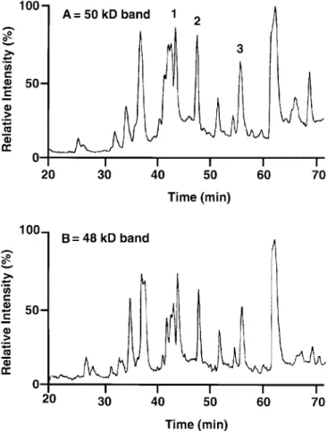
The HPLC peptide maps of the trypsin cleavage products from the two protein bands are very similar (Fig. 1), indicat-ing that there are only minor differences in amino acid com-position and/or posttranslational modification. Three peptides of the 50 kD band were sequenced (Table 3).
A computer-aided search of the Swissprot and genEMBL databases found that five amino acids of peptide 3 (Phe Asn Phe Leu Ser, Table 3) are identical to amino acids 236 to 240
of flaxseed allene oxide synthase (AOS) (22). At this stage there was no obvious homology between the peptide se-quences and the 13-HPL from green bell pepper fruit (14).
Cloning. Sense and antisense degenerate oligonucleotides
(Table 4) deduced from the peptides sequences (Table 3) were synthesized. PCR reactions were performed using the differ-ent pairs of oligonucleotides with or without cDNA as tem-plate. One main product of 160 bp was subcloned and se-quenced, and this was found to contain the three peptides, which were thus from the same peptide chain (see later). Moreover, a multiple alignment of this sequence with the AOS from flax (22), the AOS from guayule (23), the 13-HPL from green pepper (14), and the 13-HPL from Arabidopsis (15) showed the highest homology with the 13-HPL enzymes. The remainder of the cDNA from guava fruit was cloned using 3′- and 5′-RACE (see Experimental Procedures sec-tion). cDNA corresponding to the open reading frame was prepared by PCR using a proof-reading mixture of Taq/Pwo as DNA polymerase. An amplification product was obtained, subcloned, and sequenced. The complete cDNA (1467 bp) and deduced amino acid sequence are shown in Figure 2. The open reading frame encodes a total of 489 amino acids corre-sponding to a protein with a molecular weight of 54,817 Dal-tons. The isoelectric point of the protein is estimated to be 7.29. Previously, the molecular weight of the native protein was estimated by SDS-PAGE as 50 kD, and we found a pI of 6.8 using chromatofocusing. The derived amino acid se-quence has an identity of 65% with the pepper 13-HPL (14). The guava cDNA encodes four possible start sites within the first 13 amino acids (methionines 1, 6, 9, and 13) whereas the pepper gene has only two methionines, corresponding to guava-Met9 and guava-Met13.
Expression. Four different cDNA clones of the 13-HPL
(13-HPL-Met1, -Met6, -Met9, -Met13) were inserted into the E. coli expression plasmids pET30b (see Experimental Procedures section) and transformed into E. coli strain BL21(DE3). The expression level was examined at different induction temperatures, with or without addition of isopropyl-β-D-thiogalactopyranoside (IPTG) and the heme precursor, δ-aminolevulinic acid (δ-ALA). The best activities were ob-tained at 28°C with both δ-ALA and IPTG omitted. Thus, in the system used here, with the pET30 plasmid, its T7 RNA
FIG. 1. High-pressure liquid chromatography (HPLC) analysis of
puri-fied guava 13-hydroperoxide lyase (13-HPL) digested with trypsin: com-parison of the peptide maps of the 50 and 48 kD protein bands.
TABLE 3
Peptide Sequences Obtained from HPLC-Purified Tryptic Fragments
Peptidea Sequenceb
1 Asp Gly Asn Ala Ser Val Ile Phe Pro Leu Gln (Lys) 2 Asn Phe Ala Met Asp Ile Leu (Lys)
3 Phe Leu Phe Asn Phe Leu Ser (Lys)
aThese numbers correspond to the peak number obtained during purifica-tion by high-performance liquid chromatography (HPLC).
bAs trypsin cleaves specifically on the carbonyl side of lys-arg linkages, the residues in parentheses are predicted.
TABLE 4
Oligonucleotides Designed from Peptide Sequence for PCR Cloning Experiments
Oligonucleotide name Sequencea(5′-3′ orientation)
12Sa GAYGGNAAYGCNTCNGTNATHTTYCCNYT 12Sb GAYGGNAAYGCNAGYGTNATHTTYCCNYT 12Aa CTRCCNTTRCGNAGNCANTADAARGGNRA 12Ab CTRCCNTTRCGNTCRCANTADAARGGNRA 13S AAYTTYGCNATGGAYATHYT 13A TTRAARCGNTACCTRTADRA 15S TTYCTNTTYAAYTTYYT 15A AARGANAARTTRAARRA
aCodes used here for the mixed bases are: D = G, A, T; H = A, T, C; R = A, G; Y = C, T. PCR, polymerase chain reaction.
polymerase promoter, and with the cells grown in a rich medium (TB), addition of heme precursor or IPTG inducer did not help with expression of active lyase.
Proteins from the transformed and induced cells were
ana-lyzed by SDS-PAGE (Fig. 3). High levels of protein with the expected molecular weight were expressed under all culture conditions (Fig. 3; lanes 2, 3, and 4). These levels appear to account for about half of the cellular protein. We obtained
FIG. 2. Nucleotide and deduced amino acid sequence of the guava 13-HPL. The four N-terminal methionines are boxed. The location of three
peptides of known amino acid sequence are shown in bold. The enzyme is classified as CYP74B5, and the GenBank accession number is AF239670. For abbreviation see Figure 1.
lower levels of protein but the highest lyase activity when the cells were grown in the absence of δ-ALA and IPTG (Fig. 3, lane 4). By using Emulphogen or cholate detergents, most of the expressed 13-HPL protein failed to solubilize, and the in-soluble material was catalytically inactive. This likely reflects the fact that there is a very high basal induction in this sys-tem, and the cells are barely capable of handling the ex-pressed protein. Examination of the cells under the micro-scope revealed many inclusion bodies, with the highest num-ber in cells grown with IPTG. This supports the concept that the bacteria cannot handle the level of expressed protein and the main part of the translated lyase is deposited in inclusion bodies. Using IPTG as a further stimulus results in even lower recovery of correctly folded protein with catalytic activity.
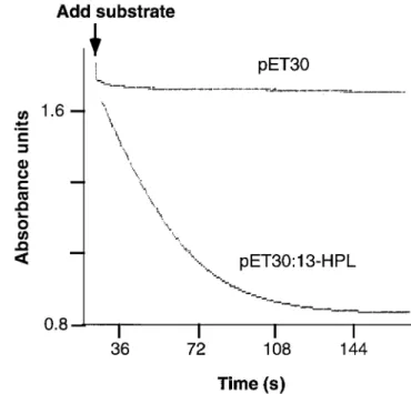
Lyase activity. The four different guava 13-HPL constructs
(-Met1, -Met6, -Met9, and -Met13) did not differ markedly in their catalytic activities in the UV assay. Figure 4 illustrates an example with pET30Met1, and also shows the lack of sig-nificant activity in the negative control in comparison to the reaction with 13-hydroperoxylinolenic acid. Approximately 10-fold lower rates of reaction were obtained with
13(S)-hy-droperoxylinoleic acid, 9(S)-hy13(S)-hy-droperoxylinoleic acid, and 15(S)-hydroperoxyeicosatetraenoic acid.
For HPLC analysis of the nonvolatile products, an aliquot of the sonicated protein preparation containing the 13-HPL was incubated with [1-14C]13(S)-hydroperoxylinolenic acid for 1 min as described in the Experimental Procedures sec-tion. Essentially all the substrate was metabolized in the 1 min of incubation, and the 1-14C label was recovered in a major polar product (Fig. 5) with a retention time expected of the
FIG. 3. Sodium dodecyl sulfate-polyacrylamide gel electrophoresis
analysis of guava 13-HPL expressed in Escherichia coli (BL21 cells). Lane 1: cells were transformed with pET30 vector alone. In the other lanes, cells were transformed with pET30:13-HPL (containing all four N-terminal methionines) cultured with δ-ALA and IPTG (lane 2), with IPTG only (lane 3), and without δ-ALA and IPTG (lane 4). Molecular weight markers (M) are in the right-hand lane. δ-ALA, δ-aminolevulinic acid; IPTG, isopropyl-β-D-thiogalactopyranoside; for other abbrevia-tion, see Figure 1.
FIG. 4. Spectrophotometric assay of the guava 13-HPL expressed in E. coli. Absorbance changes were monitored at 235 nm in potassium phosphate buffer, pH 7.6. Comparison of activity of 1 µL aliquots of bacterial lysates transformed with pET30 vector alone and with pET30:13-HPL. For abbreviations, see Figures 1 and 3.
FIG. 5. HPLC analysis of catalytic activity of guava 13-HPL expressed
in E. coli. Extracts were incubated with [1-14 C]13(S)-hydroperoxyli-nolenic acid, extracted, and aliquots injected on a Beckman Ultra-sphere ODS column (25 × 0.46 cm) and eluted with a solvent system of methanol/water/glacial acetic acid (77.5:22.5:0.01, by vol) at a flow rate of 1.1 mL/min. (A) Incubation of extracts of BL21 cells expressing vector pET30 only. (B) Incubation of extracts of BL21 cells expressing pET30:13-HPL. Radioactivity was detected on-line using a Packard Flo-One scintillation detector. For abbreviations, see Figures 1 and 3.
13-HPL-derived aldehyde, 12-oxo-9(Z)-dodecenoic acid. This product, which partly isomerized to the 10E isomer dur-ing sample workup, was resolved into the 9Z and 10E isomers by normal-phase HPLC (as described in the Experimental Procedures section). The two isomers were converted to the methoxime methyl ester derivatives and identified by GC–MS analysis (Fig. 6). The methyl (9Z)-12-oxo-dodecenoate methoxime eluted from the GC as a single peak at the reten-tion time approximately of a saturated C16fatty acid methyl ester; the 10E analog eluted 0.6 min later as a double peak of
syn and anti methoxime isomers. The 9Z isomer (assigned
based on its featureless UV spectrum, recorded before and after methoxime formation) gave a mass spectrum that was particularly close in appearance to the first reported spectrum of methyl 12-oxo-dodecenoate methoxime (24). The similar-ity in the mass spectra was noticeable particularly in terms of the pattern of abundances of the higher mass ions [M+255 (1% abundance), M − 31 at m/z 224 (14%), and m/z 192 (4%)], and the relative abundance of certain prominent lower mass ions such as m/z 98 (47%) and m/z 81 (54%). In retro-spect, the original spectrum may have been predominantly the 9Z isomer. We assigned the 10E isomer as such on the basis of the characteristics of its UV spectrum: it displayed the ex-pected enone chromophore with λmax225 nm in MeOH/H2O
(75:25, vol/vol) and extended conjugation after methoxime formation, producing a broad chromophore with λmax239 nm
in MeOH. The mass spectrum of the 10E derivative contained the same ions as in the 9Z, but there were different patterns of ion abundances (Fig. 6). For example, M+(5% relative
abun-dance) was slightly more prominent than m/z 224 (3%), whereas m/z 98 (20%) and m/z 81 (39%) were less prominent than in the 9Z isomer. This mass spectrum was a good match with another reported spectrum of the 10E isomer (25).
To detect the more volatile aldehyde product, we utilized direct GC–MS analysis of diethyl ether extracts of the reac-tion of 13-hydroperoxylinolenic acid with the expressed guava enzyme. The GC profile and mass spectrum identified the volatile product formed as 3(Z)-hexenal (Fig. 7).
DISCUSSION
An initial screening of the levels of 13-HPL in fruits and veg-etables pointed to guava as a rich source of this activity. From the subsequent isolation of the protein we found that approxi-mately a 70-fold purification was sufficient to give a homoge-neous preparation of the enzyme. Seventyfold enrichment im-plies a level of about 1–2% of the protein in the original ex-tract, consistent with guava as an abundant source of 13-HPL. The two bands isolated in the purification process are almost certainly an indication of partial proteolysis or deglycosyla-tion of the higher molecular weight protein. The difference in molecular weight (2 kD) between the two forms could be ex-plained if the heavier form is glycosylated by as few as the equivalent of 10 glucose residues. The purification of the 13-HPL from tea leaves (11) showed that the enzyme was sepa-rated with hydroxylapatite chromatography into two fractions, HPLI and HPLII with molecular weights of 53 and 55 kD. For the 13-HPL from green bell pepper fruits, Shibata et al. (12)
FIG. 6. Electron impact mass spectra of (9Z)-12-oxo-dodecenoic acid formed by reaction of the guava lyase with 13(S)-hydroperoxylinolenic acid and its 10E isomer. The two isomers were resolved by normal-phase HPLC and analyzed as the methyl ester methoxime derivative (top spectrum, 9Z; bottom, 10E). For abbreviation, see Figure 1.
showed also that 13-HPL could be resolved into two isoforms by hydroxylapatite chromatography; in this case, both iso-forms had molecular masses of 55 kD (12). Similar to the lyases of sunflower (13), the apparent molecular weight of the guava 13-HPL as determined by gel filtration is in the order of 200,000 kD, implying a tetrameric structure for the native en-zyme. This is in agreement with Olias et al. (10) who reported that 13-HPL from soybean seedlings had a native molecular mass of 240–260 kD and a subunit size of 62 kD. However, Shibata et al. (12) proposed that the 13-HPL from green bell pepper fruits is a trimer based on their measurements of the native molecular mass (170 kD) and subunit size (55 kD).
The 13-HPL of guava has a substrate specificity similar to several other reported 13-HPL enzymes. The preferred sub-strate is 13S-hydroperoxylinolenic acid. The corresponding linoleic acid 13S-hydroperoxide shows about one-tenth the rate of reaction, and the 9-hydroperoxides also are poor sub-strates. These results apparently contrast with the original data of lyase activity in guava fruit which indicated excellent recovery of the C6-aldehydes of linoleic and linolenic acids (Table 1). However, a number of factors could account for this difference that relate more to differences in assay condi-tions. The activity values in fruit comprise recovered prod-ucts whereas rates of reaction were measured with the ex-pressed enzyme. As the hexenals derived from linolenate are considerably less stable than the saturated product of linoleate, this gives a bias favoring recovery of hexanal. Sim-ilarly, changes in enzyme activity due to the different sub-strate and product concentrations and the different reaction times (<1 min, vs. 1 h) in the two assays could further influ-ence the results. It is also possible that other isoforms of hy-droperoxide lyase might exist in guava. Of the two hydroper-oxide lyases purified by Itoh and Vick (13) from sunflower hypocotyl, one resembled the guava and pepper enzymes and a second showed equal reactivity with the C18:2 analog, 13-hydroperoxylinoleic acid.
The guava cDNA sequence has about 60% amino acid
identity to the 13-HPL of bell pepper and Arabidopsis, plac-ing it in the same subfamily of cytochrome P450, CYP74B. The enzymes share about 35–40% identity with the plant AOS, which comprise the CYP74A subfamily. Within the ma-jority of cytochrome P450 families there is a conserved se-quence next to the cysteine that forms the proximal ligand to the heme (e.g., Ref. 26). In the CYP74 family, there are some unusual and distinctive sequences around this cysteine (Fig. 8). All reported 13-HPL and AOS enzymes have the se-quence Asn-Lys-Gln (NKQ) immediately before the cysteine.
FIG. 7. Electron impact mass spectrum of the volatile C6product formed by reaction of the guava lyase with 13(S)-hydroperoxylinolenic acid. The spectrum matches a reference of (3Z)-hexenal (Chemical Abstract Service No. 006789-80-6) in the NIST (National Institute of Sci-ence and Technology) 75K database on a Hewlett-Packard 5989A mass spectrometer/DOS Chemstation software, entry number 1285 (1992).
FIG. 8. Alignment of amino acid sequences around the proximal heme
ligand. The cysteinyl heme ligand is boxed. The absolutely conserved sequence NKQCA is underlined. The shaded position is usually Ala in 13-HPL, whereas in AOS enzymes the residue is the more typical P450 consensus Gly. The GenBank accession numbers of the sequences, given in parentheses, are for guava HPL (AF239670), bell pepper HPL (U51674), Arabidopsis HPL (AF087932), banana HPL (A65873, the sub-ject of U.S. patent 6,008,034, and patent EP0801133 assigned to Gi-vaudan-Roure International S.A.), alfalfa HPL (AJ249247), flax AOS (U00428), Arabidopsis AOS (AF172727), and guayule AOS (X78166). AOS, allene oxide synthase; for other abbreviation, see Figure 1.
Following the cysteine, the AOS (CYP74A) enzymes have the commonly occurring sequence Ala-Gly (Gly in this position is the P450 consensus sequence), giving the overall sequence NKQCAG. On the other hand, the lyases are distinguished by having the sequence Ala-Ala after the Cys. The second ala-nine, conserved in the lyases, occurs among less than 5% of all reported cytochromes P450. It is possible that the CAA se-quence gives a structurally significant tilt to the supporting polypeptide under the heme group. Mutation of this second alanine residue would be of interest for future experiments. Figure 8 also illustrates the absolutely conserved Val in the last position of this alignment. This is a very unusual residue in this position, which is normally represented by Gly or Ala in over 95% of all families of cytochrome P450.
There are remarkable differences in the sequences of the CYP74 family at the N-termini of the proteins (Fig. 9). Certain of the AOS, exemplified in Figure 9 by the flax and
Arab-idopsis enzymes, have a long presequence that includes a
chloroplast transit peptide. In the flaxseed AOS, as deduced by Laudert et al. (27), this includes 21 amino acids that are cleaved to give the mature protein. By contrast, the AOS of guayule has a short N-terminus, more similar to the 13-HPL enzymes. The N-terminal sequence of the guava 13-HPL has some unusual features compared to the HPL of bell pepper and Arabidopsis. There are four methionines in the first 13 amino acids. We tested the effect of deleting these sequences (albeit with an ad-ditional N-terminal sequence from the pET vector still present in each of the expressed proteins). Active lyase was obtained when either the full-length cDNA was expressed, or in the shortened cDNAs with one, two, or three methionines re-moved. The Arabidopsis 13-HPL could be expressed as
cat-alytically active enzyme after deletion of the first 28 amino acids. The shaded residues in Figure 9 indicate the most N-ter-minal amino acid in constructs that have been successfully ex-pressed with catalytically active enzyme. For the guayule AOS, the CYP74 family member with the shortest known N-termi-nus, the clear homology to the other enzymes (bold characters in Fig. 9) begins only six amino acids after the initiating me-thionine. For the CYP74 family, it has yet to be determined what is the shortest possible N-terminus that remains compati-ble with expression of catalytically active enzyme.
One of the outstanding properties of the AOS enzymes is their very high initial rate of reaction, in the order of 1000 turnovers per second. This contrasts with turnover numbers as low as 1/min for sluggish P450 reactions (28,29), and ≈3200/min for the fastest P450-hydroxylations as catalyzed, for example, by the heme domain of P450BM3 (30). The turnover number for HPL has not been reported. We were able to determine a value for the guava 13-HPL following its expression in E. coli. After partial purification of the His-tagged enzyme on a nickel affinity column, we obtained a weak UV-visible spectrum that permitted only an approxi-mate quantitation of the enzyme based on the main Soret band at ≈400 nm. Nonetheless, the value obtained was sufficient to permit calculation of the turnover number. The initial reac-tion rates indicate a turnover number for the 13-HPL of ap-proximately 2000/s, a value comparable to the AOS counter-parts in the subfamily CYP74A.
ACKNOWLEDGMENTS
This work was supported by Firmenich S.A., Geneva, Switzerland, and the Kanton of Zürich (KWF Projekt Nr. 2544.1). We thank Dr.
FIG. 9. N-terminal sequence comparison of 13-HPL and AOS enzymes. Comparison of the guava 13-HPL with
other CYP74 members. The different enzyme sequences are presented with the conserved residues aligned and shown in bold. The shaded residues indicate the N-terminal start site for the four guava constructs described here and the reported constructs of other enzymes that expressed with catalytic activity (14,15,22,23,27, 31). GenBank accession numbers are given in the legend to Figure 8. For abbreviations, see Figures 1 and 8.
Fredi Bruhlmann for helpful comments. Use of guava fruit in the commercial production of aliphatic aldehydes and alcohols from nat-ural fatty acid precursors is covered by U.S. Patent number 5,464,761 assigned to Firmenich S.A.
REFERENCES
1. Kim, I.S., and Grosch, W. (1981) Partial-Purification and Prop-erties of a Hydroperoxide Lyase from Fruits of Pear, J. Agric.
Food. Chem. 29, 1220–1225.
2. Hatanaka, A., Kajiwara, T., and Sekija, J. (1987) Biosynthetic Pathway for C6-Aldehydes Formation from Linolenic Acid in Green Leaves, Chem. Phys. Lipids 44, 341–361.
3. Croft, K.P.C., Jüttner, F., and Slusarenko, A.J. (1993) Volatile Products of the Lipoxygenase Pathway Evolved from Phaseolus
vulgaris (L.) Leaves Inoculated with Pseudomonas syringae pv. phaseolicola, Plant Physiol. 101, 13–24.
4. Dickens, J.C., Billings, R.F., and Payne, T.L. (1992) Green Leaf Volatiles Interrupt Aggregation Pheromone Response in Bark Beetles Infesting Southern Pines, Experientia 48, 523–524. 5. Hatanaka, A. (1993) The Biogeneration of Green Odor by Green
Leaves, Phytochemistry 34, 1201–1218.
6. Tressl, R., and Drawert, F. (1973) Biogenesis of Banana Volatiles, J. Agric. Food Chem. 21, 560–565.
7. Vick, B.A., and Zimmerman, D.C. (1976) Lipoxygenase and Hydroperoxide Lyase in Germinating Watermelon Seedlings,
Plant Physiol. 57, 780–788.
8. Schreier, P., and Lorenz, G. (1982) Separation, Partial Purifica-tion and CharacterisaPurifica-tion of a Fatty Acid Hydroperoxide Cleav-ing Enzyme from Apple and Tomato Fruits, Z. Naturforsch. C
37, 165–173.
9. Matsui, K., Shibata, Y., Kajiwara, T., and Hatanaka, A. (1989) Separation of 13-Hydroperoxide and 9-Hydroperoxide Lyase Activities in Cotyledons of Cucumber Seedlings, Z.
Natur-forsch. C 44, 883–885.
10. Olias, J.M., Rios, J.J., Valle, M., Zamora, R., Sanz, L.C., and Axelrod, B. (1990) Fatty Acid Hydroperoxide Lyase in Germi-nating Soybean Seedlings, J. Agric. Food Chem. 38, 624–630. 11. Matsui, K., Toyota, H., Kajiwara, T., Kakuno, T., and Hatanaka,
A. (1991) Fatty Acid Hydroperoxide Cleaving Enzyme, Hy-droperoxide Lyase, from Tea Leaves, Phytochemistry 30, 2109–2113.
12. Shibata, Y., Matsui, K., Kajiwara, T., and Hatanaka, A. (1995) Purification and Properties of Fatty Acid Hydroperoxide Lyase from Green Bell Pepper Fruits, Plant Cell Physiol. 36, 147–156. 13. Itoh, A., and Vick, B.A. (1999) The Purification and Characteri-zation of Fatty Acid Hydroperoxide Lyase in Sunflower,
Biochim. Biophys. Acta 1436, 531–540.
14. Matsui, K., Shibutani, M., Hase, T., and Kajiwara, T. (1996) Bell Pepper Fruit Fatty Acid Hydroperoxide Lyase Is a Cy-tochrome P450 (CYP74B), FEBS Lett. 394, 21–24.
15. Bate, N., Sivasankar, S., Moxon, C., Riley, J.M., Thompson, J.E., and Rothstein, S.J. (1998) Molecular Characterization of an Arabidopsis Gene Encoding Hydroperoxide Lyase, a Cy-tochrome P-450 That Is Wound Inducible, Plant Physiol. 117, 1393–1400.
16. Wan, C.Y., and Wilkins, T.A. (1994) A Modified Hot Borate Method Significantly Enhances the Yield of High-Quality RNA from Cotton (Gossypium hirsutum L.), Anal. Biochem. 223, 7–12.
17. Brash, A.R., Boeglin, W.E., Chang, M.S., and Shieh, B.-H. (1996) Purification and Molecular Cloning of an 8R-Lipoxyge-nase from the Coral Plexaura homomalla Reveal the Related Primary Structures of R- and S-Lipoxygenases, J. Biol. Chem.
271, 20949–20957.
18. Hoffman, B.J., Broadwater, J.A., Johnson, P., Harper, J., Fox, B.G., and Kenealy, W.R. (1995) Lactose Fed-Batch Overexpres-sion of Recombinant Metalloproteins in Escherichia coli BL21 (DE3): Process Control Yielding High Levels of Metal-Incorpo-rated Soluble Protein, Prot. Express. Purific. 6, 646–654. 19. Imai, T., Globerman, H., Gertner, J.M., Kagawa, N., and
Water-man, M.R. (1993) Expression and Purification of Functional Human 17 Alpha-Hydroxylase/17,20-lyase (P450c17) in
Es-cherichia coli. Use of This System for Study of a Novel Form
of Combined 17 Alpha-Hydroxylase/17,20-Lyase Deficiency, J.
Biol. Chem. 26, 19681–19689.
20. Vick, B.A. (1991) A Spectrophotometric Assay for Hydroper-oxide Lyase, Lipids 26, 315–320.
21. Brash, A.R., and Song, W.-C. (1996) Detection, Assay, and Iso-lation of Allene Oxide Synthase, Methods Enzymol. 272, 250–259.
22. Song, W.-C., Funk, C.D., and Brash, A.R. (1993) Molecular Cloning of an Allene Oxide Synthase: A Cytochrome P450 Spe-cialized for the Metabolism of Fatty Acid Hydroperoxides,
Proc. Natl. Acad. Sci. USA 90, 8519–8523.
23. Pan, Z.Q., Durst, F., Werk-Reichhart, D., Gardner, H.W., Ca-mara, B., Cornish, K., and Backhaus, R.A. (1995) The Major Protein of Guayule Rubber Particles Is a Cytochrome P450, J.
Biol. Chem. 270, 8487–8494.
24. Zimmerman, D.C., and Coudron, C.A. (1979) Identification of Traumatin, a Wound Hormone, as 12-Oxo-trans-10-dodecenoic Acid, Plant Physiol. 63, 536–541.
25. Hatanaka, A., Kajiwara, T., Sekiya, J., and Fukumoto, T. (1982) Oxygen-Isotope Effect in Enzymatic Cleavage Reaction of 13-L-Hydroperoxylinoleic acid to Hexanal and 11-Formyl-cis-9-undecenoic Acid, Z. Naturforsch. 37C, 752–757.
26. Brash, A.R., and Song, W.-C. (1995) Structure–Function Fea-tures of Flaxseed Allene Oxide Synthase, J. Lipid Mediat. Cell
Signal. 12, 275–282.
27. Laudert, D., Pfannschmidt, U., Lottspeich, F., Hollander Czytko, H., and Weiler, E.W. (1996) Cloning, Molecular and Functional Characterization of Arabidopsis thaliana Allene Oxide Synthase (CYP74), the First Enzyme of the Octadecanoid Pathway to Jasmonates, Plant Mol. Biol. 31, 323–335.
28. Guo, Z., Gillam, E.M.J., Ohmori, S., Tukey, R.H., and Guen-gerich, F.P. (1994) Expression of Modified Human Cytochrome P450 1A1 in Escherichia coli: Effects of 5′ Substitution, Stabi-lization, Purification, Spectral Characterization, and Catalytic Properties, Arch. Biochem. Biophys. 312, 436–446.
29. Gillam, E.M.J., Guo, Z., Ueng, Y.-F., Yamasaki, H., Cock, I., Reilly, P.E.B., Hooper, W.D., and Guengerich, F.P. (1995) Ex-pression of Cytochrome P450 3A5 in Escherichia coli: Effects of 5′ Modification, Purification, Spectral Characterization, Re-constitution Conditions, and Catalytic Activities, Arch.
Biochem. Biophys. 317, 374–384.
30. Capdevila, J.H., Wei, S., Helvig, C., Falck, J.R., Belosludtsev, Y., Truan, G., Graham-Lorence, S.E., and Peterson, J.A. (1996) The Highly Stereoselective Oxidation of Polyunsaturated Fatty Acids by Cytochrome P450BM-3, J. Biol Chem. 271, 22663–22671.
31. Noordermeer, M.A., van Dijken, A.J.H., Smeekens, S.C.M., Veldink, G.A., and Vliegenthart, J.F.G. (2000) Characterization of Three Cloned and Expressed 13-Hydroperoxide Lyase Isoen-zymes from Alfalfa with Unusual N-Terminal Sequences and Different Enzyme Kinetics, Eur. J. Biochem. 267, 2473–2482.
[Received February 25, 2000, and in revised form and accepted April 25, 2000]