Modifications in Early Rehabilitation Protocol after
Rotator Cuff Repair: EMG Studies
by Talia Alenabi
Department of Kinesiology
Thesis submitted in fulfillment of the requirements for the degree of Ph.D. in Science of Physical Activity
Option: Biomechanics
November 2015
i
Résumé
La déchirure de la coiffe des rotateurs est une des causes les plus fréquentes de douleur et de dysfonctionnement de l'épaule. La réparation chirurgicale est couramment réalisée chez les patients symptomatiques et de nombreux efforts ont été faits pour améliorer les techniques chirurgicales. Cependant, le taux de re-déchirure est encore élevé ce qui affecte les stratégies de réhabilitation post-opératoire. Les recommandations post-chirurgicales doivent trouver un équilibre optimal entre le repos total afin de protéger le tendon réparé et les activités préconisées afin de restaurer l'amplitude articulaire et la force musculaire. Après une réparation de la coiffe, l'épaule est le plus souvent immobilisée grâce à une écharpe ou une orthèse. Cependant, cette immobilisation limite aussi la mobilité du coude et du poignet. Cette période qui peut durer de 4 à 6 semaines où seuls des mouvements passifs peuvent être réalisés. Ensuite, les patients sont incités à réaliser les exercices actifs assistés et des exercices actifs dans toute la mobilité articulaire pour récupérer respectivement l’amplitude complète de mouvement actif et se préparer aux exercices de résistance réalisés dans la phase suivante de la réadaptation. L’analyse électromyographique des muscles de l'épaule a fourni des évidences scientifiques pour la recommandation de beaucoup d'exercices de réadaptation au cours de cette période. Les activités sollicitant les muscles de la coiffe des rotateurs à moins de 20% de leur activation maximale volontaire sont considérés sécuritaires pour les premières phases de la réhabilitation. À partir de ce concept, l'objectif de cette thèse a été d'évaluer des activités musculaires de l'épaule pendant des mouvements et exercices qui peuvent théoriquement être effectués au cours des premières phases de la réhabilitation. Les trois questions principales de cette thèse sont : 1) Est-ce que la mobilisation du coude et du poignet produisent une grande activité des muscles de la coiffe? 2) Est-ce que les exercices de renforcement musculaire du bras, de l’avant-bras et du torse produisent une grande activité dans les muscles de la coiffe? 3) Au cours d'élévations actives du bras, est-ce que le plan d'élévation affecte l'activité de la coiffe des rotateurs?
Dans notre première étude, nous avons évalué 15 muscles de l'épaule chez 14 sujets sains par électromyographie de surface et intramusculaire. Nos résultats ont montré qu’avec une orthèse d’épaule, les mouvements du coude et du poignet et même quelques exercices de renforcement impliquant ces deux articulations, activent de manière sécuritaire les muscles de
ii
la coiffe. Nous avons également introduit des tâches de la vie quotidienne qui peuvent être effectuées en toute sécurité pendant la période d'immobilisation. Ces résultats peuvent aider à modifier la conception d'orthèses de l’épaule. Dans notre deuxième étude, nous avons montré que l'adduction du bras réalisée contre une mousse à faible densité, positionnée pour remplacer le triangle d’une orthèse, produit des activations des muscles de la coiffe sécuritaires. Dans notre troisième étude, nous avons évalué l'électromyographie des muscles de l’épaule pendant les tâches d'élévation du bras chez 8 patients symptomatiques avec la déchirure de coiffe des rotateurs. Nous avons constaté que l'activité du supra-épineux était significativement plus élevée pendant l’abduction que pendant la scaption et la flexion. Ce résultat suggère une séquence de plan d’élévation active pendant la rééducation.
Les résultats présentés dans cette thèse, suggèrent quelques modifications dans les protocoles de réadaptation de l’épaule pendant les 12 premières semaines après la réparation de la coiffe. Ces suggestions fournissent également des évidences scientifiques pour la production d'orthèses plus dynamiques et fonctionnelles à l’articulation de l’épaule.
Mots clés : Épaule, Déchirure de la coiffe des rotateurs, Orthèse d’épaule, Électromyographie intramusculaire, Réadaptation, Immobilisation de l’épaule, Activités des muscles de l’épaule.
iii
Abstract
Rotator cuff tear is one of the most common causes of shoulder pain and dysfunction. The operative repair has been widely performed for symptomatic patients and many efforts have been done to improve the surgical techniques. However, the re-tear rate is still high and this affects post-repair rehabilitation strategies. Post-surgical care should balance between the restriction imposed to protect the repaired tendon and the activities prescribed to restore range of motion and muscle strength. Frequently, early after rotator cuff repair, shoulder is immobilized in a sling or abduction orthosis, but this immobilization includes elbow and wrist joints as well. In this period that may last 4-6 weeks, only passive range of motion exercises are performed. After removing the immobilizer, patients are encouraged to do active assisted and active range of motion exercises respectively to regain the full active range of motion and be prepared for the resistance exercises in the following phase of rehabilitation. Electromyography of shoulder muscles has provided scientific basis for many of rehabilitation exercises during this period. Anecdotally, the activities of less than 20% of the maximal voluntary contraction of rotator cuff muscles are considered safe for the first phases of rehabilitation after rotator cuff repair. Using this concept, the aim of this dissertation is to evaluate the activity of shoulder musculature during some movements and exercises that can theoretically be performed during the early phases of rehabilitation. Three main questions of this thesis are: 1) Do elbow and wrist mobilizations highly activate rotator cuff muscles? 2) Do some resistance exercises of arm, forearm and chest muscles produce high activity in rotator cuff muscles? 3) During active arm elevation, does the plane of elevation affect rotator cuff activity?
In our first study, we evaluated 15 shoulder muscles in 14 healthy subjects with both surface and indwelling EMG. Our results showed that while wearing a shoulder orthosis, elbow and wrist movements and even some resistance training involving these two joints, would minimally activate the rotator cuff muscles and can be considered safe. We also introduced some daily living tasks that can be performed safely during immobilization period. These findings may help to modify the design of current shoulder orthoses. In the second study, we also showed that resisted arm adduction against a low-density foam that replaced the hard wedge of orthosis
iv
would not highly activate the cuff muscles. In our final study, we evaluated the EMG of shoulder musculature during arm elevation tasks in 8 symptomatic patients with rotator cuff tears. We found that supraspinatus activity during arm elevation is significantly higher in abduction plane than in scaption and flexion planes in patients with rotator cuff tears. This suggested a plane sequences for active range of motion exercises during rehabilitation.
The findings that are presented in this dissertation, suggest some modifications in the rehabilitation protocols during the first 12 weeks after rotator cuff repair. These suggestions also provide a scientific basis for producing more dynamic and functional shoulder orthoses.
Keywords: Shoulder, rotator cuff tear, shoulder orthosis, Fine wire EMG, rehabilitation, shoulder immobilization, shoulder muscles activity
v
Résumé ... i
Abstract... iii
List of Tables ... viii
List of Figures ...ix
List of acronyms ... x
List of abbreviations ...xi
Dedication ... xii
Acknowledgement ... xiii
1 INTRODUCTION ... 1
2 LITERATURE REVIEW ... 7
2.1 ROTATOR CUFF TEARS ... 7
Etiology of rotator cuff tears ... 8
Normal Tendon ... 11
Tears in Different Views ... 14
2.1.3.1 Partial vs Full Thickness Tear ... 14
2.1.3.2 Symptomatic vs Asymptomatic ... 15
2.1.3.3 Acute vs Chronic ... 17
2.1.3.4 Uni vs Multiple tendon ... 17
Treatment: Conservative or Operative ... 18
2.1.4.1 Acute Complete Tears ... 18
2.1.4.2 Partial-Thickness Rotator-Cuff Tendon Defects ... 19
2.1.4.3 Chronic, Full-Thickness, Degenerative Tendon Defects ... 19
Surgical Options ... 21
Major Complications after Rotator Cuff Repair ... 22
2.1.6.1 Re-Tear ... 22
2.1.6.2 Shoulder stiffness ... 23
2.2 EARLY POST-OP REHABILITATION, WHAT ARE THE RATIONALES BEHIND ... 25
vi
Tendon Healing ... 25
Animal Studies ... 27
Clinical Studies ... 34
Clinical Application & Challenges ... 39
2.3 IMMOBILIZATION PROTOCOLS ... 43
Positioning: Sling vs Abduction Orthosis ... 43
Duration of Immobilization ... 45
Safe Exercises during Immobilization ... 46
2.3.3.1 Shoulder Range of Motion Exercises ... 47
2.3.3.2 Elbow and wrist exercises ... 48
2.3.3.3 Core body exercises ... 48
2.4 EMG AND REHABILITATION STUDIES ... 49
An Introduction to EMG ... 49
2.4.1.1 Molecular and chemical Basis ... 49
2.4.1.2 Detecting EMG signals: surface vs intramuscular electrodes ... 52
2.4.1.3 EMG signals: how they behave, how we treat them! ... 56
2.4.1.4 Normalization: how to interpret the EMG signals ... 58
EMG and Muscle Force ... 63
EMG and Injured Muscle ... 64
What EMG can and cannot tell us! ... 66
EMG studies on shoulder musculature ... 67
2.5 OBJECTIVES... 74
3 STUDIES ... 76
3.1 EMG OF AN IMMOBILIZED SHOULDER WHILE ELBOW AND WRIST ARE MOBILE. ... 76
3.2 RESISTANCE TRAINING DURING SHOULDER IMMOBILIZATION ... 97
3.3 SHOULDER KINEMATIC AND EMG ... 121
4 GENERAL DISCUSSION ... 141 4.1 In the first phase of rehabilitation, immobilization can be limited to shoulder joint.
vii
4.2 Some daily living tasks that involve elbow, wrist and fingers can be performed in the
first phase of rehabilitation. ... 145
4.3 Resistance exercises can be started in the first phase of rehabilitation while the shoulder is immobilized... 146
4.4 Hand gripping with maximal force, may not be safe after rotator cuff repair. ... 147
4.5 Active shoulder adduction exercises with low force do not highly activate the rotator cuff muscles ... 148
4.6 Active arm abduction imposes more stress on supraspinatus than active scaption and flexion 149 4.7 LIMITATIONS AND CHALLENGES ... 151
Kinematic studies in patients with rotator cuff pathology ... 151
Normal subjects may not represent patient population ... 152
Pre-op patients may not represent post-op patients ... 153
There is not a unified definition for ‘safe exercise’ after cuff repair ... 154
4.8 DIRECTION FOR FUTURE RESEARCHES... 156
Comparing immobilization and semi-immobilization in patients. ... 156
Determining the safe load and fatigability of shoulder muscles in patients... 157
The shape and texture of wedges for adduction exercises ... 157
Consensus on normalization method for EMG studies in patient population ... 158
5 CONCLUSIONS ... 159
6 BIBLIOGRAPHY ... 164
7 APPENDICES... i
Appendix 1: Classifications of rotator cuff tears ... i
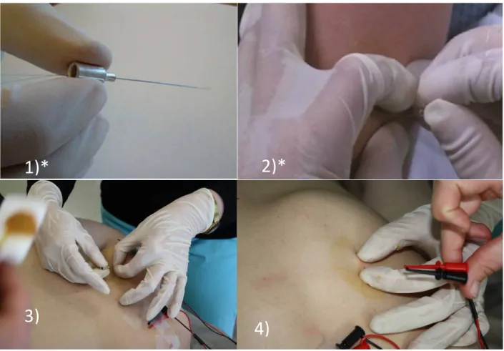
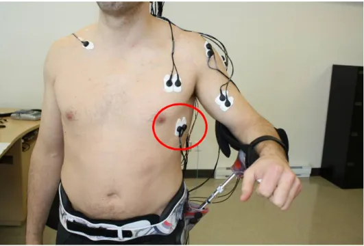
Appendix 2: Fine wire electrode insertion techniques for rotator cuff muscles ... v
Insertion placement for supraspinatus: ... v
Insertion placement for infraspinatus ...vi
Insertion placement for subscapularis ... vii
viii
List of Tables
Table 1: Animal studies concerning the effect of mobilization on tendon healing…….……..28 Table 2: Clinical studies on mobilization post rotator cuff repair…………..………...34 Table 3: Factors affecting rehabilitation protocol………...40 Table 4: Advantages and disadvantages of fine wire electrodes………...53 Table 5: EMG studies related to the first phase of rehabilitation after rotator cuff repair……67 Table 6: Suggested modifications to the early phases of rehabilitation………162
ix
List of Figures
Figure 1: Subacromial space... 9
Figure 2: Histological layers of the cuff ... 12
Figure 3: Rotator Cuff Footprint ... 13
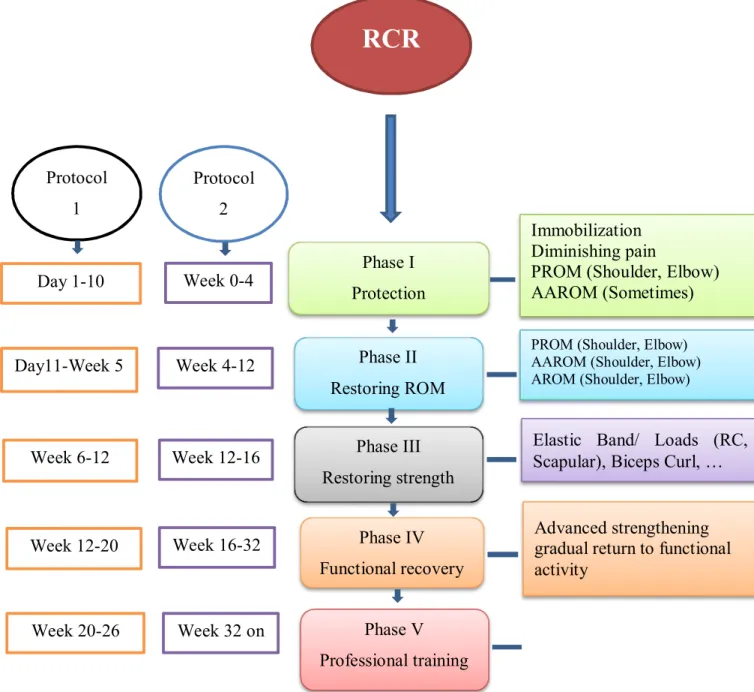
Figure 4: Schematic rehabilitation approaches, comparing two protocols with different timing ... 42
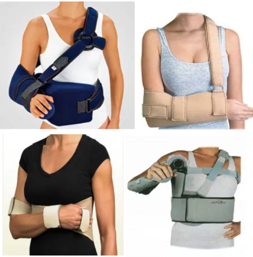
Figure 5 : Common shoulder orthoses ... 45
Figure 6: EMG Signal Cycle ... 50
Figure 7: Motor Unit... 51
Figure 8: Insertion technique for fine wire electrodes ... 53 Figure 9: location of surface electrodes for serratus anterior in respect to the arm position 53 Figure 10: Suggested modifications to the early phases of rehabilitation
x
List of acronyms
AAROM: Active Assisted Range Of Motion ADLs: Activities of Daily Living
ADM: Acellular Dermal Matrix AP: Action Potential
AROM: Active Range of Motion CPM: Continuous Passive Motion CSA: Cross Sectional Area CT: Computed Tomography ECM: Extra Cellular Matrix EMG: ElectroMyoGraphy ER: External Rotation IR: Internal Rotation
MRI: Magnetic Resonance Imaging
MU: Muscle Unit
MUAP: Motor Unit Action Potential
MVIC: Maximal Voluntary Isometric Contraction PROM: Passive Range of Motion
RC: Rotator Cuff
RCT: Randomized Clinical Trial RCR: Rotator Cuff Repair RMS: Root Mean Square US: Ultra Sound
xi
List of abbreviations
d: Day
e.g.: exempli gratia, 'for the sake of example' i.e.: id est, 'that is'
immob: Immobilization
xii
Dedication
To my beloved mother, a wonderful woman full of compassion and spirituality….
I wish her soul rest in peace in the garden of heaven.
“You are the universe in ecstatic motion.”
“You were born with wings. You are not meant for crawling, so don't. You have wings. Learn to use them and fly.”
“Your hand opens and closes, opens and closes. If it were always a fist or always stretched open, you would be paralysed. Your deepest presence is in every small contracting and expanding, the two as beautifully balanced and coordinated as birds' wings.”
Jalaleddin Muhammad Rumi
xiii
Acknowledgement
When I landed in Montreal in the summer of 2010, I was very determined to study kinesiology. As a sports physician, I could deeply understand how basic sciences are affecting our way of thinking and clinical approaches. But honestly, I was hesitating to start biomechanics - a field full of physics and mathematics- with my medical background. I was so lucky to meet Prof. Paul Allard, who is not only a great scientist but also a great human being. He introduced me the world of biomechanics, removed my fears, showed me the beauty of this field and helped me step by step to enter in this path. He is my guru in biomechanics, I would never be in this position without him.
My young and energetic supervisor, Mickael Begon taught me all the details, helped me in all aspects of my study and showed me the rights and wrongs in biomechanics. Without his support, I could hardly fulfil my job. When I start working with indwelling EMG, I could not find any expert in Montreal helping me to learn the techniques of electrode insertion. Thanks Rebecca Brookham in the University of Waterloo who shared her experiences with me. But it was Mickael who bravely let me to practice in his own body, helped me build my skills and encouraged me to teach what I learnt to my lab mates. He established a friendly atmosphere in the lab, everybody helped the others, there was always a solution for your problem, and everybody felt happy and satisfied while working hard. He is a great leader, I can easily see a bright future in front of him.
I should thank Dr. Patrice Tétreault, for all his guidance and recommendations. I learnt so many delicate notes in his shoulder clinic in Notre Dame Hospital and he kindly let me to observe his outstanding skills in the operating room. He improved my understanding of shoulder pathologies and showed me how a great surgeon designs his clinical plan. But above all these, I have been impressed by his humbleness, his passion to his patients and his sense of humanity. I took a lot of positive energy from my enthusiastic colleagues in biomechanics lab. I enjoyed accompanying them in different social activities where I could learn certain French expressions and Québec culture. They assisted me during data collections, and kindly let me to perform medical examination, intramuscular EMG and ultrasonography on their shoulders. I don’t know how to express my gratitude. I specially thank Benjamin Michaud and Fabien Dal
xiv
Maso, who were always beside me when I was getting stuck in mechanical aspects of biomechanics, and Monique Jackson who helped me with her bright critics and ideas as well as her perfect English edition of the articles.
Lastly, I would like to thank my dear husband for all his supports and encouragement. He worked hard to support the family while he was thousand miles far from our children and me. He accepted this suffering to help us live in a free country, enjoy every moment of freedom and catch the opportunities that were offered to us by this great land. Thanks my dear for all your devotions. I should also thank my two beautiful gifts of god, my dear sons Jalal and Emad. They tolerated a busy mom without nagging and were always sweet and caring. I love you guys with all my heart!
1
1 INTRODUCTION
Shoulder is a highly mobile joint with a complex joint structure. This high mobility needs a stable base. Unlike hip or knee joints, the glenohumeral joint does not have a deep socket or complex ligamentous structures that keep its stability. Stability of shoulder joint is mostly a function of muscles around the joint which work in a balanced way to press the humeral head into the glenoid. Rotator cuff (RC) muscles play an important role in stabilizing the glenohumeral joint and controlling humeral head translations. They also contribute in glenohumeral abduction, external rotation and internal rotations. It has been of upmost interest to explore the muscles responsible for shoulder stabilization and motions and identify the positions where they are most active. This type of information provided a basis for shoulder muscle training or rehabilitative movements. Although by knowing muscle’s origin and insertion and its moment arm, the muscle function can be understood, however, a muscle may be in a good anatomic position for a specific movement, but remains inactive when that motion is performed.1 Electromyography (EMG) is a generally recognized tool to evaluate muscle activity. EMG provides information on when, how much and how often a muscle is active and evaluates these items throughout a range of motion. It is also a common tool to evaluate the muscle fatigue, a condition that predisposes individual to injury. EMG studies on glenohumeral and scapular muscle activities during numerous shoulder exercises have been commonly the basis of many shoulder rehabilitation protocols including the physical therapy after rotator cuff repair which is the focus of this dissertation.
Rotator cuff tears are among the most frequently encountered causes of pain and dysfunction in the shoulder.2 Cuff tears are a burden on social and medical resources due to the high number of surgery for rotator cuff repair and long-term rehabilitation pre and post operation. For instance, in 2002, shoulder pain accounted for more than 4.5 million clinical visits in USA that resulted in 40,000 surgical procedures for rotator cuff problems.3 An annual 75,000 rotator cuff repair with a mean time off work of seven months was also reported.4 There are similar reports from different countries confirming that rotator cuff tears remain a relevant health problem.5 Despite the high prevalence of rotator cuff injuries, there are not still clear guidelines for treatments. Most surgeons in the North America regularly prescribe a trial of
non-2
operative treatment such as physical therapy and sub-acromial corticosteroid injection before considering operative repair for chronic types of symptomatic rotator cuff tears.6 The optimal duration for a non-operative treatment trial has not been clearly delineated. Likewise, evidence in relation to the best protocol for physical therapy is limited. Symptoms duration greater than one year and patient functional disability are considered the poor prognosis factors for non-operative treatment and earlier surgical intervention in these conditions are suggested.3 Expectations regarding the surgical outcomes vary. While surgeons are often interested in restoring the full ranges of motion and measured muscle strength, patients' main interests are in the relief of pain and restoration of ability to undertake activities of daily living (ADLs), employment, and recreation. A proper physical therapy program as a non-operative treatment or as a complementary treatment after operation has a tremendous importance for achieving the expected goals. The present thesis will concentrate on the early rehabilitation protocols after rotator cuff surgery.
EMG studies during different tasks and exercises have provided valuable information to be applied to shoulder rehabilitation. For example, McCann et al. (1993)7 quantified the EMG activities of shoulder muscles during the three-phases of shoulder rehabilitation program introduced by Neer (1987)8 which included passive, active and resistive exercises. They showed that EMG findings were consistent with clinical experience and low muscle activities in the early phases of the rehabilitation program have been reported. Generally, in the early phase after rotator cuff tendon repair, the patients are advised to avoid exercises that generate high rotator cuff activity. To protect the healing tendon, patient’s shoulder is immobilized for 4-6 weeks in a sling or a shoulder orthosis and only supervised passive movements are allowed. It has been shown that the activity of cuff muscles was minimal during self-assisted or helper-assisted elevation exercises.9 Limitation in dynamic rehabilitation is due to high rate of re-tear after rotator cuff surgery10-12. It is assumed that higher activity of cuff muscles may stress the healing tissues. However, in this phase of rehabilitation, exercises that activate scapular and other shoulder muscles with minimal cuff activity may be appropriate. For example, an EMG study by Smith et al. (2006)13 suggested that during periods of shoulder immobilization scapular depression and protraction exercises could potentially be safely performed to facilitate scapula-thoracic rehabilitation. Patients usually start active assisted or active movements from the
3
seventh week post operation. In this period, the plane and angle of elevations as well as arm rotations should be wisely chosen to control the stress level on the newly healing tendon. During more advanced phases of rotator cuff rehabilitation, exercises that produce moderate to higher levels of rotator cuff activity may be employed to help strengthening the cuff muscles who might become weakened or atrophied after a period of disuse.14 The studies that are presented in this dissertation are mainly related to the immobilization period and the phase of starting active range of motion exercises.
One of the important areas in post-op rehabilitation that should be addressed is the effect of long term tear on the RC muscles. Following rotator cuff tears and musculotendinous retraction induced by tendon release, serious changes such as fatty infiltration, degeneration and muscle atrophy may happen within the rotator cuff muscles.15,16 Studies on sheep models suggested that fatty infiltration and muscle atrophy progress steadily over the first 16 weeks following tendon detachment.17,15 As a majority of rotator cuff surgeries are performed on chronic tears, most patients already have advanced levels of muscle atrophy. Post-operative immobilization may even further the muscles atrophy.18 Therefore, post-op activity level is an important treatment component that needs to receive more attention for the shoulder. One of the main ideas that is followed in this dissertation is, what if full immobilisation condition changes to a semi-immobilization with a dynamic orthosis. Actually, when we started our studies, Médicus Company in Quebec was interested in developing a dynamic shoulder orthosis. Our findings provides a basis for manufacturing such dynamic orthoses.
Theoretically, the amount and pattern of mechanical loading on tendon tissue is important for tendon development and homeostasis. As natural tendon healing is insufficient, manipulation of the mechanical environment of healing tendon may exert a biologic effect for promoting a repair process that restores normal tendon structure and function.19 Disuse following immobilization has been associated with alterations in tenocyte morphology and loss of normal extracellular matrix (ECM) architecture, resulting in impaired function and healing capacity in animal tendon tissue.20 An ideal exercise program should provide a biologic stimulus to maintain tendon homeostasis and function while avoiding harmful stress. A meta-analysis study on immobilization after rotator cuff repair suggested that there is not enough evidence indicating that immobilization after repair is superior to early-motion rehabilitation in terms of
4
tendon healing or clinical outcome.21 Functional impairment in patients with rotator cuff tears may be due to muscle atrophy and weakness. According to this meta-analysis21, all patients in different studies regained their range of motion 1 year postoperatively, but in early-motion rehabilitation protocol, a significant difference in external rotation at 6 months postoperatively favoured early motion over immobilization. Besides the controversy regarding shoulder full immobilization or early motion, there is an ambiguity regarding the extent of upper limb immobilization, i.e. whether all parts of upper limb including elbow and wrist joints need to be immobilized. Only 4 weeks of elbow immobilization has been shown that significantly decreased the forces of elbow flexors.22 Early restoration of functional ability of the upper limb is the goal of treatment and is sometimes crucial for patients who are manual workers or professional athletes. This issue is addressed in our studies.
The general aim of this thesis is to study EMG activity of rotator cuff and some other shoulder muscles during certain exercises and daily living tasks to suggest different modifications to the commonly used rehabilitation protocols. Ideally the exercises that minimally activate rotator cuff muscles can be considered safe in early phases after rotator cuff repair. So, the EMG of shoulder musculature are evaluated in the following situations:
Active elbow and wrist movements
Some daily living tasks that involve elbow, wrist and fingers movements Some resistance exercises with light weights for elbow and wrist
Arm adduction exercises when active assisted exercises are allowed to be performed.
Active arm elevation in different planes and arcs of elevation
In the first part of chapter 2, the characteristics of rotator cuff tears, and their treatment strategies are elaborated. In part 2.2 the rationales behind rehabilitation protocols and the concept of tendon healing are explained. The readers who are familiar with rotator cuff tears and their management may find this two section a little bit boring but for those who have limited information about this medical problem, reading of section 2.1 and 2.2 is highly recommended. In part 2.3 the basic and clinical studies on upper limb immobilization after rotator cuff repairs
5
are summarized in two synthetic tables to present the current knowledge about the immobilization protocols. In section 2.4, the basis of EMG is explained and the reader can understand how EMG can be used as a tool to identify the safety of different rehabilitation exercises. Considering high number of papers using EMG for evaluation of muscle activity, the strong points and the limitations in using EMG are also elaborated. The methods we used in our biomechanics lab for intramuscular EMG are introduced in details in Appendix 2 to guide future researchers who are interested to work in this field. In chapter 3 the EMG activity of shoulder muscles during different movements are evaluated and certain types of movement are presented in three articles. The first article entitled “Electromyographic activity in the immobilized shoulder musculature during ipsilateral elbow, wrist and finger movements while wearing a shoulder orthosis” assesses the effect of elbow, wrist and hand mobilization on rotator cuff muscles activity. This article suggests the utilisation of a dynamic shoulder orthosis instead of traditional ones in order to increase the functionality of upper limb while imposing a safe level of load on the repaired tendon. The second article entitled “Electromyographic activity in the shoulder musculature during resistance training exercises of the ipsilateral upper limb while wearing a shoulder orthosis” introduces some resistive training exercises for early post repair period that can induce minimum level of activity within rotator cuff muscles. It also suggests another modification to common shoulder orthosis in the wedge part, using foams with different densities. Both articles have been published in the Journal of Shoulder and Elbow Surgery. The third article: “The effects of elevation plane and angle on EMG activity of shoulder musculature in patients with rotator cuff tears” intends to show how elevation plane and angle can change the pattern of muscle activity during the active arm elevation in patients with cuff tears. This type of exercises is usually prescribed for the patients 4-6 weeks post-surgery, in the second phase of rehabilitation. The latter study has been performed on symptomatic patients with rotator cuff tears who were in the waiting list for surgical repair. Considering that the studies on patient population are very few, this study can specifically demonstrate how the position of arm elevation in this patient group affect muscle activity pattern and introduces some suggestions for this type of exercises. The precise kinematic measurement which has been fulfilled by a 3D motion analysis system and the synchronization of kinematic study with EMG analysis are the strong points of this study which has been published by the journal of Clinical Biomechanics. In chapter 4 the specific objectives of this dissertation are elaborated and discussed and finally
6
in the conclusion part, some modifications to the commonly used rehabilitation protocols based on our findings are suggested. It is believed that these findings can provide a scientific basis for designing more functional shoulder orthoses and presenting some new exercises for the early periods after rotator cuff repairs.
7
2 LITERATURE REVIEW
2.1 ROTATOR CUFF TEARS
Rotator cuff tears are a common health problem. Estimates for the prevalence of tears vary widely and might differ according to the age, composition of the community or industry of the area. Lehman, et al. (1994) 23 estimated the prevalence of 6-30% of rotator cuff tears based on their cadaveric study. Ultrasonography studies during a community health check-up for general population in Japan 24 elucidated that 21.7% of 1,328 shoulders had full-thickness rotator cuff tears. This study has been supported by Yamamoto, et al.(2010) 25 who also showed that rotator cuff tear afflicts about 20% of the population regardless of the presence or absence of symptoms. Other Imaging studies reveal that 54% of asymptomatic persons over 60 years of age 26 and 65% of asymptomatic persons over 70 years of age 27 have rotator cuff defects. Massive tears account for 10-40% of all tears.5 Despite different reports of rotator cuff tear prevalence, all studies agree that the prevalence increases with age and its associated morbidity, in terms of pain and loss of function, can be severely debilitating.
Risk factors that may lead to rotator cuff tears are largely unknown. Some studies have identified increased age as the main risk factor and tears are being regarded as a normal consequence of the ageing process.27 History of trauma and the dominant arm were also reported as the main risk factors for cuff tear in general population.25 Increased adiposity28, arterial hypertension29 and smoking habit30 may also contribute to progression of rotator cuff tears. The contribution of genetic factors was suggested by Harvie, et al. (2004) 31 who identified a significantly increased risk of tears in the siblings of patients with symptomatic tears, but this concept has not been supported by further studies. Specific professions such as construction workers, office workers and musicians seem to be at higher risk for shoulder disease due to repetitive uses of the arm and working above shoulder height.32 People participating in overhead sports activities are more prone to shoulder injuries,33 because, the repetitive microtraumatic stresses placed on the athlete’s shoulder joint complex during the throwing motion may challenge the physiologic limits of the surrounding tissues including rotator cuff musculature.34 Finally, as mentioned before trauma is an important risk factor for rotator cuff tears and more than 40% of patients may report it in the history before their shoulder problems occurred.35
8
It can be seen that many factors are contributing in development of rotator cuff tears. Tears may not have a single cause but a single or multiple causal chains that may involve biological, environmental or professional risk factors. It is not clear how these risk factors work together and the magnitude of each factor on development of tear is not clear. By far, age is the most important factor for tear progression. Some of these risk factors are modifiable, some are not. Clinical studies mostly manipulate the modifiable risk factors and provide evidence for clinical approaches. However, they do little to increase our understanding of the nature of rotator cuff muscles. Basic science studies by explaining the nature and special characteristics of rotator cuff muscles and tendon can guide preventive or therapeutic interventions to be optimally timed and progressed. This dissertation presents some basic science studies to provide a basis for rehabilitation protocols in the early post-repair period.
Etiology of rotator cuff tears
With the exception of acute injuries leading to rotator cuff tears, it is generally believed that chronic impingement and tendinopathy can lead to partial tears that progress to full-thickness tears over time.36 Neer (1983)36 hypothesized that the mechanical compression and abrasion of the cuff tendons result from mechanical compression by some structure external to the tendon such as abnormal acromial morphology. These “extrinsic factors” were viewed as the main initiating factors leading to dysfunction of the rotator cuff and eventual tearing. The term “subacromial impingement” has been used to describe irritation from the antero-inferior aspect of the acromion onto the superior aspect of the rotator cuff. The subacromial space is defined by the humeral head inferiorly, the anterior edge and under surface of the anterior third of the acromion, coracoacromial ligament and the acromioclavicular joint superiorly (Figure 1). Theoretically, when the arm is elevated, the humeral head and the acromion approach each other and narrowing the subacromial space.37 Faulty posture, altered scapular or glenohumeral kinematics, posterior capsular tightness, and acromial or coracoacromial arch pathology are among the potential extrinsic mechanics that may lead to impingement. Subacromial decompression or acromioplasty is a popular operative approach attempting to alter presumed aberrant acromial morphology, and therefore eliminate impingement on rotator cuff.
9
Figure 1: Subacromial space
Inferior: humeral head, superior: under surface of the anterior third of the acromion, coracoacromial ligament and the acromioclavicular joint.
Figure is taken from:
http://thelondonshoulderpartnership.co.uk/shoulderinformation/shoulder/shoulder-impingement
However, other evidence suggested that, in most patients who have an abnormality of the rotator cuff, the primary problem is intrinsic. The ‘intrinsic impingement’, theorizes that partial or full thickness tendon tears occur as a result of the degenerative process that occurs over time with overuse, tension overload, or trauma of the tendons.38,39 Subsequently the osteophytes formation, acromial changes, muscle imbalances and weakness, and altered kinematics will follow leading to secondary subacromial impingement. However as the usual clinical examination findings reveal both tendon pathology as well as one or more extrinsic factors such
10
as osteophytes or muscle weakness, it is not clear which form comes first, tendon degeneration or changes external to the tendon? However, the orthopaedic and rehabilitation approaches mainly address the extrinsic factors because tendon degeneration has not been very well understood and so its controlling factors have not been clearly determined. I briefly explain what we know about tendon degeneration. Understanding of this issue is important because all the rationales behind the rehabilitation protocols are related to tendon pathology and healing process.
Basic science researchers tried to explain tendon degeneration. Vascular studies have described the presence of a "critical zone" in the rotator cuff, corresponded to the zone of the anastomoses between the osseous and tendinous vessels 40 or about 1 cm proximal to the insertion of the supraspinatus tendon into the humeral head.41 Cadaveric studies suggested a hypovascularity in the critical zone, and presumed hypoperfusion within this area might result in degeneration and ultimately failure of the tendon. However other vascular studies have produced contradictory findings, and histologic analysis of surgical biopsy specimens of torn rotator cuff tendon showed vascular proliferation in the biopsy specimens.42,39 Analysis by intraoperative laser doppler flowmetry 43 has also shown blood flow throughout the entire rotator cuff including hyperaemic response at the edge of the tear. Despite the controversy regarding the existence of this critical zone, we will see later that critical zone is commonly mentioned in the clinical literature. For example it is suggested that after cuff repair, immobilization in abduction position can provide better blood supply to that critical zone.44
Other basic-science researches have been directed toward cellular studies emphasising the importance of intrinsic factors. Kannus and Józsa (1991)45 evaluated the specimens obtained from the biopsy of spontaneously ruptured tendons in 891 patients and noted degenerative changes in 97% of cases. Likewise, Hashimoto et al. (2003)46 observed a high prevalence and diffuse distribution of degenerative changes in torn rotator cuff tendons without any distinct inflammatory reaction. But it was Matthews et al. (2006)47 who distinct the inflammatory process from the degenerative process in their biopsy samples of supraspinatus tendon in 40 patients with chronic rotator cuff tears. They observed that small sized tears had increased fibroblast cellularity and intimal hyperplasia, together with increased expression of leucocyte and vascular markers, retaining the greatest potential to heal. On the contrary, large and massive
11
tears showed marked oedema and degeneration with no increase in the number of inflammatory cells and blood vessels. These findings could explain why stiffness is more prevalent in small tears where higher activity of inflammatory process exists and why large degenerated tears have less capacity of healing and therefore, are more prone to re-tear.
In conclusion, the exact pathogenesis of rotator cuff tears remains unclear. It is probable that the cause of tendon pathology is multifactorial which involves extrinsic impingement from structure surrounding the cuff and intrinsic degeneration within the tendon. Generally, the degeneration - microtrauma theory is the most accepted theory for explanation of rotator cuff diseases. Age-related degeneration, vascular changes, and inflammation are all potential contributors to the intrinsic pathology of the rotator cuff. The histopathologic changes leading to rotator cuff rupture are gradual and progressive. However the exact pathway of these changes and the order of sequences are not exactly known. All these ambiguities result in difficulty of addressing the main causative factors and designing a proper preventive or therapeutic plan for rotator cuff tears. So, it is important to understand that in this dissertation we will not target the causative factors but some external factors (such as immobilization and exercises) that may affect the tendon healing are discussed.
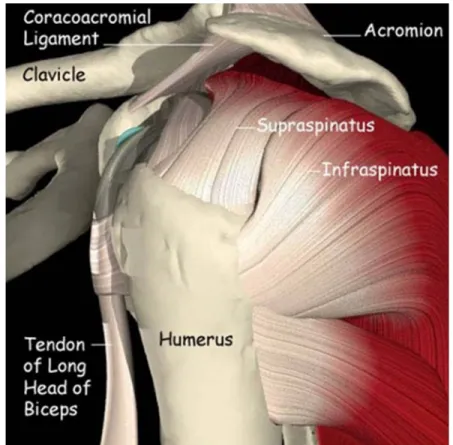
Normal Tendon
Rotator cuff consists of the tendons of the subscapularis, supraspinatus, infraspinatus, and teres minor muscles. The normal tendon of the rotator cuff has the average thickness of 10 to 12 mm.48 It is formed by the confluence of tendon, the joint capsule, the ligaments (coracohumeral and glenohumeral), all of which blend before inserting onto the humeral tuberosities. The supraspinatus and infraspinatus are not really separated and join proximal to their insertion.49 Clark and Harryman (1992)50 performed a histological analysis of the rotator cuff insertion and defined five distinct histological zones in the supraspinatus and infraspinatus portions of the rotator cuff. Figure 2 demonstrates those layers. It should be noticed that the roof of the biceps sheath is formed by fibers from layer II of the supraspinatus. A group of bundles from the subscapularis joins with fibers of the supraspinatus to serve as the floor of the biceps sheath. The rotator cuff, the coracohumeral ligament complex, and the bicipital sheath are intimately interconnected. This anatomical characteristic may help to understand why biceps pathology commonly occurs following evolvement of the tear.
12
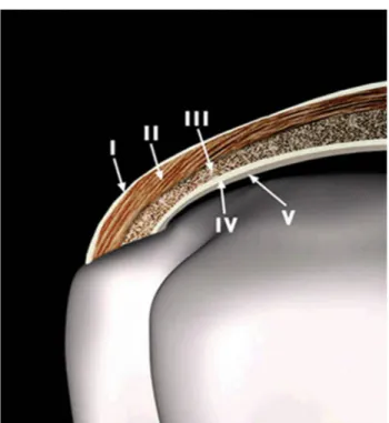
Figure 2: Histological layers of the cuff
Layer 1: 1 mm in thickness, the most superficial layer, contains large arterioles and composed of fibers from the coraco-humeral ligament, Layer II: 3-5 mm in thickness, large bundles densely packed fibers that parallel the long axis of the tendon, representing the direct tendinous insertion into the tuberosities, Layer III: 3 mm thickness, smaller bundles of collagen with a less uniform orientation, loosely organized fibers forming an interdigitating meshwork, Layer IV: loose connective tissue and thick collagen bands that merges with fibers from the coraco-humeral ligament Layer V: 2 mm in thickness, the shoulder capsule, comprises a sheet of interwoven collagen extending from the glenoid labrum to the humerus.
Figure derived with permission from: www.radsource.com
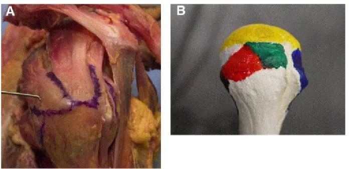
The insertion site of the rotator cuff tendon is often referred to as the footprint. According to the cadaveric study of Curtis et al. (2006),49 each rotator cuff tendon has its own, unique and measurable insertion onto the humerus. (Figure 3).
13
Figure 3: Rotator Cuff Footprint
A: intact myotendinous unit, B: supraspinatus footprint (green), infraspinatus footprint (red), subscapularis footprint (blue) and teres minor footprint (black).
Figure copied from the article written by Curtis et al.49 The permission was taken from Dr. Alan Curtis the corresponding author (in Boston Sports & Shoulder center)
The supraspinatus portion of the cuff inserts and covers the antero-superior aspect of the greater tuberosity adjacent to the articular cartilage of humeral head. Infraspinatus and teres minor are slightly away of the articular margin and create a bare area. Restoration of anatomical footprint for torn tendon is critical for surgical outcomes. Knowledge of anatomical footprint have influenced on surgical techniques aiming to increase the available surface area for repair.51 Surgical repair is more difficult for chronic tears with degeneration and muscle retraction than acute tears. In retracted situation, more tension is required to reattach the torn tendon. To protect the newly repaired tendon, the tension level should be also adjusted post-surgery and proper arm positioning in the immobilization period may help in repair integrity. This issue will be discussed in the next sections and is one of the major topics of the present thesis.
14
Tears in Different Views
Different methods of tear classification are presented in Appendix 1. This section will present some common approaches for tear definition.
2.1.3.1 Partial vs Full Thickness Tear
A partial-thickness tear is considered to be a definite disruption of the fibres of the tendon not fraying or softening of the surface and occur within the tendon without communicating with the subacromial bursa or the glenohumeral joint. The degree of tearing is described more by the depth involved in the thickness of the tendon than by the area of the tear. partial thickness tears have 3 subtypes:48
1) Bursal-sided tear: less common but frequently the most symptomatic.
2) Intratendinous tear: occurs between the superficial and deep layers of the tendon, may present as a cystic collection in the muscle.
3) Articular sided tear: 2-3 times more frequent and more symptomatic than bursal surface tears and frequently occur near the supraspinatus tendon-bone interface. In tears of the bursal surface, subacromial impingement may be responsible. Intratendinous lesions may occur in the presence of differential shear stress between the superficial and deep surface layers of the tendon and articular sided tears are mostly because of trauma to a degenerated tendon.52
With full thickness tears, the entire tendon has separated or torn from the bone. Full thickness tears can initiate on the anterior, posterior or middle portion along the width of the tendon. In almost 90% of cases, the tear is located in the anterior portion.53 A transverse tear exposes the insertion side but a longitudinal tear occurs along the torn tendon fiber. Full thickness tear can be small pinpoint, larger button hole, or involve the majority of the tendon where the tendon still remains substantially attached to the humeral head. Full thickness tears may also involve complete detachment of the tendon(s) from the humeral head and may result in significantly impaired shoulder motion and function.
15
Massive Tear is usually defined as a tear of greater than five centimeters in diameter or in terms of the amount of tendon that has been detached from the tuberosities. There is no universal agreement on the definition of a massive rotator cuff tear, however, some common definitions can be found in Appendix 1. Most massive tears involve supraspinatus and infraspinatus, but anterosuperior tears involving supraspinatus and subscapularis are also moderately common.54
2.1.3.2 Symptomatic vs Asymptomatic
The clinical manifestations of rotator-cuff tears vary widely among patients. It seems that there is no correlation between tears and symptoms as research studies have demonstrated substantial numbers of people with asymptomatic shoulders and full function have partial or full thickness rotator cuff tendon tears.25,55,56 There is considerable uncertainty why the presence of a structural full thickness tear of the rotator cuff may be associated with disabling pain and loss of function in some individuals and be asymptomatic in others. Yamaguchi et al. (2000)57 found that 40% of patients with symptomatic rotator cuff tears had also an asymptomatic tear in the contralateral shoulder. Interestingly the symptomatic tears were on average 30% larger than asymptomatic ones. The mechanism behind the evolution of pain in the setting of rotator cuff pathology is unclear. If we assume that the patient’s pain is related to his cuff tear, then it is expected that a longer duration of symptoms should correlate with a larger tear size, more muscle atrophy, and poorer active motion. However there is evidence suggesting that the severity of pain does not correlate with the severity of rotator cuff disease.58,59 These observations cast doubt on the assumption that rotator cuff tears are the source of a patient’s symptoms and suggest that pain in this patient population may be originating from other sources. The study of Flurin et al. (2007) 60 who assessed cuff integrity after arthroscopic rotator cuff repair somehow confirmed this idea. According to their study, all of the components of the Constant score were dramatically improved by surgery but better functional results in terms of activity, motion, and especially strength were obtained when the cuff remained intact on postoperative imaging studies. Interestingly, the Constant sub score for pain did not correlate with the anatomic results. Likewise, Dunn et al.(2014)59 in their multicenter study on 393 patients could not find any significant relationship between pain and the severity of the cuff disease (e.g., number of tendons torn, degree of retraction, and degree of fatty degeneration). In
16
contrast, increased comorbidities, lower education level, and race were significantly associated with pain on presentation.
Moreover, the exact tear size that causes a loss of normal shoulder biomechanics is unknown. Why some patients with rotator cuff tear have still normal function? Burkhart et al. (1993)61 described the theory of “rotator cable-crescent complex” in an attempt to explain this issue. According to this theory the intact rotator cuff inserts anteriorly on the greater tuberosity of humerus, and posteriorly closes to the inferior border of infraspinatus, so it acts like a “suspension bridge”. At the margin of avascular zone, the arching cable like thickening of the coraco-humeral ligament is located. Stress on the muscles is transferred to this cable and in case of tear, the free margin of tear corresponds to the rotator cable but the anterior and posterior margins correspond to the supports at the each ends of cable span. Therefore even in the case of damage to supraspinatus, the compressive effect of the tendon can be exerted on the humeral head through distributing tensions along the suspension bridge. Ludewig et al. (2009)62 in their 3D kinematic study demonstrated that during all humerothoracic elevation motions, glenohumeral external rotation occurred irrespective of plane. They suggested that as the tears progress into the posterior part of the rotator cuff (infraspinatus) and the patients lose external rotation strength, then the functional lose is more pronounced. These explanations can somehow clear why some patients with rotator cuff tear still have arm elevation. However, harmonizing the study population in respective to tear size or intensity of the symptoms is very challenging and two patients with similar tear characteristics may have different pain or functional scores. This issue will be demonstrated and deliberated in our third article (section 3.3).
Patients with acute, traumatic cuff tears may experience the sudden onset of weakness with elevation of the arm after an injury while symptomatic patients with chronic degenerative cuff defects may notice a gradual onset of shoulder weakness and pain, reduced functional ability including an inability to dress, attend to personal hygiene and use utensils to eat. They may also complain of nocturnal pain that affects their sleep.63,64 However, as mentioned before many degenerative rotator cuff defects are asymptomatic. It has been estimated that more than half of asymptomatic tears become symptomatic in around 3 years.65,66
It is important to consider that all surgical treatment and rehabilitation protocols are planned for symptomatic patients, not for anatomical tear. It is the patient who should be treated
17
not the tear! Therefore, when designing a rehabilitation program, it is important to address the patient’s expectations. Patients usually desire to do their daily living activities and return to a normal life as soon as possible, while clinicians may aim to restore the full range of motion and measurable cuff strength in a gradual process. So in this thesis both of these expectations are considered.
2.1.3.3 Acute vs Chronic
Acute tears are reported to make up 8% of all rotator cuff tears and are usually related to a trauma or shoulder dislocation.67 Traumatic tears are caused by a fall or trauma to an abducted externally rotated arm. They usually occur in individuals, with a mean age younger than the population affected by chronic cuff tears. Traumatic tears tend to be larger in size and also can involve the subscapularis tendon. In fact, in 50% of the cases, they are large or massive tears.65 It should be noticed that the definition of an acute tear is not always easy. Many patients report an acute event that initiated their symptoms, but many of these acute events were potentially a new injury to a shoulder that already had a rotator cuff tear. Determining if the rotator cuff tear is acute may be clinically challenging and usually requires additional investigation tools such as MRI to evaluate fatty degeneration, atrophy, and retraction.67
Chronic tears are the consequences of gradual and progressive histological changes due to extrinsic or intrinsic contributing factors which have been discussed in the previous sections.
2.1.3.4 Uni vs Multiple tendon
By far, supraspinatus is the most tendon involved; In a study with resonance magnetic arthrography, 93 cases of 105 partial thickness rotator cuff tears, and 43 cases of 93 full thickness rotator cuff tears were in supraspinatus.68 It is believed that the tear typically starts from anterior portion of supraspinatus humeral insertion near the long head of biceps and propagate posteriorly.69 However, in an ultrasound study by Kim et al. 2010,70 it was suggested that lesion arose in region 13-17 mm posterior to the long head of biceps tendon, near the junction of supraspinatus and infraspinatus tendons. Infraspinatus tear was reported in the second place with 60.4% of the cases.53 Teres Minor tear is often associated with supraspinatus and infraspinatus tears (SIT tears) following a degenerative process.71 Rupture of the subscapularis tendon is rare and the incidence was reported as 4-8%.11 Mechanism is often an
18
acute avulsion in younger patients with a hyperabduction / external rotation injury. The injury can be an isolated rupture or it may be combined with rupture of the supraspinatus.11 Considering the higher frequency of supraspinatus tear, the activity of the supraspinatus and its rehabilitation is more discussed in this dissertation.
Treatment: Conservative or Operative
Treatment for rotator cuff tendon disease ranges from conservative treatment to surgery. The decision how to treat a rotator cuff tear is based on the symptoms and its duration, examination findings, tear sizes, co-morbidities, the type and duration of previous treatment and available evidence about outcome. Most symptomatic rotator cuff injuries may be treated conservatively by using nonsteroidal anti-inflammatory drugs, corticosteroid injections, acupuncture, physiotherapy, manual therapy or functional rehabilitation. Most surgeons consider surgery after a period of failed conservative treatment. However the type and duration of this conservative treatment vary and the decision to operate is not as straightforward as it might be thought. Two review studies have highlighted this difficulty. Dunn et al. (2005)6
selected a list of orthopaedic surgeons from American Academy of Orthopaedic Surgeons directory and characterized their attitudes concerning medical decision-making about rotator cuff surgery and investigated the associations between these beliefs and reported surgical volumes. They found significant variation in surgical decision-making and a lack of clinical agreement among orthopaedic surgeons about rotator cuff surgery. Oh et al. (2007),3 in their systematic review investigated the influencing factors on decision making for rotator cuff operation. In this extensive study, they evaluated different variables such as demographic characteristics, symptoms duration, tear characteristics, indications for surgery and surgical outcomes. The conclusion of this review was that the exact indications for repair are not clear and further researches are needed to answer the question as when to operate on the rotator cuff. Therefore, the approach to the management of cuff lesions is largely based on physician’s preference and their clinical experience. The general trend is as follows:
2.1.4.1 Acute Complete Tears
Acute rotator cuff tears even if they are massive are often good candidates for repair as long as the tissue compliance is well maintained.69 It is generally accepted that full-thickness
19
rotator cuff tears will not heal spontaneously, consequently, prolonged observation and nonsurgical management allow the detached tendon to retract and resorb72 while the muscle atrophies and fatty degeneration ensues17 which in some individuals can lead to an irreparable tear. In a retrospective series,73 it was reported that tears that were repaired within the first 3 weeks of an acute injury had a greater return of motion (abduction) than those repaired after 3 to 6 weeks and those repaired from 6 to 12 weeks.
2.1.4.2 Partial-Thickness Rotator-Cuff Tendon Defects
Most partial thickness tears are treated conservatively. However, spontaneous healing of partial-thickness tears is unlikely,42,52,74 because the torn ends should contact and a good blood supply is needed. Then what is the rationale behind conservative treatment?
Fukuda et al.(2003)48 presented a modification version to Neer’s staging (see Appendix 1) which is more directly related to treatment options. They proposed that both acute oedema and haemorrhage, and chronic fibrosis and tendinitis belong in ‘modified’ stage I, with a full-thickness tear in a ‘modified’ stage III. They believe that all stage (modified)-I lesions are better to be treated conservatively. For those in stage (modified) II with partial thickness tears, if the signs and symptoms of inflammation are alleviated, and if mechanical defect of the torn tendon compensated by prime movers and intact part of cuff muscles, then a clinical ‘cure’ is achieved. Fortunately the part of the tendon remaining intact prevents retraction and muscle atrophy.69 Patients in whom the symptoms of a partial cuff tear are refractory to the conservative treatment may benefit from surgery. Some believe that the choice of treatment depends on the exact cause of lesion and the treatment should be directed towards a primary diagnosis such as impingement syndrome or instability.75 By this way, treatment of tear itself is considered secondarily.
2.1.4.3 Chronic, Full-Thickness, Degenerative Tendon Defects
While urgent repair for an acute traumatic cuff tear is mostly agreed, for treatment of atraumatic cuff tear, there is not a consensus. Full thickness tears likely progress over time with retraction of the tendon edge, which may lead to an irreparable tear. Healing is potentially hindered because of poor vascularization in certain regions as well as the intra-articular environment, in which synovial fluid may interfere with healing.67 As these tears often are considerably chronic, the physicians usually try a substantial period of non-operative
20
management to improve the mechanical dysfunction such as lack of motion or stability.69 Nonsurgical approaches include pain management, activity modification, and gentle stretching and strengthening exercises for the muscles that remain intact. Although there are not enough randomized clinical trials to assess the benefits of exercise therapy for full thickness degenerative defects, other studies such as case series or case reports have shown improvement in patients’ symptoms with exercise.76,77 However, some clinicians believe that surgical treatment is the only option for symptomatic patients with full-thickness rotator cuff tear.48
Bartolozzi et al. (1994)78 recommended that early surgery to be considered in a full-thickness tear greater than or equal to 1 cm2, symptoms lasting longer than 1 year, and functional
impairment and weakness. However, Unruh et al. (2014)58 in their prospective cohort study on 450 patients with full-thickness rotator cuff tears observed that the duration of symptoms was not related to weakness, limited range of motion, tear size, fatty atrophy, or validated patient-reported outcome measures. They concluded that using the duration of symptoms as a guide to recommend surgical repair of rotator cuff tears in order to reduce pain and improve function might not be the best approach. Masten et al. (2014)64 tried to take some guidelines for the treatment of atraumatic rotator cuff tears from the existing data. They suggested if pain or stiffness is the main symptom, surgery may not be the ideal solution. In case of weakness as the primary problem, the surgeon should determine if a durable repair is achievable; if not, repair may not be in the best interest of the patient. And finally if instability is the issue, something more than a cuff repair such as reverse total shoulder arthroplasty may be needed.
In conclusion, patients are operated in different stages as there is not a consensus on the timing of surgery, and they have usually experienced a period of conservative treatments such as physical therapy or steroid injections. Surgeon’s philosophy may affect the choice of aggressive approach. Post-surgical rehabilitation follows this philosophy and should be individualized based on patient’s situation and surgeon’s concerns. What we are presenting in this dissertation is actually a more dynamic and functional approach to the early rehabilitation after rotator cuff repair.
21
Surgical Options
Although the detailed information regarding the operation procedure is beyond the scope of this thesis, it is important to know how the integrity of repair can be affected by different surgical options. The surgery is often a combination of subacromial decompression and cuff repair which can be performed by open surgery or arthroscopy. It is believed that arthroscopic procedure may have advantages over standard open procedures including less trauma to the shoulder muscles, less pain, decreased morbidity and an earlier return of normal movement.79 In mini-open technique for the cuff repair, after arthroscopic subacromial decompression, an open repair through a smaller approach without detachment of the deltoid will be followed. Clinical outcomes for arthroscopic repair and mini-open surgery are almost similar, however, higher rate of re-rupture for larger tears that repaired arthroscopically was reported.10
For partial tears, different operative treatments such as arthroscopic surgical debridement coupled with arthroscopic sub-acromial decompression,80 arthroscopic conversion to a full-thickness tear with repair 81 or arthroscopic repair without conversion to a full-thickness tear82 have been proposed. Biceps tenodesis in case of rotator cuff repair or biceps tenotomy in case of irreparable massive tear are routinely performed by some surgeons.53
A variety of methods and suture anchors have been developed to re-attach the torn tendon to bone. The arthroscopic repair can be performed by single- or double-row of suture anchors, using conventional methods, transosseous fixation or bridging sutures. All these methods aim to establish a fibrovascular interface between tendon and bone for healing and restoration of fibrocartilaginous tendon insertion. Therefore the goal of repair is to achieve a secure tendon to bone fixation while biological healing occurs. Tendon footprint contact area and contact pressure are critical factors that contribute to biological healing at the tendon–bone interface, and subsequently, long-term repair strength.51 It was suggested that a larger area of contact between the tendon and bone may improve the biological healing process by increasing the size of the newly formed insertion site.83 All the advancements in arthroscopic surgery and improvement of suture materials, anchors and technics have been directed to enhance the strength of repair. The ideal repair should provide high initial fixation strength, minimal gap formation, and high mechanical stability until completion of the tendon-to-bone healing process.84 It has been shown that double-row repair gives better resistance to gap formation.85
22
Furthermore, smaller re-tear rate has been reported after double row technique than single row, although the clinical outcomes were similar.86 According to a recent systematic review,87 double raw repair provides superior structural healing to single-row repair. However, Apreleva et al. (2002)83 suggested that trans-osseous simple suture fixation might provide greater potential for osseous incorporation and healing at the tendon-bone interface by increasing the repair-site area.
Park et al. (2007)88 have also suggested that the mean pressurized contact area between the tendon and tuberosity insertion footprint is superior using the suture-bridge technique to that of double-row technique. Anyway, optimal method of reconstruction is highly controversial and decision-making is therefore influenced by personal preferences, past experiences, surgical volumes and perceived results. But, it should be emphasized that the type of surgical approach can affect the post-op rehabilitation program. Optimizing the biomechanical properties of the repaired tendon allows for early postoperative rehabilitation while maintaining repair integrity. Therefore, along with improvement in surgical techniques and mechanical stability, the post-surgical treatments may progress from a highly conservative rehabilitation to more dynamic and functional rehabilitation. This issue will be discussed again in this dissertation.
Major Complications after Rotator Cuff Repair
2.1.6.1 Re-Tear
The prevalence of recurrent tear varies between 16% in young subjects with non-retraced tears60 to 94% in massive cuff tears.12Bishop et al. (2006)10 assessed postoperative cuff integrity by MRI and reported that 31% of repairs in the open surgery group and 47% in the arthroscopic group were not intact. Some authors reported a significant correlation between re-rupture and poorer outcomes. For example, Harryman et al. (1991)89 in their evaluation of 105 tendon repairs noticed that the shoulders in which the repaired cuff was intact had better function during activities of daily living and a better range of active motion and strength. In their study, more than half of the repairs of a tear involving more than the supraspinatus tendon had a recurrent defect. However, improvement with respect to pain relief, range of motion and the ability to perform activities of daily living have been reported despite recurrent defects in the repaired tendon.12 Despite all the debate over the relationship of repair integrity and pain or functional outcomes, the trend is toward a better outcome with intact repair.90-92