HAL Id: tel-02887084
https://tel.archives-ouvertes.fr/tel-02887084
Submitted on 2 Jul 2020HAL is a multi-disciplinary open access archive for the deposit and dissemination of sci-entific research documents, whether they are pub-lished or not. The documents may come from teaching and research institutions in France or abroad, or from public or private research centers.
L’archive ouverte pluridisciplinaire HAL, est destinée au dépôt et à la diffusion de documents scientifiques de niveau recherche, publiés ou non, émanant des établissements d’enseignement et de recherche français ou étrangers, des laboratoires publics ou privés.
Biochemical and cellular characterization of the
interplay between glutamine metabolism, mTOR and
Notch1 signaling in cancer therapy
Tra Ly Nguyen
To cite this version:
Tra Ly Nguyen. Biochemical and cellular characterization of the interplay between glutamine metabolism, mTOR and Notch1 signaling in cancer therapy. Human health and pathology. Uni-versité de Bordeaux, 2018. English. �NNT : 2018BORD0053�. �tel-02887084�
THÈSE PRÉSENTÉE POUR OBTENIR LE GRADE DE
DOCTEUR DE
L’UNIVERSITÉ DE BORDEAUX
École Doctorale Sciences de la Vie et de la Santé Spécialité Biochimie
Par Tra Ly NGUYEN
Biochemical and cellular characterization of the interplay between
glutamine metabolism, mTOR and Notch1 signaling
in cancer therapy
Caractérisation biochimique et cellulaire de l’interaction entre le
métabolisme de la glutamine et la signalisation de mTOR et Notch1
comme thérapie contre le cancer
Sous la direction de : Dr. Raúl V. DURÁN-DÍAZ Soutenue le 24 avril 2018Membres du jury :
M. MERLIO Jean-Philippe PU à l’Université de Bordeaux Président M. SEILIEZ Iban DR à l’INRA à St Pée sur Nivelle Rapporteur Mme VASSEUR Sophie DR INSERM, CRCM Marseille Rapporteur M. VELASCO Guillermo PU à l’Université de Madrid Examinateur Mme MOREAU Violaine DR INSERM, Université de Bordeaux Examinateur M. MARAVER Antonio CR INSERM, IRCM Montpellier Examinateur M. DURÁN-DÍAZ Raúl CR INSERM, IECB Bordeaux Directeur de thèse
1
To the jury members, Pr. Jean-Philippe Merlio, Pr. Guillermo Velasco, Dr. Iban Seiliez, Dr. Sophie Vasseur, Dr. Violaine Moreau and Dr. Antonio Maraver.
Your participation in this jury is a great honor for me. I sincerely thank you for agreeing to judge my thesis work, despite the time and effort it requires. Your critical analysis will no doubt help me to improve the scientific quality of my work.
To my thesis supervisor, Dr. Raúl V. Durán
I would like to express my sincere gratitude to my supervisor. I would not have succeeded in completing my PhD without your continued guidance, your advice and your continuous support during these three and a half years. I have learned so much from you, particularly from all of our Friday meetings. Thank you for always listening to me and giving me the freedom to follow my own scientific curiosity. Your guidance was very important during the research and the preparation of this thesis. I could not imagine having a better supervisor and mentor for my PhD study.
To the members of the lab,
Thank you Clément Bodineau, my favorite labmate, for all of your help, support and positive thoughts. I really enjoy working with you, discussing science and sharing life stories. You always encourage me and correct my “perfect” French level. I will be there when your turn comes around!
Thank you Dr. Silvia Terés and Dr. Victor Villar, for the warm welcome when I first came to the lab. I took my first steps in a research lab with both of you. Thank you for all the advice during some difficult moments.
Thank you Dr. Sarah Courtois, for sharing these 3 years of PhD together and for your advice during the thesis preparation.
Thank you Dr. Marie-Julie Nokin for taking care of my projects. It was a great honor to have your solid support at the end of the thesis.
Thank you Oriane Galmar, my intern student and now PhD student. You helped me greatly during your internship and thanks for your encouragement during the thesis preparation.
Thank you Malaurie Eugénie for your kindness and your care. To the members of Royou’s team,
I would like to express my great appreciation for all the members of Royou’s team, especially Dr. Damien Goutte-Gattat, Lou Bouit, Priscillia Pierre-Elies, Dr. Cedric Landmann and Dr. James Jenkins Wallace. I had a beautiful PhD thanks to all of you. You welcomed me into your lab as if I was a member of it. You always said yes when I needed you without hesitation. Special thanks to Dr. Damien Goutte-Gattat for your advice and your technical help and to Lou Bouit for your kindness and your care. To other colleagues, I would extend my thanks:
To Mariline Yot for your support and your ability to listen to me. You are always kind with me, trying to make me feel better when difficult moments arose.
To Dr. Benjamin Drogat for your advice in critical moments and for your help during the thesis preparation. Especially for the jokes, that I don’t really understand most of the time!
To Dr. David Santamaría and Sonia San José for your scientific advice and your encouragement all the time.
To Catherine Duprat and Myriam Mederic for always being available and helping me to have a good lab life.
2
Thanks to all the collaborators who have contributed to these works. Dr. Mercedes Tomé, Dr. Benoît Rousseau, Julien Izotte, Dr. Jean-Max Pasquet, Dr. Marion Bouchecareilht, Dr. Muriel Priault, Dr. Elodie Richard and Vincent Pitard.
To Stéphanie Durrieu for sharing your knowledge and supporting me.
To Isabelle Frémaux and Dr. Florian Alonso for your support, being there when I most needed.
Thank to Dr. Nassima Meriem Gueddouda, Dr. Camila Parrot, Dr. Amy Diallo, Dr. Clémence Rabin, Dr. Vanessa Delcroix for sharing your own PhD experiences, helping me with my transition into a better PhD life!
To my friends and my family,
Thank you to, my parents, my parents-in-law and my host family for your care and your encouragement. I know it was hard for you to understand what I was doing but you still keep supporting me during this very long process.
Thank you, chị Mai for sharing these three years of PhD. We have been together through the most difficult times.
Thank you Hằng, anh Nam, chị Hương, chị Hà, anh Giang, chị Hải Anh, anh Vinh for your constant support.
Special thanks to my husband. You asked to be thanked at the beginning of the acknowledgements, but we always save the best till last (but not least!). A simple thank you is not enough. You have been here next to me for more than three years, days and nights, weeks and week-ends. Thank you for supporting me through ups and downs, for listening to all the crazy stories about my lab life. I always say, I could not have made this thesis happens without you. I cannot wait to enjoy the next step with you!
I had a beautiful PhD thank to all of you! You are all my best company for this beautiful life experience.
3
Titre : Caractérisation biochimique et cellulaire de l’interaction entre le métabolisme de la glutamine et la signalisation de mTOR et Notch1 comme thérapie contre le cancer
Résumé :
La tumorigenèse est un processus multi-étapes, constituée d'altérations génétiques qui conduisent à la transformation maligne des cellules humaines normales. Au cours de cette transformation maligne, l’activité de différentes voies oncogéniques est augmentée. Les voies de signalisation mTORC1 et Notch1 sont des voies oncogéniques bien connues qui jouent un rôle central dans la régulation de la croissance et du métabolisme cellulaires. Les traitements anti-mTORC1 et Notch1 sont approuvées en tant que thérapies anticancéreuses pour plusieurs types de tumeurs. Néanmoins, les cellules cancéreuses développent des résistances à ces inhibiteurs induisant un nombre important de rechute et donc d’échec de ces traitements. Ainsi, le but principal de ce travail est d'étudier l'inhibition des voies de signalisation mTORC1 et Notch1 dans les cellules cancéreuses afin de concevoir de nouvelles stratégies thérapeutiques anticancéreuses. En premier lieu, nous avons décrit une nouvelle classe d'inhibiteurs de mTORC1 qui présente une cytotoxicité spécifique vis-à-vis des cellules cancéreuses. Nous avons démontré que l’ICSN3250, un analogue de l'halituline marine cytotoxique, inhibe mTORC1 et induit la mort cellulaire. Le mécanisme moléculaire de cette inhibition est basé sur le déplacement de l'acide phosphatidique, un lipide activateur du complexe mTORC1, du domaine FRB de la protéine mTOR. Dans un deuxième temps, nous avons étudié le lien entre le métabolisme de la glutamine et la signalisation de Notch1 dans la leucémie lymphoblastique aiguë à lymphocytes T (T-ALL). Les changements métaboliques dans les cellules cancéreuses sont nécessaires à une prolifération cellulaire rapide et la croissance tumorale. Nous avons généré une lignée de cellule T-ALL dont la voie de signalisation Notch1 est constitutivement active et analysé les conséquences de cette activation sur le métabolisme de la glutamine. En effet, en absence de glutamine, l’activation de Notch1 induit la mort cellulaire par apoptose en perturbant l'accumulation de la glutamine synthétase, une enzyme qui permet la production de glutamine. Ce travail de thèse a donc permis de décrire de nouvelles stratégies pour cibler les voies mTORC1 et Notch1 dans le cancer. De futures investigations seront nécessaires pour étudier leur efficacité dans les thérapies anti-cancéreuses.
4
Title: Biochemical and cellular characterization of the interplay between glutamine metabolism, mTOR and Notch1 signaling in cancer therapy
Abstract:
Tumorigenesis is a multistep process, consisting of genetic alterations that drive the malignant transformation of normal human cells. During this transformation, different oncogenic pathways are upregulated. mTORC1 and Notch1 signaling are well-known oncogenic pathways which play a central role in the regulation of cell growth and metabolism. Anti-mTORC1 and Notch1 therapies are approved as cancer treatments for several types of tumor but there are still developed resistances and relapse diseases. Thus, the main aim of this work is to study the inhibition of mTORC1 and Notch1 signaling pathway in cancer cells in order to design new therapeutic anti-cancer strategies. In the first place, we reported new class of mTORC1 inhibitors which has cytotoxicity specifically towards cancer cells. We demonstrated that ICSN3250, an analogue of the cytotoxic marine alkaloid halitulin, inhibited mTORC1 and induced cell death. The molecular mechanism of this inhibition is based on the displacement of the lipid phosphatidic acid, an activator of mTORC1 complex, from the FRB domain of mTOR protein. At the second stage, we have studied the connection between glutamine metabolism and Notch1 signaling in T-cell acute lymphoblastic leukemia (T-ALL). Metabolic changes in cancer cells are advantageous for rapid cell proliferation and tumor growth. We have generated Notch1-driven T-ALL cells and analyzed the consequences of Notch1 activation on glutamine metabolism. Indeed, under glutamine withdrawal, Notch1 upregulation induced apoptotic cell death by disrupting the accumulation of glutamine synthetase, a glutamine producing-enzyme. Overall, this thesis work allowed to describe new strategies to target mTORC1 and Notch1 pathways in cancer, which need future investigations to study their efficacy in therapies.
Keywords
: mTOR, Notch1, glutamine, metabolism, cancer, therapy.Unité de recherche
Résumé en français
mTOR est une sérine/thréonine kinase très conservée qui intègre plusieurs stimuli pour réguler la croissance et le métabolisme cellulaire. mTOR forme deux complexes fonctionnellement et structurellement distincts, appelés mTORC1 et mTORC2. mTORC1 est principalement activé par la présence d'acides aminés, par des facteurs de croissance, par l'état bioénergétique de la cellule, et par la disponibilité de l'oxygène. Au niveau de ses fonctions, mTORC1 régule la synthèse des protéines, la biogenèse des ribosomes, l'absorption de nutriments et l’autophagie en réponse à des facteurs de croissance, des acides aminés, et de l'énergie cellulaire (Duran & Hall, 2012). Dans le contrôle de mTORC1 par des facteurs de croissance, le complexe de la sclérose tubéreuse (TSC) et le co-activateur de mTORC1, Rheb jouent un rôle crucial. L'un des mécanismes par lesquels la voie TSC / Rheb contrôle mTORC1 implique la production d'acide phosphatidique (PA), qui se lie directement à mTOR dans le domaine FRB et active mTORC1 en aval de TSC / Rheb. En effet, la régulation négative de la production de PA est suffisante pour diminuer l'activité de mTORC1. En raison de son rôle central dans le contrôle de la croissance cellulaire et le métabolisme, mTORC1 est activé dans de nombreux types de tumeurs pour soutenir la croissance de la tumeur. Cette régulation positive de mTORC1 constitue une étape cruciale pendant la dérégulation de la signalisation cellulaire lors de la transformation maligne.
L'objectif principal de la première partie de cette thèse est l'étude de l'effet de l'inhibition de mTORC1 par une nouvelle classe d'inhibiteurs qui cible spécifiquement les cellules cancéreuses. En raison des résultats modestes de inhibiteurs de mTORC1 pour la stratégie anti-cancer, le développement de nouveaux traitements est sous
enquêtes. Dans ce projet, nous avons étudié l'inhibition de mTORC1 par un composé synthétisé, ICSN3250, un analogue de l'alcaloïde marin cytotoxique l'halituline. Particulièrement, seules les cellules cancéreuses sont sensibles à ce composé, tandis que les cellules non cancéreuses ont montré jusqu'à 100 fois moins de sensibilité à ICSN3250, contrairement à d'autres inhibiteurs qui n'ont pas montré de sélectivité. Le mécanisme moléculaire de cette inhibition est basé sur le déplacement de PA, l’activateur de mTORC1, du domaine FRB de mTOR. En outre, ICSN3250 est capable d'affecter la capacité de PA à surmonter la régulation négative TSC2 sur mTORC1, qui est la nouveauté de notre travail dans la conception de ce nouvel inhibiteur mTOR. Ce travail a été soumis à Cancer Research en Janvier 2018 et il est en deuxième révision.
L'objectif principal de la deuxième partie de cette thèse est de fournir une compréhension fondamentale de l’interaction mécanistique entre la transformation métabolique et la dérégulation de la signalisation cellulaire pendant l’origine et la progression de la leucémie. Nous avons étudié les changements métaboliques dans des modèles cellulaires de leucémie lymphoblastique aigüe à cellules T (T-ALL) générés par l’activation de la voie Notch1 (modèles cellulaires NDALL), et la contribution de ces changements métaboliques dans la progression du cancer. Une attention particulière sera prêtée au rôle potentiel du métabolisme de la glutamine dans NDALL, et à l'interaction entre le métabolisme de la glutamine et la voie de signalisation mTORC1. La voie oncogénique la plus importante pour la transformation des cellules T est l’activation de la signalisation par la voie Notch1. Il a été constaté que des mutations conduisant à l'activation de la voie Notch1 sont présentes dans plus de 50% des patients atteints de T-ALL, ce qui souligne l'implication directe de Notch1
dans la prolifération et la survie des cellules de leucémie. Malgré le rôle oncogénique principal de la signalisation Notch dans T-ALL, l'inhibition de la signalisation Notch en utilisant des inhibiteurs de γ-sécrétase (GSI) a montré une activité anti-leucémique très modeste contre des lignées cellulaires humaines de T-ALL, exerçant principalement un effet cytostatique avec peu ou pas d'apoptose. Aussi, les premiers essais cliniques ont été limités par des effets de toxicité excessive sur l'épithélium intestinal des patients. La résistance au traitement avec les GSI peut être provoquée par la perte mutationnelle de PTEN, conduisant à l'activation constitutive de la voie PI3K/AKT/mTOR (Tzoneva & Ferrando, 2012). En fait, plusieurs indices connectent la signalisation Notch avec l'activation de la voie mTOR dans T-ALL. Curieusement, le traitement avec GSI supprime la phosphorylation de multiples protéines impliquées dans la signalisation de la voie de mTORC1, ce qui suggère un rôle mécanistique de la signalisation Notch dans l'activation de mTORC1. Notamment, le blocage simultané de la voie Notch et de la voie mTORC1 a un effet synergique dans la suppression de la croissance de NDALL. (Chan et al., 2007). Ainsi, cette inhibition simultanée a attiré notre attention comme une stratégie de co-traitement potentiel contre NDALL. Cependant, la connexion mécanistique entre les deux voies n'est pas claire.
Aujourd'hui, notre compréhension de la reprogrammation métabolique dans les leucémies de type T-ALL est très limitée, mais des études récentes ont montré des changements dans le métabolisme des acides aminés, notamment la glutamine, dans les lignées cellulaires NDALL (Basak et al., 2014). Il faut noter que parmi les changements métaboliques qui se produisent au cours de l'origine et la progression du cancer, la dépendance des cellules cancéreuses à la glutamine constitue une adaptation importante pour maintenir la demande d'énergie qui soutient la croissance et la prolifération rapide (Souba 1993). La glutamine est métabolisée par un processus
dénomé glutaminolyse, c’est-à-dire, la déamination de la glutamine pour synthétiser de l’alpha-cétoglutarate, un processus catalysé par la glutaminase et par la glutamate déshydrogénase. La glutaminase est régulée au niveau de l'expression par l'oncogène c-MYC (Gao et al., 2009), et son activité est en corrélation avec la croissance de nombreuses tumeurs (Perez-Gomez et al., 2005). Récemment nous avons démontré que l'augmentation de la glutaminolyse dans les cellules tumorales provoque également la dérégulation de la signalisation cellulaire par l’activation de la voie mTORC1 (Duran et al., 2012; Duran et al., 2013). Ainsi, la glutamine en combinaison avec la leucine active mTORC1 en augmentant la glutaminolyse et la production d’alpha-cétoglutarate. L’augmentation de la glutaminolyse stimule mTORC1, la croissance cellulaire et l’autophagie, deux processus contrôlés par mTORC1. Par contre, l'inhibition de la glutaminolyse prévient l'accumulation d'alpha-cétoglutarate et l'activation subséquente de mTORC1. Le rôle du métabolisme de la glutamine dans la dérégulation de mTORC1 dans NDALL n’est pas connu. Comme indiqué plus haut, l'implication de c-MYC (un activateur connu de la glutaminolyse) dans l’interaction entre les voies mTORC1 et Notch1 ouvre la possibilité que la glutaminolyse pourrait servir de médiateur pour l'activation de mTORC1 dans NDALL. Par conséquent, l'objectif de cette proposition est de déterminer les changements dans le métabolisme de la glutamine dans des modèles cellulaires de NDALL, établissant un rôle mécanistique potentiel de c-MYC et de l'activation de la glutaminolyse dans l’activation de mTORC1 sur ces modèles de leucémie médiés par Notch1. En outre, en suivant une approche métabolomique, nous allons étudier d'autres changements métaboliques qui se produisent lors de l'activation de Notch dans les cellules T-ALL qui pourraient être pertinents pour la progression de la tumeur. Enfin, nous allons estimer la dépendance des modèles cellulaires de NDALL sur le métabolisme de la
glutamine et sur l’activation de mTORC1, pour proposer et valider des co-traitements potentiels ciblant ces deux éléments comme une stratégie thérapeutique contre la leucémie médiée par Notch1. Pour tester si le métabolisme de la glutamine joue un rôle mécanistique dans la dérégulation de la signalisation cellulaire grâce à l'activation de mTORC1 dans NDALL, nous allons développer des modèles cellulaires de T-ALL dans lesquelles la signalisation Notch1 est suractivée. En suivant une approche multidisciplinaire impliquant la biochimie, la biologie cellulaire, la métabolomique, la bioénergétique, l’enzymologie, et la biologie translationnelle, nous allons tester si l’activation de Notch1 dans ces modèles augmente le métabolisme de la glutamine et, par conséquence, la signalisation mTORC1. Un lien mécanistique entre Notch, glutaminolyse et mTORC1 suggère que les traitements ciblant à la fois la glutaminolyse et l'activation de mTORC1 pourraient avoir un effet synergique contre les leucémies lymphoblastiques dans lesquelles la voie Notch1 est activée.
Durant cette thèse, en utilisant des approches in vitro et in vivo, nous avons montré que les cellules leucémiques médiées par Notch1 sont dépendantes au niveau de glutamine extracellulaire et ils subissent une mort cellulaire par apoptose lors du sevrage de la glutamine, qui est appelée une "dépendance à la glutamine". De plus, la surexpression de Notch1 dans les cellules leucémiques Notch1-négatives est suffisante pour induire une dépendance à la glutamine. Mécaniquement, Notch1 est capable de réguler les enzymes métaboliques du métabolisme de la glutamine, entraînant une augmentation du catabolisme de la glutamine et une diminution de l'anabolisme de la glutamine. En conséquence, cibler le métabolisme de la glutamine pourrait être considéré comme une stratégie thérapeutique contre la leucémie avec Notch1 élevée.
Dans l'ensemble, cette étude a montré deux exemples clairs ciblant le lien entre le métabolisme de la cellule et la signalisation cellulaire pour éliminer spécifiquement les cellules cancéreuses. La transduction du signal reprogramme le métabolisme cellulaire afin de remplir les besoins anaboliques et énergétiques des tumeurs, favorisant la croissance et la prolifération cellulaire. Cependant, la relation entre la signalisation cellulaire et le métabolisme n'est pas unidirectionnelle. En détectant les niveaux de métabolites intracellulaires qui affectent l'état des principales voies métaboliques, les cellules peuvent exercer un contrôle par rétroaction sur leurs réseaux de signalisation. Ces mécanismes permettent aux cellules de croître et de proliférer en accord avec leurs états métaboliques et en fonction de la disponibilité de l'environnement extracellulaire. Comprendre le mécanisme moléculaire de cette connexion aidera à comprendre comment la viabilité des cellules cancéreuses sont déterminées en réponse aux variations du niveau de nutriments environnementaux. Le traitement à base de la restriction nutritionnelle, comme ICSN3250, déplétion en glutamine ou L-asparaginase, sera développé pour cibler la connexion entre l’état métabolique et la signalisation cellulaire dans le cancer.
5
Table of contents
Table index ... 9 Figure Index ... 11 Abbreviations... 13 INTRODUCTION ... 15 1. mTORC1 signaling ... 17 1.1 mTOR discovery ... 17 1.2 Functions of mTORC1 ... 191.2.1 Building blocks for cell growth ... 19
1.2.2 Control of anabolic metabolism ... 24
1.2.3 Regulation of protein turnover ... 26
1.3 Upstream regulation of mTORC1 ... 30
1.3.1 Amino acids ... 30
1.3.2 Growth factors ... 34
1.3.3 Energy, oxygen, stress and DNA damage ... 36
1.3.4 Lipid sensing ... 37
1.4 mTORC1 inhibitors in anti-cancer therapies ... 39
1.4.1 Mechanism of rapamycin-mediated mTORC1 inhibition ... 40
1.4.2 Different classes of mTOR inhibitors ... 41
2. Glutamine metabolism ... 44
2.1 Metabolic transformation in cancer cells ... 44
2.1.1 Uptake ... 44
2.1.2 Metabolic intermediates for biosynthesis ... 46
6
2.2.1 Carbon donor ... 50
2.2.2 Nitrogen donor ... 51
2.2.3 Redox homeostasis control ... 53
2.2.4 Chromatin organization ... 54
2.3 Glutamine addiction in cancer ... 55
2.4 Therapeutic applications ... 58
2.5 Glutamine metabolism and mTORC1 pathway ... 60
3. Notch1 signaling ... 64
3.1 T-cell acute lymphoblastic leukaemia ... 64
3.1.1 Hematopoiesis and T-cell development ... 64
3.1.2 Chemotherapy resistance and relapse ... 66
3.1.3 Genetic alteration mechanisms ... 67
3.2 Notch1 signaling in T-ALL ... 69
3.2.1 Basic mechanisms of Notch signaling activation ... 71
3.2.2 Modulation of Notch1 signal transduction ... 72
3.2.3 Genes and pathways controlled by Notch1 ... 80
3.2.4 The interplay between glutamine metabolism, Notch1 and mTORC1 .... 83
3.3 Notch1-driven T-ALL therapies ... 87
3.3.1 Inhibition of Notch1 using g-secretase inhibitors ... 87
3.3.2 Inhibition of Notch1 by therapeutic antibodies ... 89
3.3.3 Blocking Peptides ... 89
3.3.4 Therapeutic targeting of downstream signaling components of the Notch pathway ... 90
Objectives of the thesis ... 93
7
Chapter one: A novel mechanism of mTOR inhibition displacing phosphatidic
acid induces enhanced cytotoxicity specifically in cancer cells. ... 97
Chapter two: Glutamine addiction induced by Notch1 activation in T-cell acute lymphoblastic leukemia. ... 99
SUMMARY & PERSPECTIVES ... 102
REFERENCES ... 108
9
Table index
Table 1. Different strategies to target glutamine metabolism in cancer. ... 59 Table 2. Synergistic effect of the combination between glutamine metabolism inhibition with different pharmacological treatments. ... 60 Table 3. Mutation frequencies in adult and pediatric T-ALLs. ... 68 Table 4. Core Components and Modifiers of the Notch Pathway. ... 73
11
Figure Index
Figure 1. The core component of mTORC1 and mTORC2 ... 18
Figure 2. Model of cap-dependent translation by mTORC1/4EBP1 and mTORC1/S6K1 axis. ... 20
Figure 3. Lipid biogenesis and lipid catabolism regulation by mTORC1. ... 22
Figure 4. Anabolic processes controlled by mTORC1. ... 24
Figure 5. Regulation of autophagy by mTORC1. ... 28
Figure 6. Summary of main upstream regulators of mTORC1 in response to activating stimuli. ... 30
Figure 7. Regulation of TORC1 by amino acids in yeast (left) ... 31
Figure 8. Rheb mediates mTORC1 activation in response to PI3K/AKT-dependent growth factor signals. ... 35
Figure 9. Phosphatidic acid-mediated mTORC1 activation. ... 39
Figure 10. Different classes of mTOR inhibitors. ... 40
Figure 11. Metabolic transformation of cancer cells. ... 47
Figure 12. Different uses of glutamine in cancer cells. ... 50
Figure 13. Glutamine as nitrogen donor for nucleotide synthesis. ... 52
Figure 14. The role of glutamine in non-essential amino acids synthesis. ... 53
Figure 15. The key role of glutamine in glutathione biosynthesis. ... 54
Figure 16. The role of glutamine-derived a-ketoglutarate in the regulation of chromatin organization. ... 55
Figure 17. Glutamine addiction in cancer cells. ... 57
Figure 18. Molecular mechanism of glutamoptosis. ... 62
Figure 19. Different progenitor cell lineages from hematopoietic stem cells during hematopoiesis. ... 65
12
Figure 20. Stages of T-cell development and T-cell-leukaemia-related oncogenes. . 66
Figure 21. Structure of Notch receptor (A), ligands and coligands (B) ... 70
Figure 22. Activation of the Notch Signaling Pathway mediated by posttranscriptional modifications and regulated proteolysis. ... 72
Figure 23. Models of ligand endocytosis in Notch signaling activation. ... 74
Figure 24. Overview of the endocytosis and trafficking of Notch receptor. ... 75
Figure 25. Glycosylation of the extracellular domain of Notch. ... 76
Figure 26. Different intrinsic mutations of Notch proteins. ... 79
Figure 27. Downstream signaling pathways of Notch1 in T-ALL. ... 80
Figure 28. The crosstalk between Notch1 and PI3K/AKT signaling pathways. ... 85
13
Abbreviations
4EBP1: eIF4E binding protein 1 ACC: acetyl-CoA carboxylase ACLY: ATP-citrate lyase AML: acute myeloid leukemia
AMPK: AMP-activated protein kinase ANK: ankyrin
APC/C: anaphase-promoting complex ARF: ADP-ribosylation factor
ATF4: activating transcription factor 4 ATG: autophagy-related protein B-ALL: B-cell acute lymphoblastic leukemia
BM: bone marrow
CAD: carbamoyl-phosphate synthetase 2, aspartate transcarbamylase,
dihydroorotase
CDK: cyclin-dependent kinase CLP: common lymphoid progenitor CMP: common myeloid progenitor CREB2: cAMP-responsive element-binding 2
CRTC2: CREB regulated transcription coactivator 2
DAP1: death-associated protein 1 DEPDC5: DEP domain-containing protein 5
DEPTOR: domain-containing mTOR-interacting protein
DG kinase: diacylglycerol kinase DLL: delta-like ligand
DNA: deoxyribonucleic acid DSL: Delta-Serrate-Lag2 EGF: epidermal growth factor eIF: eukaryotic translation initiation factor
ERK: extracellular signal-regulated kinase
ETP: early T-cell progenitor EV: empty vector
18F-FDG: 18F-fluorodeoxyglucose FASN: fatty acid synthase
FBXW7: F-box and WD repeat domain containing 7
FKBP12: FK506-binding protein of 12 kDa
FLCN: folliculin
FNIP1/2: folliculin-interacting proteins 1 and 2
FOXO: Forkhead-box Class O FRB: FKBP12-rapamycin-binding GAP: GTPase-activating protein GATOR: GAP activity toward RAGs GLUD/GDH: glutamate dehydrogenase GEF: guanine exchange factor
GFP: green fluorescent protein GLS: glutaminase
GLUL/GS: glutamine synthetase GMP: granulocyte-monocyte progenitor
GSI: g-secretase inhibitor GSSG: oxidized glutathione HAT: histone acetyltransferase HCC: hepatocellular carcinoma HD: heterodimerization
HES: hairy and enhancer of split HEY/HESR: hairy and enhancer of split-related
HIF: hypoxia inducible factor HK: hexokinase
HSC: hematopoietic stem cell IDH: isocitrate dehydrogenase IGF-1: insulin-like growth factor 1 IGF1R: insulin-like growth factor 1 receptor
IL-7: interleukin-7
IRS1: insulin receptor substrate 1 JAG: jagged
JAK: janus tyrosine kinase JMJ: jumonji
KRAS: v-Ki-ras2 kirsten rat sarcoma viral oncogene homolog
LKB1: liver kinase B1 LNR: lin12-Notch repeat LPAAT: lysophosphatidic acyltransferase LRS: leucyl-tRNA synthetases MAML: mastermind-like
MAPK: mitogen-activated protein kinase
MEP: megakaryocytic-erythroid progenitor
MIOS: WD repeat-containg protein MIO
mLST8: mammalian lethal with SEC13 protein 8
14 mRNA: messenger ribonucleic acid
MSO: methionine sulfoximine
MTHFD2: methylene-tetrahydrofolate dehydrogenase 2
mTORC1/2: mammalian target of rapamycin complex 1/2
NADPH: nicotinamide adenine dinucleotide phosphate
NECD: Notch extracellular domain NICD: Notch intracellular domain NK: natural killer
NPRL2: nitrogen permease regulator 2-like protein
NRR: negative regulatory region PA: phosphatidic acid
PARP: poly(ADP-ribose) polymerase PDCD4: programmed cell death 4 PDK1: phosphoinositide-dependent kinase 1
PEST: proline/glutamic acid/serine/threonine
PET: positron emission tomography PFK: phosphofructokinase
PGC-1a: peroxisome-proliferator-activated receptor coactivator-1a PHD: prolyl hydroxylase
PHGDH: phosphoglycerate dehydrogenase
PI: propidium idodide
PI3K: phosphoinositide 3-kinase PIKK: phosphoinositide kinase-related protein kinase PK: pyruvate kinase PLD: phospholipase D POFUT1: protein O-fucosyltransferase1 POGLUT: protein O-glucosyltransferase
PPAR: peroxisome proliferator-activated receptor
PPP: pentose phosphate pathway PRAS40: proline-rich Akt substrate of 40 kDa
PRPP: phosphoribosyl pyrophosphate PTEN: phosphatase and tensin
homolog Q: glutamine
RalA: ras-related protein A RAM: RBPjk association module RAP: rapamycin
Raptor: regulatory-associated protein of mTOR
REDD1: regulated in DNA damage and development 1
Rheb: RAS homolog enriched in brain RNA: ribonucleic acid
ROS: reactive oxygen species RSK: 90 kDa ribosomal S6 kinase SDH: succinate dehydrogenase SKP2: S phase kinase-associated protein 2
SLC: solute carrier family SQSTM1: sequestosome-1
SREBP: sterol responsive element binding protein
T-ALL: T-cell acute lymphoblastic leukemia
TAD: transactivation domain TCA: tricarboxylic acid
TFEB: transcription factor EB THF: tetrahydrofolate
TMD: transmembrane domain TSC: tuberous sclerosis complex ULK1: unc-51 like autophagy activating kinase
UPS: ubiquitine-proteasome system v-ATPase: vacuolar H(+)- adenosine triphosphatase
WDR: WD repeat-containing protein YY1: yin-yang 1
15
17
1. mTORC1 signaling
1.1 mTOR discovery
Living organisms need to coordinate the availability of nutrients with the growth of cells, tissues and organs in response to a changing environment. The mammalian target of
rapamycin (mTOR) is a key player in this coordination in eukaryotic organisms. Firstly
discovered in yeast as the direct target of the macrolide rapamycin1, mTOR is a serine/threonine kinase highly conserved from unicellular eukaryotes to humans, belonging to the PIKK (phosphoinositide kinase-related protein kinase) family. mTOR forms two functionally and structurally distinct complexes, mTOR complex 1 (mTORC1, sensitive to rapamycin) and mTOR complex 2 (mTORC2, insensitive a rapamycin)2. Probably due to its sensitivity to rapamycin, mTORC1 is the most studied among the two complexes. mTORC1 is regulated by diverse signals, including growth factors, amino acids availability, metabolic stress, oxygen availability, and by the bioenergetics status of the cell. In response to these inputs, mTORC1 regulates a broad range of major processes in the cell, stimulating anabolism (including protein and lipid synthesis) and repressing catabolic processes (such as autophagy)3. Due to its major contribution to cell growth, mTORC1 is deregulated in several disorders, including cancer, diabetes and neurodegeneration, becoming an interesting target for therapeutic approaches to improve health and lifespan3.
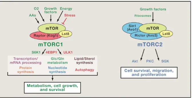
The discovery of mTOR had begun after the discovery of the macrolide antibiotic rapamycin in 1964. It was first discovered in the budding yeast Saccharomyces
cerevisiae following a screening for rapamycin resistance1,4,5. In yeast, TORC1 is composed of Tor, Lst8, Kog1 and Tco89 proteins. In mammals, mTORC1 contains mTOR, mLST8 (mammalian lethal with SEC13 protein 8), Raptor
(regulatory-18
associated protein of mTOR), PRAS40 (proline-rich Akt substrate of 40 kDa) and
DEPTOR (domain-containing mTOR-interacting protein) (Figure 1).
Figure 1. The core component of mTORC1 and mTORC2 Upstream and downstream signals of each complex are depicted.
(Modified from Blenis 2017 Cell 171(1):10-13).
Raptor serves as a complex scaffold, recruiting substrates to the kinase active site through their TOS (TOR signaling) motifs6,7. PRAS40 and DEPTOR are both mTORC1 suppressors, likely acting as competitive substrates to bind to Raptor8,9. mLST8 does not play a necessary role in mTORC1 (but specifically in mTORC2)10. In addition, structural studies showed that mTORC1 forms an obligate dimer11–13. The mechanism of action of rapamycin inhibition includes the formation of the complex rapamycin-FKBP12 (FK506-binding protein of 12 kDa). Then, the rapamycin-rapamycin-FKBP12 complex binds to the specific FRB (FKBP-rapamycin-binding) domain of mTOR and partially obstruct the active site, preventing the entry of substrates14.
19 1.2 Functions of mTORC1
1.2.1 Building blocks for cell growth
1.2.1.1 Protein synthesis and ribosome biogenesis
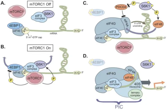
mTORC1 regulates protein synthesis and ribosome biogenesis through the phosphorylation of S6K1 (p70S6 Kinase 1) and 4EBP1 (eIF4E binding protein 1). S6K1 and 4EBP1 were the first identified mTOR substrates in metazoans and they remain as the best characterized15–18. Upon mTORC1 inhibition, S6K1 remains unphosphorylated and binds to the multi-subunit scaffold eIF3 (eukaryotic translation
initiation factor 3)19 in an inhibitory conformation. In response to growth-induced stimuli, mTORC1 gets activated and phosphorylates S6K1 on its hydrophobic motif site (Thr389). This phosphorylation liberates S6K1 from eIF3, and facilitates the subsequent activating phosphorylation of S6K1 by PDK1
(3-phosphoinositide-dependent kinase 1) on Thr22920,21. Once activated, S6K1 phosphorylates and activates numerous substrates promoting mRNA translation initiation. Among these substrates, the best characterized is the rpS6 (ribosomal protein S6), a component of 40S ribosome subunit. However, the significance of S6 phosphorylation by S6K1 in S6 functionality remains unclear22,23. S6K1 also phosphorylates and activates eIF4B, a positive regulator of the 5’ cap binding eIF4F complex24,25. In parallel, S6K1 phosphorylates PDCD4 (programmed cell death 4), an inhibitor of eIF4A, to promote the degradation of PDCD426, leading to the activation of eIF4A.
Likewise, 4EBP1 is not phosphorylated in conditions of mTORC1 inhibition, leading to its interaction with eIF4E, thus preventing eIF4E-eIF4G interaction27. The activation of mTORC1 induces the phosphorylation of 4EBP1 by mTORC1 at multiples sites (Thr37, Thr46, Thr70, Ser65), triggering the dissociation of 4EBP1 from eIF4E, allowing the binding of eIF4G to eIF4E and the recruitment of eIF4A. All these steps lead to
20
formation of the eIF4F complex (consisting of eIF4E, eIF4G and eIF4A) on the 5’-cap. In addition, the freshly formed complex recruits the 40S ribosome and the ternary complex to form the 48S translation pre-initiation complex (Figure 2).
Figure 2. Model of cap-dependent translation by mTORC1/4EBP1 and mTORC1/S6K1 axis.
(A) When mTORC1 is inactive, 4EBP1 binds to eIF4E on the mRNA 5’-cap to suppress assembly of the pre-initiation complex. (B) In response to mTORC1-activating stimuli, mTORC1 phosphorylates 4EBP1 and S6K1, inducing 4EBP1 release from eIF4E and S6K1 release from eIF3. (C) Upon the release of 4EBP1 and S6K1, the eIF4F complex, consisting of eIF4G, eIF4E and eIF4A, is assembled at the 5’-cap. In parallel, S6K also phosphorylates eIF4B, an eIF4A enhancer, and PDCD4,
an eIF4A inhibitor. (D) Binding of the 40S ribosome and the ternary complex (eIF2, Met-tRNA and GTP) at the 5’-cap with these factors to form the pre-initiation complex
and to initiate cap-dependent translation. (Modified from Magnuson et al., 2012
Biochem J. 441(1):1-21).
1.2.1.2 Lipid synthesis
mTORC1 controls lipid signaling through de novo lipid synthesis activation and lipid catabolism inhibition, in order to promote membrane synthesis for cell proliferation and long-term storage. During de novo lipid synthesis, the transcription factors SREBPs (sterol responsive element binding protein) plays an importing role in the control
21
lipogenic gene expression, necessary for lipid homeostasis, such as ACC (acetyl-CoA
carboxylase), FASN (fatty acid synthase), and SCD-1 (stearoyl-CoA desaturase 1).
mTORC1 promotes the trafficking, processing, and transcription of SREBPs28–31 (Figure 3). SREBP and its downstream biosynthetic machinery for the synthesis of fatty acids and sterols can be activated in a S6K1-dependent manner, but its molecular mechanism remains unclear28,32. Furthermore, S6K1-independent activation of SREBP involves the activity of CRTC2 (CREB regulated transcription coactivator 2), Lipin1, and p300. CRTC2, a master regulator of gluconeogenesis, inhibits the translocation of SREBP from the endoplasmic reticulum to the Golgi. Inhibition of CRTC2 upon mTORC1-mediated phosphorylation attenuates its inhibitory effect on SREBP1 maturation33. Lipin1, a phosphatidic phosphatase, acts as an inhibitor of nuclear SREBP activity. mTORC1-mediated phosphorylation of Lipin1 blocks its nuclear entry and allows SREBP-dependent gene transcription34. SREBP-1c is also acetylated by the HAT (histone acetyltransferase) p30035, and mTORC1-dependent phosphorylation of p300 is necessary for lipid synthesis through the activation of SREBP-1c36. Taken together, and through all these different pathways, SREBP mediates mTORC1-dependent lipogenesis.
The role of mTORC1 in lipid synthesis is particularly marked during adipogenesis, the biological process of mature adipocytes formation from adipose cell precursors though the enhanced synthesis and accumulation of triglycerides. This process is mediated by PPARg (peroxisome proliferator-activated receptor g), a nuclear receptor that controls fatty acid uptake, synthesis, esterification and storage in adipose cells37. PPARg expression and activity are controlled by mTORC1. The molecular mechanism is mediated by 4EBP138, and through SREBP1-dependent PPARg ligand production39.
22
Figure 3. Lipid biogenesis and lipid catabolism regulation by mTORC1. (From Thelen & Zoncu 2017 Trends in Cell Biology 27(11):833-850).
The transcription factor TFEB (bHLH leucine zipper transcription factor EB) regulates lysosome biogenesis, autophagosome formation and their fusion with the lysosome40. In addition, TFEB promotes beta-oxidation of fatty acids by upregulating the expression of PPARa and PGC1a (PPARg coactivator 1 a or peroxisome-proliferator-activated
receptor coactivator-1a)41. mTORC1-mediated phosphorylation of TFEB induces its interaction with the cytosolic protein 14-3-3 and blocks its translocation to the nucleus, repressing its transcriptional activity42–44. Thus, in parallel to the activation of lipid synthesis, mTORC1 inhibits lipid catabolism through TFEB inhibition.
1.2.1.3 Nucleotide synthesis
Nucleotides are building blocks for DNA and RNA, which are necessary for cell proliferation. Emerging evidences show that mTORC1 upregulates the synthesis of nucleotides. Through several approaches, in addition to enhancing the de novo
23
synthesis of lipids, mTORC1/S6K1-dependent SREBP activity induces the oxidative PPP (pentose phosphate pathway)28. Moreover, mTORC1 promotes the expression of PPP genes which contribute in the production of ribose moieties for the synthesis of both purine and pyrimidine nucleotides.
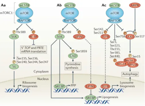
Pyrimidine nucleotide (CMP, UMP) is a nitrogen-containing base that is synthetized from glutamine, bicarbonate (HCO3-), and aspartate with ribose-5-phosphate, derived from the PPP. Phosphoproteomic and metabolomic analyses revealed that mTORC1 promotes pyrimidine synthesis through S6K1-mediated CAD (carbamoyl-phosphate
synthetase 2, aspartate transcarbamylase, dihydroorotase) phosphorylation and
activation45,46 (Figure 4). CAD catalyses the first three steps in de novo pyrimidine synthesis. The S6K1-mediated phosphorylation of CAD on S1859 induces its oligomerization, leading to an increased pyrimidine synthesis, and stimulating S phase progression. This regulation of CAD by mTORC1/S6K1 increase the pool of nucleotides available for the DNA replication and RNA synthesis that are necessary for cell growth.
Purine synthesis is a pathway that assembles carbon and nitrogen from glutamine, aspartate, glycine, bicarbonate (HCO3-) and formyl unit from the THF (tetrahydrofolate) cycle on a PRPP (5-phosphoribosyl-1-pyrophosphate) molecule to form purine nucleotide (AMP, GMP). mTORC1 promotes purine synthesis through the ATF4-dependent upregulation of MTHFD2 (methylene-tetrahydrofolate dehydrogenase 2), an important enzyme of the mitochondrial THF cycle. In response to growth-induced stimuli, ATF4 induces also the expression of other enzymes of the serine synthesis (PSAT1 and PSPH) and the mitochondrial THF cycle (SHMT2), in order to increase the production of formyl units required for de novo purine synthesis47.
24
In summary, in response to growth-induced stimuli, mTORC1 upregulates anabolic processes such as protein synthesis, lipid synthesis, and nucleotide synthesis. These mTORC1 functions are mediated by S6K1, 4EBP1 and SREBP1 which are all phosphorylated by mTORC1, inducing a signaling cascade pathway to control cell growth and proliferation.
Figure 4. Anabolic processes controlled by mTORC1.
(A) mTORC1 promotes ribosome biogenesis. (B) mTORC1 activated nucleotide and lipid synthesis. (C) mTORC1 inhibited autophagy.
(Modified from Shimobayashi & Hall 2014 Nat Rev Mol Cell Biol. 15(3):155-62). 1.2.2 Control of anabolic metabolism
Cellular metabolism is also controlled by mTORC1 in order to have enough metabolites and energy for cell proliferation. mTORC1 exerts this regulation in many different ways.
1.2.2.1 Glucose metabolism
Through oxidative glycolysis, cells use glucose for energy production and building block assembly, necessary for cell growth48,49. As a central controller of cell growth, mTORC1 promotes the shift in glucose usage from oxidative phosphorylation to
25
aerobic glycolysis, facilitating metabolic intermediates production for the biosynthesis of macromolecules. Indeed, mTORC1 increases glycolytic flux by activating both the transcription and the translation of HIF1a (hypoxia inducible factor 1 a), which induces the expression of glucose transporters GLUT1, GLUT3 and several glycolytic enzymes such as PKM2 (pyruvate kinase M2 subtype) or HK (hexokinase)50–54. Moreover, mTORC1/SREBP pathway increases the carbon flux from glucose through the oxidative PPP to generate NADPH and other intermediary metabolites needed for proliferation and growth28. Interestingly, mTORC1-dependent glucose metabolism activation leads, in certain circumstances, to glucose addiction in cancer cells55–57.
1.2.2.2 Glutamine metabolism
In addition to glucose, the amino acid glutamine (the most abundant amino acid in the blood of mammals) is a major nutrient that fuels cellular energetic to allow cell growth. The relationship between glutamine and mTORC1 is very tight, as glutamine metabolism activates mTORC158,59, and in turns glutamine metabolism is controlled by mTORC1activity60–62. The connexion between glutamine and mTORC1 will be detailed in further section of the second chapter.
1.2.2.3 Mitochondrial metabolism
Growing cells need not only glucose and glutamine, but also a number of mitochondrial intermediates to generate building blocks. Thus, it is not surprising that mTORC1 controls and stimulates mitochondrial oxidative activities through different mechanisms. In skeletal muscle tissues and cells, mTORC1 regulates the interaction of YY1 (yin-yang 1) with PGC1a, affecting the transcriptional function of this complex, which regulates the expression of mitochondrial genes involved in oxidative functions63. In addition to that, mTORC1 controls mitochondrial biogenesis and
26
function by promoting translation of nucleus-encoded mitochondria-related mRNAs through 4EBP1 inhibition64.
1.2.2.4 Other metabolic pathways
Amino acids contributing to the so-called one carbon metabolism (e.g. serine and glycine) integrate different stimuli through folate and methionine cycles, to induce cell proliferation65. This type of metabolism has been revealed to play an important role in diseases in which mTORC1 is involved, such as cancer. Indeed, mTORC1 has been reported to control serine/glycine de novo synthesis in osteosarcoma cells through the regulation of the expression of genes involved glycolysis and serine/glycine synthesis66.
Recently, mTORC1 has been shown to regulate polyamine metabolism in addition to other anabolic processes in prostate cancer67. Polyamines are small polycations, containing two, three, or four amine groups which have diverse functions such as maintaining chromatin conformation and membrane stability. The control of polyamine concentration is very tight because polyamine excess leads to hydrogen peroxide release68. mTORC1 regulates polyamines flux through phosphorylation-dependent stability of pro-AMD1 (s-adenosyl-L-methionine decarboxylase 1)67. This mTORC1-mediated induction in polyamine synthesis explains the high levels of polyamines observed in highly proliferating cells.
1.2.3 Regulation of protein turnover
In addition to inducing cell anabolism, mTORC1 controls cell growth by suppressing cell catabolism. Two major catabolic processes are known to operate downstream of mTORC1 signaling: autophagy and protein degradation through the ubiquitin-proteasome system.
27
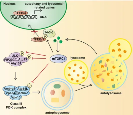
1.2.3.1 Autophagy
Autophagy is a multistep degradation process that is necessary for macromolecules recycling and maintenance of cellular homeostasis. During autophagy, cellular components are sequestered into autophagosomes (double membrane bound vesicles) that will fuse with lysosomes to form the autolysosomes in which the proteolytic degradation happens69. Autophagy is activated by AMPK (AMP-activated
protein kinase) signaling and inhibited by mTORC170. Indeed, mTORC1 regulates different steps of autophagy. The protein kinase ULK1 (Unc-51 like autophagy
activating kinase), belonging to the ULK complex, is the key downstream target of
mTORC1 in the control of autophagy71–73 (Figure 3-4). Under nutrient-rich conditions, mTORC1 phosphorylates and inhibits ULK1 at Ser757, preventing its interaction with AMPK, and thus inactivating autophagy70. Upon mTORC1 inhibition, AMPK interacts with and activates ULK1 by phosphorylating several residues (Ser555, Ser317 and Ser777). Then ULK1 will induce autophagy by activating the lipid kinase VPS34, necessary for autophagosome formation74.
In addition to ULK1 phosphorylation, mTORC1 directly phosphorylates two other autophagy-activating proteins: ATG13, a positive regulator of ULK171–73, and ATG14, a VPS34-associated component75. Therefore, mTORC1 directly phosphorylates and inhibits different mediators that positively control autophagy.
28
Figure 5. Regulation of autophagy by mTORC1.
To control autophagy in response to amino acid stimuli, mTORC1 phosphorylates ULK, ATG13, ATG14-containing Vps34 complex and TFEB. When mTORC1 is inhibited, the activation of autophagy allows the cell to maintain necessary energy
and metabolites for surviving the starvation condition. (From Rabanal-Ruiz et al., 2017 Essays Biochem 61(6):565-584).
Indirectly, mTORC1 controls autophagy through the transcription factor TFEB, which controls lysosome biogenesis and autophagy40,42–44. mTORC1-mediated TFEB phosphorylation prevents its nuclear translocation and represses the transcription of lysosomal and autophagy-related genes. In addition, mTORC1 controls autophagy in a fine-tuning manner through the HAT p30036 and NRBF2/Atg3876. Finally, DAP1 (death-associated protein 1) negatively controls autophagy when its mTORC1-mediated phosphorylation is removed upon mTORC1 inactivation. The control of autophagy by DAP1 aims at limiting the over-activation of autophagy, maintaining the homeostasis balance. The molecular mechanism of DAP1-mediated autophagy limitation is still unknown77.
29
In the context of cancer, autophagy has a dual role, preventing tumor initiation78, but in the other side supporting cell survival under metabolic stress59. Thus, the close connection between mTORC1 and autophagy allows an efficient response in function of nutrient availability, depending on the stage and context of the cancer.
1.2.3.2 Ubiquitin-proteasome system
UPS (ubiquitin-proteasome system) is a main mechanism for protein catabolism, through which proteins targeted for degradation are tagged by ubiquitine multimers and degraded by the 26S proteasome79. UPS-mediated protein degradation is tightly controlled, and its perturbation leads to diverse disease such as cancer and neurodegeneration80–83. As a major regulator of protein catabolism, mTORC1 also controls protein homeostasis through UPS inhibition. Indeed, mTORC1 inhibition increases proteasome-dependent proteolysis through an increase in protein ubiquitination without affecting the proteasome activity84. In addition to this, mTORC1 inhibition also induces proteasome abundance via ERK5 (extracellular
signal-regulated kinase 5) activation85. Paradoxically, the team of Prof Brendan Manning reported that mTORC1 activation induces an increase in cellular proteasome content through the expression of NRF1 (nuclear factor erythroid-derived 2-related factor 1). This opposite effect of mTORC1 positively regulating proteasome content was explained by the authors as a necessary mechanism for the removal of misfolded protein upon mTORC1-induced protein synthesis86. Further investigations are needed to better understand how mTORC1 coordinates these opposite effects to control proteasomal degradation of targeted proteins.
30 1.3 Upstream regulation of mTORC1
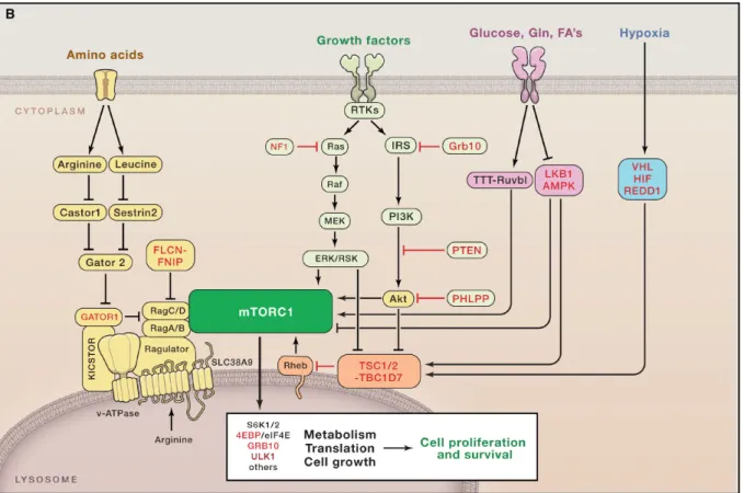
Four main upstream mechanisms of mTORC1 regulation will be described below: amino acid availability, growth factor signaling, hypoxic and bioenergetics stress, and lipid sensing (Figure 6).
Figure 6. Summary of main upstream regulators of mTORC1 in response to activating stimuli.
Tumor suppressors are in red, whose loss of expression/function occurs in cancer. Positive regulators of mTORC1 are in green, which are often activated by mutation or
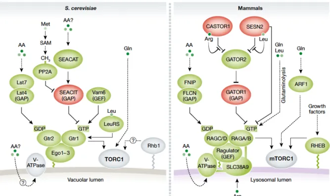
overexpression in cancer. (From Blenis 2017 Cell 171(1):10-13). 1.3.1 Amino acids
mTORC1 activation by amino acid involves predominantly the conserved Rag family of small GTPases on the surface of the lysosome. Four mammalian Rags (RagA, RagB, RagC, RagD) are localized at the surface of the lysosome independently of amino acid availability using the pentameric Ragulator complex as a scaffold87–91. In yeast, Rag orthologues are the Gtr1/2 GTPases92–94, which localize to the surface of
31
the vacuole and bind to the ternary complex Ego95,96 (analogue to the Ragulator complex) (Figure 7).
Figure 7. Regulation of TORC1 by amino acids in yeast (left) and mammals (right).
Proteins shown in green promote TORC1 activation. Proteins in red inhibit TORC1. Dashed lines indicate indirect interactions.
(From González & Hall 2017 The EMBO Journal 36:397-408).
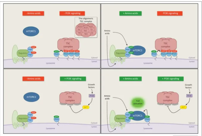
The Rag GTPases function in a heterodimeric form in which RagA or RagB interact with RagC or RagD. In conditions of amino acid availability, the subunit RagA/B is loaded with GTP, while the subunit RagC/D is GDP loaded, rendering an active conformation of the heterodimer97,98. Under this active conformation, mTORC1 binds to the Rag heterodimer through Rag-Raptor interaction, which leads to the translocation of mTORC1 to the lysosomal surface99. Once at the lysosomal surface, mTORC1 is fully activated through its direct interaction with the co-activator Rheb (RAS homolog enriched in brain). Rheb is a growth factor-stimulated small GTPase which, upon growth factor stimulus, is GTP-loaded to allow the full activation of
32
mTORC1100,101. Thus, both amino acids and growth factors are necessary to completely activate mTORC1 (see below for a full description of mTORC1 activation by growth factors).
Since the finding of the Rag GTPases as nutrient sensors for mTORC1 activation, different proteins have been identified as regulators of the nucleotide binding status of the Rags, operating either as GAP (GTPase-activating proteins) or GEF (guanine
exchange factors) of these small GTPases. GEFs regulate the replacement of GDP by
GTP, while GAPs stimulate the intrinsic GTPase activity of a related GTPase to convert GTP into GDP. In the context of the Rag GTPases, Ragulator and Gator1 have respectively GEF and GAP activity toward RagA/B. Ragulator consists of p18/Lamtor1, p14/Lamtor2, MP1/Lamtor3, HBXIP/Lamtor4 and C7orf59/Lamtor5, among which p18/Lamtor1 is a critical scaffold element for the Ragulator-Rag GTPase complex described by different structural studies88,90,91,102. Although all 5 subunits are needed for the GEF activity, the exact mechanism of Ragulator’s GEF activity is still unknown. In yeast, Ego complex (ortholog to the Ragulator) does not play the same role, as the vacuolar protein Vam6 has been proposed to be the GEF for Gtr197.
The mammalian heterotrimeric protein complex Gator1 (GAP activity toward RAGs 1) is composed of DEPDC5 (DEP domain-containing protein 5), NPRL2 (nitrogen
permease regulator 2-like protein), and NPRL3103,104. Gator1 is tethered to the lysosomal surface through the Kicstor complex (consisting of KPTN, ITFG2, C12orf66, and SZT2)105,106 and negatively regulated by Gator2 complex, consisting of SEC13 (protein SEC13 homolog), SEH1L (nucleoporin SEH1), WDR24 (WD
repeat-containing protein 24), WDR59, and MIOS (WD repeat-containg protein MIO)103,107. In the case of the RagC/D subunit, FLCN (Folliculin) and its binding partners FNIP1/2
33
(folliculin-interacting proteins 1 and 2) have been described to operate as GAPs in mammals108,109, while no GEFs have been described so far.
The question about how cells integrate the information about each amino acid availability to coordinate the response regarding the nucleotide loading state of the Rag GTPases is far to be well understood. Similarly, it is not well known how the concerted regulation of all the members of the amino acids-mediated mTORC1 activation pathway takes place. Even how amino acid availability is sensed and signalled to the Rags are still elusive, although different sensors have been described in the literature. Two main mechanisms by which mammalian cells sense amino acids have been describe, the lysosomal pathway and the cytosolic pathway. In the lysosome surface, v-ATPase (vacuolar H(+)- adenosine triphosphatase) plays an intermediate role of an “inside-out” mechanism, in which amino acids must accumulate in the lysosomal lumen to initiate signaling110. Then, amino acids inside the lumen can affect the Rag nucleotide state through the ATP hydrolysis capacity and the associated rotation of the v-ATPase. Cytosolic amino acids, such as leucine, arginine and glutamine, signal to mTORC1 mostly through the Gator1/Gator2 complexes3.
For leucine sensing, Sestrin and leucyl-tRNA synthetase are two sensors that mediate leucine signaling in mTORC1 pathway. Sestrins negatively regulate mTORC1 through Gator2 inhibition111–114 and leucine stimulation dissociates Sestrin2 from Gator2 to activate mTORC1. Furthermore, decreased leucine import due to the loss of glutamine (SLC1A5) or leucine (SLC7A5-SLC3A2) transporters impairs mTORC1 activity115. By following a mechanism similar to the one described for Sestrins, Castor1/2 were discovered as cytosolic arginine sensors116,117. Under arginine deprivation, Castor1/2 binds to Gator2 preventing Gator2-mediated mTORC1 activation. Moreover, the
34
transporter SLC38A9 have been proposed to be a lysosomal arginine sensor that also controls mTORC1 activation118–120.
In addition to the role as efflux solute for leucine import, glutamine can activate mTORC1 both in Rag-dependent and Rag-independent manners. In cooperation with leucine, glutamine is able to induce the GTP loading of Rag, thus activation mTORC1, by following a mechanism in which PHDs (Prolyl Hydroxylases Domain) have been involved58,121. In addition to that, glutamine also stimulates lysosomal translocation and activation of mTORC1 via the small GTPase ARF1 (ADP-ribosylation factor 1) and v-ATPase in a Rag-independent manner122. The metabolism of glutamine and its connection to mTORC1 will be discussed below in a specific section.
1.3.2 Growth factors
mTORC1 regulation by growth factors imply the role of the tumor suppressor TSC1/TSC2 complex (tuberous sclerosis complex), a key negative regulator of mTORC1. TSC is a heterotrimeric complex comprising TSC1, TSC2 and TBC1D7123, and its lost-of-function mutations lead to the development of tuberous sclerosis complex, or Bourneville’s disease, due to the hyperactivation of mTORC1. TSC2 has a GAP function toward the small GTPase Rheb124,125, while TSC1 acts as a scaffold to stabilize TSC2 and TBC1D7. The role of TBC1D7 is not fully understood, but its loss causes the dissociation of the complex123.
Growth factors, particularly insulin and IGF1 (insulin-like growth factor 1) inhibit TSC complex in a PI3K (phosphoinositide 3-kinase)-dependent manner. Upon TSC inhibition, the GAP activity of TSC is reduced, leading to the GTP loading of lysosomal Rheb. Then Rheb will interact with lysosomal translocated mTORC1 (induced by amino acid availability, as explained above). The mechanism of how Rheb activates mTORC1 is still elusive126,127. A structural study by cryo-electron microscopy reported
35
that Rheb binds to mTOR distally from the kinase active site and causes a global conformational change that allosterically realigns active-site residues, accelerating catalysis128.
Figure 8. Rheb mediates mTORC1 activation in response to PI3K/AKT-dependent growth factor signals.
Amino acid signaling and growth factor-PI3K signaling promote mTORC1 in parallel to fully activating mTORC1 pathway. The molecular mechanism is detailed in the text. (Modified from Dibble and Cantley 2015 Trends in Cell Biology 25(9):545-555). Growth factors and mitogen-dependent signaling pathways control mTORC1 activity via two mains mechanisms. On the one hand, through the insulin/IGF-1 pathway mediated by PI3K and PDK1, the AKT-dependent phosphorylation of TSC2 inhibits and dissociates TSC complex from the lysosomal membrane, where Rheb localizes129,130 (Figure 8). In addition to TSC2 phosphorylation, AKT phosphorylates also PRAS40 in mTORC1 to disrupt the inhibitory interaction Raptor-PRAS40, allowing
36
mTORC1 to be fully activated127,131. On the other hand, the MAPK (mitogen-activated
protein kinase) ERK and its effector RSK (90 kDa ribosomal S6 kinase), via Ras
signaling pathway, phosphorylate and inhibit TSC2132,133. Also, ERK and RSK phosphorylate Raptor to promote mTORC1 activity134,135. Besides, Wnt pathway and the inflammatory cytokine TNFa phosphorylate and inhibit TSC2 and TSC1 respectively to activate mTORC1136,137. Taking together, through Rheb and Rag GTPase family, mTORC1 signaling pathway integrates amino acid and growth factor inputs to be fully activated.
1.3.3 Energy, oxygen, stress and DNA damage
For cell growth and proliferation, in addition to positive growing signals and building blocks, cells also need energy. Energy sensing in growing cells is greatly related to glucose availability and metabolism. The sensing of the energetic status of the cell acts through the conserved AMPK pathway. In conditions of glucose deprivation, or after the inhibition of glycolysis/mitochondrial respiration, AMP/ATP ratio and ADP/ATP ratio are increased, due to a decrease in ATP synthesis. This increase in AMP/ATP ratio leads to the activation of AMPK, which promotes catabolic processes like autophagy, and inhibits anabolic processes such as protein synthesis, through mTORC1 inhibition. AMPK inhibits mTORC1 via TSC activation or Raptor inhibition138,139. Interestingly, v-ATPase-Ragulator have been shown to activate AMPK through AXIN-LKB1 on lysosome surface and therefore to inhibit mTORC1 under energy stress140. Besides, mTORC1 can sense glucose availability independently of AMPK, through the inhibition of the Rag GTPases141,142.
Growing cells also need to detect other intracellular/environmental stresses that are incompatible with growth, hypoxia or DNA damage. These stresses control mTORC1 following opposite mechanisms. In one side, redox stress upregulates mTORC1
37
through TSC1/2-Rheb pathway, by increasing the GTP-bound state of Rheb143. However, energy stress downregulates mTORC1 via p38b-PRAK-mediated Rheb inhibition144. Controversially, p38b enhances mTORC1 activity under arsenite treatment via Raptor phosphorylation145. Thus, p38 enhances or reduces mTORC1 activity in function of different environmental stresses by regulating different components of mTORC1 pathway.
In the case of oxygen availability, it is well stablished the capacity of hypoxia to inhibit mTORC1. The hypoxia-dependent inhibition of mTORC1 follows two mechanisms: an AMPK-mediated mechanisms146, and a HIF1-dependent mechanism. The second one involves the upregulation of REDD1 (Regulated in DNA damage and development 1), a HIF1 target gene which activates TSC complex147,148. Indeed, REDD1 binds to 14-3-3 protein and disrupts the interaction of TSC2/14-14-3-3-14-3-3, leading to TSC1/2 activation and mTORC1 inhibition149. In addition, it has been speculated that hypoxia could inhibit mTORC1 also through direct inhibition of PHD activity, which has been demonstrated to mediate glutaminolysis-induced mTORC1 activation121. Interestingly, and in contrast to what has been described for HIF1, HIF2 acts as an mTORC1 activator via the amino acid transporter SLC7A5, a HIF2-dependent target, in lung and liver tissues150.
1.3.4 Lipid sensing
PLD (Phospholipase D) is an enzyme involved in cell growth, and as a consequence it is upregulated in a large number of different types of tumors. In mammals, there are two isoforms of PLD: PLD1 and PLD2, both of them shown to activate mTORC1 through phosphatidic acid (PA) production and through their own expression151,152. PA is a phospholipid, known as an indicator of lipid sufficiency in dividing cells. PA can be generated by the hydrolysis of phosphatidylcholine by PLD, by LPAAT (lysophosphatidic acid acyltransferase) or DG (diacylglycerol) kinases. This is a central
38
metabolite for membrane phospholipid biosynthesis. PA-mediated mTORC1 activation connects mTORC1 to both lipids and glucose metabolism. Originally, PA and its analogues were shown to activate mTORC1 from exogenous supply153,154. The effect of PA towards mTORC1 depends on amino acids availability, and is suppressed by rapamycin treatment or by over-activation of TSC1/2. Suppression of PLD-generated PA with primary alcohols inhibit mTORC1 activity155, while PA production by other enzymes, such as LPAAT156 or DG kinase157, increases mTORC1 activity, showing that PA is physiologically necessary for mTORC1 activation. Finally, lipid sensing by mTORC1 was confirmed via de novo synthesis of PA which inhibition resulted in G1 cell cycle arrest158. PA production is also induced by Rheb, as PLD is a downstream target of Rheb (Figure 9). Rheb binds to and activates PLD in a GTP-dependent manner, then PLD-generated PA interacts and upregulates mTORC1159. Additionally, RalA (Ras-related protein A) and the GTPase ARF6 (ADP-ribosylation factor 6) are acting downstream of Rheb to induce this production induced by growth factor160–162. Furthermore, amino acids can induce PLD translocation to the lysosome and increase its activity through the class III PI3K hVps34163. Thus, PLD-derived PA contributed to nutrient mediated mTORC1 activation, and PA is necessary but not sufficient to activate mTORC1. There are different hypotheses about the mechanism of PA-mediated mTORC1 activation. These hypotheses mostly suggest two potential mechanisms: either PA interaction with the FRB domain in mTOR153 enhances mTORC1 activity, or it increases the stability of the mTORC1 complex164. Controversially, in a study that showed for the first time the anti-oncogenic role of PA-mediated PLD function, production of PA was recently shown to activate LKB1, which would result in an AMPK-mediated inhibition of mTORC1165. The balance between