HAL Id: hal-01272347
https://hal.sorbonne-universite.fr/hal-01272347
Submitted on 10 Feb 2016
HAL is a multi-disciplinary open access
archive for the deposit and dissemination of sci-entific research documents, whether they are pub-lished or not. The documents may come from teaching and research institutions in France or abroad, or from public or private research centers.
L’archive ouverte pluridisciplinaire HAL, est destinée au dépôt et à la diffusion de documents scientifiques de niveau recherche, publiés ou non, émanant des établissements d’enseignement et de recherche français ou étrangers, des laboratoires publics ou privés.
Macroglobulinemia Displaying the MYD88 L265P
Mutation Are Enriched by Epstein-Barr Virus
Immortalization
Maroulio Pertesi, Perrine Galia, Nicolas Nazaret, Maxime Vallée, Laurent
Garderet, Xavier Leleu, Hervé Avet-Loiseau, Matthieu Foll, Graham Byrnes,
Joel Lachuer, et al.
To cite this version:
Maroulio Pertesi, Perrine Galia, Nicolas Nazaret, Maxime Vallée, Laurent Garderet, et al.. Rare Circulating Cells in Familial Waldenström Macroglobulinemia Displaying the MYD88 L265P Mutation Are Enriched by Epstein-Barr Virus Immortalization. PLoS ONE, Public Library of Science, 2015, 10 (9), pp.e0136505. �10.1371/journal.pone.0136505�. �hal-01272347�
RESEARCH ARTICLE
Rare Circulating Cells in Familial
Waldenström Macroglobulinemia Displaying
the
MYD88 L265P Mutation Are Enriched by
Epstein-Barr Virus Immortalization
Maroulio Pertesi1, Perrine Galia2,3, Nicolas Nazaret3, Maxime Vallée1, Laurent Garderet4, Xavier Leleu5, Hervé Avet-Loiseau6, Matthieu Foll1, Graham Byrnes7, Joel Lachuer3,
James D. McKay1, Charles Dumontet2,3*
1 Genetic Cancer Susceptibility, International Agency for Research on Cancer, Lyon, France, 2 Hospices Civils de Lyon, Pierre Bénite, France; INSERM 1052, CNRS 5286, CRCL, Lyon, France; University of Lyon, Lyon, France, 3 ProfilExpert, Lyon, France, 4 INSERM, UMR_S 938, Proliferation and differentiation of stem cells, Paris, France; AP-HP, Hôpital Saint Antoine, Département d'hématologie et de thérapie cellulaire, Paris, France; Sorbonne Universités, UPMC Univ Paris 06, Paris, France, 5 Service des maladies du sang, Hopital Huriez CHRU, Lille, France; Centre de Recherche INSERM U837, Facteurs de persistance des cellules leucémiques, Institut pour la Recherche sur le Cancer (IRCL), Lille, France, 6 Unité génomique du myélome, Institut Universitaire du Cancer de Toulouse—Oncopole, CRCT INSERM U1037, Toulouse, France, 7 Biostatistics, International Agency for Research on Cancer, Lyon, France
*charles.dumontet@chu-lyon.fr
Abstract
TheMYD88 L265P is a recurrent somatic mutation in neoplastic cells from patients with Waldenström Macroglobulinemia (WM). We identified theMYD88 L265P mutation in three individuals from unrelated families, but its presence did not explain the disease segregation within these WM pedigrees. We observed the mutation in these three individuals at high allele fractions in DNA extracted from EBV-immortalized Lymphoblastoid cell lines estab-lished from peripheral blood (LCL), but at much lower allele fractions in DNA extracted directly from peripheral blood, suggesting that this mutation is present in a clonal cell sub-population rather than of germ-line origin. Furthermore, we observed that theMYD88 L265P mutation is enriched in WM families, detected in 40.5% of patients with familial WM or MGUS (10/22 WM, 5/15 MGUS), compared to 3.5% of patients with familial MM or MGUS (0/72 MM, 4/41 MGUS) (p = 10−7). The mutant allele frequency increased with pas-sagesin vitro after immortalization with Epstein-Barr virus (EBV) consistent with the MYD88 L265P described gain-of-function proposed for this mutation. TheMYD88 L265P mutation appears to be frequently present in circulating cells in patients with WM, and MGUS, and these cells are amenable to immortalization by EBV.
OPEN ACCESS
Citation: Pertesi M, Galia P, Nazaret N, Vallée M, Garderet L, Leleu X, et al. (2015) Rare Circulating Cells in Familial Waldenström Macroglobulinemia Displaying theMYD88 L265P Mutation Are Enriched by Epstein-Barr Virus Immortalization. PLoS ONE 10 (9): e0136505. doi:10.1371/journal.pone.0136505 Editor: Luwen Zhang, University of Nebraska -Lincoln, UNITED STATES
Received: June 16, 2015 Accepted: August 4, 2015 Published: September 9, 2015
Copyright: © 2015 Pertesi et al. This is an open access article distributed under the terms of the
Creative Commons Attribution License, which permits unrestricted use, distribution, and reproduction in any medium, provided the original author and source are credited.
Data Availability Statement: All relevant data are within the paper and its Supporting Information files. Funding: The work was funded from the Institut National du Cancer (INCA, Projet Transla 2010-077, CD) and the Fondation Française pour la Recherche contre le Myélome et les Gammapathies (FFRMG). The funders had no role in study design, data collection and analysis, decision to publish, or preparation of the manuscript.
Competing Interests: The authors have declared that no competing interests exist.
Introduction
Waldenström Macroglobulinemia (WM) is a rare B-cell lymphoproliferative disorder demon-strating an IgM monoclonal gammopathy. Monoclonal Gammopathy of Undetermined Signif-icance (MGUS) is a premalignant plasma cell disorder that precedes the development of WM, Multiple Myeloma (MM) and other related Plasma cells disorders, with a risk of progression in the order of 1.5% per year.[1] Among lymphoid malignancies, WM displays the strongest familial predisposition to develop the disease or clonal abnormalities. In a series of 257 unre-lated WM patients, Treonet al. observed a clustering of B-cell disorders among first-degree rel-atives of patients with WM, where nearly one in five WM patients had at least one first-degree relative with WM (5.1%), some form of hematological malignancy (11.7%) or Monoclonal Gammopathy of Unknown Significance (MGUS; 1.9%).[2] Similarly, in a large study of 2,144 WM/lymphoplasmacytic lymphoma patients and 8,279 population-based matched controls, Kristinssonet al. found that first-degree relatives presented a 20-fold risk of developing the same disease.[3] In addition, a recent large population-based study showed that the presence of family history is associated with significantly lower survival.[4] The existence of a familial pre-disposition is thus well documented in this disease.
MYD88 has been found to be involved in the pathogenesis of sporadic Waldenström Macroglobulinemia (WM). Treonet al. found a recurrent somatic mutation in MYD88, L265P, in the bone marrow of 26 out of 30 Waldenström patients by whole genome sequenc-ing, with an additional case identified by Sanger sequencing.[5] All patients with a family his-tory were found to carry the mutation, while it was absent in the germ-line material. The MYD88 L265P mutation has been shown to be a gain of function driver mutation in models such as ABC non-Hodgkin lymphoma cell lines, while activated MYD88 induces NF-kappa B signaling.[6,7]
Our study was initially designed to determine whether germ-line mutations inMYD88 were present in familial cases of WM, and subsequently explored the presence of the missense MYD88 L265P mutation at rare allele fractions within WM, MM families.
Materials and Methods
Patient samples
Families with at least two cases of Waldenström disease and/or IgM Gammopathy were enrolled for this study from across France. Targeted resequencing of the coding sequence of theMYD88 gene using exon capture was performed on a total of 41 cases with IgM monoclonal component (27 WM, 14 IgM-MGUS) and 5 additional family members with non-IgM mono-clonal component or full-blown multiple myeloma (4 IgG-MGUS, 1 IgA-myeloma) (S1 Fig). The ages at accrual for the WM cases are shown inS1 Table. In addition, deep Ion-Torrent based targeted resequencing for theMYD88 L265P mutation was performed on a series of 74 multiple myeloma cases and 43 MGUS cases recruited from French families with reoccurrence of multiple myeloma/MGUS, and a series of 55 French sporadic lung cancer samples recruited as described elsewhere.[8] The study was approved by the Hospices Civils de Lyon institutional review board and participants signed an informed consent form. Peripheral blood was drawn and lymphoblastoid cell lines were established as previously described.[9] DNA from lympho-blastoid cell lines after severalin vitro passages was extracted using QIAmp DNA Mini Kit (Qiagen) according to the manufacturer’s recommendations, and subsequently quantified with the Qubit fluorometer using dsDNA HS Assay (Life Technologies).
Targeted resequencing
Targeted exon capture was performed using the HaloPlex Target Enrichment kit 1–500 kb (Agilent) according to the HaloPlex Target Enrichment System-Fast Protocol Version B. Briefly, the protocol consists of the following four steps: 1) Digestion of genomic DNA in eight different restriction reactions. 2) Hybridization of restricted fragments to probes whose ends are complementary to the target fragments. During hybridization, fragments are circularized and sequencing motifs, including index sequences, are incorporated. 3) Capture of target DNA using streptavidin beads and ligation of circularized fragments. 4) PCR amplification of cap-tured target libraries.
250 ng of DNA was split among 8 different restriction digests each containing 2 restriction enzymes. The eight different digests corresponding to the same DNA sample were further pooled and used in the capture reaction with probes. HaloPlex probes are designed to hybridize selectively to fragments originating from target regions of the genome and to direct circulariza-tion of the targeted DNA fragments. During the hybridizacirculariza-tion process, Illumina sequencing motifs including index sequences (for sample multiplexing) are incorporated into the targeted fragments. Prior to pooling 48 samples, DNA was quantified using a Qubit Fluorometer (Life Technologies). Circularized fragments were then captured, ligated and amplified by PCR. Pooled libraries were further paired-end sequenced (150 bp reads) in a flow cell version 2 on a MiSeq instrument (Illumina, USA). Image analysis and base calling was performed using the Illumina RTA software version 1.13.48. The mean coverage was 375X.
Deep Ion Torrent-based targeted sequencing
PCR primers were designed (F: 5’-ggttgaagactgggcttgtc-3’, R: 5’-gcgagtccagaaccaagatt-3’) using Primer 3 Plus to amplify an amplicon of 198bp spanning the L265P on genomic DNA. 30ng of DNA were amplified by PCR using AccuStartTaq DNA Polymerase (Quanta BioSciences) according to manufacturer’s instructions. Library preparation for Ion Torrent was performed using the NEBNext Fast DNA Library Preparation Kit (New England Biolabs), template prepa-ration was done on the Ion OneTouch2 instrument using the Ion PGM Template OT2 200 Kit, followed by sequencing on an Ion Torrent PGM sequencer using the Ion PGM Sequencing 200 Kit v2 (Life Technologies).
Bioinformatics analyses, Variant analyses
Raw reads were aligned onto the reference human genome (build hg19 from UCSC database) using Bowtie 2 (version 2.0.2). Bowtie was run in local mode: consequently it does not require that the entire read aligns on the reference sequence. Reads with poor qualities were filtered out:“Illumina Passing Filter” metric was used to perform quality filtering.[10] Sequencing out-put files were mapped on hg19 reference genome with BWA, and then sorted with Picard Sort-Sam (http://picard.sourceforge.net). Picard Mark Duplicates was used to flag duplicate reads. Files were then locally realigned and base score recalibrated using the Genome Analysis Tool Kit (GATK).[11–13] Recalibrated files were used to call the variants. Both single nucleotide variants (SNV) and indels were called using GATK Unified Genotyper. All the called variants were then annotated with AnnoVar (version 2013 Aug 23).[14] Calls were subsequently filtered following a two-step approach. The first step consisted in pruning from the raw list of calls, variants that failed several quality control criteria (criteria as exonic status, segmental duplica-tions, strand bias, different starting point of reads, quality of the call, coverage from NHLBI GO Exome Sequencing Project (ESP6500) (Exome Variant Server, NHLBI GO Exome Sequencing Project (ESP), Seattle, WA), frequency in ESP6500, frequency in 1000 genomes project, frequency in a panel of 500 normal TCGA.[15,16] The second step focused on
functionality to keep only missense substitutions assessed as deleterious by either SIFT or Poly-Phen.[17,18]
Selection of samples for deep Ion Torrent sequencing, statistical
analyses
55 Lung cancer samples were selected as reference samples in order to calculate the distribution of the mutant allele fractions observed by chance. In order to calculate the minimum allele frac-tion above which samples are considered“positive” for the MYD88 L265P mutation, we first transformed the allelic fractions to log-odds which provided approximately normally distrib-uted values. We then set the threshold as 6 standard deviations above the mean, after removing the 2 most extreme values at each end of the distribution. Exceeding this threshold corresponds to rejection of the presence of theMYD88 L265P mutation at p<2x10⁻⁹.
We then applied this threshold to the Ion Torrent-based deep sequencing data on WM, MM and MGUS individuals. We did not consider individuals with a mean read depth lower than 500X, thus excluding 1 WM, 2 MM, and 2 MGUS individuals from MM families, that underperformed or failed due to technical problems. We then selected for analysis on DNA prior to EBV-immortalization (0p), after 4 passages (4p) and after 9 passages (9p) all 19 sam-ples that were classed as“positive” after fulfilling the above mentioned criteria. For a subset of 13 individuals positive for theMYD88 mutation, independent amplification and sequencing was performed for the 9p DNA sample to test the reproducibility of the allele fraction observa-tion, and the results showed strong concordance (ICC = 0.84).
The proportion of variant alleles was regressed, within each sample, against the number of passages, using random effects negative binomial model with total alleles included as the exposure.
Tests of association of MYD88 mutation presence and case status (WM vs MM) used Fish-er’s exact test.
Results
We sequenced theMYD88 gene (OMIM: 602170) using HaloPlex targeted capture on an Illu-mina MiSeq, in 41 cases with IgM monoclonal component (27 WM, 14 IgM MGUS), 5 addi-tional family members with IgG monoclonal component or full-blown multiple myeloma (4 IgG-MGUS, 1 IgA-myeloma), and 2 healthy individuals, from 20 unrelated families with at least two cases of WM or IgM MGUS (S1 TableandS1 Fig). TheMYD88 L265P mutation (NM_002468.4) was first identified in LCLs from three individuals, 1 WM and 2 IgM MGUS cases, with allelic fractions of 43.4%, 55.7% and 22.6%, respectively. The presence of this muta-tion was further confirmed using an independent PCR amplificamuta-tion and subsequent targeted Sanger sequencing. These individuals originated from three unrelated families, and the muta-tion did not segregate with the disease within the families (S2 Fig). We subsequently undertook targeted Ion Torrent-based deep sequencing (>16,000X) for the MYD88 L265P mutation in these 3 individuals using DNA extracted from blood samples not subject to EBV-immortaliza-tion (0p). TheMYD88 L265P was present in the non-immortalized DNA samples, but at con-siderably lower allelic fractions compared to DNA from LCLs (0.37%, 8.51% and 1.29%), suggesting that theMYD88 L265P mutation was more likely to be of somatic, rather than germ-line, origin.
We subsequently determined the prevalence of theMYD88 L265P mutation among WM, MM and MGUS patients from WM and MM families. We performed deep Ion Torrent-based sequencing (mean read depth 11,781X) for theMYD88 L265P mutation using DNA extracted from LCLs after 9 passagesin vitro (9p) in 38 out of 46 individuals from WM families described
above (23 WM and 15 MGUS), and in 117 individuals from a collection of familial multiple myeloma families (74 MM and 43 MGUS). We defined individuals as“positive” for the MYD88 L265P mutation when the proportion of the variant allele was in excess of the back-ground error rate (0.47%), defined using 55 lung cancer samples (Materials and Methods,S2 Table). After removing 5 under-performing samples (1 WM, 2 MM and 2 MGUS from MM families) with a mean read depth lower than 500X, we identified 15 individuals (40.5%) from the WM families (10/22 WM, 5/15 MGUS) and 4 individuals (3.5%) from the MM families (4/ 41 MGUS including 2 IgM-MGUS, 0/72 MM) positive for theMYD88 L265P variant. Individ-uals classified as carrying theMYD88 mutation were more frequent in WM families in compar-ison to MM families (Fischer’s exact test P = 1.167× 10⁻⁷). Notably, 1 out of 55 individuals
from the lung cancer series used as reference samples, was observed to have aMYD88 L265P allelic fraction of 2.5%. It is worth to note that those lung cancer patients were not selected for the absence of WM, MM or MGUS, and posterior review of the clinical status confirmed the presence of MGUS in this patient.
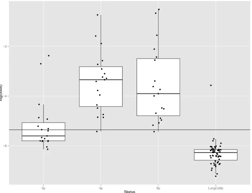
We additionally considered all 19 individuals (10 WM and 5 IgM MGUS from WM fami-lies, 4 MGUS from MM families) identified above with significant MYD88 L265P mutation allele fractions and performed deep Ion Torrent-based sequencing (mean read depth 18,000X) in DNA extracted across three time points: from blood prior to EBV-immortalization (0p), from LCLs after 4 passagesin vitro (4p) and 9 passages in vitro (9p), with the 9p points repeated for a subset of 13 samples. Eight out of 19 patients (42%) had detectable MYD88 L265P allele fractions above the detection threshold (AF 0.47–8.51%) prior to EBV-immortali-zation (0p), whereas 18 out of 19 had significant allele fractions at 4 and 9 passagesin vitro (Table 1). The proportion of the MYD88 L265P allele fraction increased within vitro passages (Fig 1) by a factor of 1.11 per passage (95% CI 1.07–1.16, p<0.001). The enrichment was more
evident between the 0p and 4p steps (S3 Fig), by a factor of 1.46 per passage (95%Ci 1.33–1.56 p<0.001), whereas samples reached a plateau or even lowered between 4p and 9p steps (0.97, 95%Ci 0.92–1.08 per passage, p = 0.29) (Table 1).
Analysis was performed on samples obtained from 19 patients (10 WM and 5 MGUS from WM families, 4 MGUS from MM families) found to carry the mutation, across three time points; from blood prior to EBV-immortalization (0p), from LCLs after 4 passagesin vitro (4p) and 9 passagesin vitro (9p), as well as in LCLs derived from 55 patients with lung cancer after 9 passagesin vitro (LungCa9p).
The black bars in the box structures represent the median value and the black dots outside the upper and lower whiskers correspond to outliers. The horizontal black line corresponds to the 0.47% threshold, as defined by the distribution of allelic fractions observed in the lung can-cer comparison group (seeMaterials and Methods). The outliers in the 0p sample group corre-spond to samples with high allelic frequency, while the outlier within the Lung Ca samples was found to be a patient with MGUS.
0p: prior to EBV-immortalization, 4p: after 4 passagesin vitro, 9p: after 9 passages in vitro, LungCa9p: lung cancer reference samples after 9 passagesin vitro.
Discussion
Our study initially aimed to investigate whether germ-line genetic variants in theMYD88 gene contribute to the genetic susceptibility to WM. We identified the presence of a rareMYD88 L265P mutation in individuals from three unrelated families. However, the presence of this variant did not explain the familial aggregation as it did not segregate with the disease within the pedigrees. Furthermore, given the observed higher variant allele fractions following EBV-immortalization and cell culture in comparison to the uncultured blood, theMYD88 L265P
mutation does not appear to be involved in genetic susceptibility to WM and is more likely of somatic origin. As such, it appears that our observation of theMYD88 L265P in DNA extracted from Lymphoblastoid cell lines is due to the presence of the mutation either in a sub-clone or in circulating free cell(s) (CFC), rather than a germline variant or cell free DNA. Indeed, the presence of clonotypic cells in peripheral blood is well described in WM. Smithet al. originally used flow cytometry to describe the presence of clonal cells in peripheral blood and suggested that it is a common finding in WM, while very rare in multiple myeloma (MM).[19] Kriang-kumet al. observed preferential use of VH3/JH4 gene families in WM and expansion of poly-clonal cells in peripheral blood.[20] Much more recently, the presence of somatic mutations in peripheral blood has been documented, suggesting that age-related clonal hematopoiesis is a common condition associated with an increased risk of hematologic cancer.[21] Our demon-stration of circulating cells harboring the somaticMYD88 L265P mutation in WM patients is consistent with these observations. However, primary cells rapidly apoptose in culture and EBV has generally been reported to infect but not to immortalize leukemic B lymphocytes.[22] It is thus unexpected that aMYD88 positive cell population derived from a patient sample would be susceptible to immortalization by EBV and proliferatein vitro over prolonged periods of time, raising the possibility that a precursor bearing the mutation but susceptible to immor-talization is present in the peripheral blood of patients and is being enriched with cell culture. Further description of the cellular characteristics of theMYD88 L265P positive clones may be informative.
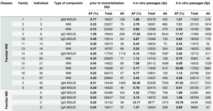
Table 1. MYD88 L265P allele fraction (AF %), given as the ratio of mutant/reference sequence reads, in samples prior to (0p) or after immortaliza-tion with EBV (4 and 9 passagesin vitro).
Disease Family Individual Type of component prior to immortalization (0p)
4in vitro passages (4p) 9in vitro passages (9p)
AF (%) Total Alt AF (%) Total Alt AF (%) Total Alt
Fa milial WM 1 1 IgM MGUS 0.77 18037 138 1.88 22478 422 1.81 11923 216 2 3 WM 0.33 23027 76 3.79 18091 685 7.21 25153 1814 2 4 WM 0.72 22352 161 3.60 13306 479 2.33 13195 308 3 6 IgG MGUS 1.29 19943 258 17.02 20819 3544 17.47 11066 1933 18 12 IgG MGUS 0.49 13014 64 0.87 15566 135 0.62 18559 115 11 14 WM 0.30 18473 56 0.44 16634 73 0.44 11912 52 13 16 WM 0.47 18797 89 2.25 13533 304 2.62 19223 503 14 19 IgM MGUS 6.30 17704 1116 6.15 17832 1097 3.25 15712 511 14 20 WM 0.34 20620 71 1.12 14142 158 0.75 6563 49 15 21 WM 0.45 14622 66 7.08 26112 1848 8.09 16425 1328 11 34 WM 0.37 18208 67 5.17 11832 612 33.96 17552 5961 16 35 WM 0.22 26473 57 0.77 16841 130 1.12 29768 334 5 37 WM 0.30 28694 87 3.42 13437 459 0.56 23214 131 5 45 IgM MGUS 8.51 16449 1400 32.40 22636 7335 37.42 20127 7531 9 46 IgM MGUS 0.30 16520 49 0.78 32414 252 0.91 29725 271 Fa milial MM A IgM MGUS 0.30 34466 104 4.32 17563 758 1.98 24381 483 B IgM MGUS 0.54 22647 123 3.19 19072 609 1.08 48700 528 C IgG MGUS 0.30 12142 36 13.17 9677 1274 10.79 9449 1020 D IgA MGUS 0.24 15617 37 1.27 16424 208 0.69 9656 67
AF: allele fraction; Total: total number of reads; Alt: number of reads with the mutant allele; WM: Waldenström disease; MM: multiple myeloma doi:10.1371/journal.pone.0136505.t001
EBV-transformed lymphoblastoid cell lines have been shown to evolve during long-term culture. Leeet al. have reported that “late” (161 passages) cells compared to “early” (4 passages) displayed phenotypic changes involving the NF-kappa B pathway and carcinogenesis-related genes.[23] Additionally, the ability of EBV to immortalize B cells may be linked to P53 content. [24] A study in diffuse large B-cell lymphoma, showed that the MYD88 L265P mutant pro-moted cell survival by triggering IRAK-mediated NF-κB signaling, acting as a gain of function driver mutation.[6] Thus, the increasing allele fraction we observe subsequent toin vitro cul-ture, and by inference the proportion of cells carrying theMYD88 L265P variant, may be due to a survival advantage conveyed to cells bearing this mutation. Our observation suggests that the establishment of lymphoblastoid cell lines could be used to enrich for certain types of rare B cell subpopulations present in patient samples.
TheMYD88 L265P mutation is highly frequently observed in the bone marrow (BM) from patients with WM, and relatively frequently in the BM from MGUS patients, while absent in
Fig 1. Distribution ofMYD88 L265P allele fractions in samples from patients with familial Waldenström Macroglobulinemia prior to or after immortalization by EBV, and in lymphoblastoid lines derived from patients with lung cancer.
doi:10.1371/journal.pone.0136505.g001
multiple myeloma patients.[25] In our series, we found this mutation to be significantly over-represented in families with a family history of WM relative to families with a family history of MM, consistent with the observation in BM samples in previous studies. This was largely due to its absence in MM patients as opposed to its presence in MGUS. Additionally, we noted the presence of theMYD88 L265P in patients with IgM or IgG-MGUS, as well as in MGUS patients from families with multiple myeloma.
Several studies have developed conventional and real-time allele-specific PCR assays (AS-PCR, ASO-RQ-PCR) for the identification of theMYD88 L265P mutation in a rapid and sensitive manner, at a lower detection limit of 0.1% to 0.25% when using BM-isolated CD19 + selected cells or BM samples.[26,27] Here, we used a combination of EBV-immortalized lymphoblastoid cell line culture and NGS techniques to detect this mutation in peripheral blood samples from WM and MGUS patients. Our findings suggest that cell culture when com-bined with sensitive mutation detection assays, may have potential as a relatively non-invasive method for detecting theMYD88 L265P mutation.
One important limitation of our study is that LCLs derived from patients with lung cancer were used as reference samples in order to calculate the detection limit of the next generation sequencing based assay for theMYD88 L265P mutation. While not available to the current study, LCLs from unaffected controls are necessary to establish a less biased estimate of the error rate of this assay, as well as the population prevalence of theMYD88 L265P mutation under such conditions.
In conclusion, our findings do not support the presence of germ-lineMYD88 mutations in familial WM. However, our results show the presence of circulating cells susceptible to immor-talization by EBV bearing theMYD88 mutation in the peripheral blood of some patients with familial WM or IgM-MGUS. Additional studies are required to determine whether the pres-ence of cells bearing theMYD88 L265P is correlated with disease progression and outcome in patients with WM or MGUS.
Supporting Information
S1 Fig. Family trees from patients with familial Waldenström disease and/or IgM MGUS. (JPG)
S2 Fig. Examples of pedigrees of families with peripheral blood cells harboring theMYD88 L265P mutation using targeted resequencing on an Illumina MiSeq.DNA samples were extracted after 9 passagesin vitro (9p), and the mutant allele fraction (%) is given as the ratio of mutant/reference reads. In the case of individual 45, DNA was extracted after 5 passagesin vitro (5p).
(JPG)
S3 Fig. Evolution ofMYD88 L265P allele fractions prior to and after immortalization by EBV.Analysis was performed on samples obtained from 19 patients positive for theMYD88 L265P mutation, across three time points; from blood prior to EBV-immortalization (0p), and from LCLs after 4 passagesin vitro (4p) and 9 passages in vitro (9p). The horizontal black line corresponds to the 0.47% threshold, as defined by the distribution of allelic fractions observed in the lung cancer comparison group (seeMaterials and Methods).
(JPG)
S1 Table. Number and age of cases with monoclonal components within Waldenström Macroglobulinemia families.n.a.: not available, WM: Waldenström Macroglobulinemia, MGUS: Monoclonal Gammopathy of Unknown Significance.aSamples not available (DOC)
S2 Table.MYD88 L265P allele fractions (AF %) in 55 lung cancer samples, given as the ratio of mutant/reference sequence reads.Total: total number of reads; Alt: number of reads with the mutant allele.
(DOCX)
Acknowledgments
The authors would like to thank the French National Cancer Institute (InCA) and the Fonda-tion Française pour la Recherche contre le Myélome et les Gammapathies monoclonales (FFRMG) for the support of this work.
Author Contributions
Conceived and designed the experiments: MP HAL JDM CD. Performed the experiments: MP PG. Analyzed the data: MF GB JL NN MV. Contributed reagents/materials/analysis tools: LG XL CD. Wrote the paper: MP JDM PG CD. Design of the study: MP HAL JDM CD. Patient accrual: LG XL CD. Lymphoblastoid cell line establishment: PG. Nucleic acid extraction: PG. Statistical analyses: MF GB. HaloPlex analyses: JL NN. Ion Torrent sequencing data analysis: MV. Manuscript writing: MP JDM PG CD.
References
1. Kyle RA, Therneau TM, Dispenzieri A, Kumar S, Benson JT, Larson DR, et al. Immunoglobulin m monoclonal gammopathy of undetermined significance and smoldering Waldenström Macroglobuline-mia. Clin Lymphoma Myeloma Leuk. 2013 Apr; 13(2):184–6 doi:10.1016/j.clml.2013.02.005PMID:
23490989
2. Treon SP, Hunter ZR, Aggarwal A, Ewen EP, Masota S, Lee C, et al. Characterization of familial Wal-denstrom's macroglobulinemia. Ann Oncol. 2006 Mar; 17(3):488–94. PMID:16357024
3. Kristinsson SY, Bjorkholm M, Goldin LR, McMaster ML, Turesson I, Landgren O. Risk of lymphoproli-ferative disorders among first-degree relatives of lymphoplasmacytic lymphoma/Waldenstrom macro-globulinemia patients: a population-based study in Sweden. Blood. 2008; 112(8):3052–3056. doi:10. 1182/blood-2008-06-162768PMID:18703425
4. Steingrímsson V, Lund SH, Turesson I, Goldin LR, Björkholm M, Landgren O, et al. Population-based study on the impact of the familial form of Waldenström macroglobulinemia on overall survival. Blood. 2015 Mar 26; 125(13):2174–5. doi:10.1182/blood-2015-01-622068PMID:25814489
5. Treon SP, Xu L, Yang G, Zhou Y, Liu X, Cao Y, et al. MYD88 L265P somatic mutation in Waldenström's macroglobulinemia. N Engl J Med. 2012 Aug 30; 367(9):826–33. doi:10.1056/NEJMoa1200710PMID:
22931316
6. Ngo VN, Young RM, Schmitz R, Jhavar S, Xiao W, Lim KH, et al. Oncogenically active MYD88 muta-tions in human lymphoma. Nature. 2011; 470(7332):115–119. doi:10.1038/nature09671PMID:
21179087
7. Walsh DE, Greene CM, Carroll TP, Taggart CC, Gallagher PM, O'Neill SJ, et al. Interleukin-8 up-regula-tion by neutrophil elastase is mediated by MyD88/IRAK/TRAF-6 in human bronchial epithelium. J Biol Chem. 2001; 276(38):35494–35499. PMID:11461907
8. Toffart AC, Timsit JF, Couraud S, Merle P, Moro-Sibilot D, Perol M, et al. Immunohistochemistry evalua-tion of biomarker expression in non-small cell lung cancer (Pharmacogenoscan study). Lung Cancer. 2014 Feb; 83(2):182–8. doi:10.1016/j.lungcan.2013.12.003PMID:24388706
9. Hammerschmidt W, Sugden B. Genetic analysis of immortalizing functions of Epstein-Barr virus in human B lymphocytes. Nature. 1989; 340(6232):393–397. PMID:2547164
10. Langmead B, Salzberg SL. Fast gapped-read alignment with Bowtie 2. Nat Methods. 2012 Mar 4; 9 (4):357–9. doi:10.1038/nmeth.1923PMID:22388286
11. Li H, Durbin R. Fast and accurate short read alignment with Burrows-Wheeler transform. Bioinformat-ics. 2009 Jul 15; 25(14):1754–60. doi:10.1093/bioinformatics/btp324PMID:19451168
12. McKenna A, Hanna M, Banks E, Sivachenko A, Cibulskis K, Kernytsky A, et al. The Genome Analysis Toolkit: a MapReduce framework for analyzing next-generation DNA sequencing data. Genome Res. 2010 Sep; 20(9):1297–303. doi:10.1101/gr.107524.110PMID:20644199
13. DePristo MA, Banks E, Poplin R, Garimella KV, Maguire JR, Hartl C, et al. A framework for variation dis-covery and genotyping using next-generation DNA sequencing data. Nat Genet. 2011 May; 43(5):491– 8. doi:10.1038/ng.806PMID:21478889
14. Van der Auwera GA, Carneiro MO, Hartl C, Poplin R, Del Angel G, Levy-Moonshine A, et al. From FastQ data to high confidence variant calls: the Genome Analysis Toolkit best practices pipeline. Curr Protoc Bioinformatics. 2013 Oct 15; 11(1110):11.10.1-11.10.33 PMID:25431634
15. Wang K, Li M, Hakonarson H. ANNOVAR: functional annotation of genetic variants from high-through-put sequencing data. Nucleic Acids Res. 2010 Sep; 38(16):e164. doi:10.1093/nar/gkq603PMID:
20601685
16. 1000 Genomes Project Consortium, Abecasis GR, Auton A, Brooks LD, DePristo MA, Durbin RM, et al. An integrated map of genetic variation from 1,092 human genomes. Nature. 2012 Nov 1; 491
(7422):56–65. doi:10.1038/nature11632PMID:23128226
17. Kumar P, Henikoff S, Ng PC. Predicting the effects of coding non-synonymous variants on protein func-tion using the SIFT algorithm. Nat Protoc. 2009; 4(7):1073–81. doi:10.1038/nprot.2009.86PMID:
19561590
18. Adzhubei I, Jordan DM, Sunyaev SR. Predicting functional effect of human missense mutations using PolyPhen-2. Curr Protoc Hum Genet. 2013 Jan;Chapter 7:Unit7.20.
19. Smith BR, Robert NJ, Ault KA. In Waldenstrom's macroglobulinemia the quantity of detectable circulat-ing monoclonal B lymphocytes correlates with clinical course. Blood. 1983; 61(5):911–914. PMID:
6403083
20. Kriangkum J, Taylor BJ, Treon SP, Mant MJ, Belch AR, Pilarski LM. Clonotypic IgM V/D/J sequence analysis in Waldenstrom macroglobulinemia suggests an unusual B-cell origin and an expansion of polyclonal B cells in peripheral blood. Blood. 2004; 104(7):2134–2142. PMID:14764523
21. Jaiswal S, Fontanillas P, Flannick J, Manning A, Grauman PV, Mar BG, et al. Age-Related Clonal Hematopoiesis Associated with Adverse Outcomes. N Engl J Med. 2014; 371(26):2488–2498. doi:10. 1056/NEJMoa1408617PMID:25426837
22. Doyle MG, Catovsky D, Crawford DH. Infection of leukaemic B lymphocytes by Epstein Barr virus. Leu-kemia. 1993; 7(11):1858–1864. PMID:8231253
23. Lee JE, Nam HY, Shim SM, Bae GR, Han BG, Jeon JP. Expression phenotype changes of EBV-trans-formed lymphoblastoid cell lines during long-term subculture and its clinical significance. Cell Prolif. 2010; 43(4):378–384. doi:10.1111/j.1365-2184.2010.00687.xPMID:20590663
24. Szekely L, Pokrovskaja K, Jiang WQ, Selivanova G, Löwbeer M, Ringertz N, et al. Resting B-cells, EBV-infected B-blasts and established lymphoblastoid cell lines differ in their Rb, p53 and EBNA-5 expression patterns. Oncogene. 1995; 10(9):1869–1874. PMID:7753563
25. Mori N, Ohwashi M, Yoshinaga K, Mitsuhashi K, Tanaka N, Teramura M, et al. L265P mutation of the MYD88 gene is frequent in Waldenström's macroglobulinemia and its absence in myeloma. PLoS One. 2013 Nov 5; 8(11):e80088. doi:10.1371/journal.pone.0080088PMID:24224040
26. Xu L, Hunter ZR, Yang G, Zhou Y, Cao Y, Liu X, et al. MYD88 L265P in Waldenström macroglobuline-mia, immunoglobulin M monoclonal gammopathy, and other B-cell lymphoproliferative disorders using conventional and quantitative allele-specific polymerase chain reaction. Blood. 2013 Mar 14; 121 (11):2051–8. doi:10.1182/blood-2012-09-454355PMID:23321251
27. Jiménez C, Chillón Mdel C, Balanzategui A, Puig N, Sebastián E, Alcoceba M, et al. Detection of MYD88 L265P mutation by real-time allele-specific oligonucleotide polymerase chain reaction. Appl Immunohistochem Mol Morphol. 2014 Nov-Dec; 22(10):768–73. doi:10.1097/PAI.0000000000000020