Sarcoptic mange and cheetah conservation in Masai Mara
(Kenya): epidemiological study in a wildlife/livestock system
FRANCIS GAKUYA1*, JACKSON OMBUI2, NDICHU MAINGI3, GERALD MUCHEMI2,
WILLIAM OGARA2, RAMÓN C. SORIGUER4and SAMER ALASAAD4,5
1
Department of Veterinary and Capture Services, Kenya Wildlife Service, Kenya 2
Department of Public Health, Pharmacology & Toxicology, University of Nairobi, Kenya 3
Department of Pathology and Microbiology, University of Nairobi, Kenya 4
Estación Biológica de Doñana, Consejo Superior de Investigaciones Científicas (CSIC), Avda. Américo Vespucio s/n 41092 Sevilla, Spain
5
Institute of Evolutionary Biology and Environmental Studies (IEU), University of Zürich, Winterthurerstrasse 190, 8057 Zürich, Switzerland
(Received 13 March 2012; revised 11 April, 13 April and 7 May 2012; accepted 8 May 2012; first published online 19 July 2012) S U M M A R Y
The sanitary control of threatened wild animals is of pivotal interest for their conservation. This task, however, is highly complex in wildlife/livestock systems. In this paper we report findings from a 2-year cross-sectional study of the epidemiology and attempted control of a Sarcoptes mite infestation in the threatened cheetah population in Masai Mara (Kenya), and discuss its interaction with sympatric wild (lion, wildebeest and Thomson’s gazelle) and domestic (dog, cattle and sheep) animals. Sarcoptes scabiei was isolated from cheetahs, Thomson’s gazelles, wildebeests, lions, cattle, goats and dogs; Psoroptes ovis, on the other hand, was only isolated from sheep. The prevalence study revealed 12·77% infection rates in cheetahs, 4·7% in dogs, 0·8% in Thomson’s gazelles, 0·8% in sheep, 0·09% in cattle, and 0·09% in goats, while it opportunistically affected lions and wildebeest. Our study revealed that prevalence of Sarcoptes mite in cheetah population was not associated with the studied geographical blocks, animal sex or the presence of affected domestic animals. Cheetah infection with S. scabiei was associated with the climatic conditions (dry more than wet season) and the balancing between the total number of Thomson’s gazelles and the prevalence of infected individuals. Apparently the high prevalence of mangy gazelles has a negative effect on cheetah; this negative effect was reduced when the number of healthy gazelles was increased. Treatment with injectable ivermectin of the clinically affected wild and domestic animals during the first year of this study was associated with much lower incidence of sarcoptic mange during the second year.
Key words: Acinonyx jubatus, Sarcoptes scabiei, Psoroptes ovis, wildlife/livestock boundary, parasite control, treatment.
I N T R O D U C T I O N
The Sarcoptes mite was regarded as one of the main infectious causes of mortality in the cheetah (Acinonyx jubatus) population in the Masai Mara ecosystem (Kenya) (Mwanzia et al.1995; Ngoru and Mulama, 2002). This threatened population is estimated at only 61 animals (Gros, 1998), sharing habitat with other wild and domestic animals that are
known to be affected by the Sarcoptes mite
(Siegmund et al. 1973; Mugera et al. 1979; Blood and Radostitis, 1989; Ngoru and Mulama, 2002; Pence and Ueckermann,2002; Kahn et al.2005). The Masai Mara ecosystem is inhabited by pastoral communities and the interaction between wildlife and livestock creates a rich platform for disease transmission in this shrinking ecosystem (Holmes, 1996). The illegal incursion of livestock into pro-tected areas occurs mostly during the dry season, which increases the chance of wildlife/livestock interaction. Major outbreaks of disease in this
interface system have been associated with droughts, when limited water and food supplies cause increased interaction between livestock and wildlife (Kock et al. 1999).
A number of mite species that affect domestic animals and wildlife (Pence and Ueckermann,2002; Kahn et al.2005; Alasaad et al.2011c) are of zoonotic importance (Bornstein et al.2001; Fischer et al.2003; Kahn et al. 2005; Navarro-Gonzalez et al. 2009; Alasaad et al. 2012). The Sarcoptes mite is a con-tagious skin disease (Zumla and Croft,1992) and is spread by direct and indirect contact with other diseased animals or objects that have been in contact with affected animals (Siegmund et al. 1973). The introduction of a single case of sarcoptic mange into overcrowded living conditions can lead to an epi-demic (Obasanjo et al. 2001) causing potentially devastating mortality in both wild and domestic animals (Bornstein et al.2001).
Sarcoptic mange has been reported in a number of different wildlife species in the Masai Mara
ecosys-tem (Mwanzia et al. 1995; Ngoru and Mulama,
2002), although a general study on the various aspects of Sarcoptes mite epidemiology and affected cheetah * Corresponding author: Department of Veterinary and
Capture Services, Kenya Wildlife Service, Kenya. E-mail: gakuya@kws.go.ke
treatment has yet to be performed. Any such epidemiological and control study would have to confront the difficulties inherent to a complex wild/ domestic animal system. All previous observational studies of mange-like skin diseases in cheetahs are from the Koiyaki and Lemek community conservan-cies in Masai Mara (Mwanzia et al.1995).
The aim of our study was to contribute to knowl-edge of the epidemiology of the sarcoptic mange in the threatened cheetah population from the wildlife/ livestock ecosystem Masai Mara, and to report on the treatment strategies undertaken thus far.
M A T E R I A L S A N D M E T H O D S
Study area
The study was carried out in the Masai Mara ecosystem (Kenya), which comprises the Masai Mara National Reserve (NR), the Mara Conservancy and the surrounding community ranches of Koiyaki, Siana, Lemek, Olkinyei and Ol Choro Orogwa. The Masai Mara N.R. and Mara Conservancy form the protected area of the Masai Mara ecosystem (10°13′ and 10°45′ S, and 34°045′ and 35°25 E), which covers approximately 1,850 km2and is located at the north-ern tip of the Serengeti National Park (Tanzania).
In the study area, all members of the ‘Big Five’ animal group (lion, leopard, African elephant, African buffalo, and Black Rhinoceros) are present. The wildebeest are the dominant herbivores, and their numbers are estimated in millions. Around July, these animals migrate north from the Serengeti plains in search of fresh pasture, and return to the south around October. The migrants are followed by predators, most notably lions and hyenas. Other herbivores are present including Thomson’s and Grant’s gazelles, impalas, topi, elands, duikers, Coke’s hartebeests, zebras and Masai giraffes. A pastoral community whose livelihood is dependent on livestock lives in the surrounding area and, as a result, interaction between livestock and wildlife is frequent.
Animal data
Observational data of mange-like skin disease was collected over a period of 2 years (November 2007– November 2009). The disease was recorded based on clinical observation, if at least 3 of the following lesions were observed in an animal: pruritus, alopecia, crust formation, skin roughening and poor body condition. The study area was divided into 8 blocks (Fig. 1). Our study was a combination of opportu-nistic and systematic (cross-sectional) sampling. Affected lions, wildebeests and dogs were opportu-nistically reported by KWS veterinarians and Masai Pastoralists. For cheetahs, Thomson’s gazelles and all domestic animals we carried out monitoring 4 times
per year (October, January, April and July).
Monitoring was carried out by 2 veterinarians and 1 technician. A distance of about 200 km per 2 days was covered per each block in each time (October, January, April and July). Domestic animals were randomly selected at both household and individual levels. The total number of herds of studied cattle, goats and sheep were 15, 20 and 32, respectively. Wild animals were observed using binoculars and the naked eyes. Since the Masai Mara area is quite secure for wildlife with high visitation, most wild animals are not shy and can be approached closely. The animals observed were cheetah (Acinonyx jubatus), Thomson’s gazelle (Eudorcas thompsonii), sheep (Ovis aries), cattle (Bos indicus), goat (Capra hircus), dog (Canis lupus familiaris), wildebeest (Connochaetes taurinus) and lion (Panthera leo). GPS co-ordinates of all individual animals and/or herds observed to have clinical signs of mange, as well as of healthy herds and animals, were recorded. The size of herds varied from 50 up to 300 animals. The seasons were divided into the dry season (January–March and July–
September) and wet season (April–June and
October–December). The studied wild and domestic species were monitored for Sarcoptes-like lesions in all blocks except block 8 (Ol Choro Orogwa), due to its inaccessibility.
A stratified random sampling method was used to identify the sampling units. Domestic animal stra-tification was based on the study blocks (community ranches) that were closest to the protected areas or to where cheetahs are known to occur.
Within the strata, mangy animals were randomly sampled. Cheetah sex and age classes (adults51 year and cubs <1year) were determined.
Capture and treatment of mange-infected animals Animals were determined to be affected through observation of clinical signs. Any animal that showed a mange-like skin condition was a candidate for treatment. Wild herbivores were captured by chemi-cal immobilization through darting using etorphine hydrochloride (M99® 9·8 mg/ml, Novartis South Africa Pty Ltd, Isando, South Africa) combined with Xylazine hydrochloride (Ilium Xylazil-100 100 mg/ml, Troy Laboratories Pty Ltd, Smithfield, Australia) while wild carnivores were captured using ketamine hydrochloride (100 mg/ml Agraket®, Agrar Holland Bv) combined with Xylazine hydrochloride. Mangy domestic animals were treated since it was easy to restrain them. The immobilized wild animals were treated in the field with 0·2 mg/kg bwt of injectable 1% Ivermectin (Kalamectin 1% w/v, Kela
NV, St Lenaartseweg, Belgium), administered
subcutaneously.
After sampling and treatment, the herbivores were revived with diprenorphine hydrochloride (M5050® 1588 Francis Gakuya and others
https:/www.cambridge.org/core/terms. https://doi.org/10.1017/S0031182012000935
12 mg/ml, Novartis South Africa Pty Ltd, Isando,
South Africa) and Atipamezole hydrochloride
(Antisedan® 5 mg/ml, Pfizer laboratories Pty Ltd, Sandton, South Africa) immediately, while the carnivores were revived using Atipamezole hydro-chloride after 30 to 45 min of immobilization to allow
Ketamine hydrochloride to wear off. Domestic
animals were re-treated after 1 month, if necessary. Treated wild animals were ear-tagged to follow their progress.
Ethics
The Committee of the Department of Veterinary and Capture Services of the Kenya Wildlife Service (KWS) approved the study including animal captur-ing and treatment protocols. KWS guidelines on Wildlife Veterinary Practice-2006 were followed. All KWS veterinaries were guided by the Veterinary Surgeons Act Cap 366 Laws of Kenya that regulates veterinary practices in Kenya.
Parasite collection and identification
Affected areas of skin from representative sam-ples randomly selected (namely: 8 cheetahs, 10
Thomson’s gazelle, 9 dogs, 51 sheep, 5 wildebeest, 2 cattle, 1 goat and 2 lions) were scraped with a scalpel until they bled to obtain hairs and crusts for parasitological examination. The scrapings were placed in universal bottles containing 70% ethanol and transported to the laboratory. A portion was removed from the alcohol and clarified in KOH to recover parasites for microscopy (Alasaad et al. 2009a). All mites were identified as S. scabiei or Psoroptes ovis on the basis of known morphological criteria (Fain, 1968; Margaret and Rusell, 1978; Sanders et al.2000).
Data analyses
R Package V.2.11.1. software was used. A (log +1) transformation had been applied to all variables prior to analysis. General Linear Models (GLMs) with binomial, quassibinomial distribution and logit link were tested following the proposal of Vicente et al. (2006). After transformation, Linear Model with R (lm function) was applied.
R E S U L T S
The clinical observation together with the examin-ation of the skin samples revealed that cheetah, Fig. 1. Map showing the study blocks in the Masai Mara ecosystem (Kenya) together with the distribution of affected animals of all species.
Thomson’s gazelle, wildebeest, lion, dog, goat and cattle all suffered from infestation by Sarcoptes scabiei, while sheep were affected by P. ovis.
The estimated total number of mangy and healthy animals in the studied area is shown inTable 1. The prevalence of mange was highest in cheetahs 12·8% (n = 47), followed by dogs 4·7 (n = 279), while the lowest was 0·09% in cattle (n = 2311) and goats (n = 1174). The prevalence of affected Thomson’s gazelle and sheep was 0·81% (n = 10 788) and 0·76% (n = 6699), respectively. Two herds of cattle (total 15 herds), 1 herd of goats (total 20 herds) and 13 of sheep (total 32 herds) were affected with mange mite giving a herd prevalence of 13·3%, 5% and 40·6% respect-ively. Only 2 lions and 5 wildebeest were found to be affected with sarcoptic mange. The prevalence of sarcoptic mange was 12·5% in female cheetahs and 9·6% in male cheetahs. The prevalence in adult cheetahs was 11·8% as opposed to cub cheetahs which was 15·3%. In both cases the differences were not statistically supported (P > 0·5). During the study period 2 cheetahs, a cub and an adult female died due to complications of mange infestation, while we did not identify any carcass of mangy Thomson’s gazelle, but we cannot discard the possibility that affected carcasses were removed by scavengers.
Affected animals were observed in 6 of the 7 blocks studied (Fig. 1). No affected animals were observed in block 6. The greatest prevalence of affected animals was in blocks 1 to 5. Affected animals of different species were observed in the different blocks, namely cheetahs in blocks 1, 3, and 5; Thomson’s gazelles in blocks 2, 3, 5 and 7; sheep in blocks 4 and 5; dogs in blocks 4 and 5; wildebeest in block 1. The affected lions were observed in block 2, while cattle and goat were observed in block 5. In terms of seasonality, affected animals were observed in blocks 1, 2, 3 and 5 during the dry season and in blocks 3 and 4 during the wet season.
Although we observed 5 affected wildebeest in 2 herds of about 100 animals which gave a prevalence of 2·0%, the sightings were from a single block at a single point in time, hence we did not carry out statistical analysis. That was the same case with the 2 lions that were observed as the study progressed. The 2 wild species were not included in the cross-sectional study. The temporal prevalence during the study period in all species is shown in Fig. 2. At the beginning prevalence increased steadily in cheetahs, Thomson’s gazelles and dogs, with the peak infestation point in thefirst 2 being reached in January 2008. The peak infestation in dogs occurred later in April 2008. There was another peak in July 2008 in cheetahs and in dogs in October 2008, while the prevalence in sheep had a different pattern. No infestation was observed in cheetahs after October 2008 or in dogs after April 2009. Prevalence in Thomson’s gazelles dropped steadily in April 2008 and was low up to April 2009.
For all studied species (with the exception of cheetah) there was no statistical association with the geographical blocks or seasons (dry or wet). The prevalence of the Sarcoptes mite in cheetah was not associated to the geographical blocks, animal sex or the presence of affected domestic animals. Cheetah infection with S. scabiei was associated with the season (dry more than wet season; F = 10·1822, P = 0·007) and the interaction between the total number of healthy gazelles and the prevalence of infected gazelles (F = 7·4489, P = 0·018). The high prevalence of mangy gazelles had a negative effect on cheetah; nonetheless this negative effect gradually decreased when the number of healthy gazelles increased. The best model was season effect with an interaction between mangy and healthy gazelle (logMangyCheetah* logMangyGazelle: logHealthy-Gazelle + Season) (For more details see Table 2 and Fig. 3).
The treated animals recovered completely within approximately 1 month. Re-treatment (1 month after the first dose) was only necessary in a few cases in domestic animals, while none of the wild animals was re-treated during the study period. As the study progressed the numbers of observed affected animals declined due to the continuous treatment of all sampled animals, and by July 2009 no positive cases in any of the species studied were found, and none of the dead animals were reported to be caused by Sarcoptes mite (Fig. 2).
D I S C U S S I O N
In this study S. scabiei was isolated in all animals that were positive for mange except sheep, where P. ovis was isolated. These observations agree with those reported by other authors who have described sarcoptic mange as the commonest in wildlife
(Pence and Ueckermann, 2002), and psoroptic
mange likewise as the commonest in sheep (Mugera et al.1979; Blood and Radostitis,1989; Kusiluka and Kambarase, 1996). Although affected sheep were consistently observed in the ecosystem throughout the study period, only Psoroptes mites were ever observed. This confirms the idea that there is no mange transmission between sheep and the other studied wild and domestic animals.
Our prevalence study of the mange-like skin disease revealed that cheetahs had the highest prevalence (12·8%) of all the studied animals.
Prevalence in female cheetahs was higher than in male cheetahs, and also higher in cubs than in adults. Although these prevalences are not significantly different, the greater prevalence in females and cubs does suggest that these two groups of cheetahs are more vulnerable since they live together and the probability of transmission is higher. No clear age structuring of scabietic wild animals was reported in many cases (Rossi et al. 2007). However, some 1590 Francis Gakuya and others
https:/www.cambridge.org/core/terms. https://doi.org/10.1017/S0031182012000935
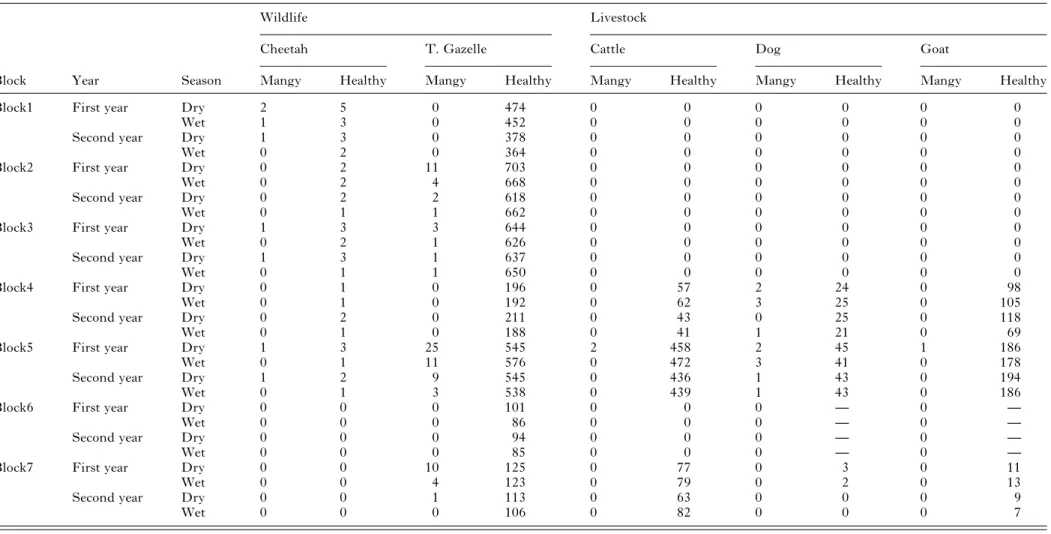
Table 1. Number of affected (mangy) and healthy animals in the Masai Mara ecosystem, stratified by block, year and season (dry and wet)
Wildlife Livestock
Cheetah T. Gazelle Cattle Dog Goat
Block Year Season Mangy Healthy Mangy Healthy Mangy Healthy Mangy Healthy Mangy Healthy
Block1 First year Dry 2 5 0 474 0 0 0 0 0 0
Wet 1 3 0 452 0 0 0 0 0 0
Second year Dry 1 3 0 378 0 0 0 0 0 0
Wet 0 2 0 364 0 0 0 0 0 0
Block2 First year Dry 0 2 11 703 0 0 0 0 0 0
Wet 0 2 4 668 0 0 0 0 0 0
Second year Dry 0 2 2 618 0 0 0 0 0 0
Wet 0 1 1 662 0 0 0 0 0 0
Block3 First year Dry 1 3 3 644 0 0 0 0 0 0
Wet 0 2 1 626 0 0 0 0 0 0
Second year Dry 1 3 1 637 0 0 0 0 0 0
Wet 0 1 1 650 0 0 0 0 0 0
Block4 First year Dry 0 1 0 196 0 57 2 24 0 98
Wet 0 1 0 192 0 62 3 25 0 105
Second year Dry 0 2 0 211 0 43 0 25 0 118
Wet 0 1 0 188 0 41 1 21 0 69
Block5 First year Dry 1 3 25 545 2 458 2 45 1 186
Wet 0 1 11 576 0 472 3 41 0 178
Second year Dry 1 2 9 545 0 436 1 43 0 194
Wet 0 1 3 538 0 439 1 43 0 186
Block6 First year Dry 0 0 0 101 0 0 0 — 0 —
Wet 0 0 0 86 0 0 0 — 0 —
Second year Dry 0 0 0 94 0 0 0 — 0 —
Wet 0 0 0 85 0 0 0 — 0 —
Block7 First year Dry 0 0 10 125 0 77 0 3 0 11
Wet 0 0 4 123 0 79 0 2 0 13
Second year Dry 0 0 1 113 0 63 0 0 0 9
Wet 0 0 0 106 0 82 0 0 0 7 —, Not estimated. 1591 Sarcop tic mange and cheetah conserva tion in Kenya . https://doi.org/10.1017/S0031182012000935 https:/www.cambridge.org/core
. University of Basel Library
, on
30 May 2017 at 13:23:23
reports indicate higher prevalences in young animals, and a decrease with advancement of age (Tikaram and Ruprah, 1968; Munang’andu et al. 2010; Alasaad et al.2012).
Our study also revealed low prevalence in
Thomson’s gazelle, which, nevertheless, is still significant since it is to our knowledge the first report of mange in free-ranging Thomson’s gazelles in Kenya. The sample size (n = 10 788) was very large and since this gazelle is a favourite prey item of the cheetah (Ngoru and Mulama, 2002; Hayward et al. 2006), there is also a high probability that trans-mission occurs between these two species. Our study further shows that, although dogs had the second highest prevalence, affected dogs were observed exclusively in market places, which suggests that there transmission to cheetahs and other wild animals is unlikely in the study area.
No association was found between Sarcoptes infection in cheetah and sympatric domestic animals, and this suggests limited, if any, interaction between wild and domestic animals in the transmission and
maintenance of this pathogen. This is in contrast with other disease models, in which the expansion of humans and their livestock into wildlife areas had resulted in increased risk of transmission (Bengis, 2002; Jones et al. 2008). However, in this study affected cheetahs were found close to affected Thomson’s gazelles, suggesting that there could be transmission of mites from Thomson’s gazelles to cheetahs during feeding. Noteworthy, this gazelle is the favourite prey item of the cheetah (Ngoru and Mulama, 2002; Hayward et al. 2006; Gakuya et al. 2011). The high prevalence of mangy Thomson’s gazelles had a negative effect on cheetahs, nonetheless this negative effect was contrasted when a high number of healthy gazelles was present. The speci fi-city of S. scabiei by its host species has been the subject of an ongoing debate (Warburton, 1920; Burgess,1994). This debate has been advanced with the advent of PCR technology and the molecular marker systems in the genetic era. Studies on the coding sequences in combination with microsatellite markers provided support for a genetic di fferen-tiation of S. scabiei (Walton et al.1999,2004; Alasaad et al.2008, 2009b,2011b). In Europe, it was shown that the Sarcoptes mite is host-taxon derived in its effect on wildlife (Rasero et al.2010), and that host-parasite specificity is temporally stable in the short term (Alasaad et al.2011a). Nevertheless, the studied wild animals in Europe lacked any clear predator-prey interaction and putative inter-specific trans-mission models such as the cheetah and the Thomson’s gazelle in this study could not be explored.
Our study also revealed greater numbers of affected cheetah in the dry as opposed to the wet season. A winter rise in the number of Sarcoptes outbreak episodes has been reported worldwide, especially in Europe, in several wild and domestic hosts (Bornstein 2001; Rossi et al. 2007). This seasonal variation is thought to be related to the fertility of Sarcoptes mites, where the maximum egg production by adult female mites occurs in the autumn, and is low to null during January to July (Sokolova et al. 1989). However, the seasonality of Sarcoptes cases in Africa is more related to the dry season, suggesting that stress factors such as the scarcity of water and grazing pastures during the dry season may lead to greater individual susceptibility to mite infection (Malan et al.1997; Munang’andu et al.2010; Alasaad et al.2012).
We noted an association between prevalence and the temporal effect in all the study animals. Mange prevalence was higher in 2007/2008 than in 2008/ 2009 and by July 2009 patent cases were hard tofind. All wild animals treated were ear-tagged and we didn’t recapture any that was affected by Sarcoptes that had been ear-tagged. However, it was difficult to estimate the recovery time of the wild animals due to environmental difficulties, and to the fact that our Fig. 2. The prevalence of Sarcoptes infestation in the
study species from Masai Mara in the period 2007–2009.
Table 2. GLM Model coefficients analysing the relationship between mangy cheetahs and seasons (wet and dry), and the total number of mangy and healthy gazelles (GzM, mangy gazelle; GzH, healthy gazelle; Seasonf [T.2], wet season. Pr(>|t|), significance level. Residual standard error: 0·117. Multiple R-squared = 0·5473. Adjusted
R-squared = 0·4266. F-statistic = 4·534. P-value = 0·01337.) Estimate Std, Error t, value Pr(> |t|) (Intercept) −0·7661 0·4517 −1·6960 0·1105 logGzM 3·4306 1·3026 2·6340 0·01879* logGzH 0·1660 0·0776 2·1390 0·04925* Seasonf[T,2] −0·1750 0·0537 −3·2600 0·00527** logGzM: logGzH −0·5445 0·2055 −2·6490 0·01822* 1592 Francis Gakuya and others
https:/www.cambridge.org/core/terms. https://doi.org/10.1017/S0031182012000935
study was based on transect visits with trimester intervals, while animals may completely recover from sarcoptic mange infection in less than 3 months. For instance, Munang’andu et al. (2010) reported a recovery prevalence of 82% in mangy African buffaloes after a single dose of ivermectin, and Goldust et al. (2012) reported a cure rate of 85·9% at a 2-week interval with a single dose of ivermectin in humans. It would be tempting to assume that treatment of clinically affected individuals belonging to a range of sympatric hosts contributed to control of sarcoptic mange at the population level but, with all evidence, the trial was not designed to test it. As a consequence, the alternative hypothesis that inci-dence of mange spontaneously decreased during the study period, as registered elsewhere (Pence and Uecke, 2002), cannot be ruled out. For further studies, radio-equipment of treated wildlife would be desirable to improve the quality of individual follow-up and adequately monitor the health of in-contact conspecifics.
In conclusion, our study shows that there is no
evidence of an association between Sarcoptes
infection in wild and domestic animals. Cheetah infection with Sarcoptes was associated with climatic stressors and the presence of mangy Thomson’s gazelles ‘the favourite prey’; however, this negative effect was contrasted when a high number of healthy gazelles was present. Cheetahs may preferentially select mangy preys, since the affected preys could have a reducedflight response compared with healthy individuals. More investigation is needed to demon-strate the efficacy of the therapeutic treatment of mangy individuals as a strategy for effective and sustainable conservation of threatened cheetahs within Sarcoptes outbreak areas.
A C K N O W L E D G E M E N T S
We thank the staff of the Department of Veterinary and Capture Services, Kenya Wildlife Service for assisting in data collection.
F I N A N C I A L S U P P O R T
The research was supported by Kenya Wildlife Service, Proyecto de Excelencia RNM 06400 (Junta de Andalucía, Fig. 3. Trellis of effects plots of the interaction logGzM (mangy gazelle), logGzH (healthy gazelle) and LogCh (mangy cheetah). All variables had been (log(x + 1) transformed. Horizontal axis: Mangy gazelles (GzM). Vertical axis: Mangy Cheetah (Ch). Each partial graph represents different values of healthy gazelles (GzH). Increasing values from left bottom corner to top right.
Spain) and Juan de la Cierva Grant (Ministerio Innovación y ciencia, Spain).
R E F E R E N C E S
Alasaad, S., Ndeereh, D., Rossi, L., Bornstein, S., Permunian, R., Soriguer, R. C. and Gakuya, F. (2012). The opportunistic Sarcoptes scabiei: A new episode from giraffe in the drought-suffering Kenya. Veterinary Parasitology185, 359–363. doi.org/10.1016/j.vetpar.2011.10.039. Alasaad, S., Oleaga, A., Casais, R., Rossi, L., Molinar-Min, A., Soriguer, R. and Gortazar, C. (2011a). Temporal stability in the genetic structure of Sarcoptes scabiei under the host-taxon law: empirical evidences from wildlife-derived Sarcoptes mite in Asturias, Spain. Parasites & Vectors 4, 1–7.
Alasaad, S., Rossi, L., Soriguer, R. C., Rambozzi, L., Soglia, D., Pérez, J. M. and Zhu, X. Q. (2009a) Sarcoptes mite from collection to DNA extraction: the lost realm of the neglected parasite. Parasitology Research104, 723–732.
Alasaad, S., Schuster, R. K., Gakuya, F., Theneyan, M., Jowers, M. J., Maione, S., Molinar-Min, A., Soriguer, R. C. and Rossi, L. (2011b) Applicability of molecular markers to determine parasitic infection origins in the animal trade: A case study from Sarcoptes mites in wildebeest. Forensic Science Medicine and Pathology. DOI 10.1007/s12024-011-9268-z
Alasaad, S., Soglia, D., Spalenza, V., Maione, S., Soriguer, R. C., Pérez, J. M., Rasero, R., Ryser Degiorgis, M. P., Nimmervoll, H., Zhu, X. Q. and Rossi, L. (2009b). Is ITS-2 rDNA suitable marker for genetic characterization of Sarcoptes mites from different wild animals in different geographic areas?. Veterinary Parasitology 159, 181–185.
Alasaad, S., Soglia, D., Sarasa, M., Soriguer, R. C., Pérez, J. M., Granados, J. E., Rasero, R., Zhu, X. Q. and Rossi, L. (2008). Skin-scale genetic structure of Sarcoptes scabiei populations from individual hosts: empirical evidence from Iberian ibex-derived mites. Parasitology Research 104, 101–105.
Alasaad, S., Walton, S., Rossi, L., Bornstein, S., Abu-Madi, M., Soriguer, R., Fitzgerald, S., Zhu, X. Q., Zimmermann, W., Uade Ugbomoiko, U. S., Pei, K. J. C. and Heukelbach, J. (2011c ). Sarcoptes-World Molecular Network (Sarcoptes-WMN): integrating research on scabies. International Journal of Infectious Diseases15, 294–297.
Bengis, R. G., Kock, R. A. and Fisher, J. (2002). Infectious animal diseases: the wildlife/livestock interface. Revue scientifique et technique. (International Office of Epizootics.) 21, 53–65.
Burgess, I. (1994) Sarcoptes scabiei and scabies. Advances in Parasitology 33, 235–292.
Blood, D. C. and Radostitis, O. M. (1989). Mange. In Veterinary Medicine. A Textbook of the Diseases of Cattle, Sheep, Pigs, Goats and Horse 7th Edn (ed. Blood, D. C. and Radostitis, O. M.), pp. 1094–1099. Saunders, Philadelphia, PA, USA.
Bornstein, S., Morner, T. and Samuel, W. M. (2001). Sarcoptes scabiei and sarcoptic Mange. In Parasitic Disease of Wild Animals 2nd Edn. (ed. Samuel, W. M., Pybus, M. S. and Kocan, A. A.). pp. 107–119. Iowa State University Press, Ames, IO, USA.
Fain, A. (1968). Étude de la variabilité de Sarcoptes scabiei avec une revision des Sarcoptidae. Acta Zoologica et Pathologica Antverpiensia47, 1–196. Fischer, K., Holt, C. D., Harumal, P., Currie, B. J., Walton, F. S. and Kemp, D. J. (2003). Generation and characterization of cDNA clones from Sarcoptes scabiei Var. hominis for an expressed sequence tag library: identification of homologues of house dust miteallergens. The American Journal of Tropical Medicine and Hygiene68, 61–64.
Gakuya, F., Rossi, L., Ombui, J., Maingi, N., Muchemi, G., Ogara, W., Soriguer, R. C. and Alasaad, S. (2011). The curse of the prey: Sarcoptes mite molecular analysis reveals potential prey-to-predator parasitic infesta-tion in wild animals from Masai Mara, Kenya. Parasites &Vectors4, 1–7. Goldust, M., Rezaee, E. and Hemayat, S. (2012). Treatment of scabies: Comparison of permethrin 5% versus ivermectin. The Journal of Dermatology39, 545–547. doi:10.1111/j.1346-8138.2011.01481.x. Gros, P. M. (1998). Status of the cheetah Acinonyx jubatus in Kenya: a field-interview assessment. Biological Conservation 85, 137–149. Hayward, M. W., Hofmeyer, M., O’Brien, J. O. and Kerley, G. I. H. (2006). Prey preferences of cheetah (Acinonyx jubatus) (Felidae: Carnivora): Morphological limitations or need to capture rapidly consumable prey before kleptoparasites arrive. Zoology270, 615–627.
Heukelbach, J. and Feldmeier, H. (2006). Scabies. Lancet 367, 1767–1774.
Holmes, J. C. (1996). Parasites as threats to biodiversity in shrinking ecosystems. Biodiversity and Conservation5, 975–983.
Jones, K. E., Patel, N. G., Levy, M. A., Storeygard, A., Balk, D., Gittleman, J. L. and Daszak, P. (2008). Global trends in emerging infectious diseases. Nature, London451, 990–993.
Kahn, C. M., Line, S., Allen, D. G., Anderson, D. P., Jeffcoh, L. B. and Quesenberry, K. E. (2005). Acariasis mite infestation. In The Merck Veterinary Manual 9th Edn., (ed. Radostitis, O. M), pp. 742–749. Merck and Co Inc. Whitehouse Station, New Jersey, USA.
Kock, R. A., Wambua, J. M., Mwanzia, J., Wamwayi, H., Ndungu, E. K., Barett, T., Kock, N. D. and Rossiter, P. B. (1999). Rinderpest epidemic in wild ruminants in Kenya 1993–1997. Veterinary Record145, 275–283.
Kusiluka, L. and Kambarase, D. (1996). Diseases caused by arthropods and fungi. In Communicable diseases of Small Ruminants in Sub-saharan Africa. Edited and published by VETAID, Centre for Tropical Veterinary Medicine, Easter Bush, Roslin, Midlothian EH259RG, Scotland, UK.
Malan, F. S., Horak, I. G., de Vos, V. and van Wyk, J. A. (1997). Wildlife parasites: Lessons for parasite control in livestock. Veterinary Parasitology 71, 137–153.
Margaret, W. S. and Rusell, L. P. (1978). Mites and ticks (Acarina) of the skin and internal organs. In Veterinary Clinical Parasitology 5th Edn, (ed. Margaret, W. S. and Rusell, L. P.), pp. 146–199. Iowa State University Press, Ames, IO, USA.
Mugera, G. M., Bwangamoi, O. and Wandera, J. G. (1979). Diseases caused by ectoparasites. In Disease of Cattle in Tropical Africa (ed. Mugera, G. M., Bwangamoi, O. and Wandera, J. G.), pp. 296–304. Kenya Literature Bureau, Nairobi, Kenya.
Munang’andu, H. M., Siamudaala, V. M., Matandiko, W., Munyeme, M., Chembensofu, M. and Mwase, E. (2010). Sarcoptes mite epidemiology and treatment in African buffalo (Syncerus caffer) calves captured for translocation from the Kafue game management area to game ranches. BMC Veterinary Research6, 1–5.
Mwanzia, J. M., Kock, R., Wambua, J., Kock, N. and Jarret, O. (1995). An outbreak of Sarcoptic Mange in the free-living cheetah (Acinonyx jubatus) in the Mara region of Kenya. In Proceedings of American Association of Zoo Veterinarians and American Association of Wildlife Veterinarians Joint Conference, pp. 105–112. Omaha, Nebraska, USA.
Navarro-Gonzalez, N., Serrano, E., Casas-Díaz, E., Velarde, R., Marco, I., Rossi, L. and Lavín, S. (2009). Game restocking and the introduction of sarcoptic mange in wild rabbit in north-eastern Spain. Animal Conservation13, 586–591.
Ngoru, B. and Mulama, M. (2002). Cheetah (Acinonyx jubatus) population status, problem and possible mitigation measures in Maasai Mara National Reserve and adjacent Group Ranches. Cheetah Conservation Project in Mara. A Report to Kenya Wildlife Service, Nairobi, Kenya. Obasanjo, O. O., Wu, P., Conlon, M., Karanfil, L. V., Pryor, P. and Moler, G. (2001). An outbreak of scabies in a teaching hospital: lessons learned. Infection Control and Hospital Epidemiology22, 13–18. Pence, D. B. and Ueckermann, E. (2002). Sarcoptic mange in wildlife. Review of the Scientific Technical Committee. Office International Epizootics (OIE), Paris, France.
Rasero, R., Rossi, L., Soglia, D., Maione, S., Sacchi, P., Rambozzi, L., Sartore, S., Soriguer, R. C., Spalenza, V. and Alasaad, S. (2010) Host taxon-derived Sarcoptes mite in European wild animals, revealed by microsatellite markers. Biological Conservation143, 1269–1277.
Rossi, L., Fraquelli, C., Vesco, U., Permunian, R., Sommavilla, G. M., Carmignola, G., Da Pozzo, M. and Meneguz, P. G. (2007). Descriptive epidemiology of a scabies epidemic in chamois in the Dolomite Alps, Italy. European Journal of Wildlife Research53, 131–141.
Sanders, A., Froggatt, P., Wall, R. and Smith, K. E. (2000). Life-cycle stage morphology of Psoroptes mange mites. Medical and Veterinary Entomology14, 131–141.
Siegmund, O. H., Fraser, C. M., Archibald, J., Blood, D. C., Handerson, J. A., Howell, D. G. and Kitchell, R. L. (1973). Mange. In The Merck Veterinary Manual 4th Edn (ed. Siegmund, O. H. and Fraser, C. M.), pp. 904–912. Published Merck and Company Inc. Rahway, New Jersey, USA.
Sokolova, T. V., Radchenko, M. I. and Lange, A. B. (1989). The seasonality of scabies morbidity and the fertility of the itch mite Sarcoptes scabiei de Geer as an index of the activity of a population of the causative agent. Vestnik DermatologiiI Venerologii63, 12–35.
Tikaram, S. M. and Ruprah, N. S. (1968). Incidence of sarcoptic mange in buffaloes in India. Tropical Animal Health and Production 18, 86–90. Vicent, J., Höfle, U., Garrido, J. M., Fernández-de-Mera, I. G., Juste, R., Barral, M. and Gortazar, C. (2006). Wild boar and red deer
1594 Francis Gakuya and others
https:/www.cambridge.org/core/terms. https://doi.org/10.1017/S0031182012000935
display high prevalences of tuberculosis-like lesions in Spain. Veterinary Research37, 107–119.
Walton, S. F., Choy, J. L., Bonson, A., Valle, A., McBroom, J., Taplin, D., Arlian, L., Mathews, J. D., Currie, B. and Kemp, D. J. (1999). Genetically distinct dog-derived and human-derived Sarcoptes scabiei in scabies-endemic communities in northern Australia. American Journal of Tropical Medicine and Hygiene61, 542–547.
Walton, S. F., Dougall, A., Pizzutto, S., Holt, D., Taplin, D., Arlian, L. G., Morgan, M., Currie, B. J. and Kemp, D. J.
(2004). Genetic epidemiology of Sarcoptes scabiei (Acari: Sarcoptidae) in northern Australia. International Journal for Parasitology34, 839–849. Warburton, C. (1920). Sarcoptic Scabies in man and animals. A critical survey of our present knowledge regarding the Acari concerned. Parasitology12, 265–300.
Weber, W. and Rabinowitz, A. (1996). A global perspective on large carnivore conservation. Conservation Biology10, 1046–1054.
Zumla, A. and Croft, S. L. (1992). Chemotherapy and immunity in opportunistic parasitic infections in AIDS. Parasitology105, 93–101.
![Table 2. GLM Model coe ffi cients analysing the relationship between mangy cheetahs and seasons (wet and dry), and the total number of mangy and healthy gazelles (GzM, mangy gazelle; GzH, healthy gazelle; Seasonf [T.2], wet season](https://thumb-eu.123doks.com/thumbv2/123doknet/14907602.656888/6.892.106.445.94.315/analysing-relationship-cheetahs-seasons-healthy-gazelles-gazelle-seasonf.webp)
