Additional femoral catheter in combination with popliteal
catheter for analgesia after major ankle surgery
S. Blumenthal
1†, A. Borgeat
1*
†, C. Neudo¨rfer
1, R. Bertolini
1, N. Espinosa
2and J. Aguirre
11Department of Anaesthesiology and2Department of Orthopaedic Surgery, University Clinic Zurich, Balgrist, Switzerland
* Corresponding author: Division of Anaesthesiology, Balgrist University Hospital, Forchstrasse 340, CH-8008 Zurich, Switzerland. E-mail: alain.borgeat@balgrist.ch
Editor’s key points
† The contribution ofsaphenous nerve afferents to pain after major ankle surgery remains unclear. † Pain scores at rest were
low with no differences between the groups at any time point.
† Pain on movement up to 48 h and at 6 months was lower in the popliteal-femoral group. † Further studies are
required to confirm these findings.
Background.The contribution of the saphenous nerve in pain after major ankle surgery is unknown. The aim of this study was to evaluate its contribution in this context.
Methods.Fifty patients were included in this prospective, randomized, controlled study. In all patients [Group P (popliteal) and Group F (popliteal+femoral)], a popliteal catheter was placed before operation and ropivacaine 0.5% (30 ml) administered via this catheter; major ankle surgery was then performed under spinal anaesthesia. In Group PF patients, an additional femoral catheter was sited before operation and ropivacaine 0.5% (10 ml) administered. Six hours after spinal anaesthesia (defined as T0), a continuous infusion of
ropivacaine 0.3% (14 ml h21) was started through the popliteal catheter until T24. Then,
the concentration was reduced to 0.2% until T48. Patients in Group PF received
continuous ropivacaine 0.2% (5 ml h21) through the femoral catheter from T0to T48. I.V.
morphine patient-controlled analgesia was used as a rescue analgesia. Pain at rest, pain with movement, adverse effects, and i.v. morphine consumption were assessed. Pain at rest and on movement was evaluated 6 months after operation.
Results. Pain at rest was comparable in the two groups. In Group PF, patients had significantly reduced pain during movement in the postoperative period (P¼0.01) and 6 months after operation (P¼0.03). Morphine consumption was significantly reduced in Group PF at T0–T24and T24–T48(P¼0.01). Adverse effects were comparable in both groups. Conclusions. The addition of continuous femoral catheter infusion of ropivacaine to a continuous popliteal catheter infusion improved postoperative analgesia during movement after major ankle surgery. This effect was still present 6 months after surgery.
Keywords:local anaesthetics, ropivacaine; nerve sheath catheters, politeal, femoral; pain, acute postoperative; regional anaesthesia; surgery, orthopaedic
Accepted for publication: 12 October 2010
Ankle arthrodesis, subtalar joint fusion, and total ankle repla-cement are known to be associated with severe and pro-longed postoperative pain.1 Pain in the territories of the
tibial and peroneal nerves has been shown to be well con-trolled by the use of a popliteal nerve catheter for continu-ous2or patient-controlled3 4local anaesthetic application.
However, pain in the area of the saphenous nerve has been reported.5 When blocked in the groin, at the level where the femoral nerve is blocked, the saphenous nerve can be blocked with a success rate of 95%.6
The aim of this prospective, randomized, controlled, and open study was to investigate the analgesic effect of a con-tinuous femoral catheter added to a concon-tinuous popliteal catheter after major ankle surgery; we hypothesized that the addition of a continuous femoral catheter would enhance the quality of postoperative analgesia. The
primary endpoint was postoperative pain intensity in the first 48 postoperative hours; secondary endpoints were post-operative morphine consumption and pain at rest and during movement 6 months after surgery.
Methods
After approval of the local ethical committee (Kantonale Ethikkommission, Gesundheitsdirektion des Kantons Zu¨rich) and written informed consent were obtained, 53 adult patients of both sexes (classified as ASA physical status I – III, aged 18–75 yr, body mass index of 20 –40 kg m22)
undergoing elective major ankle surgery involving both the lateral and medial parts of the ankle were included. Written informed consent was obtained during the preopera-tive consultation 1 week before hospital admission. Exclusion criteria were myocardial infarction within the last 6 months,
chronic pain, preoperative opioid therapy, known allergy to one of the study drugs, inability to use or understand a patient-controlled analgesia (PCA) device, infection at the nerve block puncture site, known neuropathy, and chronic liver and/or renal disease with laboratory values over twice the upper limit of normal. Secondary exclusion criteria after randomization were inappropriate catheter placement (no sensory and motor block 20 min after block performance), catheter dislocation, or accidental catheter removal before end of study completion.
The evening before surgery, patients were instructed on the use of the visual analogue scale (VAS: 0, no pain at all; 100, worst pain imaginable) and the morphine PCA device for rescue analgesia. Group assignment was performed according to a computerized randomization list: Group P received a continuous popliteal catheter for postoperative regional analgesia (without additional continuous femoral catheter); Group PF had an additional continuous femoral catheter (i.e. combined continuous popliteal and femoral catheters). Both groups had an i.v. morphine PCA for rescue analgesia (no basal rate, 2 mg morphine bolus, 10 min lock-out time).
On the day of surgery, all patients were orally premedi-cated with midazolam (0.1 mg kg21with a maximum dose of 7.5 mg) 1 h before regional anaesthesia. After arrival in the preoperative induction room, standard monitoring (elec-trocardiography, blood oxygen saturation, non-invasive arterial pressure measurement) and peripheral venous access were attained. In all patients, a continuous popliteal catheter was placed with patients in the prone position according to a technique described by our group.3 4The prox-imal end of the metal inner of the 21 G, 100 mm short-bevel needle (Polymedicw, Polyplex, Te me na, Carnie`res-sur-Seine,
France) was connected to a nerve stimulator (Stimuplex HNS 11w; B. Braun Melsungen AG, Melsungen, Germany), with the
initial setting: 1.4 mA current intensity, 0.1 ms impulse dur-ation with 2 Hz impulse frequency. The final needle position was considered successful when foot inversion3 4 was obtained with a minimal current output of 0.3–0.4 mA and with an impulse duration of 0.1 s. A 20 G perineural catheter (Polymedicw, Polyplex, Te me na, Bondy, France) was placed
with the cannula over needle technique by threading the perineural catheter 3 cm past the tip of the cannula. After subcutaneous tunnelling for 4 –5 cm, the popliteal catheter was fixed with transparent adhesive tape and connected to a 200 nm microfilter. Ropivacaine 0.5% (30 ml) (150 mg) was injected incrementally through the catheter using repeated aspiration tests.
In Group PF after performance of the popliteal catheter as described above, the puncture site for femoral block was located 5 cm below a line joining the anterior superior iliac crest spine and the pubic tubercle and 1–2 cm lateral to the femoral artery. The final needle position was considered to be correct if a ‘dancing patella’ sign was elicited with minimal current output of 0.3–0.4 mA, at 0.1 ms impulse dur-ation and 2 Hz impulse frequency. Femoral catheter place-ment was performed in a way similar to that described for
the popliteal catheter. Ropivacaine 0.5% (10 ml) (50 mg) was administered incrementally through the catheter using repeated aspiration tests. The popliteal and the femoral nerve blocks were assessed by the attending anaesthetist 30 min after the administration of the local anaesthetics.
Block success for the popliteal block was evaluated using the presence of pins-and-needles-type of paraesthesia at the tip of the first, third, and fifth toes, the degree of sensory block (cold test) was assessed over the distribution area of the tibial, deep, and superficial peroneal nerve every 5 min, for 20 min, and the degree of motor block was assessed over the distribution area of the corresponding nerves (no motor block, partial motor block, and complete motor block) every 5 min, for 20 min. In Group PF, the sensory func-tion (cold test) of the saphenous nerve at the level of the medial malleolus was checked every 5 min for 20 min. Motor weakness of the quadriceps was assessed every 5 min for 20 min. Block success was defined (i) as complete sensory block over all corresponding dermatomes (popliteal block) and over the territory of the saphenous nerve (femoral block) and (ii) complete motor block of the corre-sponding nerves for the popliteal block and at least a partial motor loss of the quadriceps for the femoral block.
Spinal anaesthesia was performed in all patients because of the use of a thigh tourniquet. Patients were positioned in the lateral decubitus position, with the operated site upward to avoid accidental removal of the perineural catheters during this procedure. A 27 G spinal needle (Whitacre, Becton Dickinson AG, Basel, Switzerland) was introduced at the level L4/5 or L3/4 using a paramedian approach. After free backflow of cerebrospinal fluid, 12.5 –15 mg isobaric bupivacaine 0.5% was slowly injected according to the patient’s age, height, and body mass index.
Acetaminophen 1 g was administered i.v. and repeated every 6 h until the end of the study. All surgeries were per-formed by the same surgeon. When necessary for intra-operative sedation, propofol was administered using a target-controlled infusion system (Deltec Croseby 3500, Laubscher, Basel, Switzerland and Diprifusor subsystem, AstraZeneca Ltd, Macclesfield, Cheshire, UK). Sedation was given according to the patient’s requests in cases of anxiety, discomfort, or patient’s wishes to be slightly sedated. Propofol i.v. infusion was started at 0.2 mg ml21at the estimated effect site and slowly titrated up to a maximum of 0.8 mg ml21until the patient was comfortable. The maximum of 0.8 mg ml21was set to avoid deep sedation with respiratory depression and loss of airway control.
After the end of surgery, patients were transferred to the post-anaesthesia care unit (PACU). When arriving in the PACU, all patients received an i.v. morphine PCA device for rescue analgesia (Pain Management Provider, Abbott AG, Baar, Switzerland) with no basal infusion, a 2 mg bolus, and lock-out time of 10 min. When the upper sensory level of the spinal anaesthesia was below the 10th thoracic der-matome, the criteria according to the modified Aldrete score were fulfilled and the postoperative pain was controlled (VAS ,30), patients were transferred to the ward.
T0 of the study was defined as 6 h after the end of
intrathecal bupivacaine administration. At T0, a
continu-ous infusion of ropivacaine 0.3% was started at a rate of 14 ml h21 until T24. At T24, the infusion rate was
reduced to 0.2% until T48. In Group PF, the continuous
local anaesthetic infusion through the femoral catheter was initiated at T0 with ropivacaine 0.2% at a rate of 5
ml h21 and continued until T48. At T48, all local
anaes-thetic infusions were stopped. If pain on the VAS at rest was below 30 within the next 3 h, the catheters were withdrawn. During the study period, additional bolus of i.v. morphine (2 mg) were applied by a member of the acute pain service if VAS was .30 despite appropriate use of the PCA device.
According to surgical prescription, patients were allowed to mobilize on the morning of the first postoperative day with crutches without weight-bearing on the operated foot. Therefore, any transient weakness of the quadriceps muscle caused by the femoral nerve catheter did not lead to delayed mobilization. Patients were allowed to partially bear weight after the fifth postoperative day.
A study nurse unaware of the purpose of the study per-formed data collection. During the night, patients were not specifically awakened for data collection. Data assessment consisted of:
(i) Postoperative pain at rest and during mobilization was assessed by means of the VAS. Pain at rest was assessed at arrival in the recovery room, at T0,
and then every 6 h until T48. Pain during mobilization
(rolling through a walking step over a flat floor without any weight including dorsal extension and plantar flexion) was evaluated at T24 and then
every 6 h until T48. Ankle pain was assessed in
detail to distinguish between pain over the medial and over the lateral aspect of the ankle.
(ii) Postoperative i.v. morphine consumption was regis-tered separately for the time periods T02T24 and
T24– T48. Total morphine requirements included
mor-phine application with the PCA device and sup-plementary morphine bolus.
(iii) Nausea and vomiting (graded with a three-point score: 0, no; 1, nausea; 2, vomiting) and pruritus (graded with a three-point score: 0, no; 1, mild, no treatment needed; 2, severe, therapy required) were recorded whenever they occurred.
(iv) The level of sedation was assessed using the Ramsay sedation score every 6 h until T48.
(v) Clinical signs of central nervous system or cardiac local anaesthetic intoxication were noted whenever they occurred.
(vi) The puncture points of perineural catheters were observed for infectious complications twice daily. Local inflammation was defined as redness, swelling, or pain on pressure at the catheter insertion site. Local infection was defined as the occurrence of pus around the catheter insertion site.
(vii) To detect the development of new neurological defi-cits, a clinical examination was performed by one of the investigators (S.B.) with special consideration to weakness, numbness, and paraesthesia in all patients on the day before surgery, 24 h after perineural cath-eters removal, and 6 months after surgery.
(viii) Patients were asked to rate their overall satisfaction with postoperative pain management on a scale ranging from 0 (not satisfied at all) to 10 (very satis-fied) 24 h after T48.
(ix) A clinical follow-up was performed 6 months after operation, during routine surgical consultation in the outpatient clinic. Patients were asked to rate their pain intensity using a VAS at rest and with movement (while walking on the floor with maximal flexion and extension). Additionally, regular pain medication was assessed. A neurological examination was performed separately by both the anaesthetist (S.B.) and the responsible surgeon.
Statistical analysis
A previous pilot study in our department (unpublished data) has shown a variation of pain severity with movement of 30% in this surgical context. A 25% pain reduction in Group PF during the first 48 postoperative hours was considered to be significant. On the basis of these data, a power analysis indicated that a sample size of 25 patients per group was sufficient to have an 80% power with a two-sided 95% sig-nificance level. Anticipating a dropout rate of 3%, 53 patients were included in the study.
Patient satisfaction scores were compared with the Mann– Whitney test. VAS scores and rescue morphine con-sumption were analysed using the Mann –Whitney test with Bonferroni’s correction for multiple repeated measurements. Adverse effects were compared using the x2 test. For all determinations, P,0.05 was considered significant. For stat-istical analysis, the software SPSS for windows, version 11.5 (SPSS Inc., Chicago, IL, USA), was used.
Results
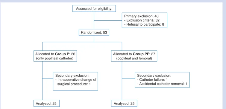
Among 53 randomized patients, 25 in each group completed the study (Table 1, Fig. 1). Mean pain at rest was similar between the two groups at all time points. No patient in either group had pain at arrival in the recovery room. Three patients in Group P reported a VAS of 15–25 when discharged to the ward compared with none in Group PF. Maximal pain at rest occurred in both groups between T12 and T30. In
Group P, three patients reported no pain at any time, com-pared with 12 patients in Group PF (P¼0.04).
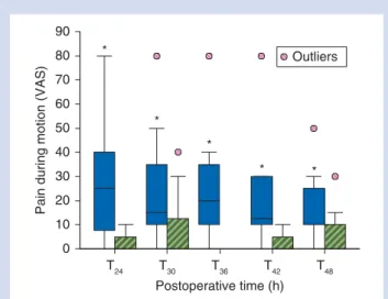
Postoperative pain during mobilization (Fig.2) was signifi-cantly lower at T24, T30, T36, T42, and T48in Group PF (P¼0.01).
All patients in Group P had pain during mobilization com-pared with 12 in Group PF, who did not report pain during mobilization at any time (P¼0.03).
In Group PF, morphine consumption was significantly lower from T0to T24and from T24to T48. Supplementary morphine
bolus administration was necessary in 52% of patients in Group PF and in 96% in Group P (P¼0.03) (Table2).
The incidence of postoperative nausea, vomiting, and pruritus was similar in the two groups. No clinical signs of local anaesthetic intoxication were reported. No sign or symptom of perineural catheter infection was observed. At T48, redness at the catheter insertion site was observed in
seven popliteal catheters in each group and eight femoral catheters in Group PF. No sign or symptom of new neurologi-cal complications was observed 24 h after catheter removal in any patient. Patient satisfaction at 24 h after catheter
removal was similar in both groups (scores 8.9 and 9.1 in Groups P and PF, respectively).
No patient was lost to follow-up 6 months after surgery, and at this time, all patients were able to walk without a walker. Pain at rest was comparable in both groups, but pain with movement (walking on the floor) was significantly lower in Group PF (Fig.3) (P¼0.03). In Group P, two patients reported no pain while walking, compared with nine patients in Group PF (P¼0.03). These 11 patients had no or low (maximal VAS of 15) pain scores during mobilization in the postoperative period. Six months after surgery no patient was using opioids, but five patients in Group P were taking anti-inflammatory agents at regular intervals, compared with one patient in Group PF. No neurological complication was observed in any patient.
Discussion
In this study, we found that the addition of a femoral cath-eter to a popliteal cathcath-eter provided significant better pain control during early mobilization. This positive effect on pain with movement was also observed 6 months after operation.
During our investigation, pain at rest was treated effec-tively in both groups. Since pain intensity at rest was already low in Group P, a much larger study size would have been necessary to demonstrate a possible significant reduction of pain at rest in Group PF.7The maximal pain at rest in our study occurred between T12 and T30. This
time-frame is comparable with the results of di Benedetto and col-leagues,5who performed a study with a popliteal catheter
Assessed for eligibility:
Randomized: 53
Primary exclusion: 40 - Exclusion criteria: 32 - Refusal to participate: 8
Allocated to Group P: 26 (only popliteal catheter)
Allocated to Group PF: 27 (popliteal and femoral)
Analysed: 25 Analysed: 25
Secondary exclusion: - Catheter failure: 1
- Accidental catheter removal: 1 Secondary exclusion:
- Intraoperative change of surgical procedure: 1
Fig 1 Patient flow diagram. Study design according to the CONSORT statement.
Table 1Patient characteristics and surgical data. Values are expressed as mean (range), mean (SD) or as absolute numbers (n)
Group P (n525) Group PF (n525) Gender (female/male) 9/16 10/15 Age (yr) 54 (22 –78) 58 (41 –80)
Body mass index (kg m22) 28 (4) 27 (4) Preoperative pain at rest 32 (9) 37 (11) Preoperative pain during
walking
49 (31) 48 (25)
Operated side (right/left) 12/13 14/11 Duration of tourniquet (min) 99 (14) 104 (18) Duration of surgery (min) 114 (37) 117 (26) Type of surgery
Ankle arthrodesis (n) 11 9
and single-shot femoral nerve block after foot surgery. However, in our study, patients had considerably less pain. This difference might be explained by the vanishing effect
of the femoral nerve single-shot block as a short-acting local anaesthetic without additives was given.8
Pain during mobilization was significantly better con-trolled in Group PF at any time. It is known that pain with movement (in contrast to pain at rest) cannot be well managed with opioids.9The saphenous nerve is the terminal sensitive cutaneous branch of the femoral nerve. It has been shown that all major nerves of the lower leg, including the saphenous nerve, contribute to the innervation of both the capsule of the ankle and the talo-calcaneonavicular joint.10 During movement, the capsule gets stressed and these movements may explain the higher pain intensities in Group P. The results of this work support this anatomical arrangement and therefore highlight the role of the saphe-nous nerve in pain during ankle mobilization.
The significantly lower postoperative morphine consump-tion in Group PF and the fact that 12 patients in this group did not need any supplementary morphine confirms the better analgesia provided by two continuous perineural catheters in this surgical context. The results in Group P are in accord-ance with the findings of Chelly and colleagues11who found a significant reduction in morphine comparing continuous popliteal catheters with morphine PCA.
The significant lower pain during mobilization at 6 months observed in Group PF can be related to the better pain control in the early postoperative period. There is more and more evidence that the intensity of postoperative pain is linked to the development of the chronification of postoperative pain. The results of this study are in accordance with this concept. Kalso and colleagues12 were among the first to demonstrate a link between the intensity of postoperative pain and chronic pain after thoracotomy. These results were also supported by Senturk and colleagues13 and Pluijms and colleagues.14 Katz and colleagues15found, in a paper looking specifically at risk factors, that early post-operative pain was the only factor that significantly predicted long-term pain. Investigations after iliac crest bone graft-ing,16hernia surgery,17hip arthoplasty,18and Caesarean sec-tions19confirmed that postoperative pain was a risk factor. The mechanisms responsible for the chronification of post-operative pain are still not well defined.20 Further studies are needed to elucidate this question.
Our observations can be criticized since we did not specify the nerve distribution, the characteristics of pain observed 6 months after operation and its impact on the patient’s daily quality of life, or the range of movement in the ankle. These secondary outcome variables should be more closely investi-gated in further prospective studies.
In the context of two combined continuous nerve blocks, the issue of local anaesthetic toxicity must be con-sidered. It was shown that the simultaneous continuous application of ropivacaine at two different sites can be per-formed without reaching toxic plasma concentration.16 21In this context, we performed a preliminary pilot study (unpub-lished results), which demonstrated that total and free (¼unbound) ropivacaine plasma concentration were well
Postoperative time (h) T48 T42 T36 T30 T24
Pain during motion (VAS)
90 Outliers 80 70 60 50 40 30 20 10 0 * * * * *
Fig 2 Postoperative pain during mobilization in patients of Group P with a popliteal catheter (blue filled bars) and in patients of Group PF with the popliteal and femoral catheters (green striped bars). Data were assessed at T24and then every 6 h until T48. Boxplots represent median, 25th and 75th percentile and the whiskers rep-resent the 10th and 90th percentile. *P¼0.01
6 months after operation
VAS pain on movement
100 Group P Group PF Outliers 80 60 40 20 0
Fig 3 Pain with movement at 6 months after operation. Pain with movement in patients of Group P and in patients of Group PF. Data were assessed 6 months after operation while patients were walking on the floor. Boxplots represent median, 25th and 75th percentile, and the whiskers represent the 10th and 90th percentiles. *P¼0.03.
Table 2 Postoperative morphine consumption (mg). Results are expressed as mean (SD). *P,0.05 between the groups
Time period Group P Group PF
0– 24 h 25 (10) 6 (5)*
below the toxic threshold defined by Knudsen and colleagues.22
It is possible that the higher plasma concentrations of ropivacaine were responsible for the better analgesia. However, Herroeder and colleagues23 infused lidocaine i.v. (bolus 1.5 mg kg21followed by a 4 h continuous infusion of 2 mg min21) or the same volume of placebo for colorectal surgery and compared pain scores. Besides different interest-ing positive effects of lidocaine, no influence on pain ratinterest-ings was found.
With regard to the site of saphenous nerve block, it could be argued that a more specific continuous saphenous nerve block in the distal part of the thigh would be more appropri-ate than a continuous femoral nerve block. This would avoid unnecessary proximal muscle weakness. However, the patients in this study were allowed to be mobilized on the morning of the first postoperative day with crutches without weight-bearing of the operated foot. Therefore, the transient weakness of the quadriceps muscle caused by the femoral nerve catheter did not lead to a delayed mobiliz-ation. Conversely, blockade of the saphenous nerve at the level of the groin using a nerve stimulator has the advantage of a higher success rate. Furthermore, the placement of a perineural catheter at this level is easier.24
We recognize that in our study, a third group with a single-shot saphenous block using ropivacaine 0.5% is lacking. This would have perhaps given information dealing with the con-troversy surrounding single-shot vs continuous infusion, although it has been shown, in different regional anaesthesia procedures, that a continuous administration of local anaes-thetic offers better analgesia compared with single-shot application.25
Pain was referred to as pain in the ankle, since it was not possible for the patients to reliably distinguish the pain over the medial aspect vs pain over the lateral aspect of the ankle, especially since the whole area was covered with dressings and a cast.
The design of this study may also be criticized since the results can be expected and are not surprising. Ankle surgery involving the medial aspect of the ankle will cause pain in the area innervated by the saphenous nerve. However, how important is the pain specifically related to the saphenous nerve in this context is not known and has never been investigated. This work emphasizes the role of this nerve in the occurrence of pain with movement. For prac-tical reasons, a real double-blinded design was not possible in this investigation—the placement of the femoral catheter was visible for both the patient and the study nurse. We cannot exclude that this point may have influenced the results.
Reviews dealing with anaesthesia for foot surgery do not mention the double-catheter technique.26–28In clinical prac-tice, the effectiveness of this ‘double catheter technique’ should be balanced against costs, workload, and a possible increase in the risk of complications.
In conclusion, we found that analgesia after major ankle surgery when performed by a combination of continuous popliteal and femoral nerve block significantly reduced
early postoperative pain with movement and postoperative morphine consumption. This positive effect was still present 6 months after operation.
Conflict of interest
None declared.
Funding
Financial support was provided solely by departmental sources.
References
1 Needoff M, Radford P, Costigan P. Local anesthesia for postopera-tive pain relief after foot surgery: a prospecpostopera-tive clinical trial. Foot Ankle Int 1995; 16: 11 –3
2 Singelyn FJ, Aye F, Gouverneur JM. Continuous popliteal sciatic nerve block: an original technique to provide postoperative analgesia after foot surgery. Anesth Analg 1997; 84: 383 –6 3 Borgeat A, Blumenthal S, Karovic D, Delbos A, Vienne P. Clinical
evaluation of a modified posterior anatomical approach to per-forming the popliteal block. Reg Anesth Pain Med 2004; 29: 290– 6 4 Borgeat A, Blumenthal S, Lambert M, Theodorou P, Vienne P. The feasibility and complications of the continuous popliteal nerve block: a 1001-case survey. Anesth Analg 2006; 103: 229– 33 5 di Benedetto P, Casati A, Bertini L, Fanelli G, Chelly JE.
Postopera-tive analgesia with continuous sciatic nerve block after foot surgery: a prospective, randomized comparison between the popliteal and subgluteal approaches. Anesth Analg 2002; 94: 996– 1000
6 Taboada M, Lorenzo D, Oliveira J, et al. Comparison of 4 tech-niques for internal saphenous nerve block. Rev Esp Anestesiol Reanim 2004; 51: 509– 14.
7 Farrar JT, Portenoy RK, Berlin JA, Kinman JL, Strom BL. Defining the clinically important difference in pain outcome measures. Pain 2000; 88: 287 –94
8 Popping DM, Elia N, Marret E, Wenk M, Tramer MR. Clonidine as an adjuvant to local anesthetics for peripheral nerve and plexus blocks: a meta-analysis of randomized trials. Anesthesiology 2009; 111: 406– 15
9 Pirec V, Laurito CE, Lu Y, Yeomans DC. The combined effects of N-type calcium channel blockers and morphine on A delta versus C fiber mediated nociception. Anesth Analg 2001; 92: 239– 43
10 Mentzel M, Fleischmann W, Bauer G, Kinzl L. Ankle joint denerva-tion. Part 1: Anatomy—the sensory innervation of the ankle joint. Foot Ankle Surg 1999; 5: 15– 20
11 Chelly JE, Greger J, Casati A, Al-Samsam T, McGarvey W, Clanton T. Continuous lateral sciatic blocks for acute postopera-tive pain management after major ankle and foot surgery. Foot Ankle Int 2002; 23: 749 –52
12 Kalso E, Perttunen K, Kaasinen S. Pain after thoracic surgery. Acta Anaesthesiol Scand 1992; 36: 96–100
13 Senturk M, Ozcan PE, Talu GK, et al. The effects of three different analgesia techniques on long-term postthoracotomy pain. Anesth Analg 2002; 94: 11 –5
14 Pluijms WA, Steegers MA, Verhagen AF, Scheffer GJ, Wilder-Smith OH. Chronic post-thoracotomy pain: a retrospective study. Acta Anaesthesiol Scand 2006; 50: 804– 8
15 Katz J, Jackson M, Kavanagh BP, Sandler AN. Acute pain after thoracic surgery predicts long-term post-thoracotomy pain. Clin J Pain 1996; 12: 50 –5
16 Blumenthal S, Dullenkopf A, Rentsch K, Borgeat A. Continuous infusion of ropivacaine for pain relief after iliac crest bone graft-ing for shoulder surgery. Anesthesiology 2005; 102: 392 –7 17 Aasvang E, Kehlet H. Chronic postoperative pain: the case of
ingu-inal herniorrhaphy. Br J Anaesth 2005; 95: 69– 76
18 Nikolajsen L, Brandsborg B, Lucht U, Jensen TS, Kehlet H. Chronic pain following total hip arthroplasty: a nationwide questionnaire study. Acta Anaesthesiol Scand 2006; 50: 495– 500
19 Nikolajsen L, Sorensen HC, Jensen TS, Kehlet H. Chronic pain fol-lowing Caesarean section. Acta Anaesthesiol Scand 2004; 48: 111– 6
20 Macrae WA. Chronic post-surgical pain: 10 years on. Br J Anaesth 2008; 101: 77– 86
21 Blumenthal S, Min K, Nadig M, Borgeat A. Double epidural cath-eter with ropivacaine versus intravenous morphine: a comparison for postoperative analgesia after scoliosis correction surgery. Anesthesiology 2005; 102: 175 –80
22 Knudsen K, Beckman Suurkula M, Blomberg S, Sjovall J, Edvardsson N. Central nervous and cardiovascular effects of i.v. infusions of ropivacaine, bupivacaine and placebo in volunteers. Br J Anaesth 1997; 78: 507– 14
23 Herroeder S, Pecher S, Schonherr ME, et al. Systemic lidocaine shortens length of hospital stay after colorectal surgery: a double-blinded, randomized, placebo-controlled trial. Ann Surg 2007; 246: 192– 200
24 Tran D, Clemente A, Finlayson RJ. A review of approaches and techniques for lower extremity nerve blocks. Can J Anaesth 2007; 54: 922– 34
25 Fredrickson MJ, Ball CM, Dalgleish AJ. Analgesic effectiveness of a continuous versus single-injection interscalene block for minor arthroscopic shoulder surgery. Reg Anesth Pain Med 2010; 35: 28–33 26 Reilley TE, Terebuh VD, Gerhardt MA. Regional anesthesia tech-niques for the lower extremity. Foot Ankle Clin 2004; 9: 349– 72 27 Singelyn FJ. Single-injection applications for foot and ankle
surgery. Best Pract Res Clin Anaesthesiol 2002; 16: 247– 54 28 Shah S, Tsai T, Iwata T, Hadzic A. Outpatient regional anesthesia