The PARP inhibitor olaparib exerts beneficial effects in mice subjected to
cecal ligature and puncture and in cells subjected to oxidative stress without
impairing DNA integrity: A potential opportunity for repurposing a clinically
used oncological drug for the experimental therapy of sepsis
Akbar Ahmada,1, Juliana de Camargo Vieirab,l, Aline Haas de Melloa, Thaïs Martins de Limab, Suely Kubo Arigab, Denise Frediani Barbeirob, Hermes Vieira Barbeirob, Bartosz Szczesnya, Gabor Tëri:?, Nadiya Druzhynaa, Elisa B. Randic, Michela Marcattia, Tracy Toliver-Kinskya, Andrâs Kissct, Lucas Liaudete, Reinaldo Salomaof, Francisco Garcia Sorianob,**, Csaba Szaboa,c,*
'Department of Anesthesiology, The University of Texas Medical Branch at Ga/veston, Ga/veston, TX, USA b Laborat6rin de Investigaçilo Médica, Faculdade de Medicina da Universidade de Silo Paulo, Silo Paulo, Brazil 'Chair of Pharmacology, Faculty of Science and Medicine, University of Fribourg, Fribourg, Switzerland d Second Department of Pathology, Semmelweis University Medical Schoo~ Budapes~ Hungary
'Department of Intensive Gare Medicine and Burns, Lausanne University Hospital Medical Center, Lausanne, Switzerland
'Division of Infectious Diseases, Department of Medicine, Hospital Silo Paulo, Escala Paulista de Medicina, Universidade Federal de Silo Paulo, Silo Paulo, Brazil
ARTICLE INFO Keywords: Sepsis Shock Multiorgan dysfunction DNA Mitochondria Cell death Treg Th17 ABSTRACT
Poly(ADP-ribose) polymerase (PARP) is involved in the pathogenesis of cell dysfunction, inflammation and organ failure during septic shock. The goal of the current study was to investigate the efficacy and safety of the clinically approved PARP inhibitor olaparib in experimental models of oxidative stress in vitro and in sepsis in vivo. ln mice subjected to cecal ligation and puncture (CLP) organ injury markers, circulating and splenic im-mune cell distributions, circulating mediators, DNA integrity and survival was measured. ln U937 cells subjected to oxidative stress, cellular bioenergetics, viability and DNA integrity were measured. Olaparib was used to inhibit PARP. The results show that in adult male mice subjected to CLP, olaparib (1-10 mg/kg i.p.) improved multiorgan dysfonction. Olaparib treatrnent reduced the degree of bacterial CFUs. Olaparib attenuated the in-creases in the levels of several circulating mediators in the plasma. ln the spleen, the number of CD4 + and CDS+ lymphocytes were reduced in response to CLP; this reduction was inhibited by olaparib treatrnent. Treg but not Thl 7 lymphocytes increased in response to CLP; these cell populations were reduced in sepsis when the animais received olaparib. The Thl 7 /Treg ratio was lower in CLP-olaparib group than in the CLP control group. Analysis of miRNA expression identified a multitude of changes in spleen and circulating white blood cell miRNA levels after CLP; olaparib treatrnent selectively modulated these responses. Olaparib extended the survival rate of mice subjected to CLP. ln contrast to males, in female mice olaparib did not have significant protective effects in CLP. ln aged mice olaparib exerted beneficial effects that were Jess pronounced than the effects obtained in young adult males. ln in vitro experiments in U937 cells subjected to oxidative stress, olaparib (1-100 µM) inhibited PARP activity, protected against the Joss of cell viability, preserved NAD+ levels and improved cellular bioenergetics. ln none of the in vivo or in vitro experiments did we observe any adverse effects of olaparib on
Abbreviations: ALB, albumin; ALT, alanine aminotransferase; AMY, amylase; BUN, blood urea nitrogen;
ea
2+, total calcium; CRE, creatinine; CLP, cecal ligation and puncture; GLU, glucose; K +, potassium; MDA, malon dialdehyde; MPO, myeloperoxidase; Na+, sodium; Nrf2, nuclear factor erythroid 2-related factor 2; NO, ni tric oxide; PAR, poly(ADP-ribose); PARP, poly(ADP-ribose) polymerase; PHOS, phosphorus; RNS, reactive nitrogen species; ROS, reactive oxygen species• Corresponding author at: Chair of Pharmacology, Faculty of Science and Medicine, University of Fribourg, Chemin du Musée 18, Fribourg, 1700, Switzerland. •• Corresponding author at: Departamento de Clinica Médica, Faculdade de Medicina da Universidade de Sao Paulo, Sao Paulo, SP, Brazil.
E-maü addresses: [email protected] (A. Ahmad), [email protected] (J.d.C. Vieira), [email protected] (A.H. de Mello),
[email protected] (T.M. de Lima), [email protected] (S.K. Ariga), [email protected] (D.F. Barbeiro), [email protected] (H.V. Barbeiro), [email protected] (B. Szczesny), [email protected] (G. Tërë), [email protected] (N. Druzhyna), [email protected] (E.B. Randi),
[email protected] (M. Marcatti), [email protected] (T. Toliver-Kinsky), [email protected] (A. Kiss), [email protected] (L. Liaudet), [email protected] (R. Salomao), [email protected] (F.G. Soriano), [email protected] (C. Szabo).
1 Equally contributed (shared first authorship).
http://doc.rero.ch
Published in "Pharmacological Research doi: 10.1016/j.phrs.2019.104263, 2019"
which should be cited to refer to this work.
nuclear or mitochondrial DNA integrity. In conclusion, olaparib improves organ function and extends survival in
septic shock. Repurposing and eventual clinical introduction of this clinically approved PARP inhibitor may be
warranted for the experimental therapy of septic shock.
1. Introduction
Sepsis is a life-threatening systemic inflammatory disease affecting several million people annually worldwide. It is associated with mor-tality rates of 20-40%. lts ftequency increases every year, in excess of the growth and aging of our population. The outcomes of sepsis are markedly worse in the elderly [1,2]. On the other hand, the female gender (in the pre-menopausal stage) tends to provide some protection against sepsis-associated mortality [3,4]. Despite extensive research and despite a large number of pivotal clinical trials targeting various pathways in immune, inflammatory and coagulation abnormalities in sepsis/septic shock, no specific pharmacological or immunological therapies are approved for the treatment of this condition.
More than two decades ago, the constitutive nuclear and mi-tochondrial enzyme poly(ADP-ribose) polymerase (PARP) emerged as a potential therapeutic target for various forms of critical illness, in-cluding sepsis. Several lines of studies, utilizing PARP inhibitors of various structural classes, as well as PARP-1-deficient mice indicated that PARP inhibition exerts beneficial effects on organ function, vas -cular function, and survival [5-19]. For instance, PARP-1 knockout mice showed, after cecal ligation and puncture (CLP), lower plasma levels of1NF-a, IL-6 and IL-10 [8]. In an intraperitoneal bacterial sepsis model, pigs treated with PARP inhibitors showed better survival after
A
150 ]i 100 ::>.s
2
::;; 50B
Lung CLP CLP CLP Olaparlb Olaparlb Olaparib 1 mgfkg 3 mg/kg 10 mg/kgLung
Olaparib Olaparib Olaparib 1 mglkg 3 mg/kg 10 mg/kg 500 400 $ ::> 300
.s
0 200 Q. ::E 100 0 250 200 ]i ::E 150 2. ~ 100 ::;: 50*
Sham*
24 h and 96 h, decreased bacteremia, attenuated 1NFa plasma levels and protected against the cardiovascular dysfunction when compared to untreated animals [9]. Moreover, treatment with a PARP inhibitor exerted a protective effect against apoptosis in the hearts of rats su b-jected to CLP [16].
Although PARP activation clearly plays a pathophysiological role in sepsis, clinical translation of this concept was not possible, because there were no clinically approved drugs to inhibit PARP. Recently, several classes of PARP inhibitors have been approved for clinical use, in the field of oncology. This development is based on an independent line of research, one that demonstrated that in cancer cells, PARP in-hibition can indu ce cell dea th and can potentia te the efficacy of various classes of anticancer agents [20,21]. One of the clinically approved PARP inhibitor drugs is olaparib (Lynparza), a competitive PARP in-hibitor that inhibits the binding of NAD+ (the substrate of PARP) into the catalytic site of PARP.
As outlined in our recent position paper, the clinical approval of PARP inhibitors opens the door for potential repurposing of these drugs for various non-oncological indications, with sepsis/septic shock being one of the prime candidates for such repurposing efforts [19]. However, additional preclinical work is needed to lay the groundwork for such repurposing efforts. As described in detail [19], such work needs to focus both on efficacy endpoints, as well as safety endpoints -chiefly,
Li ver
CLP CLP Olaparib Olaparib Olaparib
1 mg/kg 3 mg/kg 10 mg/kg
Li ver
#
CLP
Olaparib Olaparib Olaparib 1 mg/kg 3 mg/kg 10 mg/kg 400 - 300
3
.s
200 0 Q. ::;: 100 200 $ 150 ::;: 2. 100 ~ ::E 50*
*
Sham CLP Spleen #Olaparib Olaparib Olaparlb
1 mg/kg 3 mg/kg 1 O mg/l<g
Spleen
#
CLP CLP CLP Olaparlb Olaparlb Olaparlb
1 mglkg 3 mg/kg 10 mg/kg
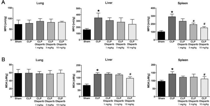
Fig. 1. Effect of olaparib on Jung, liver and spleen MPO and MDA levels in young male Balb/c mice subjected to CLP. (A): Lung, liver and spleen MPO levels
(expressed as milliunits [mU]!gram tissue) and (B): Jung, liver and spleen MDA levels (expressed as µM/gram tissue) are shown in sham mi ce (not subjected to CLP)
("Sham"), in vehicle-treated mice subjected to CLP for 24 h ("CLP'1 and in mice subjected to CLP in the presence ofvarious doses of olaparib ("CLP Olaparib 1 mg/kg",
CLP Olaparib 3 mg/kg" and "CLP Olaparib 10 mg/kg'1. Data are shown as mean ± SEM of 10 animais for each group; *p < 0.05 shows significant increase in MPO
in response to CLP, compared to the sham group; #p < 0.05 shows significant protective effect of olaparib in CLP mice compared to vehicle-treated CLP mice.
on DNA integrity, considering the fact that PARP has been long con-sidered an enzyme that has an important raie in the facilitation of DNA repair. The current report describes the results of such a project. Our experiments incorporated a standard mode! of sepsis (induced by cecal ligation and puncture) and evaluated multiple efficacy endpoints (pri-marily focusing on multiorgan injury), as well as safety endpoints (mitochondrial and nuclear DNA integrity). We have also conducted an in vitro sub-study (human monocytic cells subjected to oxidative stress) - once again, bath focusing on efficacy endpoints (cell viability, cellular bioenergetics) as well as safety endpoints (DNA integrity). The results of the current study demonstrate the efficacy of olaparib on multiple outcome variables, and do not identify any significant adverse effects of
ALP
;:::1
i
0
0 6
200 150 .... - 100 :::> 50*
ALTthe PARP inhibitor on DNA integrity. Thus, the data presented in the current report !end support for repurposing and clinical introduction of the PARP inhibitor olaparib for the experimental therapy of septic shock.
2. Materials and methods
2.1. Animais
Male or female C57BL6 mice (8-72 weeks old) were obtained from Jackson Laboratories. Animais were kept in a 12 h -12 h light / dark cycle at 21-23 'C with free access to standard chow diet.
AMY 2000
*
# # 1500 .... ; 1000 500 O Sham CLP CLP CLP CLPOlaparib Olaparib Olaparib 1 mg/kg 3 mg/kg 1 O mg/kg CLP Olaparib 1 mg/kg CLP Olaparib 3 mg/kg CLP Olaparib 10 mg/kg Sham CLP CLP Olaparib 1 mg/kg CLP Olaparib 3 mg/kg CLP Olaparib 10 mg/kg 300 200 'C "' E 100 100 80 =ë 60 "' E 40 20 10 .... ::; 6 0 :; :;
*
Sham CLP GLU Olaparib 1 mg/kg BUN CLP Olaparib Olaparib 3 mg/kg 1 O mg/kg # # CLP Olaparib Olaparib Olaparib 1 mg/kg 3 mg/kg 10 mg/kgCLP CLP CLP
Olaparib Olaparib Olaparib 1 mg/kg 3 mg/kg 1 O mg/kg 10 'C 6
"'
E 4"'
E .... ca
Olaparib 1 mg/kg CRE CLP Olaparib 3 mg/kg CLP Olaparib 10 mg/kgOlaparib Olaparib Olaparib 1 mg/kg 3 mg/kg 10 mg/kg
Albumin
CLP Olaparib Olaparib Olaparib
1 mg/kg 3 mg/kg 1 O mg/kg 20 15 'C "à, 10 E 200 150 .... 0 100 :; :; 50 2.5 2.0 - 1.5 'C tJl 1.0 0.5 0.0
*
PHOS CLP Olaparib 1 mg/kg Na+ # CLP Olaparib 3 mg/kgOlaparib Olaparib Olaparib 1 mg/kg 3 mg/kg 10 mg/kg
GLOB
CLP CLP
Olaparib Olaparib Olaparib 1 mg/kg 3 mg/kg 1 O mg/kg
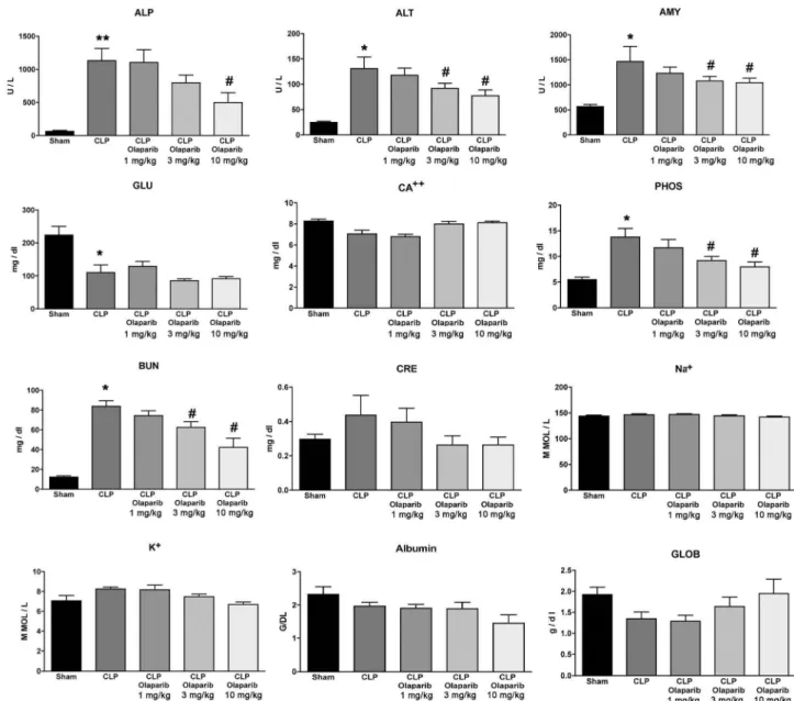
Fig. 2. Effect of olaparib on selected parameters of organ injury in young male Balb/c mice subjected to CLP. Varions physiological and organ injury marker levels: alkaline phosphatase (ALP), alanine aminotransferase (ALT), amylase (AMY), plasma glucose (GLU), plasma calcium (CA++), plasma phosphate (PHOS), plasma blood urea nitrogen (BUN), plasma creatinine (CRE), plasma sodium (Na+), plasma potassium (Na+), plasma albumin (ALB) and plasma globulin (GLOB) measured by Vetscan analysis, are shown in sham mice (not subjected to CLP) ("Sham"), in vehicle-treated mice subjected to CLP for 24 h ("CLP") and in mice subjected to CLP in the presence of varions doses of olaparib ("CLP Olaparib 1 mg/kg", CLP Olaparib 3 mg/kg" and "CLP Olaparib 10 mg/kg"). Data are shown as mean ± SEM of 10 animais for each group; *p < 0.05 shows significant increase in the respective parameter in response to CLP, compared to the sham group; #p < 0.05 shows
Fig. 3. Effect of olaparib on histological alterations injury in the lungs ofyoung male Balb/c mice subjected to CLP. Representative histological pictures are shown in
sham mice (not subjected to CLP) ("Sham"), in vehicle-treated mice subjected to CLP for 24h ("CLP") and in mice subjected to CLP in the presence ofvarious doses of
olaparib (''CLP Olaparib 1 mg/kg'', CLP Olaparib 3 mg/kg" and "CLP Olaparib 10 mg/kg'1. Pictures are selected from n = 5 animais for each group.
Fig. 4. Effect of olaparib on histological alterations injury in the liver ofyoung male Balb/c mice subjected to CLP. Representative histological pictures are shown in
sham mice (not subjected to CLP) ("Sham"), in vehicle-treated mice subjected to CLP for 24 h ("CLP") and in mice subjected to CLP in the presence ofvarious doses of
olaparib ("CLP Olaparib 1 mg/kg", CLP Olaparib 3 mg/kg" and "CLP Olaparib 10 mg/kg'1. Pictures are selected from n = 5 animais for each group.
2.2. Cecal ligation and puncture (CLP)
Acute sepsis was induced in mice by cecal ligation and puncture as previously described (22]. Briefly, mice were anesthetized by
ketamine/xylazine cocktail (i.p.), the abdomen was shaved, wiped with
70% isopropanol and a midline abdominal incision (1-2 cm) was pe r-formed. The cecum was exteriorized, ligated with a sterile silk suture
1 cm from the tip and double punctured with a 20-gauge needle. The
Fig. 5. Effect of olaparib on histological alterations in jury in the spleen ofyoung male Balb/c mi ce subjected ta CLP. Representative histological pictures are shown in
sham mice (not subjected to CLP) ("Sham"), in vehicle-treated mice subjected to CLP for 24h ("CLP") and in mice subjected to CLP in the presence ofvarious doses of
olaparib ("CLP Olaparib 1 mg/kg", CLP Olaparib 3 mg/kg" and "CLP Olaparib 10 mg/kg'1. Pictures are selected from n = 5 animais for each group.
cecum was squeezed to assure expression of a small amount of fecal material and returned to the abdominal cavity. The incision was closed with auto-clips and kept clean by povidone-iodine (Betadine). Mice were resuscitated with intraperitoneal injection of 1 ml of lactated Ringer's solution. Sham-operated mice were treated as described above with the exception of ligation and puncture of the cecum. Buprenor-phine (0.1 mg/kg; s.c. 30 min before surgery and every 12 h thereafter) was used for pain management. A sample of whole blood was collected for analysis of organ function using a comprehensive metabolic panel or for circulating mediator measurements using a multiple array system. Major organs were collected and either analyzed for immune cells by flow cytometry or snap frozen and kept at -80 'C until subsequent use (measurement of DNA integrity or MDA or MPO levels) or placed in formalin and processed for histological analysis.
For the survival study, mice were constantly monitored for 48 h. Mice that survived this period of time were euthanized by cervical dislocation. All animal procedures described in this study have been approved by the respective local Institutional Animal Care and Use Committee of the University of Texas Medical Branch and the University of Sao Paulo.
2. 3. Olaparib treatment protocol
Ail groups of mice received the following intraperitoneal treatment: vehicle (phosphate-buffered saline [PBS] with 4% dimethyl sulfoxide [DMSOJ and 5% polyethylene glycol [PEG]); olaparib, 1 mg/kg, 3 mg/ kg or 10 mg/kg (dissolved in PBS with 4% DMSO 5% and PEG). In the 24 -h protocol, the animais received two doses of olaparib, the first 30 min after the CLP and the second 8 h after CLP, and the experiment was terminated at 24 h. In the survival protocol, treatment with the PARP inhibitor was initiated at 30 min after the CLP, the second dosing was performed at 8 h after CLP, and the same dosing was repeated subsequently every 8 h. Animais were monitored for 48 h at which time point the experiment was terminated.
2.4. Complete membolic panel
Samples of whole blood were collected from septic mice, placed in lithium-heparin tubes and immediately processed for the measurement of alanine aminotransferase (ALT), albumin (ALB), alkaline phospha -tase (ALP), amylase (AMY) total calcium (Ca2 +), creatinine (CRE), glucose (GLU), phosphorus (PHOS), potassium (K + ), sodium (Na+), total bilirubin (TBIL), total protein (TP), and urea nitrogen (BUN) using the VetScan Chemistry Analyzer system (Abaxis) [22].
2. 5. Detection of circulating mediators
Blood from CLP or sham-operated mice was collected in K2EDTA
blood collection tubes and centrifuged at 4 'C for 15 min at 2,000xg within 30 min of collection. Plasma was isolated, aliquoted and stored a t - 80 'C until use. The EMD Millipore's MILLIPLEX™ MAP Mo use cytokine Magnetic Bead Panel 1 was used as described [22] for the simultaneous quantification of the following analytes: TNFa, la, IL-1[3, IL-2, IL-3, IL-4, IL-5, IL-6, IL-7, IL-9, IL-10, IL-12(p40), IL12(p70), IL-13, IL-15, IL-17, LIF, LIX, eotaxin, G-CSF, GM-CSF, KC, IP-10, MCP-1, RANTES, VEGF, MIP-la, MIP-1[3, MIP-2, M-CSF, MIG and IFNy. Data were processed using the Luminex xPONENT"' acquisition software.
90 80 ~ 70 ~ 60
"2:
50 :::l 40 (/) 30 - CLP - CLP Olaparib 1 mg/kg 20 10c
~ ïii > ·~ ::l Cf) o+-~~~~~~~--...
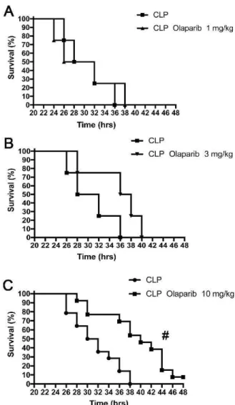
~~~~~ 10 9 8 7 6 5 4 3 2 1 20 22 24 26 28 30 32 34 36 38 40 42 44 46 48 nme (hrs) - - CLP - CLP Olaparib 3 mg/kg 20 22 24 26 28 30 32 34 36 38 40 42 44 46 48 Time (hrs) . _ CLP ... CLP Olaparib 10 mg/kg 20 22 24 26 28 30 32 34 36 38 40 42 44 46 48 Time (hrs)Fig. 6. Effect of olaparib on the survival rate of young male Balb/c mice su
b-jected to CLP. Survival rates are shown in mice subjected to CLP in the presence
of various doses of olaparib ("CLP Olaparib 1 mg/kg", CLP Olaparib 3 mg/kg"
and "CLP Olaparib 10 mg/kg"). Data are shown as mean ± SEM of 10-12 an
-imais for each group; #p < 0.05 shows significant protective effect of olaparib
in CLP mice compared to vehicle-treated CLP mice.
2. 6. Determination of tissue lipid peroxidation: malon dialdehyde assay
Tissue malon dialdehyde (MDA) levels, an index of cellular injury/ oxidative stress, were measured as described [22] in tissue homo -genates from mice subjected to 24h of CLP-induced sepsis, using a fluorimetric MDA-specific lipid peroxidation assay kit (Enzo Life Sci -ences).
2. 7. Myeloperoxidase activity assay
Myeloperoxidase activity was measured as described [22] in tissue homogenates from mice subjected to 24 h of CLP-induced sepsis, using a commercially available MPO fluorometric detection kit (Enzo Life Sci
-ences).
A
~ 120 0 ~ - 100 "C :::l 5 <.. 80"'"'
.ë
t; 60 .Ë ô 40 ô é 'Ê 20I
B
< z 0 120 ~ _100 "C:::l a<.. ao"'"'
.s
t; 60 .Ë 0 40 ôC 20 fI
c
~ 120 0 "iii 100 :§ s_g;
BO.9
t; 60 .Ë ô 40 'ë ê 'Ê 20I
Sham CLP CLP CLP CLPOlaparib Olaparib Olaparib
1 mg/kg 3 mg/kg 1 0 mg/kg 1 # # # CLP CLP Olaparib Olaparib Olaparib 1 mg/kg 3 mg/kg 1 O mg/kg
Olaparib Olaparib Olaparib
1 mg/kg 3 mg/kg 1 O mg/kg fil 140
·a
120~ ~
100 z . ~ ~ 80 ~ ~ 60 ~ ê 40 ~ 20 ::;; :{l 140 -~ - 120 ;2 ~ 100 ~e:
80 -~ ~ 601
~
40 ~ 20 ::;; :G 160 ·a. 140 0 u 5' 120 ~ ~ 100 ~ t; 80 .c 0 60 ~é 40.s
20 ~ Sham Sham ShamLung
CLP CLP CLP CLP Olaparib Olaparib Olaparib1 mg/kg 3 mg/kg 1 O mg/kg
Liver
CLP CLP CLP CLP Olaparib Olaparib Otaparib
1 mg/kg 3 mg/kg 10 mg/kg
Spleen
CLP CLP CLP CLP
Olaparib Olaparib Olaparib 1 mg/kg 3 mg/kg 1 O mg/kg 120 < ~ 100 ~ ~ 80 g fi" .: t; 60 .~~ 40 Ci~ 20 ~ 160 < z 140 0 :;; 5' 120 ~ ~ 100 .:: t; 80 0 0 60 ~é 40 ~ 20 160 ~ 140 0 ~ 5' 120 ~ ~ 100 .:: t; 80 ;~ 60 c,~ 40 ~ 20 Sham CLP Sham CLP Sham CLP CLP CLP CLP Olaparib Olaparib Olaparib 1 mg/kg 3 mg/kg 10 mg/kg CLP CLP CLP Olaparib Olaparib Olaparib 1 mg/kg 3 mg/kg 10 mg/kg CLP CLP CLP Olaparib Olaparib Olaparib
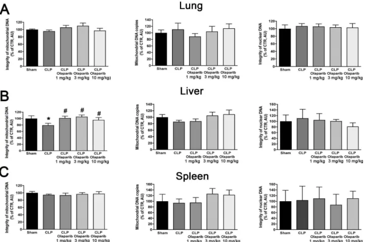
1 mg/kg 3 mg/kg 1 O mg/kg Fig. 7. Effect of olaparib on Jung, liver and spleen mitochondrial DNA integrity, mitochondrial DNA copy number and nuclear DNA integrity in young male Balb/c mice subjected to CLP. (A): Lung mitochondrial DNA integrity, mitochondrial DNA copy number and nuclear DNA integrity values (expressed as% of control); (B): liver mitochondrial DNA integrity, mitochondrial DNA copy number and nuclear DNA integrity values (expressed as% of control); (C) spleen mitochondrial DNA integrity, mitochondrial DNA copy number and nuclear DNA integrity values (expressed as% of control) are shown in sham mice (not subjected to CLP) ("Sham"), in vehicle-treated mice subjected to CLP for 24 h ("CLP") and in mice subjected to CLP in the presence of varions doses of olaparib ("CLP Olaparib 1 mg/kg", CLP Olaparib 3 mg/kg" and "CLP Olaparib lOmg/kg"). Data are shown as mean ± SEM of 10 animais for each group; *p < 0.05 shows significant change in a respective parameter in response to CLP, compared to the sham group; #p < 0.05 shows significant effect of olaparib on a given parameter in CLP mice compared to vehicle-treated CLP mice.
A
CFU BLOODB
CFU SPLEEN Fig. 8. Effect of olaparib on blood and spleenbacterial colony-forming unit (CFU) numbers
*
in young male Balb/c mice subjected to CLP.108 1000000
•
(A): Blood CFUs (expressed as CFU/ml) and (B)*
spleen CFUs (expressed as CFU/mg) are shownt
in sham mice (not subjected to CLP) ("Sham"),•
in vehicle-treated mice subjected to CLP for107 100000
24 h ("CLP") and in mice subjected to CLP in
t
....
the presence of varions doses of olaparib ("CLP..
-±-
Olaparib 1 mg/kg", CLP Olaparib 3 mg/kg" and"CLP Olaparib 10 mg/kg"). Data are shown as
106
-r
10000mean ± SEM of 10 animais for each group;
Cl
....
#
Ë T
.ê
T *p < 0.05 shows significant increases in CFUs3 :::l
IL
-T
•
IL u
..
in response to CLP, compared to the shamu
..
•
group; #p < 0.05 shows significant effect of
105 1000
•
f-T olaparib on CFU values in CLP mice compared
to vehicle-treated CLP mice.
•
..
T•
•
104 100•
# T•
•
103 10 'b~c,S
s
;.~s
;.~s
;.~ 'b~vs
s
;.~s
;.~s
;.~ «>°"' (j 'b !l.> (j 'b !l.> (j >?'b ~o, «>°"' (j 'b' !l.> (j 'b' !l.> (j >?'b' ~o,,,._'bQ.~t ,,._'bQ.~t o''b'
s-03
,,._'bQ.i>;"*' ,,._'bQ.i>;"*'s-03
- veh - 1uM - 3uM 8 10µM
§
~
- 30µM - 100uM Time (hours)Fig. 9. Effect of olaparib on E. coli bacterial growth in vitro. Data show bacterial
numbers at various time points in vehicle-treated group, and in the groups of
bacteria in the presence or various concentrations (1, 3, 10, 30, 100 µM) ola
-parib for various time points (1-5 h). Data are shown as mean ± SEM of 5
determinations per group.
2.8. Meas!ffement of nuclear Wld mitochondrial DNA integrity
Integrity of the nuclear and mitochondrial DNA, measured by the relative amount of DNA damage, was analyzed by semi-quantitative, long-amplicon PCR assays (LA-PCR) using LongAmp Taq DNA Polymerase (New England BioLabs, Ipswich, MA) in tissue homogenates as described [23]. Total DNA was isolated using DNase Blood and Tissue Kit (QIAGEN, Hilden, Germany). Briefly, damage to nuclear DNA was estimated by quantification of the PCR amplification of the 10 kb nuclear-specific DNA fragment using PicoGreen fluorescent dye to detect amplified double-stranded DNA (Quant-iT™ PicoGreen; Life
Technologies, Carlsbad, CA). Damage to the mitochondrial DNA was estimated by quantification of the PCR amplification of the 8.9 kb mitochondrial-specific DNA fragment using PicoGreen staining. Data were normalized by the secondary PCR amplification of 221 bp mi-tochondrial genome-specific fragment for correction of the multiple copies of the mitochondrial DNA. LA-PCR assay is based on premises that DNA damage inhibits progression of DNA polymerase during PCR
reaction and thus amplification of appropriate DNA fragment is nega· tively correlated with the level of the DNA damage.
3000 108 107 'tl ; 106 0 2000 Cl) 0 Ci. 10• :;:;
"'
Ë~
!!! 104 3 1000 ~ 103 u. (J (J 102 101 0 10°CLP
CLP
'
CLP
olaparib 10 mg/kg2. 9. Determination of colony-forming bacterial units (CFUs) after sepsis or
bacteremia
Blood was diluted serially in sterile saline. Spleen was homogenized
in sterile saline at 100 mg tissue/ml concentration followed by serial dilution. Fifty microliters of each dilution was plated and cultured on
LB agar plates at 37 °C. After 16 h of incubation, the number of bacterial
colonies was counted and expressed as CFUs per milliliter ofblood or as
CFUs per milligram or gram tissue.
In an in vitro satellite experiment, we evaluated whether olaparib has any direct effect on bacterial growth. In these experiments DHSa
Rcoli were cultured in liquid LB growth medium in a shaking incubator set to 37 °C and 200 RPM in the presence of various concentrations (1-1 OO µM) of olaparib. The culture density was measured a t OD60o
over 5 h.
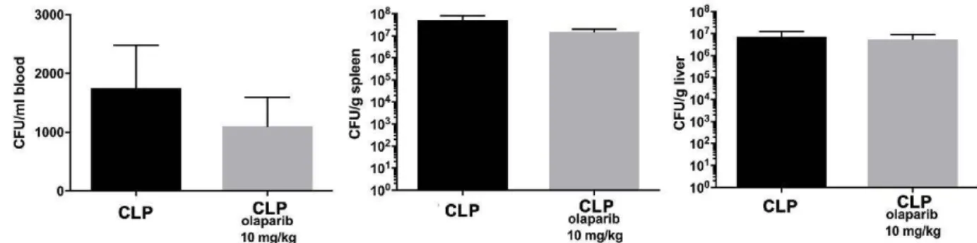
In an in vivo satellite experiment, we evalua ted whether ola parib has
any direct effect on bacterial clearance in vivo in the absence of septic shock. Male C57BL6 mice (8 weeks old) were obtained from Jackson Laboratories. Animals (either treated with olaparib, 10 mg/kg i.p. at 30 min and 8 h post-inoculation, or treated with corresponding vehicle control) were injected with 3 x 10⁸ CFU DHSa E.coli i.p. and tissues
harvested 20 h post-infection. CFUs in blood, spleen and liver were quantified as described above.
2.1 O. Histological Wlalysis
Tissues were fixed for 1 week in buffered formaldehyde solution (10% in PBS) at room temperature, dehydrated by graded ethanol and
embedded in Paraplast (Sherwood Medical, Mahwah, NJ, USA). Tissue
sections (thickness: 7 µm) were deparaffinized with xylene stained with
hematoxylin/eosin and studied using light microscopy. Slides were evaluated in a blinded fashion.
2.11. Row cytomeoy
For phenotypic characterization of cells by flow cytometry from
spleen and blood samples, we performed three panels: T cells (panel 1 ),
Treg cells (panel 2) and Thl 7 cells (panel 3). Cell suspensions were prepared and were re-suspended in flow cytometry buffer (PBS
108 107 ~ 106 Cl) ~ 105 310•
13
103 102 101 10°CLP
CLP
CLP
olaparib olaparlb
10 mg/kg 10 mg/kg
Fig. 10. Effect of olaparib on blood, spleen and liver bacterial colony-forming unit (CFU) numbers in young male Balb/c mice subjected to E. coli bacteria in vivo. Blood (expressed as CFU/ml), spleen and liver CFUs (expressed as CFU/g) are shown in vehicle-treated mice inoculated with E. coli for 24 h ("CLP'1 and in mice
subjected to CLP in the presence of 10 mg/kg olaparib. Data are shown as mean ± SEM of 6 animais for each group.
TNFa
-1
i::
_
i
06é
O Sham CLP CLP CLP CLP 11 60 i .. * *Olaparib Olaparib Olaparib
(1mglkg) (3mg/kg) (10mglkg)
ll-3
"'
Olaparib Olaparib Olaparib (1mglkg) (3mg/kg) (10mglkglIL·7
"'
Olaparib O!aparib O!aparib
t1 mg/kg) t3mglkgf (10mg/kg)
IL-12(p70)
"'
Olaparib Olaparib Olaparib
(1mglkg) (3mglkg) (10mg/kg) LIF
""
l
..
,,1
•::::
1
,,_,__,,_,'"__,m ""!!!'!!""' '---'-=O,.,..._"~"â",__,,-r
__
"
"15-0 E ~100 *Olaparib Olaparib Olaparib
(1mglkg) (3mglkg) (10mglkg)
GM·CSF
"'
Olaparib Olaparib Olaparib
{1mglkg) {3mglkg)(10mglkg) RANTES
"'
Olaparlb Olaparib Olaparlb
(1mglkg) (3mglkg) (10mg/kg)
MIP-2
Olaparib Olaparib oraparib (1 mg/kg) (3mglkg) (10mg/kg)
IL-1a
1
~
.
i
nno
Sham CLP CLP CLP CLP
*
Olaparib Olaparib Olaparib (1mglkg) (3mg/kg) (10mglkg)
IL-4
Olaparib Olaparib Olaparib (1mg/kg) (3mg/kg) (10mglkg)
IL-9
1
::•
_
...
111
·
n
0
n
O~~ •• ~.m~-"!!!Cl~P~~C~LP,-'--'-,,Cl~P~LC~l~P~
Olaparib Olaparib Olaparib
(1mglkg) (Jmglkg) (10mglkg)
IL·13
LIX
1
~
=
Î
Oô
é
Olapanb Olaparib Olapaf1b (1mglkg) (lmg/kg) (10mglkg) KC
"'"Linnn
i
10000 * 5-000 O Sham CLP CLP CLP CLP " E 6 i 'Olaparib Olaparib Olaparib
(1 mg/kg) (3mglkg)(10mg/kg)
VEGF
CLP Olaparib Olaparib Olaparib (1mglkg) (3mglkg) (10mglkg)
M-CSF
l
~,I
~
-"
,
_____,_,,
O~
,
':?'--'-=-'-0
Olaparib Olaparib Olaparib
{1mglkg) (3mglkg) (10mglkg)
*
*
IL-1~
CLP
Olaparib Olaparib Olaparib
(1mg/kg) (3mglkg) (10mglkg) IL-5 CLP Olaparib Olaparib Olaparib (1mglkg) (3mglkg) {10mglkg) IL-10 IL-2 CLP
Otaparib Olaparib Olaparib
(1mglkg) (3mg/kg) (10mglkg)
IL-6
'""
i
10000
*
•.,,,
,.._,,~ •• ~m~1
~C~LP!"-_....,,cL~P~000
LC~L~P~~C~LP,...._Olaparib Olaparib Olaparib
(1mglkg) (3mglkg) (10mglkg) IL-12(p40)
"l
8000 ~ 6000 t 4000 2000 O Shami
Oo
O
1
~
'!
i
oo
o
Olaparib Olaparib O!apaf1b Olaparib Olaparib Olaparib
·-·-~-
·-·-~-i4000
IL·15
Olaparib Olaparib Olaparib (1mg/kg) (3mglkg)(10mglkg)
Eotaxin
"' Olaparib Olaparib Olaparib (1 mg/kg) (3mglkg) (10mglkg)
IP·10
::::1
i
•,:::non
o-'-"'!"\""""• •• •m<-J•c~LP!"--'-,C~LP,.J-~CL~P~Lc=,=,-'Olaparib Olaparib Olaparib
(1 mg/kg) p mg/kg) (10 mg/kg)
MIP-1a
1
~l
_
i
Onn
Sham CLP CLP CLP CLP
4000
Olaparlb Olaparib O!aparib
(1mglkg) (3mg/kg) (10mglkg)
MIG
""
I
~300Ct
~2000i
n
' " ' L J n D
0"--~,~ •• -m~~C~LP!"-~CL~P~LC~L~P-'---~c~LP=-'--Olaparib Olaparib Olaparib
(1mglkg) (3mg/kg) (1Ctmg/kg)
IL-17
::1
*I~
i
Olaparib Olapo6~
arib Olaparib~1 mg/kg) (3mglkg) (10mglkg) G-CSF
CLP
Olaparib Olaparib Olaparib
(1 mg/kg) (3mg/kg) (10mglkg)
MCP-1
I~
O'-'--,s~h,-m-i
-C!!'L!'P L-JOAD
~C~LP,--'--'-,C~LP,-'--'-,,C~LP,-'-Olaparib Olaparib Olaparib
(1mglkg) (3mg/kg) j1Ctmglkg) MIP-1~
'"'
I
20Ct0i
41000n
nD
,,~~.~ .. -m~~C~LP~~C~L~P~~C-LP,-'-~CL~P~ Olaparib Otaparib Olaparib(1mg/kg) (3mglkg) (1Ctmglkg)
INFy
.:
1
i.
n
~
-
-
•
U
61
1
,,..__~,-.. •m'---'~cL~P~LC~L=P'---'- c-LP,.J-~CL~P-'---Olaparib Olaparib Olaparib (1mglkg) (3mglkg) (10mglkg)
Fig. 11. Effect of olaparib on the levels of varions circulating mediators (cytokines, chemokines, growth factors) in young male Balb/c mice subjected to CLP. Values
are shown in sham mice (not subjected to CLP) ("Sham"), in vehicle-treated mice subjected to CLP for 24 h ("CLP") and in mice subjected to CLP in the presence of varions doses of olaparib ("CLP Olaparib 1 mg/kg", CLP Olaparib 3 mg/kg" and "CLP Olaparib 10 mg/kg"). Data are shown as mean ± SEM of 10 animais for each
group; *p < 0.05 shows significant increase in the respective parameter in response to CLP, compared to the sham group; #p < 0.05 shows significant protective
effect of olaparib in CLP mice compared to vehicle-treated CLP mice.
Spleen
cO 0 1.5x1Q09 () 1.0•1009 ~ 0 () (!; 5.0x1Q08 0 () + ~ 2.ox10°1 )( 0 u.. 1.sx10°1 il;"'
8
1.0•1007 +i?i
5000000.0 () 0*
# 8.0•1008 + ~ 6.0x10°8 () ~ 4.0x10°8 () + M 0 2.0•10°' ().1
1.5•1007 .... ~~
1.0•1007 ~ + ~ 5000000.0 0:: ~ # 0 +.,
6.0x10°88
4.0•1008~
() (!; 2.ox10°8 0 () 0.8 0.6 Cl ~ ~ 0.4 .<: 1-0.2*
0 #8
o.o~~~~~~~- . O.J._-'--~--'---L-~----J'----200000000 + 150000000 Ci () 100000000 50000000 0 0*
"'
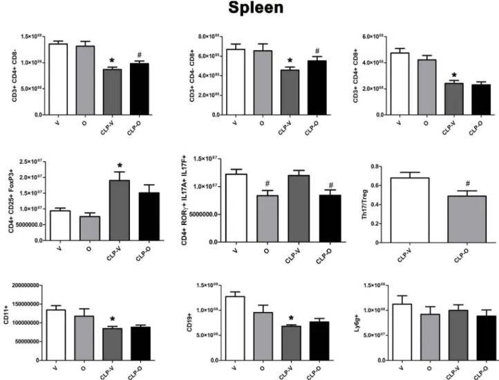
Ci + () 0Fig. 12. Effect of olaparib on the numbers ofvarious T-cell populations in the spleen ofyoung male Balb/c mice subjected to CLP. Values are shown in sham mice (not subjected to CLP, but treated with vehicle for 24 h) ("V"), in sham mice (not subjected to CLP, but treated with 10 mg/kg olaparib for 24 h) ("O") in vehicle-treated mice subjected to CLP for 24 h ("CLP-V") and in mice subjected to CLP in the presence of 10 mg/kg olaparib ("CLP-0"). Data are shown as mean ± SEM of 10 animais for each group; *p < 0.05 shows a significant change in the respective cell population in comparison to the sham group; #p < 0.05 shows significant effect of olaparib under non-CLP conditions (compared to vehicle-treated sham mice) or in CLP mice (compared to vehicle-treated CLP mice).
containing 2% BSA). Cells were stained with anti-CD3, CD4, and CDS (panel 1), anti-CD4, CD25 (panel 2) and anti-CD4 (panel 3) antibodies for 30 min in dark at 4 'C than were washed twice with flow cytometry buffer. For panel 1, cells were collected and analyzed by flow cyto-metry. Additionally, macrophages, B cells and neutrophils were simi-larly analyzed using antibodies against CDllb, CD19, and Ly6G. For panels 2 and 3, cells which were stained with surface markers were fixed and permeabilized with eBioscience's Permealization Kit, then incubated with anti-Foxp3 (panel 2) and anti-RORy, IL-17 A and IL-17 F (panel 3) antibodies for 30 min in the dark at 4 'C. Cells were then washed and analyzed by flow cytometry (Guava 8 H T; Merck-Millipore).
2.12. RT-PCR studies of miRNA expression
Total RNA was extracted from spleen and white blood cells using Trizol reagent (Invitrogen Carlsbad, California, EUA) according to the
manufacturer's specifications. Extracted RNA was eluted in RNase-free water, treated with DNase I, Amplification Grade (ThermoFisher, Waltham, MA, USA) according to the manufacturer's specifications and quantified by spectrophotometry.
cDNA was synthesized using miScript II RT Kit (Qiagen - Hilden, Germany) using HiSpec buffer chemistry, which exclusively reverse transcribes mature miRNA to cDNA; this mixture was incubated for 60 min at 37 'C and for 5 min at 95 'C to inactivate miScript Reverse transcriptase mix and placed on ice.
A functional quality contrai was performed on cDNA samples using miScript miRNA QC PCR array. qPCR was performed on a StepOnePlus Real-Time PCR System (ABI, Poster City, California, EUA). Threshold cycles (C) for PPC and RT contrai (miRTC) were examined to assess PCR and RT efficiencies, respectively. The expression of cel-miR-39 assay was also observed to confirm efficient RNA recovery. C values for all contrais were within the manufacturer's recommended range for a successful quality contrai (QC).
Spleen
(%)
60 40 20 .;, + + CO 30 CO 15 0 0 0 u + -40 .!!! uo;- u -+ .!!! ~]~ ~
202l
~ 10 u ,e~~
u ,e ~ ~ 20 .~"'
"'
0 0 10 0 5 u u u 0 ..\ 0é
qP ..\ 0 q~ss:i
..\ 0é
qP c,V <>" c." v c,V <>" 1.5 u. + 0.8 + t::*
..,
:::! Il.*
+ 0.6 )(of
li) 1.0~~
+= :::! ~ 0.4"'"
N U&·~
O ,,e u ~ 0.5 + 0 0.2...
~ 0 + u...
0 0.0 0.0 u ..\ 0 q~ qP ..\ 0 ~ qP c,V <>"c.S
c,V 25 40 20 ~ 20 ~30 ~ 15 Qi Qi Qi u 15 u u ~?!
20 ~ 10 :. 10 + +°'
"'
c
c
10 <D,.,
5 u 5 u ...J 0 0 0 ..\ 0 ~ qP ..\ 0 ~ qP ..\ 0 ~ qPc.S
c."vs
<>"r;,S
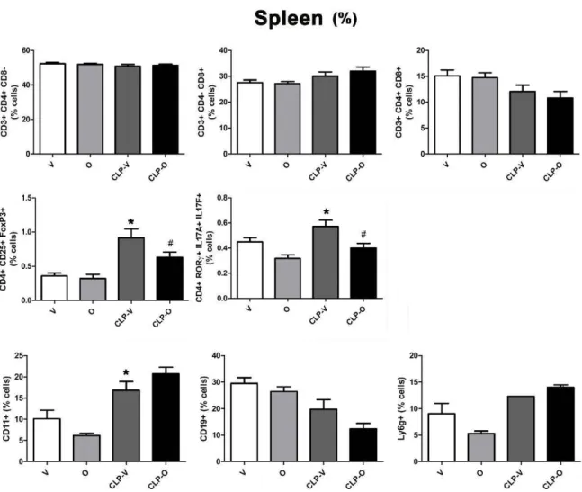
r;,VFig. 13. Effect of olaparib on the percentages ofvarious T-cell populations in the spleen ofyoung male Balb/c mice subjected to CLP. Values are shown in sham mice
(not subjected to CLP, but treated with vehicle for 24 h) ("V"), in sham mice (not subjected to CLP, but treated with 10 mg/kg olaparib for 24 h) ("0'') in vehicle
-treated mi ce subjected to CLP for 24 h ("CLP-V") and in mi ce subjected to CLP in the presence of 10 mg/kg olaparib ("CLP-0"). Data are shown as mean ± SEM of 10
animais for each group; *p < 0.05 shows a significant change in the respective cell population in response to CLP, compared to the sham group; #p < 0.05 shows
significant effect of olaparib under non-CLP conditions (compared to vehicle-treated sham mice) or in CLP mice (compared to vehicle-treated CLP mice).
Large-scale analysis of miRNA expression was performed using
miScript miRNA PCR Array Mouse Apoptosis (96 well format) (Qiagen
-Hilden, Germany). 12 plaques per organ were made. A set of controls
are included on each plate which enables data analysis using AACT
method of relative quantification, assessment of reverse transcription
performance, and assessment of PCR performance. The miScript miRNA
PCR array enables SYBR Green-based real-time PCR analysis using
StepünePlus Real-Time PCR System (ABI, Foster City, California, EUA)
as follows: for 15 min at 95 °C; 40 cycles of 15 s at 94 °C; for 30 s at
55 °C; and for 30 s at 70 °C and its software determined the number of
copies of each gene from the microarray plates.
Values of miRNA expression were normalized using the geomettic
mean calculated from the internal control genes classified according to
RetFinder Software and fold changes were calcula ted by the 2 -MCT
method. The analysis of the array was performed by the software
available in SaBiosciences' Data Analysis Center (https://www.qiagen. corn/br/ shop/ genes-and-pa thwa ys/ da ta - analysis-center-overview-page/). The most significantly altered miRNAs were confirmed by
RT-PCR. The altered miRNAs miR-15a-5p, miR-17-5p, miR-146a-5p and
miR-365-3p that were chosen for confirmation from the array; the primers were purchased from Qiagen. The cDNA was synthesized using
miScript II RT Kit and the PCR was performed using miScript SYBR
Green PCR Kit. Again, the values of miRNA expression were normalized
using the geomettic mean calculated from the previously internal
control genes classified and fold changes were calculated by the
2 - MCT method.
2.13. In vitro srudies in U937 cells subjected ro oxida.tive stress
Human monocyte histiocytic lymphoma cells (U937) were obtained
from ATCC and maintained in RPMI1640 with 10% fetal bovine serum
(Life Technologies). For resazurin and LDH assays, U937 cells were
plated in 96-well plates at 2 x 10⁴ cells/well. For DNA integrity, U937
cells were plated in 12-well plates at 2 x 10⁵ cells/well. For the NAD+
quantification assay, U937 cells were plated in 12-well plates at
3 x 10⁵ cells/well.
Blood
50000000 30000000 1000000 ,;, 40000000 0 u + 30000000...
0 u 20000000 +"'
0 20000000 u...
c u + 800000"'
0 u + 600000...
0 u 400000 #*
+ M 0 u 10000000 + 10000000 M c u + M 0 200000 u ~ 0 q~ qP c," c," 0 800000 u. + 6000000 8 + M c.. 600000 )(...
;! + 6 0 u. <...
4000000 Cl Q) + LI') 400000 N c u + 200000...
c u ;! +,..
et: 2000000 0 et: +...
~
4 :;: 1-2 0 c u 0 0 ~ 0é
qP v" v" ~ q~ pv"
vs
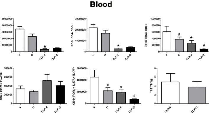
Fig. 14. Effect of olaparib on the numbers of varions T-cell populations in the blood of young male Balb/c mice subjected to CLP. Values are shown in sham mice (not subjected to CLP, but treated with vehicle for 24 h) ("V"), in sham mice (not subjected to CLP, but treated with 10 mg/kg olaparib for 24 h) ("O") in vehicle-treated mice subjected to CLP for 24 h ("CLP-V") and in mice subjected to CLP in the presence of 10 mg/kgolaparib ("CLP-0"). Data are shown as mean ± SEM of 10 animais for each group; *p < 0.05 shows a significant change in the respective cell population in response to CLP, compared to the sham group; #p < 0.05 shows significant effect of olaparib under non-CLP conditions (compared to vehicle-treated sham mice) or in CLP mice (compared to vehicle-treated CLP mice).
50 ,;, 40 0 u_ + ~ 30 ~~ ~ ~ 20 M 0 u 10 ~ 0
è
qP c," c," 2.0 +*
M c.. 1.5 )( 0 u."' += LI') Cl) 1.0 N 0 8~ + 0.5...
c u 0.0 ~ 0é
qP""
""
30 +"'
0 ~ ~ 20 0 1l u~ ;, ~ 10 0 u + 10 u....
;! +~~
~~
&-~ 0 et: +...
c uBlood
(%)
~ 0*
~ # +"'
0 0.6 u -0.4 + ~ ~8 u~ ;, ~ 0.2 0 u 0Fig. 15. Effect of olaparib on the percentages ofvarious T-cell populations in the blood ofyoung male Balb/c mice subjected to CLP. Values are shown in sham mice (not subjected to CLP, but treated with vehicle for 24 h) ("V"), in sham mice (not subjected to CLP, but treated with 10 mg/kg olaparib for 24 h) ("O") in vehicle-treated mice subjected to CLP for 24 h ("CLP-V") and in mice subjected to CLP in the presence of 10 mg/kg olaparib ("CLP-0"). Data are shown as mean ± SEM of 10 animais for each group; *p < 0.05 shows a significant change in the respective cell population in response to CLP, compared to the sham group; #p < 0.05 shows significant effect of olaparib under non-CLP conditions (compared to vehicle-treated sham mice) or in CLP mice (compared to vehicle-treated CLP mice).
A Spleen
B
Blood
Fig. 16. Effect of olaparib on spleen and blood T-cell miRNA levels in young male Balb/c mice subjected to CLP. Values are shown in sham mice (not subjected to
CLP, but treated with vehicle for 24 h) ("V"), in sham mi ce (not subjected to CLP, but treated with 10 mg/kg olaparib for 24 h) ("0'') in vehicle-treated mi ce subjected to CLP for 24 h ("CLP-V'') and in mice subjected to CLP in the presence of 10 mg/kg olaparib ("CLP-0''). Data are shown as mean ± SEM of 10 animais for each group;
*p < 0.05 shows a significant change in the respective miRNA level in response to CLP, compared to the sham group; #p < 0.05 shows significant effect of olaparib under non-CLP conditions (compared to vehicle-treated sham mice) or in CLP mice (compared to vehicle-treated CLP mice).
Cells were differentiated with PMA (100 ng/ml for 48 h), then subjected to various concentrations of hydrogen peroxide (H20 2, 300 µM, 600 µM, 1 mM) for 1 h, in the presence or absence of olaparib (1, 3, 10, 1 OO µM) pretreatment. After 1 h, cell viability was measured with the resazurin (7-hydroxy-3H-phenoxazin-3-one 10-oxide) method, or cell necrosis was detected by measuring the release of lactate dehy -drogenase (LDH) to the culture medium as described [24]. Mitochon -drial and nuclear DNA integrity was measured with the LA-PCR method as described above. Poly-ADP-ribose polymerase-1 (PARPl) enzyme and its product, poly-ADP-ribose (PAR) and actin (a loading control) were detected by Western blotting as described [25]. Total cellular NAD+ was determined using NAD/NADH Quantification Kit (Sigma) according to the manufacturer's protocol. The amount of NAD+ present in the samples was quantified in a colorimetric assay, measured at 450 nm using a microplate reader. The amount of NAD+ was normal
-ized to protein content of the samples, quantified using the PierceTM BCA Protein Assay.
Cellular bioenergetics was measured by the Extracellular Flux Analysis method as described [26]. Briefly, cells were seeded on cell culture microplates (60,000/well). After 24 h, cells were pretreated with olaparib (1, 3 or 10 µM) or its vehicle for 30 min, followed by exposure to H202 (300 µM) for 1 h. For analysis of mitochondrial
re-spiration, cells were washed twice with DMEM medium pH 7.4
sup-plemented with L-glutamine (2 mM, Gibco), sodium pyruvate (1 mM, Sigma) and glucose (10 mM, Sigma). After 1 h incubation at 37 °C in C02-free incubator, the oxygen consumption rate (OCR) after oligo-mycin (1 µM) was used to assess ATP production rate and the OCR after carbonyl cyanide-4-trifluoromethoxy phenylhydrazone (FCCP, O. 7 µM) to assess maximal mitochondrial respiratory capacity. Antimycin A (0.5 µM) and rotenone (0.5 µM) were used to inhibit the flux of electrons through complex III and I, to detect residual non-mitochondrial OCR, which is considered to be due to cytosolic oxidase enzymes. For analysis of glycolytic parameters, cells were treated with olaparib and H20 2 (as above), washed twice with phenol red-free DMEM medium pH 7.4
containing L-glutamine (2 mM), sodium pyruvate (1 mM), glucose
(10 mM) and HEPES (5 mM, Sigma). After 1 h incubation at 37 °C in C02-free incubator, proton efflux rate (PER) from basal and compen-satory glycolysis was measured. Mitochondria inhibition by rotenone (0.5 µM) and antimycin A (0.5 µM) allows calculation of the
mitochondrial-associated acidification. Subsequently, 2-deoxy-o-glu -cose (50 mM) was used to inhibit glycolysis and stop glycolytic acid
-ification. Glycolytic proton efflux rate (glycoPER) was calculated by subtracting the mitochondrial acidification to the total PER. All the
results were normalized by total protein.
2.14. Stxitistical analysis
Data are shown as mean ± SEM. One-way and two-way ANOVA with Bonferroni's multiple comparison test were used to detect differ -ences between groups. Two main type of comparisons were performed. The first type was to determine whether the insult (CLP in vivo or oxi -dative stress in vitro) has an effect on a given parameter compared to normal control values (animals not subjected to CLP or cells not su b-jected to H20 2). Statistically significant differences between these two
groups are indicated by *p < 0.05 or **p < 0.01. The second type was to detect if olaparib affected the response in the groups that were
subjected to the insult (CLP in vivo or oxidative stress in vitro), com -pared to the groups that were subjected to the same insults in the ab -sence of olaparib (Le. in the presence of olaparib vehicle). Statistically
significant differences between these two groups are indicated by #p < 0.05 or ##p < 0.01. Survival differences were analyzed by the Chi square test. Statistical calculations were performed using Graphpad Prism analysis software.
3. Results
3.1. Olaparib exern organ protective and anti-infiammatory eff ects in
organs of young adult male mice subjected to CLP, without adverse/y
affecting D NA integrity
In young adult male mi ce subjected to CLP, olaparib (1, 3 or 10 mg/ kg i.p.) concentration-dependently improved several parameters of multiorgan dysfunction (Figs. 1-3). For instance, the CLP-induced i n-creases in spleen MPO content and liver and spleen MDA levels were attenuated by olaparib (Fig. 1). In addition, the CLP-induced increases
in plasma markers ofliver and pancreas injury (ALP, ALT, amylase) and renal dysfunction (BUN) were attenuated by olaparib treatment (Fig. 2). The histopathological pictures of the lungs did not show
25001 2000 ~ 1500 ::> 1000 500
o,_-
....
Sha-~m 300 200 ~ 100 80 ALP*
CLP GLU BUN*
n
CLP Ol<1parib (1o m9'<g) CLP Ofaparib f1o mg/kg) CLP Olap;ulb (10 m9'<g) CLP Olapartb (10 m9'<g) 250 200 -' 150 ::> 100 50 10 0.5 Sham Sh111m ALT*
CLP CRE Albumin CLP CLP Olaparib (10 mglkg) CLP Olaparib (10 mgJkg) CLP 01.lparib (10 mg/kg) CLP Olapartb 110 mg/kg) 1500"
200 150 -' -' 0 100 >! ::; 50 1.5 AMY*
CLP PHOS Na+ GLOB Sham CLP CLP Olapoulb (10 mg/kg) CLP Olaparih {10mglkgl CLP Olapartb (10 m9'<g) Olaparlb (10mg/1'.g)Fig. 17. Effect of olaparib on selected parameters of organ injury in young female Balb/c mice subjected to CLP. Various physiological and organ injury marker
levels: alkaline phosphatase (ALP), alanine aminotransferase (ALT), amylase (AMY), plasma glucose (GLU), plasma calcium (CA++), plasma phosphate (PHOS),
plasma blood urea nitrogen (BUN), plasma creatinine (CRE), plasma sodium (Na+), plasma potassium (Na+), plasma albumin (ALB) and plasma globulin (GLOB) measured by Vetscan analysis, are shown in sham mice (not subjected to CLP) ("Sham''), in vehicle-treated mice subjected to CLP for 24 h ("CLP") and in mice
subjected to CLP in the presence of various doses of olaparib ("CLP Olaparib 1 mg/kg", CLP Olaparib 3 mg/kg" and "CLP Olaparib 10 mg/kg"). Data are shown as
mean ± SEM of 10 animais for each group; *p < 0.05 shows significant increase in the respective parameter in response to CLP, compared to the sham group.
marked alterations in any of the groups, with slight emphysema evident in ail a,p groups (Fig. 3). In the liver, foamy degeneration of numerous hepatocytes is evident in the CLP group; olaparib, at the 10 mg/kg dose, normalized the morphology of the hepatocytes (Fig. 4). In the spleen, CLP induced macrophage infiltration, and evidence of hemolysis was evident, with no marked differences between CLP groups with or without olaparib (Fig. 5).
Olaparib, at 10 mg/kg (but not at the two lower doses used), caused
a significant prolongation of survival of the animals subjected to ap
(Fig. 6).
CLP did not induce detectable damage in nuclear DNA in any of the
organs (liver, lung, spleen) studied, but in the liver, a significant degree
of mitochondrial DNA damage was detected, which was prevented by
olaparib treatment (Fig. 7). Olaparib treatment reduced the number of
A
E
3 LI... 10• 107 106 105•
•
•
•
CFU BLOOD
I
0 104 103 102...
1o'~~~~C~L-P----~~~~---,C-L-P~~~ Olaparib (10 mg/kg) ~ 3 u. 0B
1000000 100000 10000 1000 100 10bacteria in the plasma and spleens of mice subjected to CLP (Fig. 8). However, this effect does not appear to be a direct antibacterial action of olaparib, as in vitro incubation of E. coli with various concentrations of olaparib (1-1 OO µM) did not have any effect on bacterial growth
(Fig. 9). Moreover, in a satellite experiment of bacteremia without
septic shock, olaparib treatment failed to significantly affect bacterial CFUs in the blood, spleen or liver after an i.p. bolus of E. coli (Fig. 10). Olaparib treatment in the CLP model attenuated the increases in the levels of several circulating mediators in the plasma (e.g. 1NFa, IL-la, IL-1 [3, IL-2, IL-4, IL-6, IL-l 2p40), while others (e.g. IL-1 O,RAN1ES, VEGF) were unaffected (Fig. 11 ).
In the spleen, the number of CD4 + and CD8 + lymphocytes were
reduced in response to CLP; this reduction was attenuated by olaparib
treatment (Fig. 12). Moreover, in the spleen, the number of Treg
(CD4 + CD25 + FoxP3 +) but not Thl 7 (CD4 + RORy +ILI 7 A+ ILI 7F
+) lymphocytes increased in response to CLP; the number, as well as percentage of both of these cell populations were reduced in CLP when the animals also received olaparib (Fig. 12,13). The number of CDll b + cells in spleen were significantly decreased by CLP, but per-centages were significantly increased; olaparib had no significant effect on these responses. The number of CDl 9 + cells were significantly decreased by CLP and not affected by olaparib, and numbers and per-centages of neutrophils were unaffected by CLP and olaparib (Figs. 12
and 13).
CLP reduced the number of Thl 7 cells in the blood - but increased as a percentage, since these alterations in Thl 7 cell numbers occur against the background of an overall lymphopenia -and the number (as well as percentage) of these cells was further reduced by olaparib
(Figs. 14 andl5 ). The Thl7/Treg ratio is related to SOFA score; ele
-vated ratios have been associated with increases in SOFA scores, an indication of organ damage [27]. In the spleen, this ratio was sig-nificantly lower in CLP/olaparib group than in the CLP/vehicle group
(Fig. 12) and slightly lower in the blood, although not significantly
(Fig. 14).
CFU SPLEEN
•
1
•
•
CLP CLP Olaparib (10 mg/kg)Fig. 18. Effect of olaparib on blood and spleen
bacterial colony-fonning unit (CFU) numbers
in young female Balb/c mice subjected to CLP. (A): Blood CFUs (expressed as CFU/ml) and (B)
spleen CFU s (expressed as CFU/ml) are shown
in vehicle-treated mice subjected to CLP for 24 h ("CLP") and in mice subjected to CLP in the presence of 10 mg/kg olaparib ("CLP
Olaparib 10 mg/kg"). Data are shown as
mean ± SEM of 10 animais for each group.
In recent years, miRNAs have been identified as important
reg-ulators of a host of immune (as well as parenchymal) cell functions,
with each miRNA playingvarious regulatory roles that are context-and
cell-type as well as pathophysiological condition dependent. Large
-scale analysis of miRNA expression identified a multitude of changes in
splenic miRNA levels after CLP; olaparib treatment attenuated the
CLP-induced alterations in miR15, miRl 7, miR181 and miR365 and levels
(Fig. 16). For miRl 46, in the spleen, olaparib attenuated its CLP-
in-duced downregulation, while in the circulating leukocytes, olaparib
enhanced its CLP-induced upregulation (Fig. 16).
3.2. The beneficial effects of olaparib are absent in yowig adult female mice
subjected to CLP
In contrast to the findings in male mice, in young adult female mice
subjected to CLP, olaparib (10 mg/kg i.p.) did not attenuate blood or
splenic CFUs and did not have any significant effect on the various
circulating markers of organ injury (Figs. 17 and 18). This finding
confirms prior data showing that PARP inhibitors preferentially exert their beneficial effects in male animals subjected to various forms of
critical illness (as overviewed in [19]). The degree of the change in
several circulating markers (e.g. amylase, BUN) of injury in female mice
tended to be less pronounced in female mice than in male mice after
CLP (Fig. 2 vs. Fig. 17), perhaps indicating a lesser degree of baseline
CLP-induced multiple organ injury in female mice versus male mice.
3.3. Olaparib exerts beneficial effects on some parameters ofinjury in ggfil
male and female mice subjected to CLP
Since septic shock is, to a large extent, a disease of the elderly [1,2], and since in aged organisms some of the pathophysiological mechan
-isms are not only quantitatively but also qualitatively different from the mechanisms that occur in young adult organisms [2,22,28-30], we
have also investigated the effect of olaparib in aged mice (72 weeks old
ALP ALT AMY 1000
*
150 2000*
*
800 1500 100 _, 600 ::'. _, # ::> ::> - 1000 400 50 ::> 200 500 CLP CLP Sham CLP CLPOlaparib Olaparib Olaparlb
10m!)Jkg 10mg!kg 10mg/kg GLU CA++ PHOS 10 20 250 200 15 ;; 6 ;; :s 150 -;, 10 "'
"'
E 100 E 4 E 50 Sham CLP Sh3m CLP CLP Sham CLP CLP Olaparib Olaparib 10mgJkg 10mg/kgBUN CRE Na+
100 0.8 200 80
*
0.6 150 ::'. ;; 60 ;; _, ~ 0.4 0 100"'
E :E E 40 :E 0.2 50 20Sham CLP CLP 0.0 Sham CLP CLP Sham CLP CLP
Olapartb Olaparib Olaparib
10mg/kg 10mg/kg 10mgfkg K+ Albumin GLOB 2.5 10
e
"'
:E CLP Sham CLP CLP CLPOlaparib Olaparib Ol8parib
10mglkg 10mglkg 10mg/kg
Fig. 19. Effect of olaparib on selected parameters of organ injury in aged male Balb/c mice subjected to CLP. Various physiological and organ injury marker levels:
alkaline phosphatase (ALP), alanine aminotransferase (ALT), amylase (AMY), plasma glucose (GLU), plasma calcium (CA++), plasma phosphate (PHOS), plasma
blood urea nitrogen (BUN), plasma creatinine (CRE), plasma sodium (Na+), plasma potassium (Na+), plasma albumin (ALB) and plasma globulin (GLOB) measured
by Vetscan analysis, are shown in sham mice (not subjected to CLP) ("Sham''), in vehicle-treated mi ce subjected to CLP for 24 h ("CLP'') and in mi ce subjected to CLP in
the presence of various doses of olaparib ("CLP Olaparib 1 mg/kg", CLP Olaparib 3 mg/kg" and "CLP Olaparib 10 mg/kg"). Data are shown as mean ± SEM of 10
animais for each group; *p < 0.05 shows significant increase in the respective parameter in response to CLP, compared to the sham group; #p < 0.05 shows
significant effect of olaparib in CLP mice (compared to vehicle-treated CLP mice).
males and in females). Surprisingly, -with the exception of the pan
-creatic injury marker amylase - olaparib exetted no significant
bene-ficial effects on most organ injury markers in aged male mice (Fig. 19)
and only had modula tory effects on a small subset of circulating me
d-iators: IL-4 and IL-l 2(p70) (Fig. 20).
In contrast, in aged female mice, olaparib trea tment significantly
reduced the CLP-induced liver injury markers ALP and ALT (Fig. 21).
However, levels of the kidney injury marker BUN and pancreatic
marker amylase were not affected by olaparib after injury. Circulating
levels of the CLP-induced mediators TNFa, IL-la, MIPla, M-CSF and
MIG were significantly reduced by olaparib treatment (Fig. 22).
Neither in aged male or female mice subjected to CLP did we
ob-serve any adverse effect of olaparib on nuclear or mitochondrial DNA
integrity (Fig. 23).
Overall, the degree of the beneficial effect of the PARP inhibitor
tended to be less pronounced in the aged mice than in the young male
mice (Figs. 1-2. vs. Figs. 17-22).
TNFu IL·1a
"::
Liil
t:::
Lirr
1-
1'"
" 0 Sham CLP CLP 0 Sham CLP CLP IL-3 IL-7 Olaparib Olaparib (10m!)lkg) (10mglkg) IL-4 IL-9 CCP Olaparib (10mglkg) IL-111 IL-5 ll-10 Olaparib (10mg/kg)"'
Lto_
"'"
Lin
600 * 10000 * l400l
5(){)0 '" ' Sham CLP CLP ' Sham CLP CLP O!aparib Olaparib (10mglkg) (10mg/kg) IL-12(p70) LIF CCP Olaparib (10mglkg) IL-13"
"LlCL
i 2 0 " O Sham CLP CLP LIX Olaparlb (10mglkg)""
L.a
"'"
Lill
1000 2000 *l
.
l
500 1000 O Sham CLP CLP O Sham CLP CLP Olaparib Olaparlb (10mgllt.g) (10mglkg) GM-CSF KC ll-15 Eotaxin"'"
Lin
.
20000 ...115000•
~ 10000""
O Sham CLP CLP IP-10 Olaparib (10mglkg) El:::
Lin
~::::
L i D
~ ~ 50 l20000 o. 10(){)0 O Sham CLP CLP O Sham CLP CLP Olaparlb Olaparib (10mglkg) (10mglkgl RAN TES VEGF.::1
~
~
~
'"LIU
O Sham CLP CLP MIP-2 Olaparib (10mglkg)'""
20000Ltn
....115000 ' l10000 5000 O Sham CLP CLP Olaparib !10mallt.al M-CSF Olaparib (10mg/kg)'"
L.o_
"' l 100 * " O Sham CLP CLP Olaparib (10m!)lkg)i
MIP-1a CLP Olaparib (10mglkg)""
Lm
2000 1000 O Sham CLP CLP MIG.
"" Olaparib {10mglkg)'"'
Lirr
l1000 '" O Sham CLP CLP Olaparib (10mg/kg) IL-2 IL-6'""
LIJJ
"'" ~ 30000 l20000 "'" O Sham CLP CLP lL-12(p40) IL-17 i4000 G-CSF Olaparib (10m!)lkg)"'"
Lin
'"" i20000 '"" O Sham CLP CLP MCP-1 Olaparib (10mglkgl"'"
LtQ
20000 "§15000 ~10000 5-000 O Sham CLP CLP MIP-111 Olaparib (10mglkg)""
Un
.
6000 l4000""
O Sham CLP CLP INFy Olaparib (10mg/kg) oraparib (10mglkg)Fig. 20. Effect of olaparib on the levels ofvarious circulating mediators (cytokines, chemokines, growth factors) in aged male Balb/c mice subjected to CLP. Values are shown in sham mice (not subjected to CLP) ("Sham"), in vehicle-treated mice subjected to CLP for 24 h ("CLP") and in mice subjected to CLP in the presence of
10 mg/kg olaparib ("CLP Olaparib 10 mg/kg"). Data are shown as mean ± SEM of 10 animais for each group; *p < 0.05 shows significantincrease in the respective