HAL Id: hal-03083014
https://hal.archives-ouvertes.fr/hal-03083014
Submitted on 4 Jan 2021HAL is a multi-disciplinary open access archive for the deposit and dissemination of sci-entific research documents, whether they are pub-lished or not. The documents may come from
L’archive ouverte pluridisciplinaire HAL, est destinée au dépôt et à la diffusion de documents scientifiques de niveau recherche, publiés ou non, émanant des établissements d’enseignement et de
External pH modulation during the growth of Vibrio
tapetis , the aetiological agent of brown ring disease
A. Rahmani, C. Mathien, A. Bidault, N. Le Goïc, V. Pichereau, Christine
Paillard
To cite this version:
A. Rahmani, C. Mathien, A. Bidault, N. Le Goïc, V. Pichereau, et al.. External pH modulation during the growth of Vibrio tapetis , the aetiological agent of brown ring disease. Journal of Applied Microbiology, Wiley, 2020, 129 (1), pp.3-16. �10.1111/jam.14699�. �hal-03083014�
External pH modulation during the growth of Vibrio tapetis, the etiological agent of
1
Brown Ring Disease
2 3
Running head: External pH modulations by Vibrio tapetis
4 5
Alexandra Rahmani1#, Clémentine Mathien1, Adeline Bidault1, Nelly Le Goïc1, Christine 6
Paillard1#†, Vianney Pichereau1#† 7
1
Univ Brest, CNRS, IRD, Ifremer, UMR 6539 LEMAR, F-29280 Plouzané, France 8
†
These authors contributed equally 9
# Corresponding authors : alexandra.rahmani@univ-brest.fr ; vianney.pichereau@univ-10 brest.fr ; christine.paillard@univ-brest.fr 11 12 Abbreviations 13
BRD: Brown Ring Disease 14
d.p.i.: Days Post-Infection 15
EPFs: Extrapallial Fluids 16
FSSW: Filtered Sterilized Seawater 17
OD: Optical Density 18
Keywords: BRD, V. tapetis, R. philippinarum, pH, modulation effect, pH modifications,
19
neutralizing activity 20
Abstract
21
Aims
22
Brown Ring Disease (BRD) is an infection of the Manila clam Ruditapes philippinarum due to 23
the pathogen Vibrio tapetis. During BRD, clams are facing immunodepression and shell 24
biomineralization alteration. In this paper, we studied the role of pH on the growth of the 25
pathogen and formulated hypothesis on the establishment of BRD by V. tapetis. 26
Methods and Results
27
In this study, we monitored the evolution of pH during the growth of V. tapetis in a range of 28
pH and temperatures. We also measured the pH of Manila clam hemolymph and extrapallial 29
fluids during infection by V. tapetis. We highlighted that V. tapetis modulates the external pH 30
during its growth, to a value of 7.70. During the development of BRD, V. tapetis also influences 31
extrapallial fluids and hemolymph pH in vitro in the first hours of exposure and in vivo after 3 32
days of infection. 33
Conclusions
34
Our experiments have shown a close interaction between V. tapetis CECT4600, a pathogen 35
of Manila clam that induces BRD, and the pH of different compartments of the animals during 36
infection. These results indicate that that the bacterium, through a direct mechanism or as a 37
consequence of physiological changes encountered in the animal during infection, is able to 38
interfere with the pH of Manila clam fluids. This pH modification might promote the infection 39
process or at least create an imbalance within the animal, that would favor its persistence. 40
This last hypothesis should be tested in future experiment. 41
Significance and Impact of Study
42
This study is the first observation of pH modifications in the context of BRD and might orient 43
INTRODUCTION
45 46
The Manila clam Ruditapes philippinarum is a bivalve mollusc species of significant economic 47
interest with a world production mainly from China, followed by European countries such as 48
Italy, Spain and France (Paillard 2004b). It was imported in France in the early 70’s for 49
aquaculture. Ten years later, mortalities have been observed in clam’s cultures, caused by an 50
infection called the Brown Ring Disease or BRD (Paillard et al. 1989). Indeed, infected clams 51
were characterized by a brown deposit in the inner face of the shell between the pallial line 52
and the edge of the shell (Paillard et al. 1994). BRD is caused by a Gram negative bacterium, 53
Vibrio tapetis (Paillard and Maes 1990; Borrego et al. 1996). BRD has caused significant
54
economic losses in livestock farms on the Northern European coasts because the pathogen 55
has an optimum infection rate in cold waters at around 14°C. For example, in France, Manila 56
clam production decreased from 500 tons in 1987 to 200 tons in 1989, the aquaculture of 57
clams production being mainly located in Brittany (France), an area highly sensitive to BRD 58
(Paillard 2004b). 59
V. tapetis is a microparasite acting in two modes. In the case of BRD, the bacterium is referred
60
to as an external microparasite because it degrades the periostracal lamina of the clam to 61
enter the extrapallial compartment without invading the tissues. In this case, the pathogen 62
induces BRD in Manila clams. In some cases, when tissues are damaged, V. tapetis acts as an 63
internal microparasite by infiltrating the animal's tissues, resulting in mortality within days 64
(Paillard 2004a). 65
66
During infection by V. tapetis, the pathogen therefore colonizes the periostracum and 67
degrades it in order to infiltrate peripheral or even central extrapallial fluids (EPFs). As a 68
result, it inhibits the biomineralization of the shell, preventing the formation of calcium 69
carbonate crystals of aragonite and thus leads to the accumulation of the organic brown 70
deposit (Paillard et al. 1994). 71
Furthermore, infected clams are facing an immunodepression caused by V. tapetis. 72
Hemocytes, which are key effectors of the immune defenses of clams, are altered in BRD by 73
actin cytoskeleton disorganization causing rounding of hemocytes and downregulation of 74
pathogen recognition effectors. Consequently, infected hemocytes are no more able to 75
ensure phagocytic progress to degrade the pathogen (Choquet et al. 2003; Allam et al. 2014; 76
Rahmani et al. 2019). 77
Many factors are involved in BRD. Indeed, some alterations on the inner shell have significant 78
impacts on the functioning of the affected clams. Impeding the functioning of adductor 79
muscles and siphons, they result in abnormal positioning of the clam in the sediment and 80
prevent the proper closure of valves that can lead to mortality when fishing or directly in the 81
sediment, as clams can no longer retain their inter-valve water (Goulletquer et al. 1989; 82
Paillard 2016). 83
Shell biomineralization is characterized by the formation of Calcium-Carbonate crystals 84
(CaCO3) from calcium (Ca2+) and carbonate (CO32-) ions. CaCO3 crystals are the main 85
constituent of shellfish. In the Manila clam, the shell is composed of aragonitic crystals 86
(Taylor, J D et al. 1973; Trinkler et al. 2011). The saturation state of this mineral in seawater 87
determines whether the biomineralization reaction is favored in the direction of crystal 88
formation or dissolution. It is defined by the concentrations of Ca2+ and CO32- ions. 89
Biomineralization process is often studied in the context of ocean acidification because pH is 90
a factor that considerably influences the physiology of marine calcified organisms, and 91
saturation rate of calcium carbonate crystals through the reduction in available CO32- ions, 93
thus promoting the dissolution of the shell (Gattuso and Hansson 2011). In the mussel Mytilus 94
edulis, decrease in pH leads to a decrease in the net calcification rate, Ca2+ ion content and, in 95
general, in a change in the ultrastructure of the shell and a decrease in amino acid content (Li 96
et al. 2015). Combination of temperature and pCO2 increaseresults in a decreased shell 97
hardness in the clam Mercenaria mercenaria and in the American oyster Crassostrea virginica, 98
indicating major changes in the biomineralization process (Ivanina et al. 2013). In the brittle 99
stars Amphiura filiformis, authors showed that the animal was able to compensate, in the 100
short term, for the effects of pH decline by accelerating calcification and metabolism at the 101
expense of certain functions such as growth and reproduction because of the high energy 102
cost required for this mechanism (Wood et al. 2008). In summary, most of the studies reflect 103
that ocean acidification reduces the calcification rates of animals composed of calcitic or 104
aragonitic crystal shells by promoting the dissolution reaction of these crystals. 105
In the Manila clams, calcification, i.e. shell biomineralization reaction, occurs in extrapallial 106
fluids: ie the mantle secretes the shell components, the organic matrix (conchiolin) and the 107
calcium carbonate that form the aragonitic crystal shell (Paillard and Le Pennec 1993; Trinkler 108
2009; Trinkler et al. 2011). The periostracum (outer part of the shell) is made of a sclerotinized 109
organic matrix. Shell organic matrix is mainly composed under normal conditions of aspartic 110
acid, glycine and serine, responsible for binding with calcium ions: chelation by sulphate ester 111
groups on polysaccharide chains would initiate biomineralization. When BRD develops, 112
affected clams have deficiencies in amino acid such as glutamic acid, aspartic acid, serine, 113
and alanine, which can lead to poor binding to Calcium - Carbonate (Goulletquer et al. 1989). 114
Given the importance of pH homeostasis in the biomineralization process, it seems likely
115
that pH might play a significant role in the pathogenicity of V. tapetis in the case of BRD.
Nevertheless, the influence of pH on the development of V. tapetis, as well as on the
117
development of BRD, have never been investigated. In this paper, we chose to
118
investigate the influence of pH on the development of the pathogen and the modulation
119
of pH associated to this bacterium in clams’ fluids. Furthermore, we monitored for the
120
first time the pH modification in clam fluids during in vivo challenge by V. tapetis allowing
121
us to formulate hypothesis that pH variations due to V. tapetis might be a virulence
122
strategy of this pathogen linked to pathogenicity and to its ability to alters
123
biomineralization. This study is the first investigation of pH modifications in the context
124
of BRD.
125 126
Materials and methods 127
1) Monitoring of the pH modifications during growth of V. tapetis in culture media 128
Culture media and bacterial strain: The bacterial strain used in these experiments is V. tapetis
129
CECT4600, grown in Zobell medium (Zobell 1941) and incubated at 18°C. The first objective 130
was to monitor the growth of this strain at 7 different pH values and 4 different temperatures. 131
The 7 liquid culture media used in this experiment were obtained from Zobell media 132
supplemented with NaOH 1 M or HCl 0.1 M until the expected pH is obtained. After autoclave 133
sterilization, the pHs of the 7 media were 6.00, 6.45, 7.00, 7.55 (not modified, control medium, 134
CM), 7.86, 8.12 and 8.70, respectively. pH of the cultures/media were measured by using a 135
Mettler Toledo© InLab Micro electrode after calibration of the electrode with pH buffers 4, 7 136
and 9 at the beginning and the end of each experiment. The 4 temperatures tested were 14°C 137
(optimal infection temperature), 18°C (optimal growth temperature), 21°C (temperature at 138
which the recovery process occurs mainly) and finally 27°C (temperature at which the bacteria 139
die) (Borrego et al. 1996; Paillard et al. 2004; Trinkler 2009; Paillard 2016). 140
Growth experiments: colonies of strain CECT4600 were resuspended in 2 ml of Zobell medium
141
(unmodified pH, control medium CM). The Optical Density (OD) was measured at a 142
wavelength of 492 nm and the suspension concentration was adjusted to an OD of about 0.1, 143
i.e. to a cell density of 9.4 x 10⁷ CFU.ml-1. The suspension was diluted at 1:100 in 3 ml of Zobell 144
medium at the desired pH. The control samples were the same sterile media. 400 µL of 145
inoculated media or controls were distributed in a Bioscreen© 100-wells plate (5 replicates 146
per tested condition), allowing a sterile, thermally controlled culture with an OD 147
measurement at regular intervals under stirring. This protocol was repeated twice; each 148
experiment was run for 24 hours (except at 14°C where the experiment ran for 30 h to take 149
into account the reduced bacterial growth), in a shaking mode, with measurement of OD 150
every 20 minutes at a wavelength of 492 nm ; temperature was set up at either 14, 18, 21 or 151
27°C. T0 represents the pH measurement at the beginning of the growth experiment (T0) and 152
TF (TFinal) represent the pH measurement at the end of experiment, depending on the 153
different conditions as detailed above. 154
155
pH monitoring during the bacterial growth: Two precultures of V. tapetis CECT4600 were
156
performed in Zobell broth during 15 h at 18°C and 24 h at 14°C in order to reach an OD of 157
0.98. Each preculture was diluted 12 times (initial OD = 0.08) with fresh Zobell medium at the 158
different pHs (from 6.00 to 8.70). With these new cultures, bacterial growth was performed 159
under shaking at 14°and 18°C, respectively from the corresponding pre-culture, in three 160
replicates. The pH was measured, from 200 µl-samples, at the beginning of the experiment, 161
every hour during the exponential growth phase, and finally in the stationary phase (i.e. 48 h 162
of growth at 14°C and 24 h of growth at 18°C). 163
164
2) Monitoring of the pH in extrapallial fluids (EPFs) and hemolymph of Manila
165
clams during infection by V. tapetis
166 167
Animals: Animals used in this study were adult Manila clams (about 40 mm) from Landeda
168
(Finistère, France), kindly provided by the company SATMAR. One pool of 40 clams (for the 169
in vitro study of fluids) and 2 pools of 100 clams (for the in vivo infection study) were
170
acclimatized for 3 and 2 weeks, respectively, in oxygenated seawater at 14°C. 171
Fluids and shell sampling: The fluids chosen for this experiment were hemolymph and EPFs.
173
Hemolymph, EPFs and shell were randomly collected extemporaneously from 10 clams. 174
Hemolymphs have been collected from the adductor muscle and EPFs from the extrapallial 175
cavity of the animals as previously described (Le Bris et al. 2015). The quality of the 176
hemolymph samples was checked under the microscope (presence of pseudopods, 177
unrounded hemocytes, serum and hemocytes without microorganism contaminations) then 178
the samples were pooled and the number of hemocytes was determined by using a Malassez 179
counting grid. Shell parts were broken with a hammer to obtain small pieces and were 180
sterilized by UV (wavelength 254 nm) for 15 minutes. EPFs were also pooled. The hemocytes 181
concentration in the hemolymph pool was 5.45 x 10⁵ hemocyte per ml. 182
183
For in vitro experiments, 150 μl of FSSW, hemolymph or EPFs were placed in 1.5 ml sterile 184
tubes, with supplementation of powdered Manila clam shell, in the same amount in each 185
sample, in order to design an in vitro experiment that is as close as possible of natural 186
conditions and to allow ion exchanges between fluids and shell that might be dependent of 187
the V. tapetis growth. Then, 200 μL of a V. tapetis CECT4600 suspension in FSSW was added 188
to each tube, in order to reach a final bacteria:hemocytes ratio of 25:1 (1.37 x 10⁷ CFU.ml-1); 189
200 μl of FSSW was added for the controls. Each condition was tested in triplicates. pH 190
measures were performed at times 0 h, 3 h, 24 h, 48 h and 72 h and tubes were shaken before 191
measurement. 192
193
For in vivo infection experiments, the protocol was followed as previously described (Paillard 194
and Maes 1994). The animals were first left out of the water during the night before infection 195
to allow the valves to open spontaneously in water, and then injected with 100 μl of FSSW for 196
50 control individuals, or 100 μl of bacterial suspension (5 x 10⁸ CFU.ml-1 in FSSW) for 50 197
infected individuals, in the pallial cavity. The injected animals were left out of water for 3 198
additional hours to allow the pathogen to colonize them, and then were transferred again 199
into their respective seawater tanks. Animals were sampled at different time points after 200
infection, ie. after 15 hours, 1 day, 3 days, 7 days, 14 days and 21 days post injection (d.p.i.). 201
pH of the EPFs and hemolymph samples were measured by using the Mettler Toledo 202
microelectrode calibrated with pH 4, 7 and 9 buffers. The initial sample (T0) was taken just 203
before injection. The mode of infection does not induce mortality and allows to reproduce 204
first stage of BRD to see the effect of V. tapetis, even before the development of organic 205
deposit in the shell, highlighting the pH variations that occurs in the really first days of V. 206
tapetis infection.
207 208
Statistical analyses: Statistical analyses were performed by using a Student test or a Mann
209
Whitney Wilcoxon test, depending of data normality. 210
RESULTS 211
V. tapetis CECT4600 modulates the pH of Zobell culture medium during its growth. 212
V. tapetis was cultured in Zobell broths which pH were adjusted from 6.00 to 8.70, at 4
213
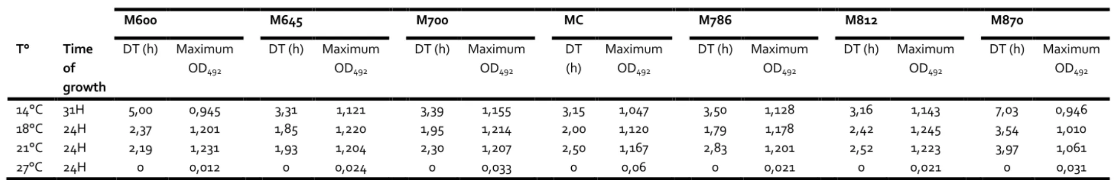
different temperatures ranging from 14°C to 27°C. The results obtained are reported in Table 214
1. First, we observed that the bacterium is able to grow at temperatures ranging from 14°C 215
to 21°C, in the pH range tested, but not at 27°C. In addition, consistently with the results 216
previously published by Borrego et al (1996), the optimal growth temperature is 18°C in the 217
unmodified medium. 218
V. tapetis has a high tolerance to pH because we observed a growth in all the tested culture
219
media. However, at 14°C, the fastest growth was obtained for the control medium (pH 7.4, 220
DT = 3.15 h), but growths were observed over a pH range from 6.45 to 8.12, with doubling 221
times from 3.15 to 3.5 and max ODs higher than 1, while the growth rates were lower at the 222
extreme pHs tested, ie. 6 and 8.7 (Table 1). The best growth at 14°C was observed in the 223
unmodified culture medium (MC, with a DT of 3.15h), and at pH 7.86 at 18°C (DT = 1.79h). 224
Interestingly, at the growth temperatures 18 and 21°C, very good growth parameters were 225
obtained for more acidic pHs, from which the lowest DTs were observed for the pH value 6.45 226
(DTs = 1.85 and 1.93 h at 18 and 21°C, respectively). 227
The pHs of the culture media have been measured at both the beginning (T0) and the end 228
(TF) of bacterial growth in all the conditions tested. Results are presented in Figures 1 to 3. In 229
the control samples (no bacteria), the pHs did not change significantly between the 230
measurements made at T0 and TF (Fig. 1 to 3). By contrast, after growth of V. tapetis at 14, 18 231
and 21°C, the pH changed during growth to reach an average value of 7.90 in all media tested 232
(Fig. 1 and 2). The pH modifications are the same at these three temperatures, allowing us to 233
show them all in the same figures (Fig. 1 and 2). We did not observe such a pH modification 234
of the culture medium at 27°C, a condition where no growth of the bacterium was observed 235
(Fig. 3). 236
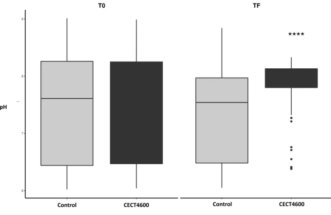
Figure 2 shows the statistical distribution of pH values measured at T0 and TF in all media. 237
The mean and variance of this distribution differed significantly during the experiment only 238
when the bacterium was able to grow (pvalue < 0.0001). Overall, these results show that V. 239
tapetis is able to modify the pH of its culture medium during its growth towards a pH close to
240
7.90. 241
We measured the evolution of medium pH all along the exponential growth phase of V. 242
tapetis at 14°C (Fig. 4) and 18°C (Fig. 5). In the control samples, the pH changed only slightly
243
during the experiment, resulting in very low slope curves (data not shown). By contrast, in the 244
experimental samples, during the growth of V. tapetis, the pHs of the media (which were 245
initially 6 to 8.7) converged to an average value of 7.71, whatever the temperature of growth 246
(Fig. 4 and 5). Therefore, our experiments show that V. tapetis is able, during its growth, to 247
modify the pH of its culture medium. More precisely, V. tapetis tends to ‘neutralize’ culture 248
media during its growth, by acidifying alkaline media and alkalinizing acidic media, to make 249
it reach a mean value of 7.7-7.9 whatever the initial pH of the culture medium. However, this 250
effect can be due to either a passive mechanism as a consequence of the bacterium’s growth 251
or an active mechanism that allow the pathogen to infect the Manila clam. 252
External pH modulation during the in vitro growth of V. tapetis CECT4600 in clams 253
extrapallial fluids or hemolymph. 254
and the hemolymph. In these experiments, FSSW was used as the reference fluid because it 257
does not allow any growth of V. tapetis. The same amount of powdered shell fragments was 258
added to each sample in order to design an in vitro experiment that is as close as possible of 259
natural conditions and to allow ions exchanges between fluids and shell that might influence 260
V. tapetis growth. The obtained results are shown in the Figure 6. Our experiments showed
261
that in FSSW, the presence of V. tapetis did not lead to any significant change in pH as 262
compare to the control, at each time point tested. By contrast, in EPFs and hemolymph, 263
exposure to V. tapetis led to a significant decrease of pH as compared to the initial conditions 264
after 3 hours of exposure (pvalue < 0.001). Indeed, for both clam biological fluids (ie. EPFs and 265
hemolymph), the pH significantly dropped from 7.6 to 7.4 after three hours of Vibrio exposure, 266
and then stabilized at approx. 7.25. However, for longer times, the pH also tended to decrease 267
in the controls, so as no significant difference appears between the control samples and the 268
samples exposed to V. tapetis for the last time points, thus revealing an interaction only in 269
the first hours of exposure. 270
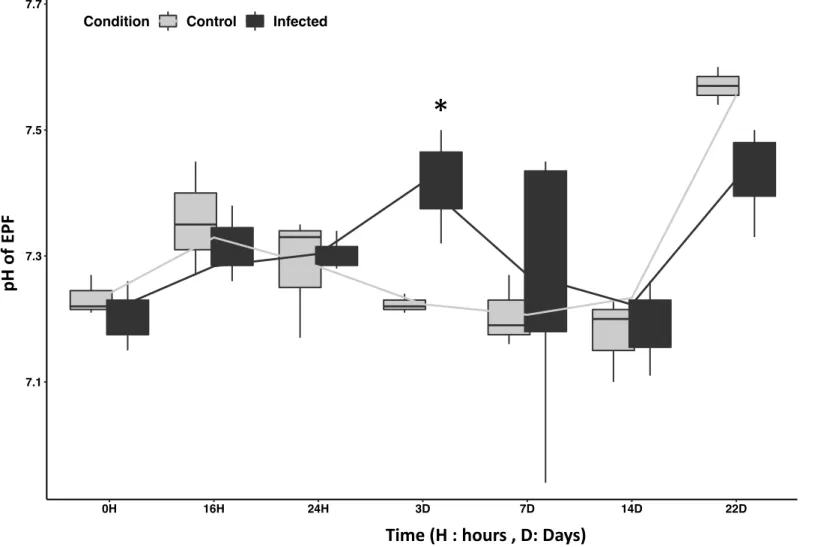
Extrapallial fluids and hemolymph pH modulation during in vivo challenge of the Manila 271
clams by V. tapetis CECT4600. 272
The previous demonstration that the presence of V. tapetis can interfere in vitro with the pH 273
of these clam biological fluids led us to hypothesize that this could happen in vivo, and that 274
the modulation of pH of these fluids could be an important aspect of the infection process. 275
Monitoring of pH evolution in EPFs and hemolymph after Manila clams infection by V. tapetis 276
is presented in Figures 7 and 8, respectively. Both fluids displayed a relatively similar pH 277
evolution following the injection of FSSW or V. tapetis in the animals. Considering the whole 278
in vivo infection experiment (22 days), we did not observe any significant difference in pH
279 280
significant difference between the pHs of the EPFs and hemolymph from control and infected 281
clams at 3 days post injection (dpi). Indeed, the pH of these fluids were higher (of approx. 0.2 282
pH unit) in both cases in clams injected with a suspension of V. tapetis, as compared to clams 283
injected with FSSW (pvalue < 0.02). Our results then tend to suggest that exposure to V. 284
tapetis induce physiological changes in challenged Manila clam that influence the pH of EPF
285
and hemolymph at 3 days of exposure to the pathogen. This also suggests that this particular 286
period should be more precisely investigated during the study of host-pathogen interaction. 287
DISCUSSION 289
pH homeostasis is an essential aspect of cell physiology. It is also one of the main 290
environmental factors controlling the biomineralization of calcified organisms. This explains, 291
at least in part, the current abundance of studies dealing with the impact of ocean 292
acidification on marine mollusks and other calcified marine organisms (Gattuso and Hansson 293
2011). The relation between decrease of pH and an increase of temperature has been studied 294
in several bivalves species, showing that these two factors can affect the immune response 295
as in the Mediterranean mussel Mytilus galloprovincialis (Matozzo et al. 2012) and was also 296
shown to influence the abundance of parasites and incidence of bacteria as in the blue mussel 297
Mytilus edulis (Mackenzie et al. 2014). Finally, pH adaptation is a crucial component of
298
virulence of many pathogenic bacteria. In addition to having to survive the acid digestive 299
barriers in some hosts, they must be able to tolerate acid stress resulting from lysosomal 300
activity after being phagocytized by immune system cells such as macrophages or hemocytes 301
(Asplund et al. 2014). 302
303
EPFs and hemolymph are involved in shell biomineralization and in immunity of the Manila 304
clam, respectively, and have therefore a crucial importance in the development of BRD. BRD 305
is typically a disease of clam biomineralization (Paillard et al. 1994). It is also a bacterial 306
disease, in which the phenomenon of phagocytosis, and more specifically the phagosome-307
lysosome fusion, has a particular importance in the animal’s immunity (Paillard 2004; 308
Rahmani et al., 2019). For all these reasons, it is surprising that the effect of pH on V. tapetis 309
has never been considered to date in the context of the BRD. In this study, we first aim to 310
characterize the relationship between external medium pH and the growth of the pathogen 311
V. tapetis.
We observed that V. tapetis, the bacterial etiological agent of BRD, is able to grow over a wide 313
range of pHs, including at a cold temperature (14°C) which is an optimum for BRD 314
development (Paillard et al. 2004). Interestingly, we showed that the bacterium, during its 315
growth, modifies the pH of its environment, increasing it in acidic media, or acidifying it in 316
alkaline ones, and systematically replaced the external pH to a value close to 7.7 during its 317
growth in a kind of ‘neutralizing activity’ in the tested range. We further characterized this 318
last point, by measuring in vitro the impact of the bacterial growth on the pH of the main 319
biological fluids of the Manila clams, ie. extrapallial fluids and hemolymph, and then in vivo, 320
the pH of these fluids in a context of infection by inducing the main mode of infection of V. 321
tapetis, allowing the pathogen to colonize extrapallial fluids. We observed that at three hours
322
after exposure to V. tapetis, the pH of the two fluids decreased significantly more than that 323
of the control in presence of powdered Manila clam shell. EPFs and hemolymph contain 324
hemocytes, which are the main cellular actors of the immune system and one of the main 325
targets of the pathogen. 326
327
In a previous transcriptomic analysis, we showed that V. tapetis is able to induce 328
deregulations of clam hemocytes physiology as reorganization of actin cytoskeleton, 329
reduction of lysosomal activity and down regulation of genes related to the complement 330
pathway (Rahmani et al. 2019). In another previous study, our team had demonstrated that 331
the cytotoxicity of the bacterium to hemocytes was maximal, in vitro, after three hours of 332
exposure to the pathogen (Choquet et al. 2003). This phenomenon is reproduced by exposing 333
Manila clam hemocytes to V. tapetis during precisely three hours. The decrease in pH in the 334
first three hours of exposure could be due to the growth of the pathogen and could be related 335
(either as a cause or a consequence) to its cytotoxic effect on clam hemocytes where the 336
influence of the bacterium is maximum. 337
338
The relationship between pH regulation and virulence is not always clear since the regulation 339
of the internal pH is a necessity for all cell types. Bacteria regulate their intracellular pH by 340
using proton pumps, or by transporting and/or metabolizing several acid or base compounds. 341
For example, Escherichia coli response to acid stress involves amino acid decarboxylase 342
antiporter pairs and proton-pumping respiratory chains complexes causing H+ ion efflux outside
343
the cell. On the contrary, during alkaline stress, E. coli was found to have a strong activation 344
of NhaA Na+/H+ antiporter, an up-regulation of ATP synthetase leading to a strong entry of
345
H+ ion into the cell (Krulwich et al. 2011). However, intestinal bacteria are facing larger pH 346
ranges than marine ecosystems. NhaA antiporter is also present in the genome of V. tapetis 347
CECT4600 according to the recently published annotation (Dias et al. 2018), as well as 348
calcium or potassium proton antiporters. Furthermore, the presence of an enzyme such as 349
arginine deiminase in the genome of this pathogen might explain alkalinization of external 350
pH observed during acidic stress as already shown in another bacterium (Budin-Verneuil et 351
al. 2006). 352
Regardless of this internal pH regulation, many bacteria modify the pH of their environment 353
as a result of their energy metabolism. This is particularly the case for many fermentative 354
bacteria producing large quantities of organic acids (e.g. lactic acid, propionic acid, formic 355
acid, etc.) leading to an acidification of their environment (Nuryana et al. 2019). Other 356
bacteria are known to alkalinize their environment, by using organic acids (e.g. lactate, 357
aspartate, glutamate) as carbon and energy sources (Stancik et al. 2002). Nevertheless, these 358
there are no case, to our knowledge, of bacteria capable of being both acidifying and 360
alkalinizing in the same culture medium, only depending on the initial pH of that medium. 361
This discovery raises many questions about the V. tapetis's adaptation mechanisms to pH 362
stress, as well as about the energy metabolism(s) it uses, and the substrates it degrades from 363
the Zobell medium, at the different pHs. This also raises important questions regarding the 364
involvement of this ‘neutralizing activity’ in the expression of its pathogenicity, in the context 365
of BRD development. The mechanisms related to this activity are for now unknown and 366
might be due to several type of mechanisms as described above. Nevertheless, this study has 367
revealed new, previously unsuspected problematics related to pH modulation, highlighting 368
the need for further analysis of genes expressed during growth in order to elucidate the 369
mechanisms linked to this phenomenon. 370
371
To better understand the close interaction between the clam and the pathogen in this 372
context, we need to refer to BRD dynamics. BRD is a chronic infection, as the pathogen does 373
not induce mortalities in the classical way of infection and can persist in infected animals 374
(Paillard 2016). Indeed, the acute phase of the infection is divided into two parts: a first one 375
characterized by an increase of V. tapetis concentration followed by the production of a 376
brown deposit on the inner part of the shell, and a second one, which is not always present, 377
characterized by shell repair that can lead to the complete remission of clams (Paillard 2016). 378
In this study, we have performed injection of V. tapetis in the pallial cavity. This mode of 379
infection allows recreating the first stages of BRD by allowing the pathogen to reproduce all 380
the early steps of BRD and then induce an acute phase of infection until formation of the 381
brown deposit. Dynamics of BRD and kinetics of V. tapetis in EPFs have previously been well 382
periostracal lamina in order to enter extrapallial fluids, and then colonizes the shell secretion 384
(Paillard and Maes 1995), leading to an increase of V. tapetis concentration in EPFs. Between 385
2 days and 7 d.p.i. the EPFs concentration of V. tapetis reaches its maximum value (Bidault et 386
al. 2015). 387
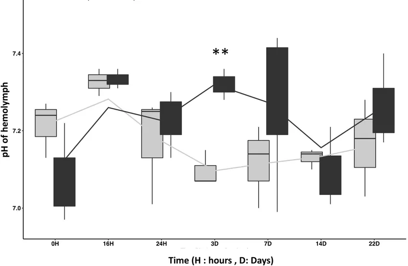
This particular period corresponds to an increase of pH in EPFs according to our study where 388
significant differences of pH have been observed in infected animals after 3 days (in vivo). It 389
should also be noted that an increase of pH has also been reported in hemolymph at 3 d.p.i., 390
thus probably revealing the changes that occurs in this compartment at the biochemical level 391
during BRD (Allam et al. 2006). 392
393
In addition, we know that during infection, major changes occur in EPFs at the enzymatic 394
level with, for example, an increase in the activity of phenoloxidase, an enzyme involved in 395
the humoral response and melanization which leads to the brown deposit production 396
(Söderhäll and Cerenius 1998). Previous studies showed that phenoloxidase activity is 397
sensitive to abiotic factors such as pH (Le Bris 2013). Indeed, phenoloxidase activity decrease 398
with pH in a range of physiological values in the Atlantic blue crab Callinectes sapidus, mostly 399
in the hemolymph compartment (Tanner et al. 2006). Thus, increasing the pH in a range of 400
physiological values in both hemolymph and EFPs could modulate phenoloxidase activity in 401
these two compartments. As phenoloxidase is related to melanization process, the increase 402
of pH can also modulate melanization, associated to the bacterial embedding within shell 403
matrix. 404
405
Considering these informations, the fact that V. tapetis is able to modulate the pH of such 406
compartments of the Manila clam in the context of BRD then questioned the impact of this 407
factor in the development of BRD itself. Indeed, pH is known to modulate the virulence of 408
many pathogenic bacteria. In Vibrio cholerae, for example, it has been shown that the protein 409
ToxR, which is responsible for the regulation of many virulence genes, is also strongly 410
involved in the response to acid stress, suggesting a close relationship between the virulence 411
and the acid stress response in this pathogen (Merrell and Camilli 2002; Lund et al. 2014). In 412
Pseudomonas aeruginosa, it has been shown that the infection was attenuated at pH 6 rather
413
than pH 7.6, and that the bacterium expresses genes that allow it to alkalinize the medium at 414
acidic pH, while at neutral pH, it preferentially expresses iron metabolism-related genes 415
(Romanowski et al. 2011). In the case of V. tapetis, we don’t know yet if pH variations are due 416
to sensing of external pH by V. tapetis that induce a change in response, maybe in relation 417
with its pathogenic activity or to the growth of the pathogen. This mechanism should be 418
investigated in a future study. 419
420
To summarize, after investigation for pH modulation during interaction between the Manila 421
clam and its pathogen V. tapetis, our results indicate that V. tapetis is able to modulate the 422
pH by a “neutralizing activity” during its growth. For the first time, we highlighted this activity 423
in both in vitro challenge and in vivo infection of Manila clam. We have determined that this 424
interaction occurs at a very precise time (3 hours exposure in vitro and 3 days in vivo). These 425
special time periods correspond to the main interactions between the pathogen and the clam 426
hemocytes. It is then likely that these close interactions might play a role in the first steps of 427
BRD development. Nevertheless, the mechanisms related to this phenomenon are, for now, 428
unknown. Our study is the first evidence that pH modulations might be a novel, and since 429
now undiscovered, mechanism that will help us to better understand host-pathogen 430
by both in vitro and in vivo challenges in order to better characterize these particular 432
interactions and to understand the mechanisms and the importance of pH modulation in the 433
pathogenic activity of V. tapetis. 434
435 436
AUTHORS CONTRIBUTIONS
437
AR designed the protocols with help of AB, NLG and CP and VP. AR performed in vitro 438
experiments on V. tapetis. AR and CM performed in vitro and in vivo experiment on Manila 439
clams challenged by V. tapetis. AR, VP and CP wrote the article (the original draft was written 440
by AR). This article was carefully reviewed by other co-authors, who all approved the final 441 version. 442 443 FUNDINGS 444
This project received grants from the H2020 European project “VIVALDI” (grant agreement 445
N°678589). This work was also supported by the “Université de Bretagne Occidentale” (UBO, 446
France), and the ”investment for the future” programs LabexMER (ANR-10-LABX-19) and 447 ISblue (ANR-17-EURE-0015). 448 449 ACKNOWLEDGMENTS 450
We warmly thank Jean François AUVRAY from the SATMAR company of Landeda (Finistère, 451
France) for providing the clams. We also warmly thank Eric DABAS for helping with 452
zootechnical support. 453
CONFLICT OF INTEREST
455
No conflict of interest declared. 456
457 458
REFERENCES
459
Allam B, Paillard C, Auffret M, Ford SE (2006) Effects of the pathogenic Vibrio tapetis on 460
defence factors of susceptible and non-susceptible bivalve species: II. Cellular and 461
biochemical changes following in vivo challenge. Fish Shellfish Immunol 20:384–397 462
Allam B, Pales Espinosa E, Tanguy A, Jeffroy F, Le Bris C, Paillard C (2014) Transcriptional 463
changes in Manila clam (Ruditapes philippinarum) in response to Brown Ring 464
Disease. Fish Shellfish Immunol 41:2–11 . https://doi.org/10.1016/j.fsi.2014.05.022 465
Asplund ME, Baden SP, Russ S, Ellis RP, Gong N, Hernroth BE (2014) Ocean acidification 466
and host–pathogen interactions: blue mussels, Mytilus edulis, encountering Vibrio 467
tubiashii. Environ Microbiol 16:1029–1039 . https://doi.org/10.1111/1462-2920.12307
468
Bidault A, Richard GG, Le Bris C, Paillard C (2015) Development of a Taqman real-time PCR 469
assay for rapid detection and quantification of Vibrio tapetis in extrapallial fluids of 470
clams. PeerJ 3:e1484 . https://doi.org/10.7717/peerj.1484 471
Borrego JJ, Castro D, Luque A, Paillard C, Maes P, Garcia MT, Ventosa A (1996) Vibrio 472
tapetis sp. nov., the causative agent of the brown ring disease affecting cultured
473
clams. Int J Syst Evol Microbiol 46:480–484 474
Budin-Verneuil A, Maguin E, Auffray Y, Ehrlich DS, Pichereau V (2006) Genetic structure 475
and transcriptional analysis of the arginine deiminase (ADI) cluster in Lactococcus 476
lactis MG1363. Can J Microbiol 52:617–622 . https://doi.org/10.1139/w06-009
477
Choquet G, Soudant P, Lambert C, Nicolas J-L, Paillard C (2003) Reduction of adhesion 478
properties of Ruditapes philippinarum hemocytes exposed to Vibrio tapetis. Dis 479
Aquat Organ 57:109–116 480
Dias GM, Bidault A, Le Chevalier P, Choquet G, Der Sarkissian C, Orlando L, Medigue C, 481
Barbe V, Mangenot S, Thompson CC, Thompson FL, Jacq A, Pichereau V, Paillard C 482
(2018) Vibrio tapetis Displays an Original Type IV Secretion System in Strains 483
Pathogenic for Bivalve Molluscs. Front Microbiol 9:227 484
Gattuso J-Pierre, Hansson Lina (2011) Ocean acidification. Oxford University Press, 485
Oxford ; 486
Goulletquer P, Héral M, Béchemin C, Richard P (1989) Anomalies de calcification chez la 487
palourde japonaise Ruditapes philippinarum: caractérisation et comparaison des 488
compositions en acides aminés de différentes parties de la coquille analysées par 489
HPLC. Aquaculture 81:169–183 490
Ivanina AV, Dickinson GH, Matoo OB, Bagwe R, Dickinson A, Beniash E, Sokolova IM 491
(2013) Interactive effects of elevated temperature and CO 2 levels on energy 492
metabolism and biomineralization of marine bivalves Crassostrea virginica and 493
Mercenaria mercenaria. Comp Biochem Physiol A Mol Integr Physiol 166:101–111
494
Krulwich TA, Sachs G, Padan E (2011) Molecular aspects of bacterial pH sensing and 495
homeostasis. Nat Rev Microbiol 9:330–343 496
Le Bris C (2013) Le système phénoloxydase: caractérisation biochimique et rôle dans la 497
réponse immunitaire chez la palourde japonaise Venerupis philippinarum exposée à 498
Vibrio tapetis. Université de Bretagne occidentale-Brest
(2015) Immune responses of phenoloxidase and superoxide dismutase in the manila 501
clam Venerupis philippinarum challenged with Vibrio tapetis – Part I: Spatio-502
temporal evolution of enzymes’ activities post-infection. Fish Shellfish Immunol 503
42:16–24 . https://doi.org/10.1016/j.fsi.2014.10.021 504
Li S, Liu C, Huang J, Liu Y, Zheng G, Xie L, Zhang R (2015) Interactive effects of seawater 505
acidification and elevated temperature on biomineralization and amino acid 506
metabolism in the mussel Mytilus edulis. J Exp Biol 218:3623–3631 . 507
https://doi.org/10.1242/jeb.126748 508
Lund P, Tramonti A, De Biase D (2014) Coping with low pH: molecular strategies in 509
neutralophilic bacteria. FEMS Microbiol Rev 38:1091–1125 . 510
https://doi.org/10.1111/1574-6976.12076 511
Mackenzie CL, Lynch SA, Culloty SC, Malham SK (2014) Future Oceanic Warming and 512
Acidification Alter Immune Response and Disease Status in a Commercial Shellfish 513
Species, Mytilus edulis L. PLOS ONE 9:e99712 . 514
https://doi.org/10.1371/journal.pone.0099712 515
Matozzo V, Chinellato A, Munari M, Finos L, Bressan M, Marin MG (2012) First Evidence of 516
Immunomodulation in Bivalves under Seawater Acidification and Increased 517
Temperature. PLOS ONE 7:e33820 . https://doi.org/10.1371/journal.pone.0033820 518
Merrell D, Camilli A (2002) Acid tolerance of gastrointestinal pathogens. Curr Opin 519
Microbiol 5:51–55 . https://doi.org/10.1016/S1369-5274(02)00285-0 520
Nuryana I, Andriani A, Lisdiyanti P, Yopi (2019) Analysis of organic acids produced by lactic 521
acid bacteria. IOP Conf Ser Earth Environ Sci 251:012054 . 522
https://doi.org/10.1088/1755-1315/251/1/012054 523
Paillard C, Allam B, Oubella R (2004) Effect of temperature on defense parameters in 524
Manila clam Ruditapes philippinarum challenged with Vibrio tapetis. Dis Aquat 525
Organ 59:249–262 526
Paillard C (2004a) A short-review of brown ring disease, a vibriosis affecting clams, 527
Ruditapes philippinarum and Ruditapes decussatus. Aquat Living Resour 17:467–475 .
528
https://doi.org/10.1051/alr:2004053 529
Paillard C (2004b) Rôle de l’environnement dans les interactions hôtes-pathogènes; 530
développement d’un modèle de vibriose chez les bivalves. Habilit À Dir Rech HDR 531
Univ Bretagne Occident Brest 532
Paillard C (2016) An ecological approach to understanding host-pathogen-environment 533
interactions: the case of Brown Ring Disease in clams. In: Oysters and Clams: 534
Cultivation, Habitat Threats and Ecological Impact 535
Paillard C, Le Pennec M (1993) Ultrastructural studies of the mantle and the periostracal 536
lamina in the manila clam, Ruditapes philippinarum. Tissue Cell 25:183–194 . 537
https://doi.org/10.1016/0040-8166(93)90018-G 538
Paillard C, Maes P (1990) Aetiology of brown ring disease in Tapes philippinarum: 539
pathogenicity of a Vibrio sp. Comptes Rendus Académie Sci Ser 3 Sci Vie 310:15–20 540
Paillard C, Maes P (1995) The Brown Ring Disease in the Manila Clam, Ruditapes 543
philippinarum. Part 1. Ultrastrucural alterations of the periostracal lamina. J
544
Invertebr Pathol 65:91–100 . https://doi.org/10.1006/jipa.1995.1015 545
Paillard C, Maes P, Oubella R (1994) Brown ring disease in clams. Annu Rev Fish Dis 4:219– 546
240 . https://doi.org/10.1016/0959-8030(94)90030-2 547
Paillard C, Percelay L, Le Pennec M, Le Picard D (1989) Origine pathogène de l’«anneau 548
brun» chez Tapes philippinarum (Mollusque, bivalve). Comptes Rendus Académie 549
Sci Sér 3 Sci Vie 309:235–241 550
Rahmani A, Corre E, Richard G, Bidault A, Lambert C, Oliveira L, Thompson C, Thompson 551
F, Pichereau V, Paillard C (2019) Transcriptomic analysis of clam extrapallial fluids 552
reveals immunity and cytoskeleton alterations in the first week of Brown Ring 553
Disease development. Fish Shellfish Immunol 93:940–948 . 554
https://doi.org/10.1016/j.fsi.2019.08.025 555
Romanowski K, Zaborin A, Fernandez H, Poroyko V, Valuckaite V, Gerdes S, Liu DC, 556
Zaborina OY, Alverdy JC (2011) Prevention of siderophore- mediated gut-derived 557
sepsis due to P. aeruginosa can be achieved without iron provision by maintaining 558
local phosphate abundance: role of pH. BMC Microbiol 11:212 . 559
https://doi.org/10.1186/1471-2180-11-212 560
Söderhäll K, Cerenius L (1998) Role of the prophenoloxidase-activating system in 561
invertebrate immunity. Curr Opin Immunol 10:23–28 . 562
https://doi.org/10.1016/S0952-7915(98)80026-5 563
Stancik LM, Stancik DM, Schmidt B, Barnhart DM, Yoncheva YN, Slonczewski JL (2002) 564
pH-Dependent Expression of Periplasmic Proteins and Amino Acid Catabolism in 565
Escherichia coli. J Bacteriol 184:4246–4258 .
https://doi.org/10.1128/JB.184.15.4246-566
4258.2002 567
Tanner CA, Burnett LE, Burnett KG (2006) The effects of hypoxia and pH on phenoloxidase 568
activity in the Atlantic blue crab, Callinectes sapidus. Comp Biochem Physiol A Mol 569
Integr Physiol 144:218–223 570
Taylor, J D, Kennedy, W J, Hall, A (1973) The Shell Structure and Mineralogy of the Bivalvia. 571
II. Lucinacea-Clavagellacea. Conclusions. Bull Br Mus Nat Hist Zool Lond 22:253– 572
294 573
Trinkler N (2009) La guérison coquillière: un mécanisme de défense de la palourde 574
japonaise Ruditapes philippinarum face au Vibrio tapetis dans le cadre de la maladie 575
de l’anneau brun. Université de Bretagne occidentale-Brest 576
Trinkler N, Bardeau J, Marin F, Labonne M, Jolivet A, Crassous P, Paillard C (2011) Mineral 577
phase in shell repair of Manila clam Venerupis philippinarum affected by brown ring 578
disease. Dis Aquat Organ 93:149–162 . https://doi.org/10.3354/dao02288 579
Wood HL, Spicer JI, Widdicombe S (2008) Ocean acidification may increase calcification 580
rates, but at a cost. Proc R Soc Lond B Biol Sci 275:1767–1773 581
Zobell C (1941) Studies on marine bacteria. I. The cultural requirements of heterotrophic 582
aerobes. J Mar Res 4:41–75 583
FIGURES AND TABLES
Figure 1: pH changes in a V. tapetis CECT4600 culture after growth at 14°C, 18° and 21°C in Zobell media which pH were initially adjusted from 6.00 (M600) to 8.70 (M870) (15 replicates) ... 27 Figure 2: pH variance of a V. tapetis CECT4600 culture after growth at 14°, 18° and 21°C in pH adjusted Zobell media from 6.00 (M600) to 8.70
(M870) (15 replicates). Left : T0 ; Right : TF or TFinal ... 28 Figure 3: pH changes in a V. tapetis CECT4600 culture after growth at 27° in Zobell media which pH were initially adjusted from 6.00 (M600) to
8.70 (M870) (15 replicates) ... 29 Figure 4: pH change in a culture of V. tapetis during the exponential growth phase at 14°C, in Zobell media which pH were initially adjusted
from 6.00 (M600) to 8.70 (M870) ; CM is the original medium where pH was not modified. ... 30 Figure 5: pH change in a culture of V. tapetis during the exponential growth phase at 18°C, Zobell media which pH were initially adjusted from
6.00 (M600) to 8.70 (M870) ; CM is the original medium where pH was not modified. ... 31 Figure 6: pH changes after V. tapetis exposure in FSSW (Filtered sterilized Seawater), EPF (extrapallial fluids) and Hemolymph (He) mixed with
powdered Manila clam shell. ** : pvalue=0.0017 ; ***=pvalue=0.0007, Student test ... 32 Figure 7: pH changes in EPFs (extrapallial fluids) of the Manila clam Ruditapes philippinarum after infection by V. tapetis. Control: injected by
FSSW ; Infected: injected by V. tapetis ... 33 Figure 8: pH changes in Hemolymph of the Manila clam Ruditapes philippinarum after infection by V. tapetis. Control: injected by FSSW ;
Infected: injected by V. tapetis ... 34 Table 1: growth parameters of V. tapetis in a range of pH media and different temperatures. DT : doubling Time ; OD : Optic Density, T° :
Figure 1: pH changes in a V. tapetis CECT4600 culture after growth at 14°C, 18° and 21°C in Zobell media which pH were initially adjusted from 6.00 (M600) to 8.70 (M870) (15 replicates)
M600 M645 M700 MC M786 M812 M870
T0
TF
Control Infected Control Infected Control Infected Control Infected Control Infected Control Infected Control Infected
6 7 8 9 6 7 8 9 pH
Catergorie Control Infected
pH evolution during growth of V. tapetis at 14,18 and 21º C between T0 and TF mesures
pH Control CECT 4600 Control CECT 4600 Control CECT 4600 Control CECT 4600 Control CECT 4600 Control CECT 4600 Control CECT 4600 T0 TFinal M600 M645 M700 CM M786 M812 M870
Figure 2: pH variance of a V. tapetis CECT4600 culture after growth at 14°, 18° and 21°C in pH adjusted Zobell media from 6.00 (M600) to 8.70 (M870) (15 replicates). Left : T0 ; Right : TF or TFinal
**** : pvalue < 0.0001, Student test
****
T0 TF
Control Infected Control Infected
6 7 8 9
pH
Catergorie Control Infected
Conditions
****
T0 TF
Control Infected Control Infected
6 7 8 9
pH
Catergorie Control Infected
****
T0 TF
Control Infected Control Infected
6 7 8 9
pH
CatergorieControlControl InfectedCECT4600
Control CECT4600 Control CECT4600
Figure 3: pH changes in a V. tapetis CECT4600 culture after growth at 27° in Zobell media which pH were initially adjusted from 6.00 (M600) to 8.70 (M870) (15 replicates)
M600 M645 M700 MC M786 M812 M870
T0
TF
Control Infected Control Infected Control Infected Control Infected Control Infected Control Infected Control Infected
6 7 8 9 6 7 8 9 pH
Catergorie Control Infected
pH evolution during growth of V. tapetis at 27º C between T0 and TF mesures
pH Control CECT 4600 Control CECT 4600 Control CECT 4600 Control CECT 4600 Control CECT 4600 Control CECT 4600 Control CECT 4600 T0 TFinal M600 M645 M700 CM M786 M812 M870
Figure 4: pH change in a culture of V. tapetis during the exponential growth phase at 14°C, in Zobell media which pH were initially adjusted T 0 10 20 30 40 50 6 7 8 9 Time (hour) pH Medium M600 M645 M700 M786 M812 M870 MC pH evolution at 14º C
during exponential growth phase of V. tapetis
pH
Time (hour)
C 0 10 20 30 40 50 4 5 6 7 8 9 pH Medium M600 M645 M700 M786 M812 M870 MCpH evolution at 14º C
during exponential growth phase of V. tapetis
C 5 6 7 8 9 pH Medium M600 M645 M700 M786 M812 M870 MC
pH evolution at 14º C
during exponential growth phase of V. tapetis
CM C 5 6 7 8 9 pH Medium M600 M645 M700 M786 M812 M870 MC
pH evolution at 14º C
Figure 5: pH change in a culture of V. tapetis during the exponential growth phase at 18°C, Zobell media which pH were initially adjusted from 6.00 (M600) to 8.70 (M870) ; CM is the original medium where pH was not modified.
T 0 5 10 15 20 25 6 7 8 9 Time (hour) pH Medium M600 M645 M700 M786 M812 M870 MC pH evolution at 18º C
during exponential growth phase of V. tapetis
pH Time (hour) C 0 10 20 30 40 50 4 5 6 7 8 9 Time (hour) pH Medium M600 M645 M700 M786 M812 M870 MC pH evolution at 14º C
during exponential growth phase of V. tapetis
C 4 5 6 7 8 9 pH Medium M600 M645 M700 M786 M812 M870 MC pH evolution at 14º C
during exponential growth phase of V. tapetis
CM C 4 5 6 7 8 9 pH Medium M600 M645 M700 M786 M812 M870 MC pH evolution at 14º C
Figure 6: pH changes after V. tapetis exposure in FSSW (Filtered sterilized Seawater), EPF (extrapallial fluids) and Hemolymph (He) mixed with EMSF FEP He T0 T3H T24H T48H T72H 7.00 7.25 7.50 7.75 8.00 7.00 7.25 7.50 7.75 8.00 7.00 7.25 7.50 7.75 8.00 Time (hour) pH
Conditions Control Infected
pH evolution in presence/absence of V. tapetis, with clam shell
in FSSW (Filtered Sterelized Sea Water), EPF (ExtraPalleal Fluids) and Hemolymph (He)
FSSW EPF Hemolymph pH o f EP F He 0H 16H 24H 3D 7D 14D 22D 7.0 7.2 7.4
Time (H = hours, D = days)
pH
Condition Control Infected
Time (hours)
**
Figure 7: pH changes in EPFs (extrapallial fluids) of the Manila clam Ruditapes philippinarum after infection by V. tapetis. Control: injected by FSSW ; Infected: injected by V. tapetis
FEP 0H 16H 24H 3D 7D 14D 22D 7.1 7.3 7.5 7.7
Time (H = hours, D = days)
pH
Condition Control Infected
pH o f EP F He 0H 16H 24H 3D 7D 14D 22D 7.0 7.2 7.4
Time (H = hours, D = days)
pH
Condition Control Infected
Time (H : hours , D: Days)
*
Figure 8: pH changes in Hemolymph of the Manila clam Ruditapes philippinarum after infection by V. tapetis. Control: injected by FSSW ; He 0H 16H 24H 3D 7D 14D 22D 7.0 7.2 7.4
Time (H = hours, D = days)
pH
Condition Control Infected
pH
o
f he
m
ol
ym
ph
He 0H 16H 24H 3D 7D 14D 22D 7.0 7.2 7.4Time (H = hours, D = days)
pH
Condition Control Infected
Time (H : hours , D: Days)
Table 1: growth parameters of V. tapetis in a range of pH media and different temperatures. DT : doubling Time ; OD : Optic Density, T° : Temperature; h: hour. M=Medium and the number is the pH (600= pH 6.00) ; MC is the original medium (unmodified pH).
V. tapetis CECT4600 growth
M600 M645 M700 MC M786 M812 M870 T° Time of growth DT (h) Maximum OD492 DT (h) Maximum OD492 DT (h) Maximum OD492 DT (h) Maximum OD492 DT (h) Maximum OD492 DT (h) Maximum OD492 DT (h) Maximum OD492 14°C 31H 5,00 0,945 3,31 1,121 3,39 1,155 3,15 1,047 3,50 1,128 3,16 1,143 7,03 0,946 18°C 24H 2,37 1,201 1,85 1,220 1,95 1,214 2,00 1,120 1,79 1,178 2,42 1,245 3,54 1,010 21°C 24H 2,19 1,231 1,93 1,204 2,30 1,207 2,50 1,167 2,83 1,201 2,52 1,223 3,97 1,061 27°C 24H 0 0,012 0 0,024 0 0,033 0 0,06 0 0,021 0 0,021 0 0,031