Biocompatible post-polymerization functionalization
of a water soluble poly(p-phenylene ethynylene)
The MIT Faculty has made this article openly available.
Please share
how this access benefits you. Your story matters.
Citation
VanVeller, Brett, and Timothy M. Swager. “Biocompatible
Post-polymerization Functionalization of a Water Soluble
Poly(p-phenylene Ethynylene).” Chemical Communications 46.31 (2010):
5761.
As Published
http://dx.doi.org/ 10.1039/c0cc01456g
Publisher
Royal Society of Chemistry
Version
Author's final manuscript
Citable link
http://hdl.handle.net/1721.1/74233
Terms of Use
Creative Commons Attribution-Noncommercial-Share Alike 3.0
Biocompatible Post-Polymerization Functionalization of a
Water Soluble Poly(p-Phenylene Ethynylene)
Journal: ChemComm Manuscript ID: Draft
Article Type: Communication Date Submitted by the
Author: n/a
Complete List of Authors: VanVeller, Brett; Massachsetts Institute of Technology, Chemistry Swager, Timothy; Massachusetts Institute of Technology,
Department of Chemistry
CREATED USING THE RSC COMMUNICATION TEMPLATE (VER. 3.1) - SEE WWW.RSC.ORG/ELECTRONICFILES FOR DETAILS
ARTICLE TYPE www.rsc.org/xxxxxx | XXXXXXXX
This journal is © The Royal Society of Chemistry [year] Journal Name, [year], [vol], 00–00 | 1
Biocompatible Post-Polymerization Functionalization of a Water
Soluble Poly(p-Phenylene Ethynylene)
Brett VanVeller and Timothy M. Swager*
Received (in XXX, XXX) Xth XXXXXXXXX 200X, Accepted Xth XXXXXXXXX 200X First published on the web Xth XXXXXXXXX 200X
5
DOI: 10.1039/b000000x
A biocompatible post-polymerization functionalization reaction takes advantage of a polymer’s structural motif for the controllable attachment of biotin as a model biosensor that responds to streptavidin.
10
Strategies for the post-polymerization functionalization (PPF) of polymers are advantageous in that they allow for tuning of a polymer’s properties without synthetically retreating to the monomer stage. Further, PPF permits the incorporation of functional groups that may be incompatible with
15
polymerization conditions. Several strategies have been reported for conjugated polymers. A number of designs involve substitution reactions with pendant halogen,1 alcohol,2 or carboxylic acid moieties,3 and application of high yielding click chemistries4 like the 1,3-dipolar cycloaddtion of alkynes
20
and azides5 or thiol-conjugate addition6 have also been
reported. Two potential drawbacks are characteristic of the above strategies: (i) an appropriately functionalized monomer specific for the intended PPF must be incorporated into the polymer synthesis––often in protected form and (ii) it can be
25
difficult to control the extent of functionalization.
We recently reported the synthesis of a rigid hydrophilic monomer (1) that––when incorporated into poly(p-phenylene ethynylenes) (PPEs)7 (P1)––leads to increased spectral purity
30
by preventing hydrophobically induced aggregate emission.8 We envisioned that the three dimensional array of vicinal hydroxyl groups might be further elaborated through periodate oxidation and reductive amination (P1→2→3, Scheme 1).9 Similar processes have been widely applied for
35
bioconjugation through periodate oxidation of carbohydrate residues, making this process compatible with existing bioconjugation schemes. Herein we report a biocompatible post-polymerization biotinylation of P1, where (i) the need for a PPF specific monomer is negated by activation of an
40
existing structural motif, and (ii) the extent of functionalization can be controlled by the equivalents of the NaIO4 reagent. Further, the improved spectral purity imparted
by the presence of 1 in 3a is not lost. In turn, this demonstrates an improved signal amplified biosensor10 45
response to fluorophore-labeled streptavidin, a tetrameric
protein with high biotin affinity (4 x 10-14 M)11 that has been applied to a variety of conjugated polymer affinitychromic3,12 and agglutination2b biosensor designs.
50
Scheme 1 Periodate oxidative activation and reductive amination
Treatment of P1 with 0.2 equivalents of NaIO4 in water
generated 1,6-dialdehyde moieties at random positions along the backbone (2, Scheme 1).13 Subsequent incubation with an
excess of amine-containing compound (a or b) in aqueous
55
alkaline solution generated the putative Schiff base, which was reduced in situ to the tertiary amine 3 with NaCNBH3
(vide infra, Scheme 2).
The azepane linkage in 3 is proposed based on two model studies. Firstly, the broad nature of the 1H NMR signals of 3a
60
overlapped with the weaker biotin signals making determination of the extent of functionalization difficult. Thus, 3b––exhibiting a strong, unobstructed pivalamide signal––was prepared under identical conditions for 3a. Integration analysis revealed a 18–20% incorporation of b
65
(Fig. S3, ESI). Therefore, while 0.2 equivalents of NaIO4
oxidant should generate 0.4 aldehyde equivalents, there appears to only be 0.2 equivalents of the incorporated amine. Secondly, acetonide protected 4––a synthetic intermediate in the synthesis of 18––was treated with periodate anion and
70
Scheme 2 a) Model reductive amination product, b) proposed mechanism
2 | Journal Name, [year], [vol], 00–00 This journal is © The Royal Society of Chemistry [year]
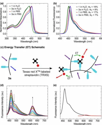
Fig. 1 (a) Absorbance spectra of 1 and 3a in water and PBS solution. (b)
Fluorescence spectra of 1 and 3a in water and PBS solution. (c) Energy transfer schematic showing how intra- and interchain exciton migration and energy transfer to TRXS can lead to amplification. (d) Addition of
5
9.15 pmol aliquots of Texas red X™-labeled streptavidin to 3.46 nmol (based on repeat unit of 3a). (e) Excitation of 1 in the presence of 100 pmol TRXS (black) and direct excitation of 100 pmol of TRXS (red).
produced the tetraaldehyde 5 (Scheme 2a). Addition of an
10
excess of butyl amine and NaCNBH3 to 4 in methanol gave 5
as the major product. Such products have been observed for bridging 1,6-dialdehydes15 and likely form via a 7-exo-trig reductive cyclization to install one amine for every dialdehyde present (Scheme 2b). Thus, we propose the PPF in Scheme 1
15
proceeds in an analogous manner, allowing for the extent of functionalization to be controlled by the molar equivalents of NaIO4.
The effect of the described PPF method on the photophysical properties of the polymer can be seen in Fig. 1a
20
and 1b. The absorbance and fluorescence maxima of 3a show excellent overlap with the parent polymer P1 in both water and PBS solution, indicating that the oxidation and reductive amination reactions leave the conjugated polymer backbone intact. The origin of the reduced quantum yield of 3a is
25
unclear. The possibility of excited state photo-electron transfer from the newly installed amine lone pairs to the polymer was examined by varying the pH but no effect was found (pH = 1–12, Fig. S5, ESI). The reduced quantum yield may be attributed to replacing diol moieties with the relatively
30
insoluble biotin, leading to a more aggregated state of the polymer and diminished quantum yield. In any event, the effect of incorporating 1 in 3a is still present as no lower energy excimer emission is observed and spectral purity is maintained.
35
The response to streptavidin in the presence of 3a is
represented schematically in Fig. 1c, where Texas Red X™-labeled streptavidin (TRXS) is able to aggregate the biotinylated polymers (3a). Amplification is achieved through the funneling of polymer excitons to the lower energy Texas
40
Red X™ dyes through intra- and interchain energy migration within the supramolecular aggregate. The results of serial additions of TRXS to 3a at room temperature in PBS solution are shown in Fig. 1d. As anticipated, a decrease in the 3a emission and a corresponding increase in dye emission was
45
observed. The amplifying effect of the polymer sensor can be seen through direct excitation of the dye (Fig. 1e, red). Finally, incubation of TRXS with P1 showed no response (Fig. 1e, black).
To better understand the nature of the interaction between
50
TRXS and 3a, we determined the Stern–Volmer quenching constant for the polymer emission (460 nm) in Fig. 1d. The Stern–Volmer plot showed positive curvature (Fig. S6, ESI), which is likely due to additional energy migration pathways within the polymer assembly16 produced by the strong biotin– 55
streptavidin association. Further, no detectable excited state lifetime change was observed with increasing TRXS concentration, indicating that static quenching is the dominant mechanism of energy transfer.
Compared with previous systems,10 a 100 fold greater KSV 60
of 2x10⁷ was found. This higher sensitivity is likely due to enhanced energy transfer through avoidance of lower energy excimers. These states––negated by the presence of 18––are
localized and perhaps too low in energy to undergo transfer to the dye.
65
In summary, a biocompatible PPF strategy has been developed, which takes advantage of existing monomer functionality and design. Further, the extent of functionalization can be controlled through the equivalents of NaIO4. Finally, a highly sensitive (KSV = 2x10
7
) turn-on
70
model biosensor based on ET between 3a and TRXS was demonstrated where the presence of 1 lead to dramatically increased sensitivity.
Financial support for this work was provided by the Natural Science and Engineering Council of Canada (NSERC). We are
75
grateful for funding from the U.S. Army through the Institute for Soldier Nanotechnologies, under Contract W911NF-07-D-004 with the U.S. Army Research Office.
Notes and references
Department of Chemistry, Massachusetts Institute of Technology,
80
Cambridge, MA, USA. E-mail: [email protected]
† Electronic Supplementary Information (ESI) available: Experimental details. See DOI: 10.1039/b000000x/
1 (a) C. H. Xue, F. T. Luo, H. Y. Liu, Macromolecules, 2007, 40, 6863; (b) C. H. Xue, V. R. R. Donuru, H. Y. Liu, Macromolecules,
85
2006, 39, 5747; (c) Y. N. Li, G. Vamvounis, J. F. Yu, S. Holdcroft,
Macromolecules, 2001, 34, 3130; (d) J. Tolosa, C. Kub, U. H. F. Bunz, Angew. Chem. Int. Ed., 2009, 48, 4610.
2 (a) C. A. Breen, T. Deng, T. Breiner, E. L. Thomas, T. M. Swager, J.
Am. Chem. Soc., 2003, 125, 9942; (b) J. N. Wilson, Y. Q. Wang, J. J.
90
Lavigne, U. H. F. Bunz, Chem. Commun., 2003, 1626.
3 S. Bernier, S. Garreau, M. Bera-Aberem, C. Gravel, M. Leclerc, J.
Am. Chem. Soc., 2002, 124, 12463.
4 H. C. Kolb, M. G. Finn, K. B. Sharpless, Angew. Chem. Int. Ed., 2001, 40, 2004.
This journal is © The Royal Society of Chemistry [year] Journal Name, [year], [vol], 00–00 | 3 5 (a) B. C. Englert, S. Bakbak, U. H. F. Bunz, Macromolecules, 2005,
38, 5868; (b) T. L. Benanti, A. Kalaydjian, D. Venkataraman, Macromolecules, 2008, 41, 8312; (c) H. B. Bu, G. Gotz, E. Reinold, A. Vogt, S. Schmid, R. Blanco, J. L. Segura, P. Bauerle, Chem.
Commun., 2008, 1320; (d) Q. Chen, B. H. Han, J. Polym. Sci., Part
5
A: Polym. Chem., 2009, 47, 2948.
6 G. C. Bailey, T. M. Swager, Macromolecules, 2006, 39, 2815. 7 B. VanVeller, T. M. Swager, Poly(aryleneethynylene)s. In Design
and Synthesis of Conjugated Polymers, M. Leclerc, J. Morin, Eds. Wiley-VCH: Weinheim, 2010; pp 175–200.
10
8 B. VanVeller, K. Miki, T. M. Swager, Org. Lett., 2010, 12, 1292. 9 G. T. Hermanson, Bioconjugation Techniques. Elsevier Science: San
Diego, 1996.
10 J. Zheng, T. M. Swager, Chem. Commun., 2004, 2798. 11 N. M. Green, Methods Enzymol., 1990, 184, 51.
15
12 E. Geiger, P. Hug, B. A. Keller, Macromol. Chem. Phys., 2002, 203, 2422.
13 1H NMR analysis of 2 showed little bias regarding which diol moiety
(endo or exo) was oxidized. 14 Based on three experiments.
20
15 (a) G. F. Painter, A. Falshaw, H. Wong, Org. Biomol. Chem., 2004,
2, 1007; (b) V. Bonnet, R. Duval, V. Tran, C. Rabiller, Eur. J. Org. Chem., 2003, 4810; (c) A. Robinson, G. L. Thomas, R. J. Spandl, M. Welch, D. R. Spring, Org. Biomol. Chem., 2008, 6, 2978; (d) P. R. Brooks, S. Caron, J. W. Coe, K. K. Ng, R. A. Singer, E. Vazquez, M.
25
G. Vetelino, H. H. Watson, D. C. Whritenour, M. C. Wirtz, Synthesis, 2004, 175; (e) A. H. Fray, D. J. Augeri, E. F. Kleinman, J. Org.
Chem., 1988, 53, 896; (f) P. Stoy, J. Rush, W. H. Pearson, Synth.
Commun., 2004, 34, 3481.
16 (a) I. A. Levitsky, J. S. Kim, T. M. Swager, J. Am. Chem. Soc., 1999,
30
121, 1466; (b) Satrijo, A.; Swager, T. M., J. Am. Chem. Soc., 2007, 129, 16020.
We are herein submitting a manuscript entitled “Biocompatible Post-Polymerization
Functionalization of a Water Soluble Poly(p-Phenylene Ethynylene)” for consideration as a
publication in Chemical Communications. The approach given takes advantage of existing
biocompatible reactivity for the functionalization of polymers in a predicatble manner. Further,
the modified polymers were ealuated within the context of a biotin–streptavidin model biosensor.
80x41mm (300 x 300 DPI)
S1
Supporting Information for
Controllable Biocompatible Post-Polymerization
Functionalization of Poly(p-Phenylene Ethynylene)s and
Highly Sensitive Detection of Streptavidin
Brett VanVellerand Timothy M. Swager*
Department of Chemistry, Massachusetts Institute of Technology, Cambridge, Massachusetts 02139 Email: [email protected]
Contents
Page
Materials
S2
General Experimental
S2
Synthetic Procedures
Synthesis of 3a
S2
Synthesis of 3b
S2
Synthesis of 5
S3
Synthesis of 6
S3
NMR Spectra
Figure S1:
1H and
13C spectra of 5
S4
Figure S2:
1H and
13C spectra of 6
S5
Figure S3:
1H spectrum of 3b
S6
Figure S4:
1H spectrum of 3a
S6
UV-vis and Fluorescence
Table S1: Summary of photophysical data of 3a
S7
General protocol for energy transfer assays in PBS:
S7
Figure S5: Effect of pH on quantum yield for 3a
S7
S2
Materials: Silica gel (40 µm) was purchased from SiliCycle. All solvents used for
photophysical experiments were spectral grade. Pd(PPh
3)
4was purchased from Strem
Chemicals, Inc. All other reagent grade materials were purchased from Aldrich, TCI
America, and Alfa Aesar, and used without further purification.
Experimental:
NMR Spectroscopy:
1H and
13C NMR spectra for all compounds were acquired in CDCl
3,
D
2O and DMF-d
7on a Bruker Avance Spectrometer operating at 400 and 100 MHz,
respectively. The chemical shift data are reported in units of δ (ppm) relative to residual
solvent.
Gel Permeation Chromatography (GPC): Polymer molecular weights were determined
using a triple detection method for calibration with poly(acrylic acid) standards on a
Viscotek TDA 305-040 instrument equipped with two Viscotek A-MBHMW-3078
columns and analyzed with light scattering and refractive index detectors. Samples were
dissolved in 5% NH
4OH.
Absorption and Emission Spectroscopy: Fluorescence spectra were measured on a SPEX
Fluorolog-τ3 fluorometer (model FL-321, 450 W Xenon lamp) using right-angle
detection. Ultraviolet-visible absorption spectra were measured with an Agilent 8453
diode array spectrophotometer and corrected for background signal with a solvent filled
cuvette. Fluorescence quantum yields of #### in both water and 1X PBS were
determined relative to perylene and are corrected for solvent refractive index and
absorption differences at the excitation wavelength.
Lifetime measurements: Time resolved fluorescence measurements were performed by
exciting the samples with 160 femtosecond pulses at 390 nm from the double output of a
Coherent RegA Ti:Sapphire amplifier. The resulting fluorescence was spectrally and
temporally resolved with a Hamamatsu C4780 Streak Camera system.
S3
Synthetic Procedures
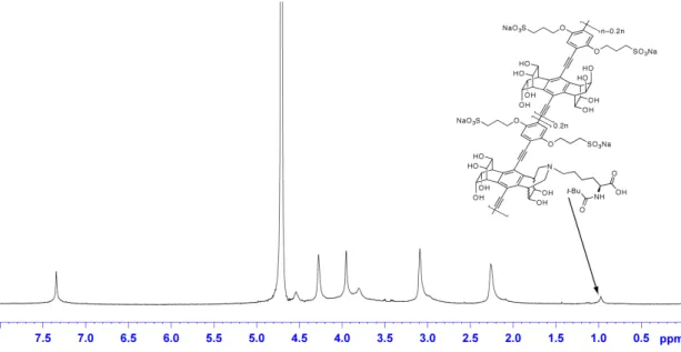
Biotin functionalization, synthesis of 3a: Polymer 1 (11.8 mg, 14.6 µmol based on
repeat unit) was dissolved in 4 mL of H
2O and NaIO
4(2.92 µmol in 0.2 mL) was added
dropwise under vigorous stirring. After 30 min, a (Biotin-PEG
3-NH
2, 3 mg, 7 µmol in 1
mL of 0.2M Na
2HPO
4) was added and the reaction was stirred for 20 min. A solution of
NaCNBH
3(15 mg, 239 µmol in 1 mL of 40 mM Na
2HPO
4) was added and the reaction
stirred for 3 hours. The reaction was dialyzed against water with 5 changes of water and
lyophilized to yield 3a. GPC gave M
n= 38,474, PDI = 3.4.
1H NMR (600 MHz, D
2O):
δ7.44 (s, 2H), 4.64 (broad, 4H), 4.37 (broad, 4H), 4.05 (broad, 4H), 3.89 (broad, 4H),
3.80-3.30 (biotin, PEG), 3.18 (broad, 4H) 2.35 (broad, 4H), 1.30-0.90 (biotin).
Piv-Lysine functionalization, synthesis of 3b: Prepared using identical conditions as
above for 3a except that b (Piv-Lys-NH
2) was used in place of a. GPC gave M
n= 49,073,
PDI = 4.9.
1H NMR (600 MHz, D
2O): δ7.42 (s, 2H), 4.61 (broad, 4H), 4.35 (broad, 4H),
4.03 (broad, 4H), 3.88 (broad, 4H), 3.16 (broad, 4H), 2.33 (broad, 4H), 1.05 (broad, tBu,
1.6–1.8*).
*Based on three experiments and corresponds to 18–20%.
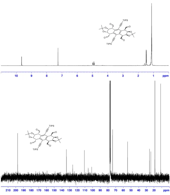
Synthesis of tetraaldehyde 5: A solution NaIO
4(0.200 g, 0.935 mmol) in 10 mL of
water was added to a solution of 4 (0.200 g, 0.248 mmol) in 10 mL of THF. Solid
TBAIO
4(54 mg, 0.124 mmol) was added directly and the solution was refluxed for 30
min. After cooling, the reaction was partitioned between EtOAc and brine and the
organic phase collected. The aqueous layer was washed with fresh EtOAc and the
combined organic layers were dried over Na
2SO
4and concentrated in vacuo. The residue
was eluted through a silica gel plug using EtOAc to give 5 (95%).
1H NMR (400 MHz,
CDCl
3): δ 9.65 (d, J=2, 4H), 4.98 (nfo, actual ddd, J=6.6, 2.8, 2, 4H), 4.89 (dd, J=6.6,
2.8, 4H), 1.48 (s, 6H), 1.46 (s, 6H), 1.12 (s, 42H).
13C NMR (125 MHz, CDCl
3) δ 197.6,
134.1, 125.9, 110.7, 105.7, 101.2, 73.9, 54.5, 25.9, 24.2, 18.8, 11.4. HRMS (EI) calcd. for
C
46H
66O
8Si
2[M+H] 803.4369, found 803.4344.
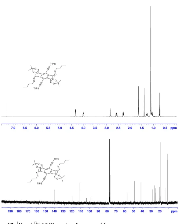
Synthesis of amine 6: To a solution of 5 (0.150 g, 0.186 mmol) in 10 mL of MeOH was
added butyl amine (82 mg, 1.12 mmol). After stirring for 10 min at room temperature,
NaCNBH
3(0.250 g, 3.98 mmol) was added and the mixture was refluxed for 3 hours.
Once cool, 1 mL of sat. NaHCO
3was added and the solvent was removed in vacuo. The
residue was partitioned between DCM and sat. NaHCO
3. The organic layer was dried
over Na
2SO
4and evaporated to dryness. Silica gel chromatography (EtOAc:Hex, 8:2)
provided 6 (65%) as a white solid.
1H NMR (400 MHz, CDCl
3): δ 4.33 (dd, J=4.0, 2.4,
4H), 4.00 (m, 4H), 2.84 (d, J=12, 4H), 2.58 (dd, J=11.8, 7.5, 4H), 2.29 (br t, 4H),
1.64 (s, 6H), 1.40 (s, 6H), 1.28 (m, 4H), 1.12 (br s, 42H), 1.06 (m, 4H), 0.74 (t, J
=7.4, 6H).
13C NMR (125 MHz, CDCl
3