'Deadman' and 'Passcode' microbial
kill switches for bacterial containment
The MIT Faculty has made this article openly available.
Please share
how this access benefits you. Your story matters.
Citation
Chan, Clement T Y, Jeong Wook Lee, D Ewen Cameron, Caleb J
Bashor, and James J Collins. “‘Deadman’ and ‘Passcode’ Microbial
Kill Switches for Bacterial Containment.” Nature Chemical Biology
12, no. 2 (December 7, 2015): 82–86.
As Published
http://dx.doi.org/10.1038/nchembio.1979
Publisher
Nature Publishing Group
Version
Author's final manuscript
Citable link
http://hdl.handle.net/1721.1/108106
Terms of Use
Article is made available in accordance with the publisher's
policy and may be subject to US copyright law. Please refer to the
publisher's site for terms of use.
“Deadman” and “Passcode” microbial kill switches for bacterial
containment
Clement T. Y. Chan1,†, Jeong Wook Lee1,†, D. Ewen Cameron1,†, Caleb J. Bashor1, and James J. Collins1,2,3,4,*
1Institute for Medical Engineering & Science, Department of Biological Engineering, and Synthetic
Biology Center, Massachusetts Institute of Technology (MIT), Cambridge, Massachusetts 02139, USA
2Harvard–MIT Program in Health Sciences and Technology, Cambridge, Massachusetts 02139,
USA
3Broad Institute of MIT and Harvard, Cambridge, Massachusetts 02142, USA
4Wyss Institute for Biologically Inspired Engineering, Harvard University, Boston, Massachusetts
02115, USA
Abstract
Biocontainment systems that couple environmental sensing with circuit-based control of cell viability could be used to prevent escape of genetically modified microbes into the environment. Here we present two engineered safe-guard systems: the Deadman and Passcode kill switches. The Deadman kill switch uses unbalanced reciprocal transcriptional repression to couple a specific input signal with cell survival. The Passcode kill switch uses a similar two-layered transcription design and incorporates hybrid LacI/GalR family transcription factors to provide diverse and complex environmental inputs to control circuit function. These synthetic gene circuits efficiently kill Escherichia coli and can be readily reprogrammed to change their environmental inputs, regulatory architecture and killing mechanism.
With the advent of synthetic biology, genetically modified microorganisms have been increasingly used for biomedical, industrial and environmental applications1–6. Deployment of these engineered microbes in large scales and open environments calls for the
development of safe and secure means to restrain their proliferation. Pioneering
Users may view, print, copy, and download text and data-mine the content in such documents, for the purposes of academic research, subject always to the full Conditions of use:http://www.nature.com/authors/editorial_policies/license.html#terms
*Corresponding author. ; Email: [email protected] phone: 617-324-6607 fax: 617-253-7498
†These authors contributed equally to this work
ACCESSION CODES
DNA sequences have been submitted to Genbank with accession numbers KT893253, KT893254, KT893255, KT893256, KT893257, KT895272, KT895273, KT895274, KT895275, KT895276 and KT895277.
AUTHOR CONTRIBUTIONS
C.T.Y.C, J.W.L., D.E.C., C.J.B. and J.J.C. designed the study, analyzed data and wrote the paper. C.T.Y.C. and J.W.L. performed the experiments.
HHMI A
uthor Man
uscr
ipt
HHMI A
uthor Man
uscr
ipt
HHMI A
uthor Man
uscr
ipt
biocontainment systems used metabolic auxotrophy in which target cells could only grow in the presence of an exogenously supplied metabolite7,8, and the recent creation of an E. coli strain with an altered genetic code enabled production of synthetic auxotrophy strains in which an exogenous supply of non-natural amino acids is required for cell survival9,10. Traditional metabolic auxotrophy strains are hampered by the potential for inadvertent complementation by crossfeeding or by the presence of the metabolite in heterogenous environments, and synthetic auxotrophy systems rely on extensive genome-wide engineering that may be impractical for many industrial production and biotherapeutic microbes.
Furthermore, they are intrinsically difficult to reprogram for different environmental conditions, potentially limiting their application. An alternative approach to biocontainment is to use gene circuits to maintain essential gene expression or block toxin gene expression under the assigned biocontainment conditions7,11–14. Upon loss of the biocontainment signal, the circuit blocks essential gene expression or induces toxin gene expression to kill the cell. These circuits offer the promise of complex environmental signal integration but are hindered by a relative lack of programmable environment sensors to enable their use under non-laboratory conditions15.
Here we design and construct two programmable biocontainment circuits in E. coli – a Deadman kill switch that uses a transcription-based monostable toggle design to provide rapid and robust target cell killing, and a Passcode circuit that uses hybrid LacI/GalR family transcription factors (TFs) to construct complex environmental requirements for cell survival. We use a tripartite strategy of TF protein engineering to detect diverse input signals, robust circuit design to provide signal processing, and redundant toxin-induced and protease-mediated cell killing mechanisms. The resulting biocontainment systems are modular, flexible and extensible, and should prove useful across many industrial and biotherapeutic applications.
RESULTS
Deadman circuit development
We developed the Deadman kill switch to serve as a passively activated biocontainment system for engineered microbes. Similar to pioneering biocontainment systems in E. coli12 and Pseudomonas putida16, the Deadman circuit uses a small molecule binding transcription factor to produce a “survival” state in which repression of toxin production is linked to the presence of a specific environmental signal. Upon loss of the environmental signal, the circuit switches to the “death” state in which de-repressed toxin production kills the cell. To increase the robustness of these biocontainment states, the Deadman circuit uses a genetic “toggle switch” architecture in which reciprocal repression by the LacI and TetR
transcription factors form transcription states that are maintained by the circuit’s linked feedback loops17,18 (Supplementary Results, Supplementary Fig. 1). To create a circuit in which the “death” state is dominant in the absence of the survival signal, we altered the ribosome binding site (RBS) strengths of LacI and TetR to favor TetR expression in a single-copy plasmid (Supplementary Fig. 2 and Online Methods). In the resulting monostable circuit, the presence of the TetR inhibitor anhydrotetracycline (ATc) is required to maintain the circuit in the subordinate LacI+ “survival” state (Supplementary Fig. 3). Incorporation of
HHMI A
uthor Man
uscr
ipt
HHMI A
uthor Man
uscr
ipt
HHMI A
uthor Man
uscr
ipt
toxin genes into the TetR+ state creates a kill switch where the presence of ATc is required to block toxin expression and cell death.
We included additional palindromic LacI operator sites in the toxin gene promoter to minimize leaky toxin expression19 and introduced a transcriptional terminator upstream of the promoter to insulate the gene from spurious transcription (Supplementary Fig. 4). To accelerate the circuit’s switching dynamics, we fused a degradation tag to the C-terminus of LacI that is specifically recognized by mf-Lon20, a heterologous protease under control of a LacI-dependent promoter (Supplementary Fig. 5a). Upon removal of ATc, TetR repression of lacI allows expression of mf-Lon, which targets LacI for degradation to create a positive feedback loop that accelerates the switch to the TetR+ state (Supplementary Fig. 5b). Importantly, single-cell analysis of these circuits by flow cytometry showed a monomodal distribution of cells in the LacI+ and TetR+ state, demonstrating stable circuit expression across the cell population (see 0 and 6 hour data in Supplementary Fig. 5c).
Deadman kill switch characterization
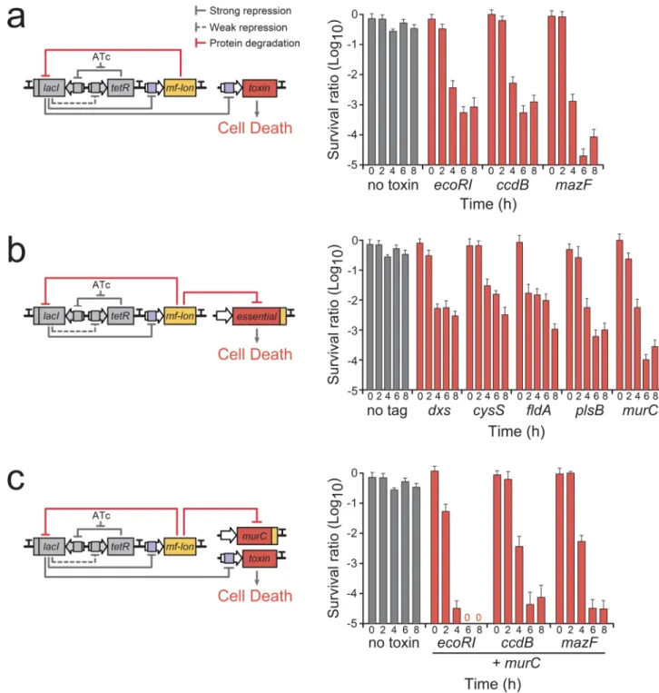
To identify an efficient mechanism to kill the host cells upon circuit activation, we tested several toxin genes that directly damage the host cell’s DNA or RNA. We chose to test the endonuclease ecoRI21, the DNA gyrase inhibitor ccdB22 and the ribonuclease-type toxin mazF23 because they are well-characterized, are native to E. coli, and provide a range of killing mechanisms. The toxin genes were independently incorporated into the Deadman circuit, and a range of RBS strengths were tested for each toxin to optimize cell death upon circuit activation24 (Supplementary Fig. 6). Upon removal of ATc, the toxins produced 3–5 logs of killing within 6 hours as measured by colony forming units (CFUs) (Fig. 1a). To increase the robustness of the circuit and provide an independent method of circuit-dependent cell death, we used mf-Lon protease to not only degrade LacI but also target essential proteins for degradation (Fig. 1b). We attached the mf-Lon degradation tag pdt#1 to the 3’ end of five essential genes whose protein products are particularly sensitive to mf-Lon degradation20, and we then measured cell viability following removal of ATc (Fig. 1b). Among the tested essential gene targets, the peptidoglycan biosynthesis gene murC provided the strongest and fastest cell death phenotype (survival ratio < 1 × 10⁻⁴ within 6 hours). To determine if the toxin- and mf-Lon-mediated killing mechanisms produce synergistic effects, we created Deadman circuits containing each of the toxins in combination with the mf-Lon-MurC targeting module (Fig. 1c). In each instance, the combinatorial approach provided more effective biocontainment, and in particular, coordinated EcoRI expression and mf-Lon-mediated MurC degradation resulted in cell killing below the limit of detection (survival ratio < 1 × 10⁻⁷) 6 hours after removal of ATc (Fig. 1c). Furthermore, the Deadman circuit’s design provides an additional fail-safe mechanism which bypasses the circuit’s sensor system to directly activate toxin expression to cause cell death. Direct derepression of the subordinate TF, in this case derepression of LacI with isopropyl
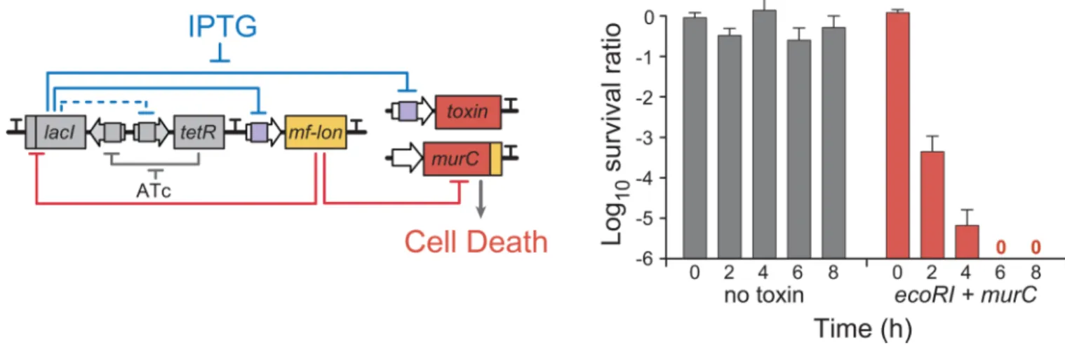
β-D-1-thiogalactopyranoside (IPTG), activates toxin production and cell death irrespective of the presence of the programmed survival signal (Fig. 2).
HHMI A
uthor Man
uscr
ipt
HHMI A
uthor Man
uscr
ipt
HHMI A
uthor Man
uscr
ipt
Hybrid transcription factor design
To extend the versatility and modularity of this system, we built a second circuit, called the Passcode circuit, which uses hybrid LacI/GalR family TFs to expand the range and
complexity of environmental signals used to define biocontainment conditions. This survival “passcode” can be easily reprogrammed to restrict cell growth to a new environment or to limit knowledge of the growth conditions to authorized personnel. To build hybrid LacI family TFs, we first identified the boundaries of the environmental sensing modules (ESMs) and DNA recognition modules (DRMs) found in LacI family members (Supplementary Figs. 7–11). Similar to previous studies25,26, we generated hybrid TFs that use the small molecule input defined by the hybrid’s ESM to regulate the promoter defined by the hybrid’s DRM (Fig. 3a and Supplementary Fig. 12).
To construct the hybrid TFs, we used the cellobiose-responsive TF, CelR, from
Thermobifida fusca and the galactose-responsive TF, GalR, and IPTG-responsive LacI from E. coli. We fused the ESMs from CelR and GalR to the DRM of LacI to generate the hybrid TFs CelR-LacI and GalR-LacI. To test their functionality, these hybrid TFs or native LacI were used to control GFP expression from a promoter containing lacO operator sites recognized by the LacI DRM. The hybrid TFs allowed strong GFP expression upon exposure to the small molecule input defined by their ESM and showed almost no response to the other inputs (Supplementary Figs. 11b and 12b). We fused the LacI, GalR and CelR ESMs to the DRM of ScrR from Klebsiella pneumoniae and used the resulting hybrid TFs to regulate a promoter containing scrO operator sites. As predicted from their design, these hybrid TFs only respond to the input defined by their ESM (Supplementary Figs. 11b and 12c), although it is interesting to note that the GalR ESM shows distinct inhibition by high levels of IPTG as previously reported27 (Supplementary Fig. 13). Importantly, the DRMs used in these hybrid TFs provided similar specificity, as they regulated promoters containing their cognate operator sites but not other LacI family operator sites (Supplementary Fig. 14). Similar to previous work27, we found that co-expression of hybrid TFs containing the same DRM could be used to regulate a single promoter, creating an AND logic gate function (Supplementary Fig. 15).
Development of the Passcode kill switches
We used these hybrid TFs to create a series of Passcode circuits that contain a single transcriptional architecture but respond to distinct combinations of environmental inputs to control gene expression and cell survival. The Passcode circuits contain the output module (in this case, gfp) under control of a TF (hybrid C) whose expression is controlled by an AND gate formed by two TFs (hybrid A and hybrid B) (Supplementary Fig. 16). This serial arrangement, made possible by the orthogonality of the hybrid DRMs and ESMs, creates the condition that both of the inducers recognized by hybrid A and hybrid B (inputs a and b, respectively) must be present to allow expression of hybrid C to repress gfp expression. Loss of input a or input b or the presence of input c allows gfp expression, causing cell death if gfp is replaced by a toxin gene.
To test the functionality and modularity of this circuit architecture, we created three versions of the Passcode circuit that respond to different combinations of input signals to control
HHMI A
uthor Man
uscr
ipt
HHMI A
uthor Man
uscr
ipt
HHMI A
uthor Man
uscr
ipt
output expression (Fig. 3a). For example, in one Passcode circuit (Fig. 3b, left column), we used GalR-LacI (A) and CelR-LacI (B) to control expression of LacI-ScrR (C), which in turn represses toxin expression. In this circuit, loss of galactose (input a) or cellobiose (input b) allows GalR-LacI or CelR-LacI to bind the lacO operator, blocking LacI-ScrR expression, thereby enabling toxin expression and causing cell death. Any exposure to IPTG (input c) releases LacI-ScrR repression of toxin expression, thereby killing the cell as well.
Importantly, the passcode combinations for cell survival and cell death can be reprogrammed simply by rearranging the ESMs of the three TFs to rewire the connections between the environmental sensing and transcriptional regulation.
These Passcode circuits were first evaluated with GFP as the output module in all eight combinations of the three environmental inputs. All three circuits allowed high level GFP expression in all conditions except that designated by the desired three input combination (Supplementary Fig. 16b), and single-cell fluorescence showed a monomodal population distribution under all conditions (Supplementary Fig. 16c). GFP was then replaced with the ecoRI and mf-Lon-MurC toxin modules described for the Deadman switch above (Fig. 3a), and toxin expression levels were optimized by testing a range of calculated RBS strengths24 (Supplementary Fig. 17). Hybrid C, which directly controls toxin expression in the circuit, was also engineered in the same manner to optimize circuit performance (see Online
Methods). Each kill switch circuit was tested in E. coli using eight combinations of input signals, and cell survival was measured by CFU count at multiple time points
(Supplementary Fig. 18). As seen in Fig. 3b, only circuits that received the proper survival code allowed the host cells to survive (each survival condition is highlighted in green). Furthermore, inclusion of both the ecoRI and mf-Lon toxin modules in the Passcode circuit caused the cell survival ratio to drop below 1×10⁻⁶ for all non-passcode conditions.
Long-term circuit stability
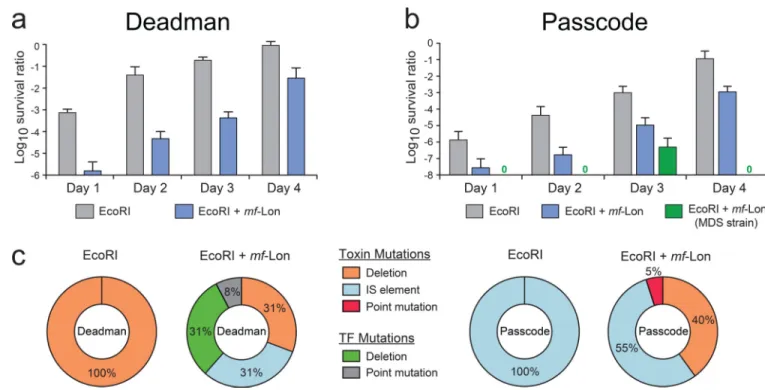
To measure the long-term stability and robustness of the Passcode and Deadman kill switches, we passaged cells containing the circuits for four days under survival conditions and periodically tested subsets of cells for circuit function under non-permissive conditions. Both the Deadman and Passcode circuits showed reduced killing efficiency over time, and sequence analysis of cells that escaped biocontainment predominantly showed inactivating mutations in the toxin genes (Fig. 4 and Supplementary Figs. 19–21). The noted exception was independent TetR mutations in the two-toxin Deadman circuit where TetR inactivation repressed toxin expression even in the absence of the ATc survival signal. It is important to note, however, that these “escapees” are still sensitive to IPTG-mediated fail-safe circuit activation as described above (Fig. 2). Genome-encoded insertion-sequence (IS) elements28, particularly IS1 and IS5, caused a large percentage of inactivating mutations in the one-toxin and two-toxin Passcode circuits. Deletion of these IS elements and other genome repair mechanisms in E. coli reduced the Passcode “escapee” rate by 3–5 logs after four days, demonstrating that increased stability of the host genome will augment the functionality of these biocontainment systems (Fig. 4b and Supplementary Fig. 19). As the toxin genes were the main target for circuit inactivation, inclusion of additional redundant killing systems into each circuit should further reduce the escapee rate.
HHMI A
uthor Man
uscr
ipt
HHMI A
uthor Man
uscr
ipt
HHMI A
uthor Man
uscr
ipt
DISCUSSION
The Deadman and Passcode switches provide robust information processing circuits to couple environmental signals with conditional survival of the microbial host. The Deadman kill switch described above is based on a monostable circuit that passively activates toxin gene expression in the absence of the small molecule input ATc. Since ATc is not normally found in nature, engineered cells that escape biocontainment will trigger cell death to prevent the spread of the organism or its genetic content into the surrounding ecosystem. Unlike auxotrophy-based biocontainment where the environmental signal is an intrinsic feature of the system9,10, the environmental sensing and cell killing systems are decoupled in the Deadman switch. This circuit relies on two main elements for functionality: (1) the orthogonality of the TFs to create a toggle switch, and (2) their relative activity under induced expression. As such, the Deadman circuit is highly modular, and the environmental signal detected by the circuit may be altered by replacing TetR with a wide range of transcription factors, including more than 80,000 annotated TetR family members29 as well as orthogonal LacI/GalR family members including hybrid TFs as described for the Passcode switch. In addition, the Deadman circuit has an additional fail-safe mechanism which activates toxin production and cell death in the presence of IPTG, enabling exogenous control over the microbe’s survival even as the cell uses the circuit to monitor its
environment.
Similar to the Deadman switch, the Passcode circuits are based on a two-layered transcriptional repression design. To build hybrid TFs, we identified the conserved
boundaries of the ESMs and DRMs within the LacI/GalR family members LacI, GalR, CelR and ScrR. The resulting environmental sensing and DNA binding modules provide
independent control of the sensory input and regulatory output of each hybrid TF. Previous pioneering work used the boundary between the conserved regulatory domain and HH motif to create hybrid TFs25,26, but some of these hybrids required additional protein engineering and mutagenesis to become functional. Here we identify a discrete boundary between the conserved HH and HTH motifs to create independent environmental sensory and DNA binding domains that can be efficiently combined without further protein engineering. The modularity provided by these hybrid TFs dramatically expands the number and range of environmental signals that can be used to control biocontainment systems such as the Deadman and Passcode circuits described here, as the ESM and DRM boundaries defined in this study may be used to incorporate sensing modules from many of the ~29,000 LacI/GalR family members30 that detect diverse environmental signals.
These hybrid TFs may also be used to functionalize other synthetic circuits, including the Deadman switch, to respond to different environmental signals. Moreover, the regular use of LacI and TetR in other bacteria31,32 suggests that these circuits may be readily transferred to other microbes, including industrial production strains. Replacement of the antibiotic resistance cassettes in these plasmids with well characterized selection systems that use toxin-antitoxin modules or auxotrophy complementation should also enable their use in biotherapeutic applications4,33.
HHMI A
uthor Man
uscr
ipt
HHMI A
uthor Man
uscr
ipt
HHMI A
uthor Man
uscr
ipt
In summary, we have established two circuit-based microbial kill switches that constrict host cell survival to an environment defined by specific input signals. Unlike existing
biocontainment systems with fixed survival conditions that are difficult to modify, the Deadman and Passcode kill switches are inherently customizable, both in the environmental conditions that control circuit activation and in the output modules that control cell fate. In addition to its use as a biocontainment system, the Passcode circuit may find particular utility as a tool for intellectual property protection, where unauthorized growth of strains without the appropriate “passcode” molecules would induce cell death. With the proper choice of toxins, such as the endonuclease EcoRI described here, the Passcode circuit could be used to not only kill the host cell but also degrade its genome and accompanying plasmids to deter attempts at reverse-engineering the strain of interest. Use of hybrid TFs that respond to proprietary small molecule inputs may further secure the strain against theft, even if its genome is sequenced.
ONLINE METHODS
Analysis of protein sequences and crystal structures
ClustalW235 was used for protein sequence alignment of GalS, GalR, AscG, RbsR, PurR, GntR, LacI, and MalI from E. coli; CelR from T. fusca; ScrR from V. alginolyticus (ScrR-V); and ScrR from K. pneumonia (ScrR-K). Protein crystal structure analysis was performed with PyMol 1.5.x using Protein Data Bank (PDB) entries 1EFA, 1LBG, 1LBI, 1LBH, 1QPZ, and 1TLF36–40.
Strains
E. coli MG1655ΔlacI was used to perform functional analysis of hybrid TFs as shown in Supplementary Figures 10–13. In this strain, transcription from the pLtetO-1 promoter driving TF expression is constitutive because it does not contain tetR. E. coli MG1655Pro, which produces high levels of LacI and TetR11, was used in hybrid TF analysis when LacI regulation of pLlacO-1 was a desired feature (Supplementary Fig. 14). In these assays, the TetR inhibitor anhydrotetracycline (ATc; 100 ng/mL) was included in the media to ensure TF expression from the pLtetO promoter. E. coli MG1655ΔlacI was the parental strain for all circuit characterization and was created through P1 phage transduction of lacI::kanR from the Keio collection41 into E. coli MG1655 (ATCC 47076). Flp recombinase, expressed on pCP20, was used to remove the kanR cassette42. To construct E. coli strains containing Lon recognition tags on the essential genes dxs, cysS, fldA, plsB or murC, the pdt#1 mf-Lon recognition tag from each corresponding gene in the EPD library20 was transferred to MG1655ΔlacI by P1 phage transduction and the kanR cassette was removed as above. P1 phage transduction was used to convert E. coli MDS42pdu34 (Scarab Genomics) for use in the Passcode switch analysis. Specifically, lacI::kanR and recA::kanR deletions from the Keio collection41 and murC-pdt#1 from the EPD library20 were independently transferred to MDS42pdu by P1 phage transduction, and the accompanying kanamycin cassettes were removed by FlpE-mediated excision using pECA102.
HHMI A
uthor Man
uscr
ipt
HHMI A
uthor Man
uscr
ipt
HHMI A
uthor Man
uscr
ipt
Cell growth and media
Luria-Bertani (LB) media was used for all experiments, and the following antibiotics and inducers were included when appropriate: ampicillin (50 µg/ml), chloramphenicol (10 µg/ ml), kanamycin (50 µg/ml), ATc (100 ng/ml), IPTG (1 mM), galactose (20 mM) and cellobiose (5 mM). For the Deadman switch, single colonies grown on LB agar plates containing ATc were inoculated into liquid cultures containing ATc for growth overnight at 37°C with shaking. Similarly, cells harboring each of the three Passcode switches were picked from plates with the survival combination of inputs and inoculated into their respective survival liquid media. Overnight cultures were inoculated 1:20,000 into 96-well plates and grown at 37°C and 900 rpm for further tests.
Plasmid construction
All plasmids were constructed using conventional molecular cloning protocols43 and Gibson Assembly44. E. coli NEB Turbo (New England BioLabs Inc.) was used for cloning purposes, and all primers were purchased from IDT. To create the Deadman switch pDM1 (Genbank accession number KT893253), genetic elements from the toggle pECJ320 were cloned into the conditionally amplified single-copy plasmid pBAC/oriV45, and the lacI and tetR RBS strengths were modified as described in the following section. To provide increased control over the promoter controlling mCherry expression, the T1 terminator from rnpB (Registry of Standard Biological Parts BBa_J61048) was inserted upstream (Supplementary Fig. 4a), and three palindromic lac operator sites19 were inserted around the –35 and –10 region of the promoter (pDM2, GenBank accession number KT893254). The M. florum protease gene mf-lon was cloned under control of a LacI-regulated promoter (pDM2L; GenBank accession number KT893255). The resulting plasmid served as the base Deadman circuit, and
mCherry was cloned to yield pDM3 and ecoRI, ccdB and mazF were cloned to make the toxin variants (see Supplementary Table 1).
Hybrid TF genes (lacI-galR LG36-LG46, galR-lacI, celR-lacI, lacI-scrR, galR-scrR, and celR-scrR) were constructed by overlap extension PCR to fuse the environmental sensing modules (ESMs) and the DNA recognition modules (DRMs) of the designated genes. The hybrid TFs were cloned into pTR, a derivative of pKE2-MCS containing the pLtetO-1 promoter and T0 terminator from pZA1146, using restriction sites BamHI and BsrGI. Transcription from the pLtetO-1 promoter driving TF expression is constitutive because the E. coli strains used in this study did not contain tetR. Reporter plasmids (pREPORT) were constructed from the plasmid pZA1246, with mcherry or gfp inserted downstream of the pLlacO promoter using KpnI and HindIII. To test hybrid TFs that contain the ScrR DRM, pLlacO-1 was replaced with pLscrO-1 or pLscrO-2 using Gibson Assembly method44. For implementation of both LacI/pLlacO-1 and GalR-ScrR/pLscrO inducible expression systems in the same cells (Supplementary Fig. 14), the pLlacO-1-mCherry-T1 cassette was
subcloned into pTR using restriction sites, NheI and SalI.
The Passcode circuit was developed using a two-plasmid system. Plasmid pPasscode (GenBank accession numbers KT895272, KT895273 and KT895274), derived from pKE2_MCS17, was constructed to contain the hybrid TF circuit, and pToxin (GenBank accession numbers KT895275, KT895276 and KT895277), derived from pZA1246, was
HHMI A
uthor Man
uscr
ipt
HHMI A
uthor Man
uscr
ipt
HHMI A
uthor Man
uscr
ipt
constructed to contain the toxin output module under control of the pLscrO promoter. For pPasscode, three promoter-hybrid TF-terminator fragments were used to construct each hybrid TF circuit version, as listed in Supplementary Table 1. For version 1 of pPasscode (pPasscode1), in which LacI-ScrR is used as hybrid C, the promoter pLscrO-2 was utilized to control the expression of toxin gene(s) in pToxin. For pPasscode2 that contains GalR-ScrR, the promoter pLscrO-1 was used for toxin control in pToxin. For pPasscode3 that contains CelR-ScrR, the promoter pLscrO-1 was used to control the expression of mf-Lon and the promoter pLscrO-2 was used to control the expression of ecoRI. For Passcode circuits that contain two toxin gene systems, the DNA fragments pLscrO-mf-Lon-terminator and pLscrO-ecoRI-terminator were incorporated into pToxin using Gibson Assembly (Supplementary Table 1). For Passcode circuit characterization, pPasscode was first transformed into the desired E. coli strain and grown in media containing the “passcode” combination of the three inputs (IPTG, galactose and cellobiose). Plasmid pToxin, which contains the toxin gene(s), was then transformed into the cells to complete the Passcode circuit.
RBS strength optimization for monostable toggle construction
RBS calculator algorithm24 was used to identify RBS variants that produce a range of LacI and TetR expressions (Supplementary Table 1). Cells containing each toggle RBS variant were grown overnight in the presence of ATc, transferred to media without ATc, and then measured for mCherry expression by flow cytometry after 6 hours. Toggle variant 5, which showed the largest change in mCherry fluorescence upon loss of ATc, was chosen for use in the Deadman circuit (Supplementary Fig. 2). To quantify the relative LacI and TetR expression levels, mCherry was fused to the C-terminus of LacI or TetR to yield pBAC-LC and pBAC-TC, respectively (GenBank accession numbers KT893256 and KT893257). RBS variants for LacI and TetR were then cloned into pBAC-LC and pBAC-TC, respectively, and a SpectraMax M5 microplate reader (Molecular Devices) was used to measure mCherry fluorescence with excitation and emission wavelengths of 587 nm and 610 nm, respectively, with an emission filter cutoff at 610 nm. mCherry fluorescence was normalized to cell growth (OD600).
RBS strength optimization for toxin expression
To optimize cell death dynamics upon Deadman or Passcode circuit activation, a range of predicted RBS strength variants24 was generated for each toxin (Supplementary Table 1). For the Deadman kill switches (Supplementary Fig. 6), RBS variants and the corresponding toxin genes ecoRI, ccdB, and mazF, were cloned into pDM2L using Gibson Assembly (Supplementary Table 1). Overnight cultures were grown in the presence of ATc and then transferred into media with ATc (survival condition) or with IPTG (induced death
condition). A SpectraMax M5 microplate reader (Molecular Devices) was used to measure cell growth (OD600) every 15 min for 15 hours, and the cell growth ratios of the induced
death state to the survival state were calculated at 15 hours.
For Passcode kill switches, RBS variants (Supplementary Table 1) and the corresponding toxin genes ecoRI and mf-lon were cloned into pREPORT to replace gfp and tested for optimal expression under regulation by the hybrid TFs LacI-ScrR, GalR-ScrR and
CelR-HHMI A
uthor Man
uscr
ipt
HHMI A
uthor Man
uscr
ipt
HHMI A
uthor Man
uscr
ipt
ScrR. Plasmids containing each RBS-toxin variant were transformed into cells constitutively expressing LacI-ScrR, GalR-ScrR, or CelR-ScrR, grown overnight without inducers, and then transferred into media with or without the appropriate inducer (1 mM IPTG, 20 mM galactose, or 5 mM cellobiose for cells containing LacI-ScrR, GalR-ScrR, or CelR-ScrR, respectively). Cell growth analysis was performed as described for the Deadman circuit above, and the cell growth ratio was calculated at 12 hours. Representative data are shown in Supplementary Figure 17.
RBS strength optimization for ScrR ESM-containing TFs
A range of RBS variants was tested to optimize the expression of ScrR ESM-containing TFs (see TF “C” in Fig. 3a) in the Passcode circuits (Supplementary Table 1). Cells with the Passcode circuit harboring RBS variants were transformed with the indicated pToxin plasmid, grown overnight under survival conditions (see Supplementary Fig. 16 for the appropriate inducers for each circuit), and then transferred to media with all 8 combinations of the three inducers (IPTG, galactose, and cellobiose). Performance of each circuit was determined by CFU count after 8 hours of exposure as described in the following section.
Survival assays
Colony forming unit (CFU) cell viability assays were used to measure functionality of the Deadman and Passcode circuits. Overnight cultures were grown under the survival conditions (Deadman: with ATc, Passcode: with survival “passcode” inputs) and were transferred into fresh LB medium with or without the survival signal(s). For the Passcode circuit, all eight combinations of the three inputs were tested (+/− IPTG, +/− galactose and +/− cellobiose). Samples were collected every two hours, serially diluted in PBS over a 7-log range, and spotted (5 µL) onto a square plate containing LB agar with the appropriate survival signal(s). CFU and survival ratios were calculated as previously reported11: CFU/mL = (number of colonies) × (dilution factor)/0.005 mL, survival ratio (log10) = log
{(CFU/mL without the survival signal)/(CFU/mL with the survival signal)}.
Long-term growth analysis
Cells containing the Deadman and Passcode kill switches were passaged under survival conditions for 4 days (Deadman: 100 ng/mL ATc; Passcode: unique inducer for each Passcode circuit; see Supplementary Fig. 16). Sub-populations of these cells were transferred 1:20,000 into media with or without the survival signal(s) (Deadman: no ATc; Passcode: no inducers), and samples were collected at 8 hours after inoculation, serially diluted 1:10 in PBS over a 7-log range, and spotted (5 µL) onto LB agar plates with the appropriate survival signal(s). CFU and survival ratios were calculated as described above.
Escapee genetic analysis
Cells containing independent Deadman and Passcode circuit transformants (n=20 for each circuit) were grown under survival conditions (Deadman: 100 ng/mL ATc; Passcode: unique inducer for each Passcode circuit; see Supplementary Fig. 16). The cells were then
transferred to media without the survival signal(s) for 8 hours and then placed on LB agar plates containing the appropriate survival signal(s). Deadman circuits were isolated from
HHMI A
uthor Man
uscr
ipt
HHMI A
uthor Man
uscr
ipt
HHMI A
uthor Man
uscr
ipt
surviving cells by amplification with Phusion high-fidelity DNA polymerase (NEB), and Passcode circuits were isolated by plasmid DNA purification, and the circuits were then sequenced by Quintara Biosciences (Boston, MA).
Flow cytometry assay
Cells containing Passcode circuits were grown as described for each experiment, and at the appropriate time were fixed in 2% paraformaldehyde in PBS and then diluted 1:10 in PBS for analysis. mCherry and GFP fluorescence measurements were performed using a BD FACSAriaII (BD Biosciences) or a BD LSRFortessa™ flow cytometer (BD Biosciences). Flow cytometry data were gated by forward and side scatter to eliminate multi-cell aggregates, and the geometric mean of mCherry and GFP fluorescence distributions were calculated using FlowJo software (Treestar). At least 10,000 events were collected for each measurement.
Supplementary Material
Refer to Web version on PubMed Central for supplementary material.
ACKNOWLEDGMENTS
We thank Jihoon Han for assistance with molecular cloning. This work was supported by funding from the Defense Threat Reduction Agency grant HDTRA1-14-1-0006, Office of Naval Research MURI grant N000141110725, Air Force Office of Scientific Research grant FA9550-14-1-0060, and the Howard Hughes Medical Institute.
REFERENCES
1. Moe-Behrens GH, Davis R, Haynes KA. Preparing synthetic biology for the world. Frontiers in microbiology. 2013; 4:5. [PubMed: 23355834]
2. Bacchus W, Aubel D, Fussenegger M. Biomedically relevant circuit-design strategies in mammalian synthetic biology. Molecular systems biology. 2013; 9:691. [PubMed: 24061539]
3. Olson EJ, Tabor JJ. Post-translational tools expand the scope of synthetic biology. Current opinion in chemical biology. 2012; 16:300–306. [PubMed: 22766485]
4. Wright O, Delmans M, Stan GB, Ellis T. GeneGuard: A Modular Plasmid System Designed for Biosafety. ACS synthetic biology. 2014
5. Chappell J, et al. The centrality of RNA for engineering gene expression. Biotechnology journal. 2013; 8:1379–1395. [PubMed: 24124015]
6. Cameron DE, Bashor CJ, Collins JJ. A brief history of synthetic biology. Nature reviews. Microbiology. 2014; 12:381–390.
7. Wright O, Stan GB, Ellis T. Building-in biosafety for synthetic biology. Microbiology. 2013; 159:1221–1235. [PubMed: 23519158]
8. Steidler L, et al. Biological containment of genetically modified Lactococcus lactis for intestinal delivery of human interleukin 10. Nature biotechnology. 2003; 21:785–789.
9. Rovner AJ, et al. Recoded organisms engineered to depend on synthetic amino acids. Nature. 2015; 518:89–93. [PubMed: 25607356]
10. Mandell DJ, et al. Biocontainment of genetically modified organisms by synthetic protein design. Nature. 2015; 518:55–60. [PubMed: 25607366]
11. Callura JM, Dwyer DJ, Isaacs FJ, Cantor CR, Collins JJ. Tracking, tuning, and terminating microbial physiology using synthetic riboregulators. Proceedings of the National Academy of Sciences of the United States of America. 2010; 107:15898–15903. [PubMed: 20713708]
HHMI A
uthor Man
uscr
ipt
HHMI A
uthor Man
uscr
ipt
HHMI A
uthor Man
uscr
ipt
12. Contreras A, Molin S, Ramos JL. Conditional-suicide containment system for bacteria which mineralize aromatics. Applied and environmental microbiology. 1991; 57:1504–1508. [PubMed: 16348490]
13. Cai Y, et al. Intrinsic biocontainment: Multiplex genome safeguards combine transcriptional and recombinational control of essential yeast genes. Proceedings of the National Academy of Sciences of the United States of America. 2015; 112:1803–1808. [PubMed: 25624482]
14. Gallagher RR, Patel JR, Interiano AL, Rovner AJ, Isaacs FJ. Multilayered genetic safeguards limit growth of microorganisms to defined environments. Nucleic acids research. 2015; 43:1945–1954. [PubMed: 25567985]
15. Voigt CA. Genetic parts to program bacteria. Current opinion in biotechnology. 2006; 17:548–557. [PubMed: 16978856]
16. Jensen LB, Ramos JL, Kaneva Z, Molin S. A substrate-dependent biological containment system for Pseudomonas putida based on the Escherichia coli gef gene. Applied and environmental microbiology. 1993; 59:3713–3717. [PubMed: 8285679]
17. Litcofsky KD, Afeyan RB, Krom RJ, Khalil AS, Collins JJ. Iterative plug-and-play methodology for constructing and modifying synthetic gene networks. Nature methods. 2012; 9:1077–1080. [PubMed: 23042452]
18. Gardner TS, Cantor CR, Collins JJ. Construction of a genetic toggle switch in Escherichia coli. Nature. 2000; 403:339–342. [PubMed: 10659857]
19. Sadler JR, Sasmor H, Betz JL. A perfectly symmetric lac operator binds the lac repressor very tightly. Proceedings of the National Academy of Sciences of the United States of America. 1983; 80:6785–6789. [PubMed: 6316325]
20. Cameron DE, Collins JJ. Tunable protein degradation in bacteria. Nature biotechnology. 2014; 32:1276–1281.
21. Cheng SC, Kim R, King K, Kim SH, Modrich P. Isolation of gram quantities of EcoRI restriction and modification enzymes from an overproducing strain. The Journal of biological chemistry. 1984; 259:11571–11575. [PubMed: 6088551]
22. Smith AB, Maxwell A. A strand-passage conformation of DNA gyrase is required to allow the bacterial toxin, CcdB, to access its binding site. Nucleic acids research. 2006; 34:4667–4676. [PubMed: 16963775]
23. Zhang Y, et al. MazF cleaves cellular mRNAs specifically at ACA to block protein synthesis in Escherichia coli. Molecular cell. 2003; 12:913–923. [PubMed: 14580342]
24. Salis HM. The ribosome binding site calculator. Methods in enzymology. 2011; 498:19–42. [PubMed: 21601672]
25. Meinhardt S, Swint-Kruse L. Experimental identification of specificity determinants in the domain linker of a LacI/GalR protein: bioinformatics-based predictions generate true positives and false negatives. Proteins. 2008; 73:941–957. [PubMed: 18536016]
26. Meinhardt S, et al. Novel insights from hybrid LacI/GalR proteins: family-wide functional attributes and biologically significant variation in transcription repression. Nucleic acids research. 2012; 40:11139–11154. [PubMed: 22965134]
27. Shis DL, Hussain F, Meinhardt S, Swint-Kruse L, Bennett MR. Modular, multi-input
transcriptional logic gating with orthogonal LacI/GalR family chimeras. ACS synthetic biology. 2014; 3:645–651. [PubMed: 25035932]
28. Sousa A, Bourgard C, Wahl LM, Gordo I. Rates of transposition in Escherichia coli. Biology letters. 2013; 9:20130838. [PubMed: 24307531]
29. Cuthbertson L, Nodwell JR. The TetR family of regulators. Microbiology and molecular biology reviews : MMBR. 2013; 77:440–475. [PubMed: 24006471]
30. Finn RD, et al. Pfam: the protein families database. Nucleic acids research. 2014; 42:D222–D230. [PubMed: 24288371]
31. Ramos JL, et al. The TetR family of transcriptional repressors. Microbiology and molecular biology reviews : MMBR. 2005; 69:326–356. [PubMed: 15944459]
32. Cebolla A, Vazquez ME, Palomares AJ. Expression vectors for the use of eukaryotic luciferases as bacterial markers with different colors of luminescence. Applied and environmental microbiology. 1995; 61:660–668. [PubMed: 7574604]
HHMI A
uthor Man
uscr
ipt
HHMI A
uthor Man
uscr
ipt
HHMI A
uthor Man
uscr
ipt
33. Mignon C, Sodoyer R, Werle B. Antibiotic-free selection in biotherapeutics: now and forever. Pathogens. 2015; 4:157–181. [PubMed: 25854922]
34. Csorgo B, Feher T, Timar E, Blattner FR, Posfai G. Low-mutation-rate, reduced-genome Escherichia coli: an improved host for faithful maintenance of engineered genetic constructs. Microbial cell factories. 2012; 11:11. [PubMed: 22264280]
METHODS ONLY-REFERENCES
35. Larkin MA, et al. Clustal W and Clustal X version 2.0. Bioinformatics. 2007; 23:2947–2948. [PubMed: 17846036]
36. Bell CE, Lewis M. A closer view of the conformation of the Lac repressor bound to operator. Nature structural biology. 2000; 7:209–214. [PubMed: 10700279]
37. Friedman AM, Fischmann TO, Steitz TA. Crystal structure of lac repressor core tetramer and its implications for DNA looping. Science. 1995; 268:1721–1727. [PubMed: 7792597]
38. Lewis M, et al. Crystal structure of the lactose operon repressor and its complexes with DNA and inducer. Science. 1996; 271:1247–1254. [PubMed: 8638105]
39. Schumacher MA, Choi KY, Lu F, Zalkin H, Brennan RG. Mechanism of corepressor-mediated specific DNA binding by the purine repressor. Cell. 1995; 83:147–155. [PubMed: 7553867] 40. Glasfeld A, Koehler AN, Schumacher MA, Brennan RG. The role of lysine 55 in determining the
specificity of the purine repressor for its operators through minor groove interactions. Journal of molecular biology. 1999; 291:347–361. [PubMed: 10438625]
41. Baba T, et al. Construction of Escherichia coli K-12 in-frame, single-gene knockout mutants: the Keio collection. Molecular systems biology. 2006; 2:2006 0008.
42. Datsenko KA, Wanner BL. One-step inactivation of chromosomal genes in Escherichia coli K-12 using PCR products. Proceedings of the National Academy of Sciences of the United States of America. 2000; 97:6640–6645. [PubMed: 10829079]
43. T M, EF F, J S. Molecular Cloning: A Laboratory Manual. Cold Spring Harbor Laboratory Press. 1982; 545
44. Gibson DG, et al. Enzymatic assembly of DNA molecules up to several hundred kilobases. Nature methods. 2009; 6:343–345. [PubMed: 19363495]
45. Wild J, Hradecna Z, Szybalski W. Conditionally amplifiable BACs: switching from single-copy to high-copy vectors and genomic clones. Genome research. 2002; 12:1434–1444. [PubMed: 12213781]
46. Lutz R, Bujard H. Independent and tight regulation of transcriptional units in Escherichia coli via the LacR/O, the TetR/O and AraC/I1-I2 regulatory elements. Nucleic acids research. 1997; 25:1203–1210. [PubMed: 9092630]
HHMI A
uthor Man
uscr
ipt
HHMI A
uthor Man
uscr
ipt
HHMI A
uthor Man
uscr
ipt
Figure 1. Deadman kill switch
(a) Deadman circuit control of toxin gene expression. Cell viability was measured by CFU count following removal of the survival signal (ATc) and is displayed as a ratio of cells without ATc to cells with ATc at each time point. (b) Deadman circuit control of targeted essential protein degradation. Inclusion of the mf-lon specific pdt#1 tag on the specified essential gene causes mf-Lon-mediated degradation of the essential protein upon Deadman circuit activation. (c) Combined control of toxin expression and targeted essential protein degradation increases Deadman-induced cell death. In particular, targeted MurC degradation
HHMI A
uthor Man
uscr
ipt
HHMI A
uthor Man
uscr
ipt
HHMI A
uthor Man
uscr
ipt
and EcoRI expression reduced cell viability to below the limit of detection (< 1 × 10⁻⁷) after 6 hours (indicated by a “0”). All data points represent mean ± S.D. of three biological replicates.
HHMI A
uthor Man
uscr
ipt
HHMI A
uthor Man
uscr
ipt
HHMI A
uthor Man
uscr
ipt
Figure 2. The fail-safe mechanism for Deadman circuit activation
To demonstrate active control over host cell viability, cells grown under survival conditions (with ATc) were exposed to 1 mM IPTG to directly induce EcoRI and mf-Lon expression. Cell viability was measured by CFU count and is displayed as a ratio of cell survival with and without IPTG at each time point. Data points represent the mean ± S.D. of three biological replicates.
HHMI A
uthor Man
uscr
ipt
HHMI A
uthor Man
uscr
ipt
HHMI A
uthor Man
uscr
ipt
Figure 3. Passcode kill switch
(a) Passcode circuit schematic and logic gate behavior. Cell survival requires the continued presence of inputs a and b and the absence of input c. Loss of input a or b or the addition of input c cause the passcode circuit to activate toxin expression, leading to cell death. (b) Three versions of the Passcode kill switch were used to control expression of ecoRI, mf-lon-mediated MurC degradation (mf-lon), or both ecoRI and mf-lon. Cells containing each circuit were placed in each of eight possible combinations of the three input molecules, and cell viability was measured by CFU count after 8 hours. In each condition, cell survival is
HHMI A
uthor Man
uscr
ipt
HHMI A
uthor Man
uscr
ipt
HHMI A
uthor Man
uscr
ipt
displayed as a ratio of cells in that condition to cells in the “survival” condition highlighted in green. Cell survival below the limit of detection (< 1 × 10⁻⁷) is indicated by a “0”. All data points represent mean ± S.D. of three biological replicates.
HHMI A
uthor Man
uscr
ipt
HHMI A
uthor Man
uscr
ipt
HHMI A
uthor Man
uscr
ipt
Figure 4. Long-term circuit stability
(a) and (b) Cells with Deadman or Passcode circuits containing one toxin (EcoRI) or two toxins (EcoRI and mf-Lon) were passaged under survival conditions for 4 days, and sub-populations of cells were periodically switched to nonpermissive media (Deadman: no ATc, Passcode: no inducer) for eight hours. The survival ratio is the ratio of cells that survive in the death state to those in the survival state. Data points represent the mean ± S.D. of six biological replicates. The passcode circuit was also passaged in E. coli MDS42pdu ΔrecA (MDS strain), which lacks recombinogenic and mobile genomic elements34. Deadman and Passcode circuits that do not contain toxin modules displayed increased stability throughout the 4 day experiment (see Supplementary Figs. 20–21). (c) Cells containing Deadman and Passcode circuits that survived exposure to their respective death states were isolated, and the entire circuit and toxin(s) were sequenced to identify the inactivating mutations. Toxin gene disruption by genome-encoded insertion-sequence (IS) elements and large deletions were the predominant cause of circuit inactivation. In the two-toxin Deadman circuit, inactivating TetR mutations allowed continued LacI expression and repression of toxin genes in non-biocontainment conditions (see Fig. 1 for a visual aid).