Cognitive dysfunction and prefrontal synaptic
abnormalities in a mouse model of fragile X syndrome
The MIT Faculty has made this article openly available.
Please share
how this access benefits you. Your story matters.
Citation
Krueger, D. D. et al. “Cognitive dysfunction and prefrontal
synaptic abnormalities in a mouse model of fragile X syndrome.”
Proceedings of the National Academy of Sciences 108 (2011):
2587-2592. ©2011 by the National Academy of Sciences.
As Published
http://dx.doi.org/10.1073/pnas.1013855108
Publisher
National Academy of Sciences (U.S.)
Version
Final published version
Citable link
http://hdl.handle.net/1721.1/65898
Terms of Use
Article is made available in accordance with the publisher's
policy and may be subject to US copyright law. Please refer to the
publisher's site for terms of use.
Cognitive dysfunction and prefrontal synaptic
abnormalities in a mouse model of fragile X syndrome
Dilja D. Krueger, Emily K. Osterweil, Stephanie P. Chen, Lynne D. Tye, and Mark F. Bear1
Howard Hughes Medical Institute, Picower Institute for Learning and Memory, Department of Brain and Cognitive Sciences, Massachusetts Institute of Technology, Cambridge, MA 02139
Edited by Richard L. Huganir, The Johns Hopkins University School of Medicine, Baltimore, MD, and approved January 3, 2011 (received for review September 17, 2010)
Among the hallmark phenotypes reported in individuals with fragile X syndrome (FXS) are deficits in attentional function, inhibitory control, and cognitiveflexibility, a set of cognitive skills thought to be associated with the prefrontal cortex (PFC). However, despite substantial clinical research into these core deficits, the PFC has received surprisingly little attention in preclinical research, particularly in animal models of FXS. In this study, we sought to investigate the molecular, cellular, and behavioral consequences of the loss of the fragile X mental retardation protein in the PFC of Fmr1 KO mice, a mouse model of FXS. We identify a robust cogni-tive impairment in these mice that may be related to the deficits in cognitiveflexibility observed in individuals with FXS. In addition, we report that levels of proteins involved in synaptic func-tion, including the NMDA receptor subunits NR1, NR2A, and NR2B; the scaffolding proteins PSD-95 and SAPAP3; and the plastic-ity-related gene Arc, are decreased in the prefrontal cortex ofFmr1 KO mice and are partly correlated with behavioral performance. Finally, we report that expression of c-Fos, a marker of neuronal activity, is decreased in the PFC ofFmr1 KO mice. Together, these data suggest thatFmr1 KO mice may represent a valuable animal model for the PFC-associated molecular, cellular, and behavioral abnormalities in FXS and that this model may be useful for testing the efficacy of therapeutic strategies aimed at treating the cogni-tive impairments in FXS.
behavior
|
operant|
orbitofrontal|
NMDA receptorF
ragile X syndrome (FXS) is the most common form of inherited mental retardation and a leading known cause of autism (1). It is caused by loss of theFmr1 gene product fragile X mental retardation protein (FMRP), an mRNA-binding protein involved in translational regulation (2, 3). FMRP is thought to repress the synthesis of proteins required for protein synthesis-dependent synaptic plasticity (4, 5). In FXS, the absence of FMRP is hypothesized to result in unrestricted synthesis of plasticity-related proteins (6, 7), impairing the ability of synapses to appropriately undergo plasticity in an activity-dependent and stimulus-specific manner. In support of this hypothesis, mice with a deletion in theFmr1 gene (Fmr1 KO mice) display aberrant forms of plasticity (4) and an increase in immature dendritic spines that presumably reflects an abnormal synaptic connectiv-ity (8). Together, these synaptic alterations are thought to un-derlie the cognitive and behavioral phenotypes that are the hallmark features of FXS.Among the most common symptoms reported in FXS are deficits in attentional function, inhibitory control, and cognitive flexibility (9), cognitive skills that have all been linked to the prefrontal cortex (PFC) and associated fronto-striatal networks (10, 11). Anatomical and imaging studies of individuals with FXS have identified structural alterations in PFC, and numerous fMRI studies have shown aberrant patterns of neural activity in fronto-striatal pathways during cognitive tasks (12). Together, all of these results suggest that the fronto-striatal network is one of the key brain systems impaired in FXS (9).
Despite extensive clinical research into this core deficit in FXS, only a few studies have specifically addressed the role of the PFC and related cognitive functions in animal models of FXS. In the PFC ofFmr1 KO mice, abnormalities were detected in the density of dendritic spines (13), the induction of spike-timing–dependent plasticity (14), and the response to dopami-nergic signaling (15–17), and subtle cognitive impairments in these mice have also been reported (18–20). However, sub-stantial further research will be crucial both in elucidating the molecular mechanisms by which loss of FMRP expression affects PFC function in FXS and in developing animal models suitable for screening potential drug treatments that target the core cognitive deficits. In the current study, we address this issue by developing a behavioral paradigm that identifies a robust cog-nitive impairment in the Fmr1 KO mice, as well as by in-vestigating the accompanying molecular and cellular alterations in the PFC of these mice.
Results
Fmr1 KO Mice Are Impaired in the Acquisition of a Visuospatial Discrimination Task. A major aim of our study was to identify behavioral deficits in Fmr1 KO mice that represent a model for the core cognitive phenotypes observed in FXS. To this end, we first tested mice in a behavioral task used to assess sustained attention and inhibitory control in rodents, thefive-choice serial reaction time task (5CSRTT) (21). This paradigm uses an op-erant test chamber equipped with five nose-poke holes, i.e., apertures that can detect a nose-poke response by the mouse, and a food magazine into which a food reward can be delivered. In our experiment, Fmr1 KO mice and wild-type (WT) litter-mates werefirst trained to perform a nose-poke response in an illuminated aperture to obtain a food response (seeSI Materials and Methods for details of the training phases), and they were then subjected to two final tests, one to measure sustained at-tention and one to measure inhibitory control (SI Materials and Methods). We found thatFmr1 KO mice did not differ from their WT littermates in either of the twofinal tests, suggesting that, under our conditions, these mice do not display deficits in sus-tained attention or inhibitory control (Fig. S1 A and B). In-terestingly, however, we observed a significant and very selective deficit in one of the pretest training phases (Fig. S1C;SI Mate-rials and Methods), and we therefore decided to further pursue this impairment.
Author contributions: D.D.K. and M.F.B. designed research; D.D.K., E.K.O., S.P.C., and L.D.T. performed research; D.D.K. and E.K.O. analyzed data; and D.D.K., E.K.O., and M.F.B. wrote the paper.
Conflict of interest statement: Mark Bear has a financial interest in Seaside Therapeutics, Inc.
This article is a PNAS Direct Submission.
Freely available online through the PNAS open access option.
1To whom correspondence should be addressed. E-mail: [email protected].
This article contains supporting information online atwww.pnas.org/lookup/suppl/doi:10. 1073/pnas.1013855108/-/DCSupplemental.
NEUROS
To this end, we trained mice on a shortened paradigm in-cluding only thefirst two training phases of the original 5CSRTT (Fig. 1A). In the first phase, all five apertures were illuminated
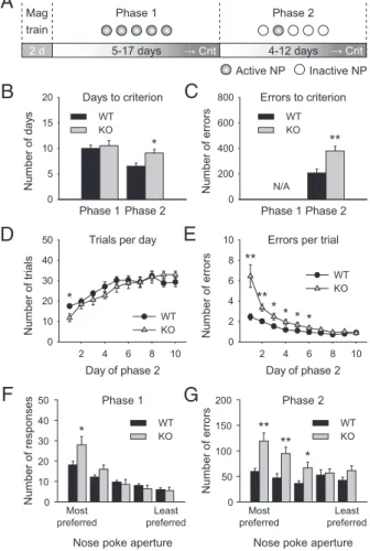
and active, and a nose-poke response in any of the apertures resulted in the delivery of a food reward.Fmr1 KO mice showed no significant difference in the number of days taken to acquire this task (Fig. 1B; one-way ANOVA for genotype, F1,24 < 1), suggesting that they are not impaired in the acquisition of an appetitive instrumental response under the current experimental conditions. In the second phase, only one of thefive apertures was illuminated and active, and only a response in this aperture was rewarded.Fmr1 KO mice took significantly longer to acquire this phase of the task (Fig. 1B; one-way ANOVA for genotype, F1,22= 7.11,P < 0.05), and they made significantly more errors than WT mice (Fig. 1C; one-way ANOVA for genotype, F1,22= 12.52,P < 0.01). Further analysis of this phase revealed thatFmr1 KO mice did not differ significantly from WT littermates in the number of trials completed per session, although there was a trend toward a genotype× training day interaction (Fig. 1D; repeated measures ANOVA for genotype, F1,24 < 1; genotype × training day in-teraction, F9,216= 1.67,P = 0.1). However, they made signifi-cantly more errors per trial in thefirst days of training (Fig. 1E; repeated measures ANOVA for genotype,F1,24= 14.3,P < 0.001; genotype× day interaction, F9,216= 8.76,P < 0.001). These data suggest that Fmr1 KO mice are impaired in the acquisition of a visuospatial discrimination task, but not in the performance of the visuospatial discrimination itself, as indicated by the fact that both groups reach equal levels of performance by the end of a 10-d training period (Fig. 1E; post hoc analysis for day 10, P = 0.824). Fmr1 KO Mice Display a Stronger Spatial Preference in Responding. To further explore the cause of this impairment, we investigated the pattern of nose-poke responses by spatial location, analyzing each of thefive apertures individually. On the basis of the total number of responses made in the last 2 d of phase 1 (i.e., the days in which the mouse was considered to have acquired the task), the apertures were ranked by preference for each in-dividual mouse. Both genotypes showed a spatial preference for certain apertures over others in phase 1 (Fig. 1F; main effect of aperture, F4,72= 58.82, P < 0.001). However, Fmr1 KO mice showed a stronger spatial preference; i.e., they made significantly more responses in the most preferred aperture than did the WT mice (Fig. 1F; main effect of genotype, F1,18 < 1; genotype × aperture interaction,F4,72= 7.06,P < 0.001; post hoc analysis for the most preferred aperture,P < 0.05). Using the same prefer-ence ranking, the number of errors to criterion in phase 2 was assessed for each nose-poke aperture (Fig. 1G; main effect of genotype, F1,18 = 10.7, P < 0.005; genotype × aperture in-teraction,F4,72= 4.06,P < 0.01). WT mice made similar num-bers of errors to criterion in each aperture, suggesting that the slight spatial preference displayed in phase 1 did not influence performance in phase 2. Fmr1 KO mice, on the other hand, made significantly more errors in the previously preferred apertures. These data suggest that a lack of cognitiveflexibility in theFmr1 KO mice may contribute to the delay in acquisition of the visuospatial discrimination task described here.
Behavioral Impairments Are Accompanied by Decreases in Synaptic Markers in Orbitofrontal Cortex and Medial Prefrontal Cortex ofFmr1 KO Mice.A central question in FXS is how loss of translational regulation by FMRP affects the molecular composition of the synapse, and how this in turn results in the cognitive impairments seen in FXS. To begin to address this issue, we investigated whether expression of synaptic proteins was altered in the PFC of the same Fmr1 KO mice that had undergone behavioral training. We isolated two subregions of PFC (Fig. 2A)—the orbitofrontal cortex (OFC) and the medial prefrontal cortex (mPFC)—both of which are required for various aspects of be-havioral flexibility in rodents (22–24). The global profile of protein expression inFmr1 KO mice was similar to that of WT mice, as assessed by a total protein stain (Fig. 2B). Interestingly,
Nu m b e r o f d a ys Nu mb e r o f e rro rs Nu m b e r o f tria ls N u m b er of er ro rs
Mag Phase 1 Phase 2
2 d 5-17 days 4-12 days Active NP Inactive NP
A
B
C
D
E
Days to criterion Phase 1 Phase 2 0 5 10 15 20 WT KO Errors to criterion Phase 1 Phase 2 0 200 400 600 800 WT KOTrials per day
Day of phase 2 2 4 6 8 10 0 10 20 30 40 50 WT KO
Errors per trial
Day of phase 2 2 4 6 8 10 0 2 4 6 8 10 WT KO * ** * ** ** N/A * ** * Num b er of responses
Nose poke aperture
Num b er of errors
F
Phase 1G
Most preferred Least preferred 0 10 20 30 40 50 WT KO * Phase 2 0 50 100 150 200 WT KO Most preferred Least preferred ** ** *Nose poke aperture train
Crit Crit
Fig. 1. Fmr1 KO mice are impaired in the acquisition of a visuospatial
dis-crimination task. (A) Schematic illustrating the design of the behavioral task,
which consists of three components: magazine training (“Mag”) to
habitu-ate the mice to the operant conditioning chambers; phase 1, in which allfive
nose-poke (“NP”) apertures are active (“correct response”); and phase 2, in
which only one nose-poke aperture is active and the other four are inactive
(“error”). Mice were trained on each of the two acquisition phases until their
performance reached a predefined criterion (“crit”; see Materials and
Methods for details). (B) Number of days taken to reach criterion on each phase. Fmr1 KO mice were not impaired in the acquisition of phase 1, but
they required significantly more time than their WT littermates to reach
criterion on phase 2 (one-way ANOVA, P< 0.05, n = 12). (C) Cumulative
number of errors to criterion. Fmr1 KO mice made significantly more errors
than their WT littermates in phase 2 (one-way ANOVA, P< 0.01, n = 12). This
measure is not applicable for phase 1 because all responses were considered correct and errors were not possible. (D) Number of trials completed per day
during phase 2. There was no significant difference between genotypes as
assessed by repeated measures ANOVA, although post hoc analysis revealed
a small but significant decrease in the number of trials completed by KO on
thefirst day of phase 2. (E) Average number of errors made per trial on each
day of phase 2. Fmr1 KO mice made significantly more errors per trial on the
first 6 d of phase 2 (repeated measures ANOVA for genotype, P < 0.001;
genotype× day interaction, P < 0.001, n = 10). (F) Total number of responses
per nose-poke aperture in the last 2 d of phase 1, ranked by preference for
each mouse. Fmr1 KO mice show a significantly stronger preference for their
most preferred aperture (repeated measures ANOVA, genotype× aperture
interaction, P< 0.001, n = 10). (G) Total number of errors to criterion per
nose-poke aperture in phase 2, ranked by the preference shown in phase 1.
Fmr1 KO mice made significantly more errors in their most preferred
aper-tures (repeated measures ANOVA genotype× aperture interaction, P < 0.01,
n = 13). Error bars represent SEM; *P< 0.05, **P < 0.01.
however, a number of proteins associated with postsynaptic function were significantly decreased in both OFC (Fig. 2 C and D; Table 1) and mPFC (Fig. 2E; Table 1) of Fmr1 KO mice, whereas none of the proteins examined here were significantly increased in the absence of FMRP. The most robust and con-sistent decreases were those in the NMDA receptor subunits NR1, NR2A, and NR2B, the postsynaptic scaffolding proteins SAPAP3 and PSD-95, and the plasticity-related molecule Arc (Table 1). In contrast, no significant changes were observed in another synaptic marker, SNAP25, or in the cytoplasmic protein actin, suggesting that the decreases may be specific for a subset of components of the postsynaptic apparatus. Together, these data suggest that there is a decrease in either the number or the complexity of postsynaptic terminals in the OFC and mPFC of Fmr1 KO mice, which in turn may be relevant for the cognitive impairment described above.
Behavioral Performance Correlates with Levels of NR2A and NR2B in mPFC.To further investigate which of the above molecular alter-ations may be most relevant for the cognitive impairment in the Fmr1 KO mice, we analyzed whether expression levels of any of the postsynaptic proteins were directly correlated with behavioral
performance (Fig. 3). This analysis revealed that levels of NR2A in mPFC, but not in OFC, were negatively correlated with the number of days taken to reach criterion (Fig. 3A; r = −0.59, P < 0.001), indicating that the mice with the lowest levels of these subunits also took the longest to acquire the visuospatial dis-crimination task. A similar, albeit substantially weaker, correlation was observed for NR2B in mPFC (Fig. 3B; r = −0.32, P < 0.05). Interestingly, levels of NR1 were not correlated with behavioral performance in either OFC or mPFC (Fig. 3C; r = −0.1, P = 0.5). It therefore appears that the decreases in NR2A and NR2B levels in the mPFC may be particularly important for impairment in the acquisition of the current visuospatial discrimination task. Synaptic Proteins Are Altered in Behaviorally NaiveFmr1 KO Mice. Two potential explanations may account for the relationship be-tween synaptic protein levels and behavioral performance: The molecular alterations may have preceded the cognitive deficit or, conversely, they may have occurred as a consequence of the be-havioral training. To distinguish between these possibilities, we isolated tissue from OFC and mPFC of WT andFmr1 KO mice that were the same age as the previous subjects, but that had not received any behavioral training or handling before dissection. We found that the decreases in NR2A, NR2B, and NR1 were present in both OFC and mPFC of the behaviorally naiveFmr1 KO mice, suggesting that these changes precede the cognitive impairments observed in the mice (Table 1). Similarly, SAPAP3, PSD-95, and Arc were decreased basally in OFC of Fmr1 KO mice, but not in mPFC, consistent with data from behaviorally trained mice. Conversely, the control proteins SNAP25 and actin were not significantly changed. These data imply that the mo-lecular alterations described in this study are a direct consequence of the loss of FMRP expression in the PFC and may in turn contribute to cognitive dysfunction in theFmr1 KO mice. Synaptic Proteins Are Altered in Synaptoneurosomes from Be-haviorally NaiveFmr1 KO Mice.To confirm that the changes ob-served in the whole-homogenate preparation reflect alterations in Std WT KO 250 150 100 75 50 37 25 mPFC
B
C
NR1 NR2A NR2B SAPAP3 PSD95 Arc SNAP25 Actin WT KO Pe rc e n t WT (OFC)D
E
** *** *** * ** NR1 NR2A NR2B SAPAP3PSD95 Arc SNAP25 Actin WT KOA
0 60 80 100 120 140 0 60 80 100 120 140 P e rcent W T ( m P F C ) * * * * NR1 NR2A NR2B SAPAP3PSD95 Arc SNAP25 Actin OFCFig. 2. Expression of proteins related to synaptic function is decreased in
OFC and mPFC of Fmr1 KO mice. (A) Schematic illustrating the brain regions dissected for biochemical analysis [brain-slice images adapted from the Mouse Brain Library (42)]. (B) Representative blot showing staining for total protein levels in OFC of WT and KO mice. No major differences were
ob-served in the pattern of protein bands in Fmr1 KO mice.“Std,” standard.
Numbers represent the molecular weight in kilodaltons of the standard bands. (C) Representative immunoblots from OFC of WT and KO mice for
each of the proteins included in D and E. Quantification of protein levels in
OFC (D) and mPFC (E) of WT and KO mice (n = 18–24). Significant decreases
were observed in a number of proteins in Fmr1 KO mice. Error bars represent
SEM; *P< 0.05, **P < 0.01, ***P < 0.001.
Table 1. Expression of proteins associated with postsynaptic function is reduced in both homogenate and
synaptoneurosomes ofFmr1 KO mice Trained
(homogenate)
Naive
(homogenate) Naive (SNS) % WT n P value % WT n P value % WT n P value OFC NR1 85.5 22 0.003 90.6 22 <0.001 92.7 22 0.026 NR2A 82.1 20 <0.001 88.6 23 0.006 82.9 21 <0.001 NR2B 97.0 21 0.647 88.3 24 0.019 85.6 22 0.003 SAPAP3 84.5 22 <0.001 85.9 23 0.036 86.1 22 0.012 PSD95 86.2 22 0.017 90.2 29 0.026 91.9 22 0.048 Arc 89.2 22 0.004 91.8 22 0.033 91.1 16 0.032 SNAP25 97.9 22 0.593 102.5 22 0.449 95.3 24 0.249 Actin 103.1 23 0.494 104.5 23 0.547 104.1 22 0.806 mPFC NR1 89.7 23 0.025 90.4 21 0.024 87.0 22 <0.001 NR2A 88.8 22 0.015 86.3 22 0.004 86.6 22 <0.001 NR2B 85.8 22 0.012 85.8 23 0.025 86.8 21 0.013 SAPAP3 89.4 22 0.015 87.3 23 0.019 90.8 21 0.107 PSD95 93.4 22 0.100 94.9 23 0.308 99.4 23 0.915 Arc 95.6 22 0.447 96.9 20 0.342 99.3 16 0.882 SNAP25 104.2 23 0.290 97.6 27 0.438 94.5 22 0.160 Actin 103.9 18 0.683 97.1 20 0.687 99.2 22 0.763
For each protein, data are expressed as percentage of WT levels in Fmr1
KO. n represents the number of animals per group. Significance was
deter-mined by paired Student’s t test.
NEUROS
receptor composition at individual synapses, we repeated our molecular analysis using a synaptoneurosome (SNS) preparation that is enriched in pre- and postsynaptic structures (25). SNS were prepared from the same set of behaviorally naive WT andFmr1 KO mice used for the whole-homogenate preparation. We found that the expression pattern of NR1, NR2A, NR2B, PSD-95, SAPAP3, and Arc was virtually the same in SNS and in whole-homogenate samples (Table 1), implying that these changes re-flect alterations in the complexity of individual synapses. Levels of c-Fos Expression Are Decreased Basally in OFC and mPFC of Fmr1 KO Mice. In light of our findings that synaptic protein composition is altered basally in OFC and mPFC ofFmr1 KO mice, we investigated whether these changes are accompanied by differences in the basal expression of c-Fos, a marker used to assess neuronal activity (26). Sections were prepared from OFC and mPFC of WT andFmr1 KO mice, and they were then an-alyzed for expression of c-Fos and the neuronal marker NeuN by immunohistochemistry (Fig. 4A). Our data revealed that the percentage of c-Fos–expressing neurons was significantly lower inFmr1 KO mice than in WT mice both in OFC (Fig. 4B; P < 0.05) and in mPFC (Fig. 4C; P < 0.05). These data suggest that neuronal activity is decreased basally in the PFC of Fmr1 KO
mice, which is likely to contribute to the other phenotypes de-scribed in this study.
Discussion
In the present study, we report the identification of a robust cognitive deficit in the Fmr1 KO mouse model of FXS, as well as accompanying changes in synaptic composition and neuronal activity in the PFC of Fmr1 KO mice. Our findings have two important implications. First, the behavioral task developed here provides a tool to screen drug candidates for efficacy in treating the impairments in higher cognitive function that are central to FXS. Second, the observation that postsynaptic proteins, and particularly NMDA receptor subunits, are decreased in the PFC of adult Fmr1 KO mice contributes significantly to our un-derstanding of the molecular mechanisms by which loss of FMRP expression may cause cognitive dysfunction.
The development of reliable behavioral paradigms that can be used to model the symptoms of neurodevelopmental disorders such as FXS and autism in mouse models is important for the validation of new therapeutics (27). A number of behavioral assays have been used to study the efficacy of putative treatment strategies inFmr1 KO mice, including tests of anxiety, suscepti-bility to seizures, sensorimotor gating, sociasuscepti-bility, and fear memory (7, 13, 28, 29). On the basis of these assays, exciting insights have been gained regarding the potential for developing small molecule therapeutics for FXS. However, none of these studies to date have directly addressed the higher cognitive phe-notypes that are the hallmark features of FXS, partly due to a paucity of suitable animal models. Given that these phenotypes are likely to have molecular substrates and developmental tra-jectories that are very different from those for behavioral traits such as anxiety and sociability, it will be essential to establish whether results obtained from tests of the latter can be general-ized. Our study aimed to provide a measure of cognitive dys-function inFmr1 KO mice for this purpose. We found that Fmr1 KO mice are not impaired in the acquisition of an appetitive in-strumental response, consistent with a previous report (30). In contrast,Fmr1 KO mice were significantly impaired in the sub-sequent acquisition of a visuospatial discrimination in a manner that is consistent with an inability either toflexibly respond to changing reward contingencies or to shift attention from one perceptual dimension (spatial location) to another (visual cue). Both of these cognitive skills are impaired in individuals with FXS (31, 32), and both have been linked to the PFC (22, 23). It is therefore likely that the behavioral phenotype identified here reflects a deficit in PFC function in the Fmr1 KO mice, providing a useful paradigm for further research in this area.
In accordance with this assumption, significant decreases were observed in a number of synapse-associated proteins in the OFC and mPFC ofFmr1 KO mice. This finding was initially surprising NR2A***
Percent WT protein levels
Day s to cri teri o n 0 50 75 100 125 150 0 5 10 15 WT KO NR2B*
Percent WT protein levels
Day s to cri teri o n 0 50 75 100 125 150 0 5 10 15 WT KO NR1
Percent WT protein levels
Day s to cri teri o n 0 50 75 100 125 150 0 5 10 15 WT KO
A
B
C
r = -0.32 r = -0.59 r = -0.1Fig. 3. Levels of NR2A and NR2B in mPFC are correlated with behavioral performance. (A–C) Correlation between the number of days taken to reach
cri-terion on the visuospatial discrimination task and the levels of NR2A (A), NR2B (B), and NR1 (C) in mPFC (expressed as percentage of WT average). A significant
correlation was observed for NR2A (A: Pearson correlation coefficient, r = −0.59, P < 0.001, n = 40) and NR2B (B: r = −0.32, P < 0.05, n = 40), but not for NR1 (C:
r =−0.1, P = 0.5, n = 44). WT c-Fos WT NeuN WT merged KO c-Fos KO NeuN KO merged Pe rc e n t W T OFC WT KO 0 20 40 60 80 100 120 140
A
B
* Pe rc e n t W T mPFC WT KO 0 20 40 60 80 100 120 140C
*Fig. 4. c-Fos expression is decreased in OFC and mPFC of Fmr1 KO mice. (A)
Representative image of c-Fos and NeuN staining in OFC of WT (Left) and KO
(Right) mice. (B and C) Quantification of the number of c-Fos–positive
neurons (normalized to the total number of neurons per image and expressed as percentage of WT levels) in OFC (B) and mPFC (C) of WT and KO
mice. A significant reduction in the number of c-Fos–positive neurons was
observed in both OFC (paired Student’s t test, P < 0.05, n = 8) and mPFC (P <
0.05, n = 8). Error bars represent SEM; *P< 0.05.
because FMRP is thought to be a translational repressor, the absence of which should cause levels of target proteins to in-crease rather than dein-crease (6, 7). Indeed, several of the proteins identified here are known to be directly or indirectly regulated by FMRP, including NR2A (33), SAPAP3 (15, 34), PSD-95 (35), and Arc (36), and previous studies have reported that, under different conditions, these proteins are either increased or un-altered (15, 34, 37, 38). It is likely that the decreases observed in our study reflect an impoverished postsynaptic architecture due to long-term developmental alterations at synapses in the pre-frontal cortex, rather than an acute effect of the loss of FMRP-mediated translational regulation. This theory is consistent with reports of an increased density of longfilopodial-like spines in the PFC ofFmr1 KO mice (13, 14), which are likely to contain a more immature, and hence less complex, postsynaptic apparatus. In-terestingly, the decrease in levels of the NMDA receptor subunits NR2A and, to a lesser extent, NR2B in the mPFC appears to be especially relevant for the cognitive impairments observed here. Although the details of the relationship between NR2A/B levels and behavioral performance have yet to be determined, it is in-teresting to note that both cognitive flexibility and attentional shifting are dependent on NMDA receptor function and can be impaired by pharmacological antagonism of NMDA receptors (39, 40). Similarly, levels of SAPAP3 were consistently decreased in both regions of PFC. Given that loss of SAPAP3 in mice has pre-viously been linked to aberrations at corticostriatal synapses and repetitive behavioral phenotypes reminiscent of obsessive-com-pulsive disorder (41), it is possible that the changes in SAPAP3 levels observed here may also partly contribute to the cognitive phenotype in theFmr1 KO mice.
Parallel to the alterations in synaptic composition, we also observed a decrease in the percentage of c-Fos–positive neurons in both OFC and mPFC ofFmr1 KO mice, indicating a reduction in basal neuronal activity. Interestingly, a recent study showed that spike-timing–dependent plasticity is altered in mPFC of Fmr1 KO mice due to a reduction in L-type calcium channel function and decreased reliability of postsynaptic calcium signaling (14). It is conceivable that the same developmental processes that result in the impoverished synaptic architecture identified above may also affect expression of L-type calcium channels, which in turn may contribute to the decreased c-Fos expression and neuronal activation observed in our study. Overall, it thus appears that loss of FMRP function leads to reduction of synaptic and neuronal function at multiple levels, and that in combination, these alter-ations may result in substantial cognitive impairments such as the one identified in our study. By inference, similar molecular and cellular abnormalities could potentially underlie human phetypes believed to be associated with PFC dysfunction—most no-tably the perseverative behaviors and restricted interests that are a hallmark of FXS and autism.
Materials and Methods
Subjects. Male Fmr1 KO mice (The Jackson Laboratory) and WT littermates were bred on a C57BL/6 background and used for experiments at age 2–4
mo (seeSI Materials and Methodsfor details). In all experiments, yoked
WT-KO pairs that were processed simultaneously and under identical conditions were used. Experimenters were blind to genotype at all times throughout the experiment. All experiments were approved by the Massachusetts In-stitute of Technology Committee on Animal Care and followed the National Institutes of Health Guide for the Care and Use of Laboratory Animals. Behavioral Training. Behavioral experiments were conducted in operant
conditioning chambers (Med Associates) equipped with five nose-poke
apertures and a pellet dispenser and food magazine. Before each experiment, mice were food-restricted to 85% free-feeding body weight and given two daily magazine training sessions, each lasting 10 min, in which each entry into the food magazine was reinforced by one food pellet. Subsequently, mice
were trained on one of two behavioral experiments (1): a 5CSRTT (seeSI
Materials and Methodsfor details) or (2) a visuospatial discrimination task derived from the training phase of the 5CSRTT.
For the visuospatial discrimination task, mice (n = 14 pairs) were trained in two phases to perform a nose-poke response to a visual cue.
Phase 1. In this phase, all five nose-poke apertures were illuminated and active, and mice were trained on an instrumental response in daily 15-min sessions. Each trial began with the illumination of the apertures, and a
re-sponse in any aperture resulted in delivery of a food reward (“correct
re-sponse”). At this time, the lights in the apertures were turned off and the
magazine light was turned on. A trial was considered completed when the food pellet was retrieved, at which time the magazine light was turned off and the aperture lights were turned back on. Mice were required to
com-plete at least 15 trials per session for two consecutive days (“criterion”) to
advance to the next training phase.
Phase 2. This phase was identical to the previous phase, except that only one nose-poke aperture was illuminated and active at any given time. A response
in this aperture resulted in delivery of a food reward (“correct response”),
whereas a response in any other aperture had no programmed consequence
(“error”). For each trial, a different nose-poke aperture was chosen to be
illuminated and active in a pseudorandomized manner. Mice reached
cri-terion for this phase when they completed>15 trials per session and made
more correct responses than errors (i.e.,>50% accuracy rate) for two
con-secutive days. Data were recorded as the number of days to criterion, the cumulative number of errors to criterion, the number of trials per day, and the number of errors per trials performed across days of training. Tissue Preparation for Biochemical Analysis. Behaviorally trained mice (n = 26 pairs) were killed 24 h after the last behavioral training session, and whole-homogenate samples from OFC and mPFC were isolated. Behaviorally naive
mice (n = 25–30 pairs) were killed without any prior handling, and tissue
homogenates and synaptoneurosomes were prepared using a protocol
modified from Hollingsworth et al. (25). See SI Materials and Methods
for details.
Immunoblotting. Protein levels in whole homogenate and SNS were
de-termined using standard immunoblotting procedures. Each lane contained 5–
10μg total protein because this amount of total protein was found to yield
signals in the linear range for our target proteins (Fig. S2). SeeSI Materials
and Methodsfor further details.
Immunohistochemistry. Mice (n = 8 pairs) were transcardially perfused with 4% paraformaldehyde, and 50-μm brain sections were stained for c-Fos and NeuN by using standard immunohistochemistry procedures. Images were acquired with an Olympus laser-scanning confocal microscope, and image
analysis was conducted on raw images using ImageJ software. See SI
Materials and Methodsfor further details.
Statistical Analysis. Statistical analysis for behavioral experiments was con-ducted using one-way or two-way repeated measures ANOVA, and post hoc
analysis was performed using Scheffé’s test. Group comparisons for the
biochemical analysis were conducted using two-tailed paired Student’s t
tests. Relationships between behavioral data and protein levels were
ana-lyzed using Pearson’s correlation coefficient. SeeSI Materials and Methods
for further details.
ACKNOWLEDGMENTS. We thank Kathleen Oram, Erik Sklar, Suzanne Meagher, Zachary Cohen, Tamara Tasoff, and Kristyn Maiorca for excellent technical and administrative assistance. This work was supported in part by grants from the FRAXA Research Foundation, the National Institute of Men-tal Health, the National Institute of Child Health and Human Development, the Simons Foundation, and the Cathy M. Comeau Memorial Fund.
1. Penagarikano O, Mulle JG, Warren ST (2007) The pathophysiology of fragile X syndrome. Annu Rev Genomics Hum Genet 8:109–129.
2. Bassell GJ, Warren ST (2008) Fragile X syndrome: Loss of local mRNA regulation alters synaptic development and function. Neuron 60:201–214.
3. Kelleher RJ, III, Bear MF (2008) The autistic neuron: Troubled translation? Cell 135: 401–406.
4. Huber KM, Gallagher SM, Warren ST, Bear MF (2002) Altered synaptic plasticity in a mouse model of fragile X mental retardation. Proc Natl Acad Sci USA 99:7746–7750.
NEUROS
5. Bear MF, Huber KM, Warren ST (2004) The mGluR theory of fragile X mental retardation. Trends Neurosci 27:370–377.
6. Qin M, Kang J, Burlin TV, Jiang C, Smith CB (2005) Postadolescent changes in regional cerebral protein synthesis: an in vivo study in the FMR1 null mouse. J Neurosci 25: 5087–5095.
7. Dölen G, et al. (2007) Correction of fragile X syndrome in mice. Neuron 56:955–962. 8. Comery TA, et al. (1997) Abnormal dendritic spines in fragile X knockout mice:
Maturation and pruning deficits. Proc Natl Acad Sci USA 94:5401–5404.
9. Reiss AL, Hall SS (2007) Fragile X syndrome: Assessment and treatment implications. Child Adolesc Psychiatr Clin N Am 16:663–675.
10. Chudasama Y, Robbins TW (2006) Functions of frontostriatal systems in cognition: Comparative neuropsychopharmacological studies in rats, monkeys and humans. Biol Psychol 73:19–38.
11. Royall DR, et al. (2002) Executive control function: A review of its promise and challenges for clinical research. A report from the Committee on Research of the American Neuropsychiatric Association. J Neuropsychiatry Clin Neurosci 14:377–405. 12. Lightbody AA, Reiss AL (2009) Gene, brain, and behavior relationships in fragile X syndrome: Evidence from neuroimaging studies. Dev Disabil Res Rev 15:343–352. 13. Liu ZH, Chuang DM, Smith CB (2010) Lithium ameliorates phenotypic deficits in
a mouse model of fragile X syndrome. Int J Neuropsychopharmacol 25:1–13. 14. Meredith RM, Holmgren CD, Weidum M, Burnashev N, Mansvelder HD (2007)
Increased threshold for spike-timing-dependent plasticity is caused by unreliable calcium signaling in mice lacking fragile X gene FMR1. Neuron 54:627–638. 15. Wang H, Kim SS, Zhuo M (2010) Roles of fragile X mental retardation protein in
dopaminergic stimulation-induced synapse-associated protein synthesis and sub-sequent alpha-amino-3-hydroxyl-5-methyl-4-isoxazole-4-propionate (AMPA) receptor internalization. J Biol Chem 285:21888–21901.
16. Ventura R, Pascucci T, Catania MV, Musumeci SA, Puglisi-Allegra S (2004) Object recognition impairment in Fmr1 knockout mice is reversed by amphetamine: Involvement of dopamine in the medial prefrontal cortex. Behav Pharmacol 15: 433–442.
17. Wang H, et al. (2008) FMRP acts as a key messenger for dopamine modulation in the forebrain. Neuron 59:634–647.
18. D’Hooge R, et al. (1997) Mildly impaired water maze performance in male Fmr1 knockout mice. Neuroscience 76:367–376.
19. The Dutch-Belgian Fragile X Consortium (1994) Fmr1 knockout mice: A model to study fragile X mental retardation. Cell 78:23–33.
20. Moon J, et al. (2006) Attentional dysfunction, impulsivity, and resistance to change in a mouse model of fragile X syndrome. Behav Neurosci 120:1367–1379.
21. Robbins TW (2002) The 5-choice serial reaction time task: Behavioural pharmacology and functional neurochemistry. Psychopharmacology (Berl) 163:362–380. 22. Dalley JW, Cardinal RN, Robbins TW (2004) Prefrontal executive and cognitive
functions in rodents: Neural and neurochemical substrates. Neurosci Biobehav Rev 28: 771–784.
23. Schoenbaum G, Roesch MR, Stalnaker TA, Takahashi YK (2009) A new perspective on the role of the orbitofrontal cortex in adaptive behaviour. Nat Rev Neurosci 10: 885–892.
24. Gourley SL, Lee AS, Howell JL, Pittenger C, Taylor JR (2010) Dissociable regulation of instrumental action within mouse prefrontal cortex. Eur J Neurosci 32:1726–1734.
25. Hollingsworth EB, et al. (1985) Biochemical characterization of afiltered synap-toneurosome preparation from guinea pig cerebral cortex: Cyclic adenosine 3 ′:5′-monophosphate-generating systems, receptors, and enzymes. J Neurosci 5:2240– 2253.
26. Sheng M, Greenberg ME (1990) The regulation and function of c-fos and other immediate early genes in the nervous system. Neuron 4:477–485.
27. Silverman JL, Yang M, Lord C, Crawley JN (2010) Behavioural phenotyping assays for mouse models of autism. Nat Rev Neurosci 11:490–502.
28. Yan QJ, Rammal M, Tranfaglia M, Bauchwitz RP (2005) Suppression of two major Fragile X Syndrome mouse model phenotypes by the mGluR5 antagonist MPEP. Neuropharmacology 49:1053–1066.
29. Bilousova TV, et al. (2009) Minocycline promotes dendritic spine maturation and improves behavioural performance in the fragile X mouse model. J Med Genet 46: 94–102.
30. Frankland PW, et al. (2004) Sensorimotor gating abnormalities in young males with fragile X syndrome and Fmr1-knockout mice. Mol Psychiatry 9:417–425.
31. Scerif G, Cornish K, Wilding J, Driver J, Karmiloff-Smith A (2004) Visual search in typically developing toddlers and toddlers with Fragile X or Williams syndrome. Dev Sci 7:116–130.
32. Wilding J, Cornish K, Munir F (2002) Further delineation of the executive deficit in males with fragile-X syndrome. Neuropsychologia 40:1343–1349.
33. Edbauer D, et al. (2010) Regulation of synaptic structure and function by FMRP-associated microRNAs miR-125b and miR-132. Neuron 65:373–384.
34. Schütt J, Falley K, Richter D, Kreienkamp H-J, Kindler S (2009) Fragile X mental retardation protein regulates the levels of scaffold proteins and glutamate receptors in postsynaptic densities. J Biol Chem 284:25479–25487.
35. Zalfa F, et al. (2007) A new function for the fragile X mental retardation protein in regulation of PSD-95 mRNA stability. Nat Neurosci 10:578–587.
36. Park S, et al. (2008) Elongation factor 2 and fragile X mental retardation protein control the dynamic translation of Arc/Arg3.1 essential for mGluR-LTD. Neuron 59: 70–83.
37. Giuffrida R, et al. (2005) A reduced number of metabotropic glutamate subtype 5 receptors are associated with constitutive homer proteins in a mouse model of fragile X syndrome. J Neurosci 25:8908–8916.
38. Li J, Pelletier MR, Perez Velazquez J-L, Carlen PL (2002) Reduced cortical synaptic plasticity and GluR1 expression associated with fragile X mental retardation protein deficiency. Mol Cell Neurosci 19:138–151.
39. Amitai N, Markou A (2010) Disruption of performance in the five-choice serial reaction time task induced by administration of N-methyl-D-aspartate receptor antagonists: Relevance to cognitive dysfunction in schizophrenia. Biol Psychiatry 68: 5–16.
40. Neill JC, et al. (2010) Animal models of cognitive dysfunction and negative symptoms of schizophrenia: Focus on NMDA receptor antagonism. Pharmacol Ther 128:419–432. 41. Welch JM, et al. (2007) Cortico-striatal synaptic defects and OCD-like behaviours in
Sapap3-mutant mice. Nature 448:894–900.
42. Rosen G, et al. (2000) The mouse brain library @ www.Mbl.Org. International Mouse Genome Conference 14:166.