HAL Id: hal-01742032
https://hal.archives-ouvertes.fr/hal-01742032
Submitted on 27 Mar 2018
HAL is a multi-disciplinary open access
archive for the deposit and dissemination of
sci-entific research documents, whether they are
pub-lished or not. The documents may come from
teaching and research institutions in France or
abroad, or from public or private research centers.
L’archive ouverte pluridisciplinaire HAL, est
destinée au dépôt et à la diffusion de documents
scientifiques de niveau recherche, publiés ou non,
émanant des établissements d’enseignement et de
recherche français ou étrangers, des laboratoires
publics ou privés.
Monoclinic symmetry of twin-free nanocrystals in the
BiScO3 -PbTiO3 solid solution as revealed by
aberration-corrected TEM
Teresa Hungría, Florent Houdellier, Miguel Algueró, Alicia Castro
To cite this version:
Teresa Hungría, Florent Houdellier, Miguel Algueró, Alicia Castro. Monoclinic symmetry of twin-free
nanocrystals in the BiScO3 -PbTiO3 solid solution as revealed by aberration-corrected TEM. Physical
Review B: Condensed Matter and Materials Physics (1998-2015), American Physical Society, 2010, 81
(10), �10.1103/PhysRevB.81.100102�. �hal-01742032�
Monoclinic symmetry of twin-free nanocrystals in the BiScO
3-PbTiO
3solid solution
as revealed by aberration-corrected TEM
Teresa Hungría,1,
*
Florent Houdellier,2Miguel Algueró,1 and Alicia Castro1 1Instituto de Ciencia de Materiales de Madrid (ICMM), CSIC, Cantoblanco, 28049 Madrid, Spain2Centre d’Elaboration de Matériaux et d’Etudes Structurales (CEMES), CNRS, 29 Jeanne Marvig, 31055 Toulouse, France
共Received 15 December 2009; revised manuscript received 8 January 2010; published 11 March 2010兲
Ferroelectric perovskite solid solutions with morphotropic phase boundary共MPB兲 have been studied exten-sively as best candidates for electromechanical transduction due to their exceptionally high piezoelectric coefficients. However, despite the high number of publications about these oxides, there is still ambiguity about the actual symmetry of the phases at the MPBs. Here, we present a detailed analysis of the crystalline structure of 0.39BiScO3-0.61PbTiO3 twin free nanocrystals, using the combination of aberration-corrected high-resolution electron microscopy and multislice simulations. Our study indicates the presence of a monoclinic symmetry for crystals with an average size of 15 nm. This is strong evidence in favor of the actual existence of this polymorph at the MPB, which has been recently questioned. It also shows that perovskite distortion is still present in nanocrystals, which is important from the point of view of the size limit of ferroelectricity and consistent with previous electrical measurements in nanostructured ceramics.
DOI:10.1103/PhysRevB.81.100102 PACS number共s兲: 77.80.⫺e, 61.05.⫺a, 61.46.⫺w, 68.37.Og
Ferroelectric perovskite solid solutions with a morphotro-pic phase boundary共MPB兲 between ferroelectric polymorphs of rhombohedral and tetragonal symmetries present the high-est known piezoelectric coefficients.1 This is the case of
PbZr1−xTixO3 共PZT兲 共MPB at x⬃0.47, d33⬃300 pC N−1兲,
which is the basis of commercial, high sensitivity piezo-electric ceramics; a mature and ubiquitous technology.2
Other examples are the relaxor-ferroelectric solid solutions 共1−x兲Pb共Mg1/3Nb2/3兲O3− xPbTiO3 共PMN-PT兲 and
共1−x兲Pb共Zn1/3Nb2/3兲O3− xPbTiO3 共PZN-PT兲, of which
rhombohedral single crystals with composition close to the MPB共x⬃0.35 and 0.10, respectively兲 show ultrahigh piezo-electricity along the 具001典 direction 共d33⬎2000 pC N-1兲.3
A convincing mechanism for the very high electrome-chanical response of these MPB materials was not proposed until 2000, after the report of a monoclinic Cm phase at the MPB of PZT.4 This phase provided a polarization rotation
path between the rhombohedral共polarization along the 具111典 direction兲 and tetragonal 共polarization along the 具001典 direc-tion兲 phases within the 共110兲 plane. It was immediately shown theoretically and experimentally that high strain re-sulted from the polarization rotation under the electric field.5,6 Recently, several works reported the existence of
monoclinic phases with space groups Pm and Cm at the MPBs of the PZN-PT and PMN-PT systems.7–10
However, the existence of the monoclinic phases was re-cently questioned after the experimental observation of a sig-nificant decrease in the ferroelectric/ferroelastic domain size in PZT at the MPB down to the nanoscale11and of a
corre-lation between the nanoscale domain configurations and the monoclinic distinctive features in the diffraction data.12 It
followed a demonstration by diffraction theory that nanotwin superlattices of either rhombohedral or tetragonal phases give place to effective adaptive monoclinic phases of space groups Cm and Pm, respectively, with cell parameters that are intrinsically related to those of the rhombohedral and tetragonal phases.13,14In this model, an effective polarization
rotation can still take place under the electric field by
rear-rangement of the nanodomains, which has been experimen-tally observed.15,16
Nevertheless, no matter how sound and consistent these latter experimental and theoretical results are, they do not allow the conclusion that the monoclinic phases do not exist. The controversy can only be ended by studying the actual symmetry of a twin free crystal at the MPB, which is not accessible with standard methods. Indeed, only local tech-niques derived from the transmission electron microscopy are suitable to determine without ambiguity the symmetry of small nanodomains. This was very recently done for PbZr0.54Ti0.46O3 in ceramics with domains of 30–100 nm
width using convergent beam electron diffraction 共CBED兲, and results indicated that the average crystal symmetry of the probed volume had to be monoclinic.17 However, it is well
known that CBED techniques cannot be used for symmetry determination in very thin crystal where dynamical effects are not sufficient to provide clear symmetry in the diffraction disks.
In our case, we have studied the crystal symmetry of twin free nanocrystals of 0.39BiScO3-0.61PbTiO3 共composition
in the MPB region of the solid solution兲 as small as ⬃10–30 nm in diameter using aberration-corrected high-resolution electron microscopy共ACHREM兲. We have chosen to work with nanocrystals because it is well known that twin formation is energetically unfavorable below a critical size.18
共1−x兲BiScO3− xPbTiO3 is a high Curie temperature,
ferro-electric perovskite solid solution with MPB that has a lot of similarities with PZT; monoclinic Cm phase at the MPB, and no local cationic order or chemically inhomogeneous regions.19–21 Coexistence of monoclinic Cm and tetragonal P4mm phases has been reported for 0.6⬍x⬍0.7, with
mainly monoclinic phase; 85%, for x = 0.61.20 ACHREM is an advanced microscopy technique that has allowed a step forward in qualitative and quantitative electron microscopy, providing genuine atomic resolution, so there is no doubt that it opens a range of novel possibilities in materials research.22–25
Perovskite phase 0.39BiScO3-0.61PbTiO3 nanocrystals were obtained by mechanosynthesis from about 3 g of a stoi-chiometric mixture of analytical grade Bi2O3, Sc2O3, PbO,
and TiO2 共anatase兲. The initial mixture was placed in the
stainless-steel vessel of a planetary mill共Fritsch Pulverisette 6兲 with five steel balls 2 cm in diameter and 35 g in weight, the grinding bowl being rotated at 300 rpm. The mecha-nochemical treatment was carried out in oxygen in order to avoid the Bi2O3 reduction, for times up to 35 h.
Crystallographic evolution during mechanical activation of the initial mixture was investigated by x-ray powder dif-fraction 共XRD兲 with a Bruker AXS D8 Advance diffracto-meter between 5 and 60° 共2兲, with 2 increments of 0.1° and counting time of 1.5 s/step. The Cu K␣ doublet 共=0.15418 nm兲 was used in these x-ray experiments. As can be observed in the XRD pattern of the final product of the mechanochemical activation process from the stoichio-metric mixture 关Fig.1共a兲兴, the 0.39BiScO3-0.61PbTiO3
per-ovskite was mechanosynthesized and isolated as a single phase after the mechanical treatment. As in previous works, mechanosynthesis has shown to be a suitable preparation route to obtain nanocrystalline oxides with perovskite-type structure.26–28 The value of the crystal size calculated from
the XRD data using the Scherrer formula29is about 10 nm.
Crystal size obtained from the XRD was confirmed by conventional TEM 共Philips CM20FEG microscope working at 200 kV兲, which provides reliable size distributions for such nanocrystalline powders. For all the TEM studies the powder sample was prepared in the same way: first it was crushed in an agate mortar and suspended in n-butanol. After ultrasonic dispersion, a droplet was deposited on a copper grid supporting a perforated carbon film. A low magnifica-tion TEM image and the Feret diameter distribumagnifica-tions from an ensemble of more than 200 nanosized crystals for the mecha-nosynthesized 0.39BiScO3-0.61PbTiO3 are shown as an
ex-ample in Figs.1共b兲 and1共c兲, respectively.
In order to carry out the advanced structural characteriza-tion, mechanosynthesized perovskite was also investigated by ACHREM. In this case, electron microscopy observations were performed using the SACTEM Toulouse, a TECNAI
F20共FEI兲 equipped with field-emission gun 共FEG兲 operating up to 200 kV, objective-lens spherical aberration corrector CEOS 共Ref. 30兲 and a Gatan USC1000 2kX2k
charge-coupled device camera. The microscope conditions were set and measured by the corrector software using the classical Zemlin-tableau method.31 Aberrations are corrected up to
high-order 共A1, B2, A2, A3, S3兲 with a C3 value less than
1 m. The microscope conditions have been optimized both for 200 and 100 kV. Indeed all the experiments were per-formed at 100 kV due to the high instability of the sample using high energy electron beam.
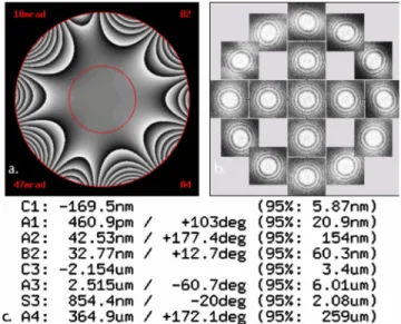
Conventional high-resolution transmission electron mi-croscopy is a very efficient method for studying the struc-tural properties of crystals at the nanoscale. However, a fine interpretation of the HREM contrast in terms of symmetry, atomic positions, etc., of the crystal requires high control of the microscope’s imaging conditions. Indeed the atomic fringes contrast can be strongly affected by the phase modi-fication of the diffracted beam coming from the uncorrected objective. To overcome these difficulties, focal series of HREM pictures are normally implemented in order to calcu-late and suppress numerically these phase perturbations, us-ing dedicated software. In the case of ACHREM, we can have a perfect control of the high-order aberrations coeffi-cients which furthermore allowed us to map the phase modi-fication added by the system with the residual aberrations 共see Fig.2兲. Focal series should be implemented to suppress
aberrations up to fifth-order astigmatism. Concerning our sample, we have found that these high-order aberrations have not a sufficient effect to modify the atomic contrast observed. The use of aberration correction for imaging nanocrystal-line samples has important advantages: 共1兲 minimized delo-calization of the image contrast, which results in the obser-vation of fine details coming from high spacial frequencies components of the diffraction pattern 共2兲 enhanced image contrast and field of view by correcting off-axial coma.32
As it is known, comparison between experimental fringes contrast and simulated ones for a define structure is very
FIG. 1. 共a兲 X-ray diffraction patterns, 共b兲 low magnification TEM image, and 共c兲 crystal size distribution of the
0.39BiScO3-0.61PbTiO3sample prepared by mechanosynthesis. FIG. 2. 共Color online兲 Example of a Zemlin tableau used to
adjust the elements of the aberration corrector taking from a par-tially amorphous region close to the nanocrystal of interest.
HUNGRÍA et al. PHYSICAL REVIEW B 81, 100102共R兲 共2010兲
problematic using conventional HREM due to the large amount of parameters which must be involved in the simu-lation. Thanks to the corrector, they could be easily included in the simulation. In order to retrieve the crystalline structure of the different twin free nanocrystals, multislice simulations of several ACHREM pictures of the mechanosynthesized perovskite were performed using different input structures based on literature 共rhombohedral R3m, tetragonal P4mm and monoclinic Cm兲.17,19,20 Table I summarizes the lattice
parameters obtained from XRD data of the mechanosynthe-sized perovskite by least-squares methods by assuming these three symmetries.
Figure2depicts an example of the Zemlin tableau31used
to determine the value of high-order aberrations inserted in the selected multislice simulations. The thickness has been determined assuming a spherical shape of each nanocrystal with a lateral size of 10–30 nm.
Images with a remarkable high contrast and resolution were obtained, leading to high quality fast Fourier transform 共FFT兲 for each individual nanocrystal, even when it was nec-essary to set the corrector and the microscope at 100 kV acceleration voltage to avoid degradation phenomena under the beam. Through the comparison between the FFT and the simulated electron-diffraction pattern of the three possible polymorphs 共tetragonal, rhombohedral, and monoclinic兲 it was possible to retrieve the crystallographic orientation 共zone axis兲 of the structures under analysis. An example is shown in Fig.3where the detail of the HREM contrast taken on a particle of about 20 nm in size can be observed. The lower inset shows the FFT belonging to the nanosized crystal from which the zone axes of each polymorph were obtained: 关001兴 in the case of the tetragonal P4mm, 关111兴 for the rhom-bohedral R3m and 关011兴 for the monoclinic Cm. As can be observed in the three upper insets belonging to the simula-tions of each structure in the above-mentioned orientasimula-tions, the coarse structure 共low spatial frequencies which contrib-utes to the HREM contrast兲 fits in the three cases. However, the main advantage of using ACHREM comes from the pos-sibility to observe the fine structure of the contrast, coming from the contribution of high spatial frequencies of the dif-fraction pattern. Indeed, in the aberration-free conditions, the transfer function of the microscope stays flat even for high spatial frequencies components without contrast inversion usually observed in uncorrected microscope.33 Then, all the
spatial frequencies are equally transmitted by the optical
sys-tem without any delocalization. As a result, due to the con-tribution of these diffracted beams, very small interference fringes, directly related to the object fine atomic structure, induced fine details as, for example, can be observed in Fig.
3 where secondary spots appeared in the middle ground of the image, which can then be directly compared with simu-lated contrast performed by the multislice algorithm using the chosen structure and the microscope conditions given by the corrector. In the case of the nanocrystal shown in Fig.3, it is possible to see that only in the case of the monoclinic symmetry the fine structure of the simulation fits perfectly. Furthermore, at 100 keV with a phase plate change less than
/4 on a tilt range of 20 mrad, it is possible to have access to aberration-free fine fringes of 0.185 nm. This value is sufficiently important to assess the reliability of the symme-try comparison results regarding small variations of the mi-croscope conditions. In order to be sure, systematic simula-tions have been carried out, and the symmetry is not affected by the artifacts such as illumination tilt which add off-axial coma and thickness in a range of +/−10 nm, what is large enough in this kind of nanocrystalline samples.
This nanocrystal is not an isolated case, and it is possible to find numerous examples with sizes between 10 and 30 nm with monoclinic symmetry and randomly oriented. Figure4
depicts the HREM micrograph of a 15 nm crystal together with the simulation of the Cm polymorph with关100兴 orien-tation. It was also possible to identify some particles with tetragonal symmetry; however the great majority of the cases present the monoclinic one what is in good agreement with
TABLE I. Structural data obtained from XRD data of the mechanosynthesized 0.39BiScO3-0.61PbTiO3 by least-squares
methods. Space group Tetragonal P4mm Rombohedral R3m Monoclinic Cm A共nm兲 0.399 0.572 0.568 B共nm兲 0.399 0.572 0.550 C共nm兲 0.407 0.698 0.404 ␣ 90 90 90  90 90 90.2 ␥ 90 120 90
FIG. 3. HREM image of a crystal of 20 nm of the 0.39BiScO3-0.61PbTiO3sample. Lower inset: FFT of the nanosized crystal. Upper insets: simulation of the three polymorphs in differ-ent zone axis 共Tet=tetragonal symmetry, zone axes 关001兴; Rhom = rhombohedra symmetry, zone axes 关111兴; and Mon=monoclinic symmetry, zone axes关011兴兲.
that obtained by Rietveld analysis of XRD data.20Therefore,
these results clearly revealed the monoclinic symmetry of twin free nanocrystals in the BiScO3-PbTiO3 solid solution,
and they are a strong evidence that monoclinic phases at MPBs are not the result of the nanotwin superlattices of the other polymorphs.
Results here reported are also relevant to another highly topical issue: the size limit for ferroelectricity. This is a co-operative phenomenon, and thus the existence of a funda-mental size limit below which ferroelectricity vanishes is expected.34As far as perovskite nanoparticles are concerned,
tetragonal distortion has been found to persist in BaTiO3and
PbTiO3 nanoparticles as small as 26 and 20 nm,
respectively.35,36 This parameter is directly associated with
the spontaneous polarization, which was indirectly measured for PbTiO3 particles of 28 nm.37Also, local ferroelectricity
has been demonstrated in BaTiO3 nanoparticles of 25 nm.38
In relation to MPB perovskites, predominant tetragonal phase has been reported for PZT nanoparticles of 30 nm by Rietveld analysis of XRD data.39Unlike in PZT, ferroelectric order does not develop in nanoscale relaxor based materials, and instead the high-temperature relaxor state is stabilized.40,41 In this work and in the case of MPB
BiScO3-PbTiO3, monoclinic symmetry is found for
nano-crystals as small as 10 nm.
In summary, our study indicates the existence of the monoclinic polymorph in the MPB region of the BiScO3-PbTiO3 solid solution, which has been revealed in
crystals with size between 10 and 30 nm. This is strong evidence in favor of the actual existence of this phase in MPBs between rhombohedral and tetragonal phases of ferro-electric perovskite solid solutions, a controversial issue within the ferroelectric materials field. This result also indi-cate that the polar phase persist in the nanoscale, which is in good agreement with our recent results where ferroelectric switching was clearly demonstrated for nanostructured ce-ramics of this system prepared by spark plasma sintering.42
These results compare favorably with those reported for re-laxor based MPB systems, for which the high-temperature relaxor state was stabilized at room temperature as a size effect, and no ferroelectric long-range order develops.
This work has been funded by MEC共Spain兲 through the Projects No. MAT2007-61884 and No. MAT2008-02003/ NAN. T.H. is indebted to the CSIC 共MICINN兲 of Spain for the “Junta de Ampliación de Estudios” contract 共Reference No. JAEDOC082兲. Technical support by I. Martínez 共ICMM兲 is also acknowledged.
*Corresponding author. FAX: ⫹34 913720623; thungria@icmm.csic.es
1W. Cao and L. E. Cross, Phys. Rev. B 47, 4825共1993兲. 2N. Setter, Piezoelectric Materials in Devices共EPFL, Lausanne,
2002兲.
3S. E. Park and T. R. Shrout, J. Appl. Phys. 82, 1804共1997兲. 4B. Noheda et al., Phys. Rev. B 61, 8687共2000兲.
5H. Fu and R. E. Cohen, Nature共London兲 403, 281 共2000兲. 6R. Guo et al., Phys. Rev. Lett. 84, 5423共2000兲.
7B. Noheda et al., Phys. Rev. Lett. 86, 3891共2001兲. 8J. M. Kiat et al., Phys. Rev. B 65, 064106共2002兲. 9B. Noheda et al., Phys. Rev. B 66, 054104共2002兲.
10A. K. Singh and D. Pandey, Phys. Rev. B 67, 064102共2003兲. 11L. A. Schmitt et al., J. Appl. Phys. 101, 074107共2007兲. 12K. A. Schonau et al., Phys. Rev. B 75, 184117共2007兲. 13Y. U. Wang, Phys. Rev. B 74, 104109共2006兲. 14Y. U. Wang, Phys. Rev. B 76, 024108共2007兲. 15Y. M. Jin et al., Phys. Rev. Lett. 91, 197601共2003兲. 16K. A. Schonau et al., Phys. Rev. B 76, 144112共2007兲. 17R. Schierholz et al., Phys. Rev. B 78, 024118共2008兲. 18G. Arlt, J. Mater. Sci. 25, 2655共1990兲.
19R. E. Eitel et al., J. Appl. Phys. 96, 2828共2004兲. 20J. Chaigneau et al., Phys. Rev. B 76, 094111共2007兲. 21B. Kim et al., J. Appl. Phys. 105, 114101共2009兲.
22K. W. Urban, Nature Mater. 8, 260共2009兲. 23K. W. Urban, Science 321, 506共2008兲.
24M. Hÿtch et al., Nature共London兲 453, 1086 共2008兲. 25C. L. Johnson et al., Nature Mater. 7, 120共2008兲. 26T. Hungría et al., Chem. Mater. 17, 6205共2005兲. 27M. Algueró et al., Chem. Mater. 19, 4982共2007兲. 28T. Hungría et al., Nanotechnology 19, 155609共2008兲.
29B. D. Cullity, Elements of X-ray Diffraction; Series in
Metal-lurgy and Materials共Addison-Wesley, Reading, MA, 1967兲.
30H. Rose, Optik共Stuttgart兲 85, 19 共1990兲. 31F. Zemlin et al., Ultramicroscopy 3, 49共1978兲.
32F. Houdellier et al., Advances in Imaging and Electron Physics
共Elsevier Inc., San Diego, CA, 2008兲, Vol. 153, Chap. 6.
33J. C. H. Spence, Experimental High-Resolution Electron
Micros-copy共Oxford University Press, New York, 1980兲.
34N. A. Spaldin, Science 304, 1606共2004兲.
35S. Chattopadhyay et al., Phys. Rev. B 52, 13177共1995兲. 36M. B. Smith et al., J. Am. Chem. Soc. 130, 6955共2008兲. 37E. K. Akdogan et al., J. Appl. Phys. 97, 084305共2005兲. 38S. Ray et al., Small 2, 1427共2006兲.
39C. Liu et al., J. Am. Chem. Soc. 123, 4344共2001兲. 40J. Carreaud et al., Phys. Rev. B 72, 174115共2005兲. 41M. Algueró et al., Small 3, 1906共2007兲.
42M. Algueró et al., Appl. Phys. Lett. 94, 012902共2009兲.
FIG. 4. HREM image of a crystal of 15 nm of the 0.39BiScO3-0.61PbTiO3perovskite. Lower inset: FFT of the nano-sized crystal. Upper inset: Simulation of the monoclinic polymorph with the关100兴 orientation.
HUNGRÍA et al. PHYSICAL REVIEW B 81, 100102共R兲 共2010兲