Gilles Edan
Enseignement du DES de neurologie
14 Mars 2014
Maladie
de Whipple
Manifestations neurologiques
Rare et méconnue
L’évolution spontanée de la maladie est longue,
marquée par des épisodes de rémission et de rechutes,
évoluant jusqu'à la mort en absence de traitement
antibiotique
Fréquence du neuro -W hipple dans le cadre de la
maladie de W hipple dans la littérature:
de 5 % jusqu
’à 90% d’après les examens post-mortem
inaugural dans 20%
Généralités
Germe dit émergent Tropheryma
whipplei, à gram positif de la
famille des actinomyces, à
multiplication intra cellulaire,
probable germe commensal du
tube digestif
Il existe un facteur génétique
probable (plus fréquent chez les
hommes et chez les porteurs du
HLA B-27).
Quelques vignettes pour illustrer
la grande diversité des tableaux
neurologiques
Etiologie
S h o u l d we s ys t e m a t i c a l l y p e r f o r m c e n t r a l n e r vo u s s ys t e m
i m a g i n g i n p a t i e n t s wi t h W h i p p l e
’s endocarditis ?
B e s n a r d S , C a d y A , F l e c h e r E , F i l y R , R e v e s t M , A r v i e u x C , D o n n i o P Y,
M i c h e l e t C ,
Ta t t e v i n P
.
Am J M e d 2 0 1 0 ; 1 2 3 ( 1 0 ) : 9 6 2
.
Homme de 58 ans , hospitalisé pour perte de
poids, 37°8, dépression
Endocardite triscuspidienne (echo)
LCR Normal
Dg BJéjunale : macrophages PAS +, PCR +
Homme de 56 ans, dyspnée,
Perte de poids, fièvre
Tr mnésiques
LCR : 17 cel. PCR –
Valve PCR +
T
R
A
I
T
E
M
E
N
T
p
a
r
c
e
f
t
r
i
a
x
o
n
e
p
d
t
2
-4
s
e
m
a
v
e
c
g
e
n
t
a
2
s
e
m
,
s
u
i
v
i
1
a
n
a
v
e
c
T
S
.
E
v
o
l
u
t
i
o
n
f
a
v
o
r
a
b
l
e
Tt: ceftriaxone pdt 2-4 sem avec genta 2 sem, suivi 1 an avec bactrim
Évolution favorable.
W h i p p l e ' s d i s e a s e : M u l t i p l e s ys t e m i c a n d n e u r o l o g i c a l
r e l a p s e s
R . O . D O M Í N G U E Z A ,
C . M Ü L L E R A , I . D A V O L O S A , P. M A C K E I T H B , E . A R I A S C , A . L .
T A R A T U T O C
N e u r o l o g i a . 2 0 1 3 ; 2 8 ( 1 ) : 5 7 - 6 4
Figure 2. (1) Biopsy sample of brain and intraventricular adhesion tissue subjected to
periodic acid Schiff test (PAS+), revealing lymphocytes and macrophages containing material consistent with TW bacteria. (2) Polymerase chain reaction (PCR) test; TW detected with 16s ribosomal RNA sequencing.
These oligonucleotides can detect fragments of 160 base pairs in the presence of bacterial DNA. The white arrow indicates the intense white band in the column indicating our patient
sample, compared to a positive control.
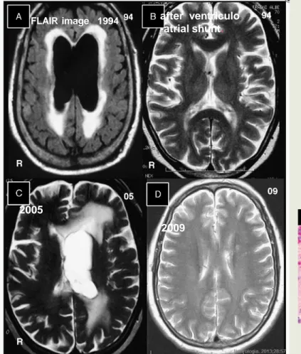
•,né 1928, Mal de Whipple 1991
•A 66 ans (1994): tr marche avec hydrocéphalie (A)
Traité par dérivation et Bactrim (B)
• À 72 ans (2005) , épisode confusionnel (C)
Traité par dérivation et Bactrim
A 77 (2010) , asymptomatique (D)
FLAIR image 1994
,
after ventriculo
-atrial shunt
.
2005
M R I o f r e c u r r e n t i s o l a t e d c e r e b r a l W h i p p l e ' s d i s e a s e
F u l d e m Y ı l d ı r ı m D ö n m e z, E s r a M e l t e m K a y a h a n U l u , C e y l a B a ş a r a n ,
M ü g e Ü n l ü k a p l a n , A r zu U y u ş u r, M a h i r Y ı l d ı r ı m , E s r a Ö zg ü l
D i a g n I n t e r R a d i o l 2 0 1 0 ; 1 6 : 11 2- 11 5 .
H 68 ans, tableau aigu avec céphalée, nausée,
Fièvre 39°, confusion, dysarthrie, paraplégie
Crises d’épilepsie suivi d’un état mutique
Biopsie cérébrale:macrophages PAS+,PCR –
Tt ceftriaxone et Bactrim
Récupération à 1 an
W h i p p l e ' s d i s e a s e : P r e s e n t a t i o n o f a n u n u s u a l c a s e
w i t h i s o l a t e d c e r e b r a l i n vo l ve m e n t
S . L O M B A R D O G A L E R A A ,
, E . R O L D Á N R O M E R O A , F. B R AV O
R O D R Í G U E Z A
N e u r o l o g i a . 2 0 1 3 ; 2 8 ( 5 ) : 3 1 7 - 3 2 4 .
Figure 1. CT without contrast (A) performed upon arrival of the patient shows a hypodense area affecting the left caudate and lentiform nuclei and the internal capsule. Note the hypodense area extending contralaterally along the edge of the corpus callosum. MR images taken 18 hours later revealed hypointensities in the same regions described by the CT scan in T1-weighted sequences and
hyperintensity in DP-weighted sequences. Administration of paramagnetic contrast (D–F) yielded distinct, heterogeneous and mainly peripheral areas of enhancement clearly showing the lesion's contralateral extension through the
anterior commissure (arrow in D).
•Femme 30 ans, pas d’ATCD ,épisode aigu avec
céphalée, paresthésies bracchio-faciales droites,
•Puis aboulie, hypersomnie, incontinence urinaire
•Biopsie N caudé droit : macrophages PAS +,PCR +
• Evolution favorable sous traitement
I s o l a t e d C N S W h i p p l e d i s e a s e w i t h n o r m a l b r a i n M R I
a n d f a l s e - p o s i t i ve C S F 1 4 - 3 - 3 p r o t e i n .
V i c t o r W S u n g , M i c h a e l J L y e r l y, K e n n e t h B F a l l o n , K h u r r a m B a s h i r
B r a i n a n d B e h a v i o r : 2 0 1 2 : 2 ( 6 ) ; 8 3 8- 8 4 3
.
Free Tropheryma whipplei organisms in neuropil (left) adjacent to a perivascular region where numerous intracellular T. whipplei organisms are present within macrophages (×1000, PAS with diastase). Macrophages having distended, pale basophilic cytoplasm in cerebral cortex (×200, hematoxylin and eosin). MRI images obtained on admission. (A) Axial FLAIR image that is unremarkable, without any significant hyperintensities. (B) Axial T1-weighted postcontrast image that is unremarkable, showing no abnormal areas of enhancement.
Homme de 42 ans
avec un tableau sur 18 mois
de démence progressive
paralysie supranucléaire,
myoclonie,
ataxie ,
à IRM et CSF normaux
14-3-3 positif !
décédé
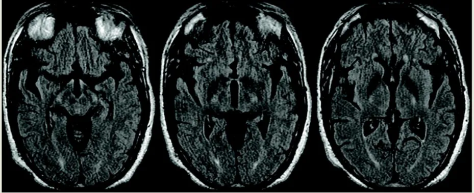
Axial noncontrast FLAIR and fast spin-echo T2 MR images as well as postgadolinium axial
and coronal T1 images demonstrate a 9-mm focus of enhancing abnormal T2 signal intensity
without mass effect within the inferomedial aspect of the hypothalamus bilateral...
Black D et al. AJNR Am J Neuroradiol 2010;31:1493-1497
©2010 by American Society of Neuroradiology
Homme de 54 ans, tr. Neurologiques, d’évolution subaigu associant hypersomnie,
perte de poids, dysarthrie, confusion et démarche ataxique
Dg : PCR + LCR et BJ + prélèvement endoscopique .
Evolution favorable sous Tt
Axial noncontrast FLAIR MR images demonstrate bilaterally symmetric T2 signal-intensity
abnormality without mass effect, involving the corticospinal tracts, brain stem, and brachium
pontis.
Black D et al. AJNR Am J Neuroradiol 2010;31:1493-1497
©2010 by American Society of Neuroradiology
Homme de 59 ans, tableau progressif sur quelques mois avec marche ataxique,
quadriparésie (fauteuil roulant) dysarthrie, dysphagie, diplopie, tr cognitifs
PCR +CSF ; BJ –
Axial noncontrast FLAIR and axial and coronal T1 postgadolinium images demonstrate
enhancing abnormally increased T2 signal intensity in the hypothalamus and anteromedial
aspect of the right temporal lobe, including the hippocampal head.
Black D et al. AJNR Am J Neuroradiol 2010;31:1493-1497
©2010 by American Society of Neuroradiology
Homme de 38 ans , ATCD whipple digestif diagnostiqué et traité à 28 ans,
Tableau aigu avec céphalée, diplopie et crises hallucinatoires olfactives
PCR + LCR, PCR – sérum
Axial noncontrast FLAIR images demonstrate symmetric high T2 signal intensity in the
hypothalami and corticospinal tracts.
©2010 by American Society of Neuroradiology
Homme 40 ans, ATCD Whipple digestif à 35 ans traité
Tableau progressif avec déclin cognitif depuis 6 mois, hypothermie, paralysie
de verticalité, myorythmies oculo masticatoires
©2010 by American Society of Neuroradiology