Angioimmunoblastic T-cell lymphoma is the most
common T-cell lymphoma in two distinct French
information data sets
Reliable information regarding the current prevalence of peripheral T-cell lymphoma (PTCL) entities is missing. Herein we report on the frequency of PTCL entities in France between 2010 and 2013. Using Lymphopath, a national lymphoma network established by the French National Cancer Agency, which covers about 70 % of all lymphomas currently diagnosed in France, we found that PTCL comprised 6.5 % of non-cutaneous lymphomas with angioimmunoblastic T-cell lymphoma (AITL) being the most frequent (739 cases; 36 %), followed by peripheral T-cell lymphoma not otherwise specified (PTCL-NOS) (550 cases, 27%). These data were verified in an independent data set from a transnational research consortium active in three European countries. In comparison to epidemiologic data reported previously, we show that AITL is by far the most common PTCL subtype. In light of the results of recent molecular findings highlighting the heterogeneity of T-cell lymphomas and the advent of targeted therapies, these data have important implications for both basic and clinical research.
Peripheral T-cell lymphomas (PTCLs) represent diverse and complex diseases, estimated to represent an overall 10-15% of all lymphomas worldwide, with the highest inci-dence rates occurring in Asia.1,2 The relative prevalence of PTCL entities delineated according to the criteria of the REAL (1994) /WHO (2001) classification systems, was eval-uated in multiple institutions in the late 1990s and early 2000s, based on retrospective cohorts of patients. In those
series, PTCL-NOS (a “by default” diagnosis for cases not ful-filling the criteria for other more specific entities) was the most frequent entity, followed by anaplastic large cell lym-phoma (ALCL) and AITL, while extranodal entities, in gen-eral, accounted for a small proportion of the cases.1,3,4
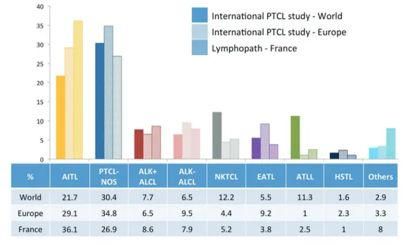
This worldwide epidemiology was most recently addressed by the International PTCL study, which reviewed more than 1,300 patients diagnosed with PTCL between 1990 and 2002 in North America, Europe and Asia.5 In this cohort, PTCL-NOS was the most common diagnosis (25,9%), followed by AITL (18,5%), representing 30.4% and 21.7% of non-cutaneous PTCL, respectively (Figure 1).5 This study also confirmed geographic variations in the dis-tribution of PTCL entities, notably demonstrating that the highest frequencies of AITL and enteropathy-associated T-cell lymphoma (EATL) are in Europe.
Here, we provide recent data obtained from a large prospective survey in France. The prevalent analysis of PTCL was derived from data collected through Lymphopath, a national network of 33 expert reference centers for hematopathology which was established by the French National Cancer Agency in 2010. Non-expert pathologists are encouraged to refer every newly diagnosed lymphoma for review to a Lymphopath center. Diagnoses provided by experts, following slide review and additional ancillary tech-niques performed in the reference center, are entered into a central database. Of the 31,401 non-cutaneous lymphomas reviewed in Lymphopath over four years (2010-2013), there were 2,046 cases of PTCLs (6.5%) which comprised: 739 AITL (36.1%), 550 PTCL-NOS (26.9%), 176 ALK+ ALCL (8.6%), 162 ALK- ALCL (7.9%), 107 extranodal NK/T-cell lymphomas, nasal-type (5.2%), 77 EATL (3.8%), 52 adult T-cell leukemia/lymphoma (2.5%) and 20 hepatosplenic T-T-cell lymphomas (1.0%). The remaining 163 cases (8%) included
haematologica 2015; 100:e361
L
ETTERS TO THE
E
DITOR
Figure 1. Relative frequency of non-cutaneous PTCL entities according to the International peripheral T-cell lymphoma (PTCL) project (worldwide (n=1314) and European (n=450) statistics)5and in the Lymphopath registry (France) (n=2046). ALCL: anaplastic large cell
lym-phoma; ALK: anaplastic lymphoma kinase; ATLL: adult T-cell leukemia/lymlym-phoma; EATL: enteropathy-associated T-cell lymlym-phoma; HSTL: hepatosplenic T-cell lymphoma; NK/T: extranodal NK/T-cell lymphoma; PTCL-NOS: peripheral T-cell lymphoma, not otherwise specified. For the International T-cell lymphoma project, the percentage of the different non-cutaneous PTCL entities have been adjusted, after exclusion of cases not confirmed to be T-cell lymphomas (for the international statistics) and exclusion of categories corresponding to cutaneous lymphomas and “other disorders”.
74 cases of NK/T-cell leukemias (15 aggressive NK-cell leukemias, 14 T-prolymphocytic leukemias and 45 large granular lymphocyte leukemias), and 89 other, or unclassi-fiable cases. Thus, in Lymphopath, AITL represents the most frequent non-cutaneous PTCL entity (36.1%) followed by PTCL-NOS (26.9%), which is distinctly less recurrent.
In comparison to the European data of the International PTCL study (Figure 1), the current findings differ principally with respect to the relative prevalence of AITL and PTCL-NOS.5 The reason for this discrepancy is unclear. The inter-national study comprised a total of 450 European cases col-lected from eight submitting centers (two centers from Spain and Italy, and one center each from France, Germany, UK, and Norway), whereas the current analysis encompass-es a much higher number of casencompass-es, encompass-estimated to be com-prised of more than 70% of all new lymphoma diagnoses in France. Yet, despite the coverage not being exhaustive, it thus far represents the most recent and largest prospectively collected series validated by expert hematopathologists, with unrestricted access to ancillary diagnostic tools.
Although a selection bias inherent to the design of the international study (retrospective collection, limited number of submitting centers, university medical centers only) can-not be excluded, the differences observed may in fact reflect geographical differences within Europe. An overrepresenta-tion of EATL in Nordic countries is well recognized, but this largely rare disease has only minimal impact on global sta-tistics. Interestingly, according to the data collected by the national Swedish Lymphoma Registry over a 10-year period (2000-2009), the 755 PTCL (cutaneous and non-leukemic) registered in Sweden comprised 34% PTCL-NOS, 29% ALCL, only 14% AITL, and 9% EATL, a distri-bution also markedly different from both the published European statistics and from our findings, thus further sug-gesting geographic variations.6
Another potentially conflicting difference is that the inter-national study cohort comprises cases diagnosed between 1990 and 2002 and reviewed with reference to the 2001 WHO classification, while the Lymphopath dataset is derived from a more recent observation period (2010-2013). In the
haematologica 2015; 100:e362
L
ETTERS TO THE
E
DITOR
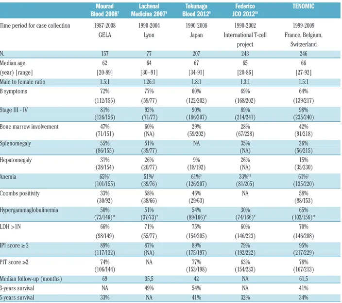
Table 1. Clinical characteristics of 246 AITL patients with follow-up data from the Tenomic collection with comparison with other published
series.
Mourad
Lachenal
Tokunaga
Federico
TENOMIC
Blood 2008
7Medicine 2007
8Blood 2012
9JCO 2012
10Time period for case collection 1987-2008 1990-2004 1990-2008 1990-2002 1999-2009
GELA Lyon Japan International T-cell France, Belgium,
project Switzerland
N. 157 77 207 243 246
Median age 62 64 67 65 66
(year) [range] [20-89] [30–91] [34-91] [20-86] [27-92]
Male to female ratio 1.5:1 1.26:1 1.8:1 1.3:1 1.5:1
B symptoms 72% 77% 60% 69% 64%
(112/155) (59/77) (122/202) (168/202) (139/217)
Stage III - IV 81% 92% 90% 89% 98%
(126/156) (71/77) (186/207) (214/241) (235/240)
Bone marrow involvement 47% 60% 29% 28% 42%
(71/151) (NA) (59/202) (67/228) (91/218) Splenomegaly 55% 51% NA 35% 26% (86/155) (39/77) (NA) (56/215) Hepatomegaly 31% 26% 9% 26% 15% (38/154) (20/77) (18/192) (NA) (35/230) Anemia 65%† 51%‡ 61%‡ 33%‡‡ 61%‡ (101/155) (39/76) (126/207) (81/205) (135/220) Coombs positivity 33% 58% 46% NA 58% (30/92) (38/66) (29/63) (88/153) Hypergammaglobulinemia 50% 51% 54% 30% 65% (73/146)* (37/73)¥ (89/166)¥ (74/166)¥ (102/156)* LDH >1N 66% 71% 75% 60% 70% (98/149) (55/77) (154/205) (146/223) (146/208) IPI score ≥ 2 89% 87% 89% 79% 95% (117/132) (NA) (175/197) (192/222) (217/229) PIT score ≥2 74% NA 77% 63% 78% (106/144) (153/198) (154/233) (167/213)
Median follow-up (months) 69 35,5 42 NA 61,5
3-years survival NA 49% 54% NA 41%
5-years survival 33% NA 41% 32% 34%
interim, it was discovered that AITL is associated to CD10 expression and derived from T follicular helper (TFH) cells. These perceptions were incorporated into the description of AITL in the 2008 WHO classification,2
and since then CD10 and novel markers associated to TFH differentiation have been validated for diagnostic use and increasingly incorpo-rated into routine practice.7
Thus, consideration must be given to whether the higher prevalence of AITL observed recently might be linked in some way to the availability of novel ancillary tools to support the diagnosis.
Confronted with the lack of a similar systematic database antedating Lymphopath in France, we used another non-overlapping set of PTCL patients to compare the distribu-tion of PTCL entities over an earlier time period. This set of patients was collected through Tenomic, a transnational research consortium on T-cell lymphomas involving several LYSA (Lymphoma Study Association) centers in France, Belgium and Switzerland. The Tenomic database (approved by the ethical committee “CPP Ile-de-France IX 08-009”) is a retrospective and prospective collection of PTCL samples with available frozen tissue, and corresponding clinical annotations (Online Supplementary Methods), including a sub-set of patients enrolled in GELA/LYSA studies. All Tenomic cases are reviewed by at least two expert hematopatholo-gists belonging to the consortium, and classified according to the 2008 WHO criteria. The Tenomic database comprised 623 non-anaplastic PTCL cases diagnosed between 1999 and 2009, reviewed after 2008. Remarkably, within the lim-its of this retrospective collection, there was an even higher ratio of AITL (n=293) to PTCL-NOS (n=171) in the Tenomic dataset (1.7:1) than in that of Lymphopath (1.35:1). According to the detailed records available for 236 AITL cases (Online Supplementary Methods and Online Supplementary Table S1), the characteristic morphological features of AITL (polymorphic cellular infiltrate (100%), arborizing vessels (98%) blast cells (94%), clear cells (69%) and sinus sign (72%)), were present in most cases. In addi-tion, an expansion of follicular dendritic cells (FDCs), regarded as a hallmark of AITL,7
was present in 93% of the cases, and EBV-positive blasts were evidenced in 77%. The percentages of cases expressing CD10 (83%) and the TFH-associated molecules PD1 (78%), CXCL13 (76%) and BCL6 (62%) in the neoplastic cells were comparative to those pre-viously reported.7
In addition, among the 246 patients who had available follow-up data, the frequencies of B symp-toms (67%), advanced stage disease (98%), IPI score>2 (94%), hypergammaglobulinemia (64%) and positive Coombs' test (41%) (Table 1), were overall similar to those recorded in other series,8-11
providing evidence that the pathological AITL diagnoses were concordant with the clin-ical and biologclin-ical features of that entity. The five-year over-all survival (OS) for the entire group, whose treatment char-acteristics are detailed in the Online Supplementary Table S2, was 34% (IC 95% [27%-40%]) (Online Supplementary Figure S1), which is also in agreement with other series.8-11
Interestingly, 43 of the 293 Tenomic AITL cases (14.6%) had initially been diagnosed as PTCL-NOS, and reclassified as AITL cases by expert review. The pathological and clini-cal features (shown in Online Supplementary Tables S3 and S4) of these reclassified cases did not differ from those of the entire cohort, suggesting that the apparent “increase” in AITL frequency may at least partly reflect the underrecogni-tion or underdiagnosis of this entity in previous years.
This report further confirms the poor long-term outcome for AITL patients, and in view of the high prevalence of this disease there are essential, but as yet unfulfilled, needs for new therapeutic options. Importantly, the response to novel agents in AITL patients may be distinctively different from that seen in other PTCL patients.12
In that respect, the recent findings of recurrent mutations in genes coding for enzymes controlling DNA methylation (TET2, IDH2 and DNMT3),13,14
and in RHOA (coding for a small GTPase),15offer rationale for the use of demethylating agents and/or specific inhibitors in AITL patients.16 As a whole, the data presented herein are highly relevant at a time when there is a shift towards the development of indi-vidualized therapies in PTCL, and should be taken into con-sideration for the design of future clinical studies.
Laurence de Leval,1*Marie Parrens,2Fabien Le Bras,3
Jean-Philippe Jais,4Virginie Fataccioli,5Antoine Martin,6
Laurence Lamant,7Richard Delarue,8 Françoise Berger,9
Flavie Arbion,10Céline Bossard,11Marie-Christine Copin,12
Danielle Canioni,13Frédéric Charlotte,14Gandhi Damaj,15Peggy
Dartigues,16Bettina Fabiani,17Albane Ledoux-Pilon,18Karine
Montagne,19Thierry Molina,13Martine Patey,20
Patrick Tas,21Michel Peoch,22Barbara Petit,23Tony Petrella,24
Jean-Michel Picquenot,25Thérèse Rousset,26
Marie-Christine Rousselet,27Isabelle Soubeyran,28
Sylvie Thiebault,29Olivier Tournilhac,30Luc Xerri,31
Christian Gisselbrecht,32Corinne Haioun,3Georges Delsol,7and
Philippe Gaulard5,33,34
L.d.L. and M.P. equally contributed to the study
1Institut de Pathologie, Centre Hospitalier Universitaire Vaudois,
Lausanne, Switzerland; 2Département de Pathologie, Hôpital Pessac,
Bordeaux, France; 3Unité Hémopathies Lymphoïdes, Hôpital Henri
Mondor, Assistance publique-Hôpitaux de Paris (AP-HP), F-94010 Créteil, France; 4Département de Statistiques, Hôpital Necker, AP-HP,
Paris, France; 5Département de Pathologie, Hôpital Henri Mondor,
APHP, Créteil, France; 6Service d’Anatomie et Cytologie Pathologiques,
Hôpital Avicenne, APHP, Bobigny, France; 7Département de Pathologie,
Hôpital Purpan, Toulouse, France; 8Service d’Hématologie, Hôpital
Necker, AP-HP, Paris, France; 9Département de Pathologie, Hôpital
Lyon-Sud, Pierre-Benite, France; 10Service d’Anatomie et Cytologie
Pathologiques, Hôpital Bretonneau, Tours, France; 11Service d’Anatomie
et Cytologie Pathologiques, Hôpital Hôtel Dieu, Nantes, France;
12Centre de Biologie-Pathologie, CHRU, Lille, France; 13Département de
Pathologie, Hôpital Necker, AP-HP, Paris, France; 14Département de
Pathologie, Hôpital La Pitié-Salpétrière, AP-HP, Paris, France; 15Service
d’Hématologie, CHU de Caen, France; 16Département de Pathologie,
Institut Gustave Roussy; université Paris Sud-11, Villejuif, France;
17Département d’Anatomie Pathologique, Hôpital Saint-Antoine,
AP-HP, Paris, France; 18Service de Pathologie, Hôtel Dieu,
Clermont-Ferrand, France; 19Service de Pathologie, Hôpitaux de Brabois,
Vandoeuvre, France; 20Laboratoire Central d’Anatomie et de Cytologie
Pathologiques, Hôpital Robert Debré, Reims, France; 21Service
d’Anatomie et Cytologie Pathologiques, CHU Pontchaillou, Rennes, France; 22Service d’Anatomie et Cytologie Pathologiques, CHRU de St
Etienne, France; 23Département de Pathologie, CHU Dupuytren,
Limoges, France; 24Service d’Anatomie et Cytologie Pathologique, CHU
Dijon, France; 25Département de Pathologie, centre Henri Becquerel,
Rouen, France; 26Laboratoire d’Anatomie et Cytologie Pathologiques,
Hôpital Gui de Chauliac, Montpellier, France; 27Département de
Pathologie cellulaire et Tissulaire, CHU, Angers, France; 28Département
de Pathologie, Institut Bergonié, Bordeaux, France; 29Service de
Pathologie, Centre Hospitalier Mulhouse, France; 30Service de Thérapie
Cellulaire et d'Hématologie clinique adulte, EA3846, Inserm CIC-501, Université d'Auvergne CHU Clermont-Ferrand Hôpital Estaing, Clermont-Ferrand, France; 31Département de Biopathologie, Institut
Paoli-Calmettes, Marseille, France; 32Service d’Hématologie, Hôpital
Saint-Louis, AP-Hp, Paris, France; and 33INSERM U955, Créteil,
France, and 34Université Paris Est, Créteil, France
Appendix: list of Lymphopath expert pathologists
G. Delsol, L. Lamant, P. Brousset, C. Laurent, CHU Purpan de Toulouse; P. Gaulard, C. Copie-Bergman, J. Moroch, N. Ortonne, Hôpital Henri Mondor, Créteil; J. Briere, V.Meignin, Hôpital Saint-Louis, Paris; T. Molina, N. Brousse, D. Canioni, S. Fraitag, Hôpital Necker, haematologica 2015; 100:e363
Paris; B. Fabiani, JF Flejou, Hôpital Saint Antoine, Paris; F. Charlotte, E. Labouyrie, Hôpital La Pitié Salpêtrière, Paris; A.Martin, Hôpital Avicenne, Bobigny; S.Prevot , C. Guettier, M. Raphaél, Hôpital A. Béclère, Clamart/ Kremlin Bicêtre; J. Bosq, P. Dartigues, Institut Gustave Roussy, Villejuif; V. Costes, T. Rousset, CHU de Montpellier ; A.de Mascarel, M. Parrens, B. Vergier, CHU de Bordeaux; I. Soubeyran, Institut Bergonié , Bordeaux; F. Berger, A. Traverse-Glehen, B. Balme, CHU de Lyon Sud; Dr C Chassagne-Clément, Dr A Decouvelaere, Dr A Fouchardière, Centre Léon Bérard, Lyon; B. Fabre, CHU de Grenoble ; M. Peoc’h , CHU de Saint Etienne ; A. Pilon, P. Dechelotte, F. Franck, CHU de Clermont-Ferrand ; L. Xerri, B. Chataille, Institut Paoli Calmettes, Marseille ; I. Peyrotte, JF Michiels, P. Hofman, O. Vire, CHU de Nice ; A. Moreau, C. Bossard, CHU de Nantes ; M-C. Rousselet, A. Croué, CHU d’Angers ; P. Tas, CHU de Rennes ; F. Arbion, A. de Muret, CHU de Tours ; I. Quintin-Roue, CHU de Brest ; MC Copin, B. Bouchindhomme, C. Delattre, CHU de Lille ; H. Sevestre, CHU d’Amiens ; J-M. Picquenot, A. François, P. Couville, Centre Henri Becquerel, Rouen; M. Galateau-Salle, CHU de Caen ; L. Martin, T. Petrella, CHU de Dijon, S. Degano-Valmary, CHU Besançon; F. Plénat, CHU de Nancy ; M. Patey, CHU de Reims; S. Thiebault, CH de Mulhouse ; M. Delage, B. Petit, CHU Limoges. Participants to the Tenomic consortium: A. Martin, Hôpital Avicenne, Bobigny, France; I. Soubeyran, P. Soubeyran, Institut Bergonié, Bordeaux, France; P. Dechelotte, A. Pilon, O.Tournilhac, Hôtel-Dieu, Clermont Ferrand, France ; P. Gaulard, C Copie-Bergman, MH Delfau, A Plonquet, F Le Bras, J Dupuis, C. Haïoun, Hôpital H Mondor, Créteil, France; T. Petrella, L. Martin, JN Bastié, O Casasnovas CHU, Dijon, France; B. Fabre, R. Gressin, D. Leroux, MC Jacob CHU, Grenoble, France; L. de Leval, B. Bisig, A. Cairoli, CHUV, Lausanne, Suisse; C. Bonnet, CHU Sart-Tilman, Liège, Belgique; M.C. Copin, B. Bouchindhomme, F. Morschhauser, CHU, Lille, France; B. Petit, A. Jaccard, Hôpital Dupuytren, Limoges, France; F. Berger, B. Coiffier, CHU Sud, Lyon, France;T. Rousset, P. Quittet, G. Cartron, Hôpital Gui de Chauliac-St Eloi, Montpellier, France; S. Thiebault, B. Drenou, Hôpital E. Muller, Mulhouse, France; K. Montagne, C. Bastien, S. Bologna, CHU de Brabois, Nancy, France; C. Bossard, S. Le Gouill, Hôtel-Dieu, Nantes, France; J. Brière, D. Sibon, C. Gisselbrecht, Hôpital St Louis, Paris, France; B. Fabiani, A Aline-Fardin, P. Coppo, Hôpital Saint-Antoine, Paris, France; F. Charlotte, J. Gabarre, Hôpital Pitié-Salpétrière, Paris, France; T. Molina, J. Bruneau, D. Canioni, V. Verkarre, E Macintyre, V. Asnafi, O. Hermine, R. Delarue, JP Jaïs, Hôpital Necker, Paris, France ; M. Parrens, JP Merlio, K. Bouabdallah, Hôpital Haut Lévêque, Bordeaux, France; S.Maugendre-Caulet, P. Tas, F. Llamas-Gutierrez T. Lamy, CHU Pontchaillou, Rennes, France; JM Picquenot, F. Jardin, C. Bastard, Centre H Becquerel, Rouen, France; M. Peoch', J. Cornillon, CHU, Saint Etienne, France; L. Lamant, G. Laurent, L. Ysebaert, Hôpital Purpan, Toulouse, France; J.Bosq, P. Dartigues, V. Ribrag, Institut G Roussy, Villejuif, France; M. Patey, A. Delmer, Hôpital R. Debré, Reims, France; JF Emile, K. Jondeau, Hôpital Ambroise Paré, Boulogne, France; MC Rousselet, M Hunault, CHU, Angers, France ; C. Badoual, Hôpital européen Georges Pompidou, Paris; C. Legendre, S. Castaigne, AL Taksin, CH Versailles, Le Chesnay, France; J. Vadrot, B Joly, A. Devidas, CH Sud francilien, Corbeil, France; G. Damaj, Service d’Hématologie, CHU Caen, France; P Dessen, G Meurice, Institut G Roussy, Villejuif, France; M Delorenzi, E Missiaglia, MP Dobay, Swiss Institut of Bioinformatics, Lausanne, Suisse; F Radvanyi, E Chapeaublanc, Institut Curie, Paris, France; S Spicuglia, CIML, Marseille, France; J Soulier, Hôpital St Louis, Paris, France ; C Thibault, IGBMC, Illkirsch, France ; V. Fataccioli, project coor-dinator, GH Henri-Mondor Albert-Chenevier, APHP, Créteil, France
Acknowledgments: the authors thank Karen Leroy from the Tumor tissue Bank (Plateforme de Ressources Biologiques, GH Henri Mondor, AP-HP, Créteil, France), the LYSA platform for their expert assistance with histological techniques and statistical analyses and reporting, Marc Maynadier, Mylène Dandoit (CHU Dijon), Nadia Lansalot-Amara (CHU Toulouse), Magali Marie-Sainte (GH Mondor, Créteil), Séverine Lepuil (CHU Pessac) for the Lymphopath database.
Funding: this study was supported in part by grants from the Institut
National du Cancer (INCa), a Programme Hospitalier de Recherche Clinique (PHRC 2007 – Code Projet : AOM 07236), the Fondation pour la Recherche Médicale (DEQ 2010/0318253), the Association pour la Recherche Thérapeutique, Génétique et Immunologique dans les Lymphomes (ARTGIL) and the Plan Cancer Research Programme (Belgium, LdL). The Lymphopath network is granted by the Institut National du Cancer (INCa)
Correspondence:
Laurence.deLeval@chuv.ch/philippe.gaulard@hmn.aphp.fr doi:10.3324/haematol.2015.126300
Key words: peripheral T-cell lymphoma, angioimmunoblastic T-cell lymphoma, prevalence; epidemiology; France.
Information on authorship, contributions, and financial & other disclo-sures was provided by the authors and is available with the online version of this article at www.haematologica.org.
References
1. Anderson JR, Armitage JO, Weisenburger DD. Epidemiology of the non-Hodgkin's lymphomas: distributions of the major subtypes differ by geographic locations. Non-Hodgkin's Lymphoma Classification Project. Ann Oncol. 1998;9(7):717-20.
2. Swerdlow S, Campo E, Harris N, et al. WHO Classification of Tumours of Haematopoietic and Lymphoid Tissues. Lyon: IARC Press, 2008.
3. Melnyk A, Rodriguez A, Pugh W, et al. Evaluation of the Revised European-American Lymphoma classificaiton confirms the clinical relevance of immunophenotype in 560 cases of aggressive non-Hodgkin's lymphoma. Blood. 1997;89: 4514-4520.
4. Lopez-Guillermo A, Cid J, Salar A, et al. Peripheral T-cell lymphomas: initial features, natural history, and prognostic factors in a series of 174 patients diagnosed according to the R.E.A.L. Classification. Ann Oncol. 1998;9(8):849-855.
5. Vose J, Armitage J, Weisenburger D. International TCLP. International peripheral T-cell and natural killer/T-cell lymphoma study: pathology findings and clinical outcomes. J Clin Oncol. 2008;26(25):4124-4130. 6. Ellin F, Landstrom J, Jerkeman M, et al. Real-world data on prognostic
factors and treatment in peripheral T-cell lymphomas: a study from the Swedish Lymphoma Registry. Blood. 2014;124(10):1570-1577. 7. Attygalle AD, Cabecadas J, Gaulard P, et al. Peripheral T-cell and
NK-cell lymphomas and their mimics; taking a step forward - report on the lymphoma workshop of the XVIth meeting of the European Association for Haematopathology and the Society for Hematopathology. Histopathology. 2014;64(2):171-199.
8. Mourad N, Mounier N, Briere J, et al. Clinical, biologic, and pathologic features in 157 patients with angioimmunoblastic T-cell lymphoma treated within the Groupe d'Etude des Lymphomes de l'Adulte (GELA) trials. Blood. 2008;111(9):4463-4470.
9. Lachenal F, Berger F, Ghesquieres H, et al. Angioimmunoblastic T-cell lymphoma: clinical and laboratory features at diagnosis in 77 patients. Medicine (Baltimore). 2007;86(5):282-292.
10. Federico M, Rudiger T, Bellei M, et al. Clinicopathologic characteris-tics of angioimmunoblastic T-cell lymphoma: analysis of the interna-tional peripheral T-cell lymphoma project. J Clin Oncol. 2013; 31(2):240-246.
11. Tokunaga T, Shimada K, Yamamoto K, et al. Retrospective analysis of prognostic factors for angioimmunoblastic T-cell lymphoma: a multi-center cooperative study in Japan. Blood. 2012;119(12):2837-2843. 12. Moskowitz AJ, Lunning MA, Horwitz SM. How I treat the peripheral
T-cell lymphomas. Blood. 2014;123(17):2636-2644.
13. Lemonnier F, Couronne L, Parrens M, et al. Recurrent TET2 mutations in peripheral T-cell lymphomas correlate with TFH-like features and adverse clinical parameters. Blood. 2012;120(7):1466-1469. 14. Cairns RA, Iqbal J, Lemonnier F, et al. IDH2 mutations are frequent in
angioimmunoblastic T-cell lymphoma. Blood. 2012;119(8):1901-1903. 15. Sakata-Yanagimoto M, Enami T, Yoshida K, et al. Somatic RHOA mutation in angioimmunoblastic T cell lymphoma. Nat Genet. 2014;46(2):171-175.
16. Cheminant M, Bruneau J, Kosmider O, et al. Efficacy of 5-Azacytidine in a TET2 mutated angioimmunoblastic T cell lymphoma. Br J Haematol. 2015;168(6):913-916.
haematologica 2015; 100:e364