AN ALGORITHM FOR INTRINSIC
GAIN AND OFFSET STABILIZATION OF
PULSE HEIGHT SPECTRA
byJiang-hong Lu
Submitted to the
DEPARTMENT OF ELECTRICAL ENGINEERING AND COMPUTER SCIENCE in partial fulfillment of the requirements for the degree of
MASTER OF SCIENCE at the
MASSACHUSETTS INSTITUTE OF TECHNOLOGY January, 1991
© Jiang-hong Lu 1991
The author hereby grants to MIT permission to reproduce and to distribute copies of this thesis document in whole or in part
Signature of Author
Department of Electrical Eggineerinnd Computer Science January 15, 1991
Certified by
-Y-ProfessSr George C. Verghese
Thesis Supervisor (Academic)
Certified by
Dr. Charles C. Watson
esis Su pep"Schumberger-Doll Research)
Accepted by
-' Professor Arthur C. Smith
Chairman, Department Committee on Graduate Students
OF TECHC .' IG
APR 0
3
1991
Table of Contents
D edication...1
Acknowledgement ... 2
A bstract...3
Chapter 1 Introduction...4
Chapter 2. Problem Background... ... ... 7
2.1 Scintillator Counter ... 7
2.2 Multiply Scattered Gamma-Ray Devices ... 9
2.3 Gain and Offset Variations ... 10
2.4 Conventional Measurements for Gain and Offset...12
Chapter 3. Spectral Analysis and Numerical Approach ... 16
3.1 The D ata... 16
3.2 Principal Components ... 18
3.3 PHS Reconstructions with Principal Components ... 21
Chapter 4 The Algorithm...25
4.1 Some Preliminaries on Gain and Offset Changes ... 26
4.2 Estimation Algorithms ... ... 28
4.2a Least Square Error Estimation.. ... 30
4.2b Maximum A Posteriori (MAP) and Maximum Likelihood (ML) Estimation...34
4.2c Connections Between the Estimation Algorithms...40
4.3 The Cost Function Behavior ... ... 41
4.3a The Mapping Subroutines... .... 43
4.3b The Truncation Effect ... 49
4.4 The Minimization Algorithm .... ... ... ...52
Chapter 5 Performance Analysis ... ... 53
5.1 Preliminaries on Performance Analysis... 53
5.1a The D ata ... 53
5.1b Noise Adding...53
5.2 Performance on scan sd133 and sd32 ... 59 5.2a Tests on Noise Free Data ... ... 60
5.2a-1 Performance Test on Gain Estimation over the Entire Scan ... 60 5.2a-2 Performance Test on Offset Estimation over the
Entire Scan ... 67
5.2a-3 Performance Test on Gain Estimation on a Single
Spectrum ... ... .. ...70 5.2a-4 Performance Test on Offset Estimation on a Single
Spectrum ... ... 71 5.2a-5 Performance Test on a Single Spectrum with two
V ariables ... 71 5.2b Noise Added at 600 ft/hr Logging Speed ... ... 72
5.2b-1 Performance Test on Gain Estimation over the Entire Scan ... 72 5.2b-2 Performance Test on Offset Estimation over the
E ntire Scan ... 75 5.2b-3 Performance Test on Gain Estimation on One
Spectrum ... .. ... ... ...76 5.2b-4 Performance Test on Offset Estimation over One
Spectrum ... ... ... 77 5.3 Performance on scan sdl33a and sd36a...78 5.3a Tests on Noise Free Data... ... .... 79
5.3a-1 Performance Test on Gain Estimation over Scan
sdl33a ... 79 5.3a-2 Performance Test on Offset Estimation over the
Entire Scan... 82 5.3b Noise Added at 1800 ft / hr Logging Speed ... 83
5.3b-1 Performance Test on Gain Estimation over the Entire Scan ... 84 5.3b-2 Performance Test on Offset Estimation over the
Entire Scan ... ... 85 Chapter 6 Conclusions ... ... 88
References ... 89
Appendix 1 Three Mapping Subroutines...90 Appendix 2 Subroutine File ... 95
Dedication
To My Parents, Grandpa and Jiqing with Love
Acknowledgement
I am very grateful to my thesis supervisor, Professor George Verghese, for his guidance and encouragement.
I am very indebted to my thesis supervisor. Dr. Charles Watson, who supervised me through out the thesis project. I admire his enthusiasm in research and his meticulous scholarship. I have learned a tremendous amount from Charles during my internship at Schlumberger-Doll Research.
I thank Tarek Habashy and Joel Groves for their guidance and support for my work at Schlumberger-Doll Research.
I thank Darwin Ellis, Joseph Chiaramonte, James Grau, David Rossi, Raymond Kocian, Dianne Hughes and Sheila Monheit for helping me with the computer work.
I thank Joseph Doucet, Felix Chen, Peggy Dow, Qing-huo Liu, Christian Straley, Yupai Hsu and all other friends at Schlumberger-Doll Research for their friendship and support.
AN ALGORITHM FOR INTRINSIC
GAIN AND OFFSET STABILIZATION OF
PULSE HEIGHT SPECTRA
byJiang-hong Lu
Submitted to the
DEPARTMENT OF ELECTRICAL ENGINEERING AND COMPUTER SCIENCE on January 15, 1991 in partial fulfillment of the requirements for the degree of
MASTER OF SCIENCE
in Electrical Engineering and Computer Science
Abstract
Multichannel pulse height spectra (PHS) are often used as property measurements of rock formations in well-logging. Gain and offset errors occur due to the many factors, and need to be corrected. The conventional method is to use stabilization source for calibration. No method of intrinsic gain and offset correction has been reported. An estimation algorithm is developed for intrinsic gain and offset stabilization.
Results from testing the estimation algorithms showed the estimated gain and offset fit the actual gain and offset well.
Thesis Supervisor: Title:
Thesis Supervisor: Title:
Dr. Charles C. Waston
Research Scientist, Schlumberger-Doll Research Dr. George C. Verghese
Chapter 1 Introduction
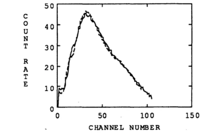
Gamma ray pulse height spectra (PHS)1 have been used for a long time in radiation detection, such as underground nuclear bomb detections in the 1940's, and environmental radiation measurements [8] in the past thirty years. In well logging, pulse height spectra obtained from multiply scattered gamma-ray devices are used for measurements of rock formation properties[3]. A typical gamma ray device consists of a radioactive source emitting gamma rays2 into the rock, and a scintillation detector recording the numbers of accumulated photons at certain energy levels from the emitted gamma rays which are scattered back from the rock. In the measuring process, a gamma ray device is put in a well against the rock formation and then moved up smoothly along the formation. Over every small distance (usually 1/4 inch) scanned by the device, a pulse height spectrum is accumulated. Some of the spectra are shown in Figure 1.1. The interaction between the photon flux of gamma rays and the rock formation is primarily determined by the rock's electron density and the Compton and photoelectric cross sections. Therefore, the pulse height spectra accumulated from a rock formation can be used to determine the rock's density and effective atomic number.
Gamma ray pulse height spectra are measured by scintillation counters. A scintillation detector records detected photons into different energy channels which forms the multichannel pulse height spectra. The photon energy and the channel number have the following approximately linear relationship:
.E=GC+O
where E is the energy deposited by the photons in the scintillation counter, G is energy gain, C is channel number, and O is energy offset. Gain and offset are parameters related to the scintillation counter. Gain is the energy variation range per channel, and offset is the background energy due to the DC voltage level applied to the scintillation detector.
1 Measured by scintillation detectors. See Section 2 for detail. 2 Gamma rays are formed by high energy photons.
During a measurement, both gain and offset could vary due to changes in many factors. The temperature of the device is often a very significant factor. Other factors can be
the electronic circuitry due to the aging of the device, the electromagnetic field in the device,
mechanical shocks and so forth. The relationship between the gain and offset variations and
the changes in the influencing factors is generally complex and unknown. Gain and offset variations in the detector system are typically more significant in well logging than in the laboratory measurements. Corrections for gain and offset are essential before the pulse
height spectra can be made useful.
Over the years, people in laboratories and well logging fields have developed many different ways for gain and offset corrections during the measuring process. The conventional method is to use a gain calibration source, but this has some unavoidable shortcomings discussed in Section 2.
In this study we will attempt to develop an alternative procedure for the gain and offset stabilization of the multiply scattered gamma ray devices used in borehole logging. From Dr. Watson's research [13], we know that even though a gamma ray pulse height spectrum's shape (see Figure 1.1) depends on the physics properties of the rock formation, every spectrum can be expressed as the sum of five or six basis spectra3. These basis
spectra are associated with each device at a certain gain and offset. If a spectrum measured by the same device cannot be reconstructed well by the basis spectra, the spectrum must have different gain and offset values than the values associated with the basis spectra. We already have a Fortran subroutine which adjusts a spectrum with different gain and offset values. To make gain and offset corrections of a spectrum, we can use the subroutine to adjust the spectrum with different combinations of gain and offset until we find the gain and offset values which give the best basis spectra reconstruction of the spectrum. This gain and offset are then our estimated values.
Our algorithm which does not rely on a gain stabilization source is an alternative to the conventional technique. Our goal is to implement the algorithm and test its performance. In Chapter 2, we discuss the background of gain and offset variation problems, the conventional method for making gain and offset corrections, and the structure of tools
used in well logging. In Chapter 3, we analyze the characteristics of the pulse height spectra and present our approach to gain and offset adjustment. In Chapter 4, we address our estimation algorithm for gain and offset. In Chapter 5, we show the performance of our estimation algorithm. Finally, in Chapter 6 we draw conclusions on the research.
1.5 1 0.5 0 50 CHANNEL NUMBER 100
Chapter 2. Problem Background
In this chapter, we examine the structure of scintillation counters and gamma-ray devices used in borehole logging and discuss the factors that might cause variations in gain
and offset. We then study the conventional method for gain and offset determination and
explain why more work needs to be done on gain and offset stabilization.
2.1 Scintillator Counter
The detection of ionizing radiation by scintillation light produced in certain materials is one of the oldest radiation detection techniques on record. The scintillation process remains one of the most useful methods available for the detection and spectroscopy of a wide assortment of radiations [6]. A typical scintillation counter is illustrated schematically in Figure 2.1. The device consists of a crystal scintillator detector, a photomultiplier tube and a circuit for photon energy resolution.
A crystal scintillator detector converts high energy photons into low energy light photons. Usually, photons emitted from a radiation process, such as gamma rays, possess very high energies. When a high energy photon enters a crystal scintillator detector, the photon interacts with an electron inside the crystal and transfers part of its energy to the electron, which excites the electron to a new energy state in which the electron is called a secondary electron. The photon then interacts with another electron, loses more energy to excite the electron. The process goes on until the photon is absorbed or escapes from the crystal. The secondary electrons release the absorbed energy by emitting photons which are usually light photons with relatively low energies. Typically, a high energy photon is converted into a few hundreds light photons.
A scintillator crystal should convert the kinetic energy of charged particles into detectable light with a high scintillation efficiency. For absolute measurements of photon energy, it is most convenient if the conversion is linear -- the light yield should be proportional to deposited energy over as wide a range as possible. The discovery of thallium activated sodium iodide in the early 1950s began the age of modem spectroscopy
of gamma rays. The most notable property of NaI(T1) is its excellent light yield, which is among the highest of any known scintillation material to secondary electrons. Its response to electrons (and any gamma rays) is close to linear over most of the significant energy range which includes the energy band from 10 keV to 700 keV that we are interested in.
Gamma NaI Photomultiplier Tube Circuitry Pulse Height
Ray (PT spectra
(PHS)
Crystal Scintillator
Figure 2.1: Schematic of a scintillation counter
The photomultiplier tube (PMT) converts light signals that typically consist of no more than a few hundred photons into a usable current pulse which is recorded by the circuits. The conversion is accomplished in three steps which are similar to the conversion in a crystal scintillator. First, a light photon from the crystal scintillator is absorbed at the photocathode raising an electron to an excited state. Then the electron escapes from the photocathode through photoelectric emission and enters the electron multiplier where the energy deposited by the electron results in the emission of more electrons producing an avalanche. Finally, the secondary electrons form a electric pulse at the collector,.which is recorded into a particular channel of a multichannel analyzer according to its amplitude (pulse height). Some of the pulse height spectra are shown in Figure 1.1.
Due to the approximately linear conversion in both the scintillator and the photomultiplier tube, the relationship between the energy deposited by a gamma ray and the recording channel number is also approximately linear [5]:
E=GC+O < 2.1 >
where E represents the energy deposited by the high energy photon (keV), G represents energy gain (keV / channel), C represents channel number and 0 represents offset (keV).
2.2 Multiply Scattered Gamma-Ray Devices
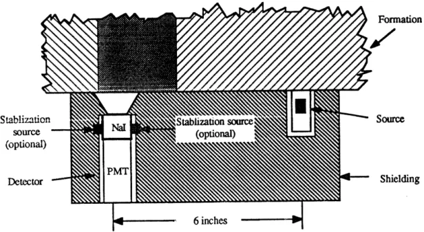
In well logging, the interaction between gamma rays and the rock formation is measured to determine the electron density and the average photoelectric cross section of the rock. Figure 2.2 shows a schematic of a typical multiply scattered gamma ray device used in laboratories, which is formed by a radioactive source emitting 662 keV gamma rays into the rock, and a scintillation counter detecting the gamma rays scattered back at energies greater than 10 keV. In between the radioactive source and the scintillator detector is shielding which prevents gamma rays from directly entering the scintillator without travelling through the formation. Near the detector are two optional stabilization sources for gain and offset calibration4. As the device moves along the formation, a pulse height spectrum is obtained for every spatial interval by integrating over a uniform time interval. Examples of typical spectra are shown in Figure 1.1. Typically, the integrating spatial interval is about a quarter of an inch, and the integrating time interval is no greater than one second. In a multichannel pulse height spectrum, every channel contains the number of photons (counts) at a certain energy level. If we divide the counts by its integrating time interval, we get the count rate (counts / second) which will be used in the rest of our analysis.
A multiply scattered gamma ray device used in well logging is moved up through a
borehole against the rock formation to make measurements. Logging speed (inch / second) describes how fast the device moves in a borehole during measurements. The device is usually similar to the one shown in Figure 2.2.
Formation
Stablization Stablization source Source
source (optioI
(optional)
Detector PM Shielding
6 inches
Figure 2.2: Schematic of a multiply scattered gamma ray device
2.3 Gain and Offset Variations
Many factors, especially temperature of a scintillator detector, count rate and mechanical shocks, can cause gain and offset to vary. Gain changes are dominated by changes in the photomultiplier tube. One very important type of gain change is the dynamic change due to large variations in the throughput of the photomultiplier, for the photomultiplier is not necessarily a linear amplifier. Count rate is determined by the throughput in the scintillator. The precise relationship between gain and count rate is usually unknown. Offset changes are primarily due to changes in the DC voltage level ("ground" potential) in various parts of the circuit. There can also a contribution from "dark current" at high temperatures (eg. 1500 C). A DC voltage applied to a photo multiplier tube or a capacitor can cause a current ("dark current") when the temperature of the device is high enough. Offset changes are usually not as serious as gain changes. In well logging, it is common to make corrections only for gain.
0 50 100 CHANNEL NUMBER
Figure 2.3: Effect of gain change on a spectrum The offset for both spectra is 0.0 keV. The gain for the original spectra (solid curve) is 3.2 keV/channel. The dashed curve is the equivalent spectrum of the original when gain is 4.0 keV/channel. 60 50 40 30 20 10 0 0 50 CHANNEL NUMBER 1:0
Figure 2.4: Effect of offset change on a spectrum The gain for both spectra is 3.2 keV/channel. The offset for the original spectra (solid curve) is 0.0 keV. The dashed curve is the equivalent spectrum of the original when offset is 3.2 keV.
For a device, the gain and offset could vary significantly during a measurement either in a laboratory or a logging well. A one hundred percent change in gain or offset is quite common in laboratories. In well logging, the variations are much greater. As we know from Equation < 2.1 >, the apparent energy deposited by a high energy photon into
the detector is determined by the gain and offset of the device and the channel number from the measurement. If the gain and offset are wrong, the energy of the detected particles would be misinterpreted. Therefore, it is imperative to recognize changes in gain and offset for any measurement. A change in gain either "squeezes" or "stretches" the spectrum and a change in offset simply shifts the spectrum. Effects of gain and offset changes on a spectrum are shown in Figure 2.3 and Figure 2.4.
2.4 Conventional Measurements for Gain and Offset
For any precise measurements, the detector system is calibrated by measuring the gain and offset of the device. A technique of using one or more stabilization sources to do the calibration is very effective and widely used in laboratory measurements and well logging.
In the laboratory, radioactive materials of known radiation energies are used as stabilization sources. For example, cesium ('3 7Cs) emits 662 keV gamma rays, and
americium ( 24 1Am) emits 60 keV gamma rays. When measuring gain and offset of the detector on a multiply scattered gamma ray device, put the stabilization sources cesium and americium in front of the scintillator detector and get a pulse height spectrum. A good measurement should result in a calibration spectrum similar to the one shown in Figure 2.5. The spectrum should have two clear peaks around 60 keV (El) and 662 keV (E2). From the
spectrum, we know the channel numbers Cl and C2 where the peaks occur. The
relationship between the energy E and channel number C is not absolutely linear. However, the non-linearity is not important for well logging applications. Empirically, two point calibration is adequate for gain and offset using Equation < 2.1 >:
El = G C1 + O < 2.2 > E2 = G C2 + O < 2.3 >
If offset is under control and only gain needs to be measured, either one of the two peak points can be used.
from
A"
WF
rron
CS
CHANNEL IAMBER
Figure 2.5: A calibration spectrum
C, C).
CHANNAEL I )AM e-R
Figure 2.6: Poorly measured calibration spectrum
Very often, poor measurement results as shown in Figure 2.6 occur due to inadequate statistics. The results can be improved by using longer integrating time intervals in order to record more photons which might make the peaks stand out more. In well logging, however, the logging speeds cannot be reduced indefinitely for both technical and economical reasons. Moreover, an increase in integrating time interval causes a reduction in
spatial resolution. Also some of the photons from the source deposit only part of their energy and escape from the detector, which causes background noise in the low energy levels of the spectrum.
Gain and offset corrections are usually straightforward in the laboratories, because conditions can be controlled so that gain and offset changes occur slowly compared to the
time duration of a measurement. In the laboratory measurements, the device can be moved
very slowly so that an increase in integrating time interval will not reduce spatial resolution. Therefore, the laboratory data have better statistics.
%
Te
t 3o
o o loo Ica
CtH AmVNE L VIKM 8
Figure 2.7: A PHS with a cesium stabilization source
In the fields of well logging, the same technique is applied for gain and offset calibration. As shown in Figure 2.2, one or more stabilization sources are placed at the scintillator detector. If cesium is used as both the device source and the stabilization source, the measured spectrum should be similar to the one shown in Figure 2.7. Gamma rays from the stabilization source enter the detector directly and form a peak in the spectrum
around 662 keV, which can be used for gain calibration. Gamma rays from the device source have to travel through the rock formation and part of their energies are lost in the formation. Therefore, the gamma rays from the device source form the lower energy part of the spectrum, approximately from 10 keV to 500 keV. Usually, proper hardware design
can reduced offset variations so that an offset calibration measurement is not essential. On the other hand, gain is much more difficult to control. Typically in well logging, only a gain correction measurement is performed.
In well logging, measurements obtained from multiply scattered gamma ray devices
by using stabilization sources cannot perform as well as in the laboratories for the
following reasons.
1. The tool is often operated at the ambient borehole temperature which varies with depth and may go up to 1500 C or higher. The wild temperature change causes both gain and offset to vary significantly and makes it hard to accomplish gain and offset calibration.
2. Measurements in well logging are made very rapidly compared to the laboratory measurements, which results in poor statistics in gain calibration peak.
3. The crystal scintillator is usually small, thus the background noise at low energy levels from the stabilization source is large, and the sensitivity of the measurement is degraded.
4. The tool is being moved up or down the borehole and is subject to mechanical shocks. Fast and dynamic changes in the measurements are hard to handle with the stabilization source technique.
We are motivated by the need to improve the stabilization of gamma ray spectral measurements under well logging conditions. In our research, we try to develop another technique for gain and offset stabilization without using the stabilization sources.
Chapter 3. Spectral Analysis and Numerical
Approach
We mentioned in the Introduction that any pulse height spectrum obtained from a multiply scattered gamma ray device (see figure 2.2) can be expressed as the sum of a set of basis spectra with the same gain and offset as the measured spectrum. The shape of a PHS can vary significantly due to the physical variations in density and effective photoelectric factor 6 Pe of the measured rock formation. But as long as the gain and offset do not change, the PHS can be well represented in terms of the same set of basis spectra.
Gain and offset changes affect the "shape" of a pulse height spectrum, which usually results in a dilation and translation of the spectrum (see figure 2.3 and 2.4). A pulse
height spectrum with gain G and offset O can be mapped into the equivalent spectrum with gain G' and offset O' which are different from G and 0. The mapping can be done with some subroutine7. We hope that the changes in gain and offset cause enough change in the shapes of PHS to be recognized.
We want to find a set of basis spectra for a multiply scattered gamma ray device with known gain and offset. Then with the help of the mapping subroutine, we can estimate the gain and offset of a PHS by mapping it from possible gains and offsets into the gain and offset of the basis spectra and then reconstructing the PHS with the basis spectra. The best reconstruction should occur when the PHS is mapped from its true values of gain
and offset.
3.1 The Data
We use lab data for algorithm development and performance testing. Our algorithm is intended to improve the gain and offset stabilization for field data obtained from borehole logging. However, field data have unknown changes in gain and offset, which make it
6 By convention, the photoelectric factor of a material is defined as P. (ze 1(z , where Z is the atomic
number.
impossible to test the performance of our algorithm. Field data also consists of a higher level of counting noise. The gain and offset for lab data are known, or the changes in gain and offset are small. Therefore, we can map the lab data into other gain and offset values or add counting noise (usually Poisson noise), and then use the original data for calibration.
Our analysis is based on the ensemble of pulse height spectra acquired by Dr. Arthur Becker in the laboratory at Schlumberger-Doll Research. The type of device shown in Figure 2.2 were used to scan a synthesized rocklike formation to obtain the PHS. In Figure 2.2, the spacing between the source and the scintillator detector is 6 inches. The three scans in the ensemble used in our analysis are all obtained from the device shown in Figure 2.2.
The laminated formation used for data acquisition was synthesized from 35 sheets
of 9 different rock or rocklike materials varying in density from 1.8 to 3.0 g/cc and in the
effective averaged photoelectric factor8, Pe, from 0.2 to 6. These materials were randomly layered transversely to the scan direction. PHS were accumulated at 0.25 inch intervals along the face of this formation in order to adequately sample possible spectral variations due to property transitions. In the analysis, 240 spectra per scan are used and each spectrum consists of 94 channels9. In Figure 2.2, the scan was made with the device immediately adjacent to the face of the formation, which is called no stand-off. Two other scans were made with the same device and formation by inserting thin sheets of different materials between the device and the face. The thin sheets were 0.21 inch thick plexiglass and 0.26 inch thick barite rubber. The three scans form the ensemble of measurements for the device. The ensemble has uniform gain and offset of 3.2 keV/Channel and 0.0 keV respectively. Table 1 shows the file names of the data used in our analysis.
The ensemble of measurements give fairly complete coverage of spatial variations in the direction of the scan, but limited variations in the directions orthogonal to the scan
8 By convention, the photoelectric factor of a material is defined as P - (z / IOa .', where Z is the atomic
number.
9 The original PHS acquired by Dr. A. Becker consists of 99 channels in every spectrum. The first five
channels in most of the spectra are zeros. Therefore, we only include the last 94 channels for each spectrum in our data files.
direction. In our study, we will use all scans in the the ensemble. In Chapter 5, we show some analysis on scan sd133, sdl33a in Table 1.
Table 1 Scan File Names
Materials inserted Scan File Name
No Stand-off sd133
plexiglass (0.21 in.) sd133a
barite rubber (0.26 in.) sdl33c
3.2 Principal Components
From the previous discussion, we want to find a set of basis spectra for a multiply scattered gamma ray device at certain gain and offset. One such set can be formed by the principal components which were discussed in Dr. Watson's paper [13].
First, we define each spectrum from an ensemble as a column vector z of dimension n.10. Then the spectral covariance matrix is formed by averaging over the scans:
Rzz ((z -(z) (z - (z))T)= U M UT < 3.1 >
where the brackets () signify an ensemble average. Because the covariance matrix Rz is real and symmetric, it can be diagonalized as indicated above. The matrix U is orthogonal in which each column, ui, is an eigenvector of Rzz. These eigenvectors are also known as the principal components of the ensemble. The plot of the first four principal components of scan sd133 are in Figure 3.1. The matrix M is diagonal and contains the eigenvalues of
Rzz, (i). These eigenvalues are also the variances of the principal components and thus all
the 9i's are non-negative. By convention, the 9i's are ordered by descending magnitude. The plot of the variances is in Figure 3.2.
0.4 0.2 0 -0.2 -0.4 100 CHANNEL NUMBER
Figure 3.1: The first four principal components from scan sd133
50
COMPONENT NUMBER
100
Figure 3.2: The logarithm variance of scan sd133
The magnitude of spectral variances decrease rather quickly over the first few components and the magnitude of the rest of the components are negligible. Let z be a spectrum which has the same variance for the principal components. If we use just the first m principal components to estimate the spectrum z , we get
m
m = aiui + (z)
i= 1 < 3.2 >
e2(Zm) = ((^ M -z)(m -z)) < 3.3 >
is minimized at:
ai = UT (z- (z). < 3.4 >
where ai is obtained by projecting vector z on principal component ui. The minimum square error is then:
n es =(c (zsn -z) z -z))= I 9i
i = m+1
The mean square error is negligible when m is between 4 and 6 because the first few principal components contain almost all the variance of z. Figure 3.3 and Figure 3.4 show two spectra from scan sd133 and their reconstructions with four principal components computed from scan sd133. From the two plots, we see that the reconstruction errors are very small even though the two spectra were measured from formations with different densities.
The above phenomena can be interpreted as follows. The first few principal components contain the independent modes of variation in PHS while the rest of the principal components contain mostly noise. Therefore, we can use the first few principal components to form the set of basis spectra.
120 C O 100 U N 80 T R 60 A T 40 E 20 0 0 50 100 CHANNEL NUMBER
Figure 3.3: The first spectrum from scan sd133 (solid curve) and its reconstruction (dashed curve) from the first four principal components computed from ensemble sd133.
60 C 0 50 U N 40 T R 30 A T 20 E 10 0 0 50 100 CHANNEL NUMBER
Figure 3.4: The 60th spectrum from scan sd133 (solid curve) and its reconstruction (dashed curve) from the first four principal components computed from ensemble sd133.
3.3 PHS Reconstructions with Principal Components
From the previous section, a PHS can be well reconstructed with only a few principal components. In fact, if all n independent principal components are used to reconstruct a n-dimensional PHS, the reconstruction should be perfect under all possible circumstances. However, the condition for having good reconstructions with only a few principal components is that the gain and offset for the PHS and the principal components must be the same. As long as this condition holds, we can get good PHS reconstructions from only four or six principal components no matter how the shapes of the PHS might change due to the physical property variation of the rock formation. Figure 3.5 and Figure 3.6 show two spectra from scan sd133a and their reconstructions with the first four principal components from scan sd133. Although the spectra in scan sd133a are not included in the computation of the principal components, good reconstructions are obtained. For all the spectra and principal components, the gain is 3.2 keV / channel and the offset is 0.0 keV.
Once the gain and offset of a PHS do not match with the gain and offset of the principal components, the PHS cannot be well reconstructed with only a few principal components. In Figure 3.7, the solid spectrum is the obtained by mapping the first spectrum in scan sd133 into a new gain (2.9 keV / channel) and offset (-3.2 keV). The reconstruction of the mapped spectrum with the first four principal components from sd133 is the dashed curve. Compared with Figure 3.3, the reconstruction in Figure 3.7 does not match the original well. Another example of bad reconstruction is shown in Figure 3.8 where the solid curve is obtained by mapping the sixtieth spectrum in sd133 into a 2.9 keV/channel gain and -3.2 keV offset. Compared with Figure 3.4, the reconstruction in Figure 3.8 does not match the original well.
50
CHANNEL NUMBER
100
Figure 3.5: The 70th spectrum from scan sdl33a (solid curve) and its reconstruction (dashed curve) from the first four principal components computed from ensemble sd133.
120 100 80 60 40 20 50 CHANNEL NUMBER 100
Figure 3.6: The 150th spectrum from scan sd133a (solid curve) and its reconstruction (dashed curve) from the first four principal components computed from ensemble sd133.
100
50 100 150
CHANNEL NUMBER
Figure 3.7 Solid spectrum: The first spectrum in sd133 mapped into gain = 2.9 keV/channel, offset = -3.2 keV; Dashed spectrum: reconstruction from the first four principal components computed from sd133.
0 50 100 150
CHANNEL NUMBER
Figure 3.8 Solid spectrum: The sixtieth spectrum in sd133 mapped into gain = 2.9 keV/channel, offset = -3.2 keV; Dashed spectrum: reconstruction from the first four principal components computed from sd133.
Based on the PHS reconstruction behavior, we can determine whether or not the gain and offset of a PHS matches with the gain and offset of a set of principal components:
If a PHS can be well reconstructed with a few (four to six)
principal components, then the PHS and the set of principal components have the same gain and offset.
If a PHS cannot be well reconstructed with a few principal components, then the PHS and the set of principal components have different gain and offset.
Further, we can estimate the gain and offset of a PHS based on the reconstruction behavior. Given a pair of gain and offset (g, o), a set of principal components for the PHS can be computed. Given a PHS with unknown gain and offset, reconstructions can be computed with different set of principal components with known gain and offset (g, o) until a good enough reconstruction is found. The gain and offset for the set of principal components which give the good reconstruction are then the estimated gain and offset for the PHS.
In implementing the estimation, instead of computing the principal components for different pairs of gain and offset (g, o), we keep the principal components with standard gain and offset (go oo) unchanged. Because all the PHS used in our analysis are acquired
with gain 3.2 keV / channel and offset 0.0 keV, we use this gain and offset as our standard gain and offset. Given a PHS with unknown gain and offset, we mapped the
PHS from a pair of guessed gain and offset (g, o) into the standard gain and offset, and
compute the reconstruction of the mapped PHS with the principal components. If our guessed gain and offset (g, o) match the gain and offset for the PHS, we should get a good reconstruction.
Chapter 4 The Algorithm
The problem we are trying to solve in our research is: given a pulse height spectrum measured by a gamma ray device, determine the gain and offset for the PHS without using any stabilization radiation sources.
From the discussion in Chapter 3, a PHS can be well reconstructed with a few (four to six) principal components if and only if the gain and offset for the PHS and the set of principal components match. In this chapter, we develop an estimation algorithm for gain and offset based on the PHS reconstruction behavior. Our algorithm includes the following steps:
Step 1. Define a set of four principal components with the standard gain and offset (go oo)1 1.
Step 2. For every pair of gain and offset (g, o) in a given range, map the given PHS from (g, o) into the standard gain and offset (go, o).
Step 3. Reconstruct the mapped PHS with the set of principal components defined in Step 1.
Step 4. Define cost functions which determine how good a reconstruction is. A cost function is minimized at the best reconstruction.
Step 5. Search for the pair of gain and offset at which the principal components give the best reconstruction. The resulted pair of gain and offset is our estimation for the given PHS.
First, we discuss some of the preliminaries on PHS gain and offset changes. Then we introduce our estimation algorithm, the problems encountered in implementation and how we solve the problems.
11 We define the standard gain and offset as: go = 3.2 keV / channel; o00 = 0.0 keV. All PHS in Table 1 are acquired with the standard gain and offset.
4.1 Some Preliminaries on Gain and Offset Changes
A pulse height spectra measured by a scintillator detector contains multichannel representing different energy range. Let f(E) be the number of photons with energy E detected per unit energy per second. Then f(E)dE is the number of photons with energy between E and E+dE detected within a second, the pulse height spectrum in energy space. A pulse height spectrum is registered in "channel space" by means of a coordinate transformation:
E=gx+o < 4.1-1 >
where x is the channel number, g is gain (keV/channel) and o is offset (keV). Take the derivative of equation < 4.1-1 >,
dE= g dx < 4.1-2 >
Therefore, we get the following:
f(E) dE = g f(g x + o) dx - z(x) dx
where
z(x) M g f(g x + o)
z(x) is the pulse height spectrum in channel space with gain and offset spectra in our data ensemble are PHS in channel space.
(g, o). All the
Now suppose that the pulse height spectrum is acquired with a different pair of gain and offset (g', o'), the following is true:
f(E) dE = g f(g x + o) dx = g' f(g x + o') dx' E= gx +o= g x +o' < 4.1-5 > < 4.1-6 > < 4.1-3 > < 4.1-4 > and
must also be true. Let
h(x') = g' f(g' x' + o') < 4.1-7 >
h(x') is then the pulse height spectrum with gain and offset (g', o'). With equation <4.1-4> and <4.1-7>, we get the following from equation <4.1-5>:
z(x) dx = h(x) dx' < 4.1-8 >
From equation <4.1-6>, we get
x=g'x'+o -o = gj '+'-o 9 9 a and dx= g dx' < 4.1-9 > < 4.1-10 >
Substitute x and dx in equation < 4.1-8 > with equation < 4.1-9 > and < 4.1-10 >, we get the transformation from a PHS z(x) with the pair of gain and offset (g, o) to a equivalent PHS h(x') with another pair of gain and offset (g', o'):
h(x') =1 z(1- x+ 0'- ) =g- z(x)
9 9 9 9 < 4.1-11 >
This is the transformation used in the spectrum mapping program mentioned earlier.
The transformation in < 4.1-11 > is reversible, that is, if we transform h(x') from gain and offset (g', o') to (g, o), we get back the PHS z(x). Let z'(x) be the PHS with gain and offset (g, o) transformed from h(x') with (g', o'), then
z'(x) - g h (x + -- o )
g g g <4.1-12 >
gx+o-o = g +O-___o
x x +
equation < 4.1-12 > becomes
z'(x) =- h( [ x+' I' o )= h(x) = z(x) <4.1-12
g a g < 4.1-12 >
Therefore, we proved z'(x) = z(x). Note that z'(x) = z(x) is true only when z(x) is defined at all real values of x for x e [ Xm, x- .
4.2
Estimation Algorithms
Three estimation algorithms for estimating the gain and offset of a pulse height spectrum are discussed in this section. The three algorithms are: least square error (LSE) estimation, maximum likelihood (ML) estimation and maximum a posteriori (MAP) estimation. We used LSE and ML estimations in our study. Before discussing the estimation algorithms, we define the estimation problem in a mathematical manner.
Let h(x) be an observed pulse height spectrum (number of photons per channel per second) where x is the channel number. The gain and offset (g, o) for h(x) are unknown. Usually, PHS obtained from well logging contains noise most of which are due to the counting process. Let z(x, g, o) be the noise free part of h(x), then
h(x) = z(x, g, o) + noise(x) < 4.2-1 >
Let z(x) be the equivalent PHS of z(x, g, o) with standard gain and offset (go. oo). From the transformation in equation < 4.1-11 >, we have
z(x, g, o) = - z( x + x+o- oo)
go go go < 4.2-2 >
As discussed in the previous chapter, z(x) can be fully constructed as:
z(x) = aiui(x)+((x))4
where uj's are the principal components with standard gain and offset (go. oo), a,'s are the
corresponding reconstruction coefficients, W(x)) is the average PHS with (go, oo), and m is between four to six. If we take the transformation in equation <4.1-11> on both sides of <4.2-3>, we get:
z(x. g, o) = au i(x. g, o)+(x g, o)
i-l < 4.2-4 >
where u i (x. g. o) is the transformed principal component,
ui (x, g, o) = ug +o - o
go go go < 4.2-5 >
and { (x, g, o)) is the average PHS with gain and offset (g, o),
( (x g. o)) = (z( +- + - ))
< 4.2-6 >
Equation < 4.2-4 > can be written as
z(x, g, o) = U(x, g, o) a + (z (x, g, o)) < 4.2-7 >
where U(x, g, o) is a matrix that contains m principal components,
U(x, g, o) = [u (x, g, o) 2(x, g, 0) ... Um(X, g, o0)] < 4.2-8 >
and a is a vector that contains the reconstruction coefficients,
a=[a1 a2 ... am]T < 4.2-9 >
The PHS h(x) can then be written as:
h(x) = U(x, g, o) a + ( z(x, g, o)) + noise(x) < 4.2-10 >
Now the estimation problem becomes: given the observed pulse height spectrum h(x) , determine the gain and offset (g, o) and the reconstruction coefficient vector a.
In the three estimation algorithms, we show how the reconstruction coefficient
vector a is computed, and how the cost functions are defined as the criterion for gain and
offset estimation.
4.2a Least Square Error Estimation
For a given pulse height spectrum h(x) with unknown gain and offset (g', o'), the estimated PHS with m principal components is:
g(x, o) = U(x, g, o) a + ( z(x, g, o)) < 4.2a-1 >
Without taking the noise part of h(x) into account, the reconstruction coefficient vector a is computed so that s2 () ,the square error between h(x) and z(x, g, o) defined below is
minimized.
82 (= (- h)T -h)
< 4.2a-2 >
From Chapter 3, a is then the projection coefficients for projecting h(x) onto the principal components.
a = U(x, g, o)T(h(x)-(z(x, g, o))) < 4.2a-3 >
If (g, o), the gain and offset for the principal components and the mean spectrum matches with (g', o'), then the estimated PHS z(x. g, o) should match with the given PHS h(x) well. The mean square error (z) is a good criterion for judging how well the
estimated PHS z(x, g. o) fits the original PHS h(x). However, 82 6 weights every channel in the PHS equally while the counts in different channels, and therefore, the noise level could vary significantly. In the weighted least square error estimation, we modified equation <4.2a-2> and define the cost function as the weighted mean square error:
E (g. o)= 1 (z(x, g. o) - h(x) )
N1., z(x, g, o) < 4.2a-4 >
where 9(x, g, o) is the weighting function. We will see in section 4.2b that ^(x. 0 o) is
approximately the noise variance of h(x) in each channel x. The reconstruction coefficient vector defined by equation <4.2a-3> does not necessarily minimize the above cost function E2 (g, o). Further investigations need to be done on the computation of a. In Section 4.2b, we introduce another way of computing a.
The pair of gain and offset (g, o) that minimizes e2 (g, o) is our estimated gain and offset for (g', o'). For computing the cost function < 4.2a-4 > at each pair of gain and offset (g, o), the transformed principal component matrix U(x, g. o) and the transformed average PHS (z(x g. o)) are required. However, instead of computing a and E2 by transforming the principal components and average spectrum from the standard gain and offset (go, oo) to (g, o), it is numerically simpler to leave U(x) and (z(x)) at (go. oo) and transform h(x) from (g, o) to (go. oo). Because of the discrete nature of the channels in the spectra, and the need to interpolate when performing the transformation (see Section 4.3), the a and £ computed at (g, o) may not be numerically identical to the ones computed at (go, 00), but we believe that these effects will not be very important for our estimation algorithms, Thus, let ho(x) be the PHS transformed from h(x) with (g, o) into ho(x) with the standard gain and offset (go, oo)
ho(x) = go h (gox + oo
-g s g < 4.2a-5 >
Now transform both sides of equation <4.2a-3 > from gain and offset (g, o) into the standard gain and offset (go, oo). On the left side, a should remain approximately the same after the transformation. On the right side, U(x, g, o) becomes U(x) which is the matrix contains the principal components u'S with the standard gain and offset, h(x) becomes ho(x), and (z(x, g, o)) becomes (z(x)). Therefore, equation < 4.2a-3 > becomes the following after the transformation:
Let ^ (x) be the transformation of ^(x, g, o)
(x) -= ^(g x +o00-0o, g,o)
Take the transformation on both sides of equation < 4.2a-1 >, we get
z (x) = U(x) a + (z(x)) < 4.2a-7 >
We can then keep the principal component matrix U(x, g, o) and average spectrum (z(x, g, o)) with (go, oo00) unchanged and compute the cost function with the transformed PHS:
N 2
S(g~~1 (^(x) - ho(x))
N,-1 z(x) < 4.2a-8 >
In the implementation of the least square error estimation as well as the estimation algorithms in the next sections, only the given PHS is transformed.
For a given PHS h(x), the estimation for gain and offset with the least square error estimation algorithm is done in the following steps:
1. Take a guess for the gain and offset, (g, o).
2. Map h(x) from (g, o) into (go, oo) to get the spectrum ho(x):
ho (x) = go h ( x + o -o )
3. Reconstruct ho(x) with the first m (m is four) principal components to get z (x):
z (x) = U(x) a +(z (x))
where a is the reconstruction coefficient vector:
4. Compute the cost function, that is the weighted mean square error:
N (^(x) - ho(x) )2
N .1 z(x)
where N is the number of channels in z(x) and (x).
5. Find (g, o) that minimizes the above cost function. This step will be discussed later in this chapter.
Figure 4.1 shows an example for implementing the above five steps. Figure 4.2 shows a plot of the cost function vs gain which has a minimum around the true gain.
50
CHANNEL NUMBER
100
Figure 4.1: Estimation for gain and offset
h(x): the given PHS with unknown gain and offset.
he (x): the transformation of h(x) from (g, o) into (go, o). 7(x): the principal components estimation of ho (x).
5 4 3 2 1 2.8 3 3.2 3.4 3.6 gnl
Figure 4.2: The cost function (< 4.2a-8 >) vs. gain for a spectrum in ensemble sd133.
4.2b Maximum A Posteriori (MAP) and Maximum Likelihood (ML)
Estimation
The weighted least squares algorithm proposed in the proceeding section is closely related to maximum likelihood (ML) estimation of the gain and offset. ML estimation, in turn, can be generalized to maximum a posteriori (MAP) estimation if prior information on the probability distribution of the gain, offset and the coefficient vector a for principal component reconstruction are available.
Let h(x) be the observed PHS containing Poisson noise, and x is channel number. The gain and offset (g, o) for h(x) is unknown. The MAP estimation problem becomes: given h(x), determine g, o and a such that the conditional probability p(g, o,alh(x)) is maximized.
From Bayes' theorem:
p ( g, o, a I h (x)) = p(h I g, o, a) p(g, o, a)
p(h) < 4.2b-1 >
Taking the logarithm of both sides, the problem is equivalent to maximizing:
In[ p(g, o, a Ih (x))] = In[ p(h I g, o, a)] + In [ p(g, o, a)] - In p(h)] < 4.2b-2 >
The last term on the right side does not depend on g, o or a, therefore it can be dropped. Further, we can assume that g, o and a are independent, and the joint probability for g, o, and a is the product of the unconditional individual probabilities:
p(g, o, a) = p(g) p(o) p(a). <4.2b-3 >
and
In [ P( g, o, a) = In [ P(g)] + n[ P(o)] +In[ P(a)]. <4.2b-4 >
The MAP estimation problem becomes: finding g, o and a which maximize
-C2 (g, o, a) = In [ P(h I g, o, a)] + In[ P(g)] + InC P(o)] + In[ P(a)]
which is equivalent to find g, o, and a which minimize the following:
ElPs (g, o, a) =- { In [ P(h I g, o, a)] + In[ P(g)] + In[ P(o)] + In[ P(a)] )
< 4.2b-5 >
< 4.2b-6 >
Equation < 4.2b-6 > is then the cost function for maximum a posteriori (MAP) estimation.
The first term on the right side of equation < 4.2b-5 >, In [ P(h g, o, a)], is the
logarithm of the likelihood function and the remaining three terms are prior distributions. If we maximize only the likelihood function, we are making a maximum likelihood (ML) estimation. Therefore, the cost function for maximum likelihood estimation is:
et.k (g, o, a) = - In [P(h I g, o, a)] < 4.2b-7 >
Now the cost functions for MAP and ML estimation are defined. Next, we consider different terms in the cost functions separately.
For the likelihood function, let's examine the spectrum h(x). Usually, PHS obtained from the well logging field contain noise. Recall equation < 4.2-1 >,
h(x) = z(x, g, o) + noise(x)
where z(x, g, o) is the noise free part of h(x) and noise(x) is the noise part which is zero mean and uncorrelated between channels. Most of the noise comes from the counting process, and therefore we can use the Poisson distribution to model the noise. If we average h(x) over the noise, we get:
< 4.2b-8 >
Therefore, h(x) is distributed as a Poisson random variable with mean z(x, g, o):
p(h I g, o, a) = z e-. h!
In[p(h I g o, a)] = [h(x) In[ z(x,g. o) -z(x.g,o) -In[h(x)] ]
.1s
< 4.2b-9 >
< 4.2b-10 >
Recall equation < 4.2-7 >,
z(x, g, o) = U(x, g, o) a + ( (x. g, o)) < 4.2b-11 >
the reconstruction coefficient vector a dependence of z(x, g, o) separates from the gain and offset (g, o) dependence, and z(x, g, o) is linear in a. For any pair of gain and offset (g, o), let us determine the optimal reconstruction coefficient vector a that maximizes the likelihood function In [ p(h I g, o, a) ]
8 In[p(h I g, o, a)] N h(x) 8 z(x,g,o) 8 z(x,g,o)
aai
X.-
z(x, g, o)ai
a
ai
< 4.2b-12 > 1 5 i< m, ai is a component of a.and
From equation < 4.2b-11 >,
a
z(x,g,o) = u i(x, g, o)aai
Equation < 4.2b-12 > becomes: " [h(x) ui (x. g, o)-ui(x, g, o) z(x, g, o) ] 0 x.1 z(x. g, o)We can approximate the denominator z(x, g, o) with h(x), and use the definition in equation
< 4.2b-11 > to get:
[ h(x)-(z(x, g, o) ui (x, g. o)-ui (x, g o) U(x,g,o)a
]
0fL h(x)
Let H be a diagonal matrix whose diagonal elements are h(x):
H h0 h(2)
Equation < 4.2b-13 > can now be written as a matrix equation:
UT(x, g, o) H'1 [h(x)-(z(x, g, o) ) - U(x, g, o) H-1 U(x, g, o) a = 0
which gives:
a( g, o) = [ UT(x, g. o) H' U(x, g, o) ]' U(x. g, o) H' [ h(x) - (z(x, g, o) )]
< 4.2b-13 >
< 4.2b-14 >
a(g, o) is the optimal estimate of a for any given gain and offset (g, o). Using a(g, o), we can estimate the noise free PHS z(x, g, o) as:
2(x, g, o) = U(x,g,o)a(g,o)+(z(x, g, o) )
< 4.2b-15 >
In[p(h I go, a)] = h(x) In [(x, g, o-(x. go - In [ h(x)!]]
a.- < 4.2b-16 >
In implementing the estimation algorithm, we compute the likelihood function differently than equation < 4.2b-16 >. As discussed in the least square estimation, instead of computing the cost functions with the principal components and average spectrum transformed into with different gain and offset (g, o), we compute the cost function with the principal components U(x) and average spectrum (z(x)) at the standard gain and offset. And we use ho(x) , the transformation of h(x) from (g, o) into the standard gain and offset:
ho(x) =g h(gx +o0-o )
9 9 9 < 4.2b-17 >
As a result, the above equations are rewritten correspondingly as
ho(x) = z(x) + noiseo(x)
z(x)= U(x) a + (z (x))
H= 0 )ho(2)
Then the optimal a is
a(g, o) = [UT(x) I1 U(x) l UT(x) I'[ ho(x) - z(x) )] < 4.2b-18 >
and the estimated noise free PHS with the standard gain and offset is
^(x) = U(x) a(g, o) +(z(x)) < 4.2b-19 >
We expect that there may be numerical effects associated with applying the Poisson model to ho(x) instead of h(x), but these have not been investigated.
H' =constant 0 1
If we replace H with H', equation <4.2b-18> becomes the same as <4.2a-6>. We call the
a computed from <4.2a-6> the projection coefficient vector, and a(g, o) computed from
<4.2b-18> the optimal coefficient vector. We expect a(g, o) to give better estimation results than a. However, in Chapter 5 the estimation performance tests show that a(g, o) is not any better than a. The reason is unknown.
With z(x) defined in equation <4.2b-19>, the likelihood function is approximately (see the discussion following equation <4.2a-4>):
N
In[ p(ho I g, o, a) = [o(x) In[(x)- ^(x) -In [ho(x)]]
.x < 4.2b-20 >
The cost function (< 4.2b-7 >) for maximum likelihood estimation becomes
dE (g, O)= - In[ p(ho I g, o, a) ] = - 7 [ ho(x) In[x) -(x) -In [ ho(x)! ]
< 4.2b-21 >
For maximum likelihood estimation, in some of our tests the projection coefficients (< 4.2a-6 >) will be used to replace a(g, o):
a = U(x ( ho(x) -(z(x))) < 4.2b-22 >
The cost function (< 4.2b-6 >) for maximum a posteriori estimation of gain and offset can be formed with the likelihood function (< 4.2b-21 >) and the apriori information
N
EtP (go)= - [ ho(x) In [(x)] -(x) - In[ ho(x)] + In [ P(g)] + In ( P(o)]
x-l
In our study, we only implemented the maximum likelihood estimation.
For a given PHS h(x), the estimation for gain and offset with ML estimation algorithm is done in the following steps:
1. Take a guess for the gain and offset, (g, o).
2. Compute a or a(g. o) and z (x)
3. Compute the cost function for ML estimation1 2.
4. Find (g, o) that minimizes the cost function. This step will be discussed later in this chapter.
4.2c Connections Between the Estimation Algorithms
The procedures for the three estimation algorithms discussed above are the same. The difference among the estimation algorithms is the definition of the cost functions and the computation of reconstruction coefficient vector a. The reconstruction coefficient vector can be either the projection coefficient vector as in equation < 4.2b-22 >, or the optimal coefficient vector as in equation < 4.2b-18 > that maximizes the likelihood function.
Once a is defined, we can get the reconstructed PHS with the principal components and then computed the cost functions. The cost function for the weighted least square estimation is defined in equation < 4.2a-8 >:
CZ (g. 0)= (Z(X) ho(x2
N1 z(x) < 4.2c-1 >
The cost function for maximum likelihood estimation is defined in equation <4.2b-25>:
L. (g =- ln[p(hol g, o, , =- [ o(x~ h) in (x)] (x) - n [ ho(x)! ] X-i
12 The cost function for maximum likelihood estimation is defined in equation <4.2b-21>. In implementing the algorithm, we used the simplified cost function in equation <4.2c-2> in Section 4.2c.
When ho(x) >> and I( x) - x)<< I (x), as is generally the case for our PHS, the Poisson distribution approaches a normal distribution and the likelihood function can be approximated as:
_ [o(x) in [-(x) -2(x) ln C[ ho(x)!] , [(x)--x - - n [22 (x)]
.u -. 2z(x) 2
The cost function for maximum likelihood estimation can the be written as:
.
(
o)
=
(g,
o) +
in
[
2[,(X)2 2 .Ku < 4.2c-2 >
and the cost function for maximum a posteriori estimation (equation < 4.2b-23 >) can be written as:
E2A(g, o) = Ma (g, o) + In[ P(g)] + In[ P(o)] < 4.2c-3 >
4.3 The Cost Function Behavior
A cost function should be smooth and have a clear global minimum around the true
gain and offset. However, in the implementation, the cost functions show some strange behaviors as shown in Figure 4.3 and 4.4. In Figure 4.3, the cost function has glitches, but there is a global minimum. In Figure 4.4, it is not clear if the cost function has a global minimum or not. The cost function decreases quickly as gain reaches small values. These problems are possibly caused by the mapping subroutine that implements the PHS transformation from one pair of gain and offset to another, or by truncation effects. These effects will be discussed in this section.
0.2
15 r
0.05
-10 -5 0 5 10
of
Figure 4.3 Cost function vs offset.
The first spectrum in sd133 is mapped into gain = 3.2 keV/channel, offset = 1.6 keV. The cost function for least square error estimation for offset is computed at different offset. The minimum occurs at ofl = 1.2 keV
200 150 / 100 50 2 4 6 8 gain
Figure 4.4 Cost function vs. gain
The fiftieth spectrum in sd133 is mapped into gain = 4.0 keV/channel, offset = 0.0 keV. The cost function for least
square error estimation for gain is computed at different gain. The minimum occurs at gain = 4.1 keV/channel.
4.3a The Mapping Subroutines
In implementing the estimation algorithms, the mapping subroutines transform a
PHS from one pair of gain and offset to another with the transformation in equation <4.1-11>:
h(x ) = z(F x + o - o )= z(x)
g g g g <4.3a-1>
Where z(x) is the spectrum with gain and offset (g, o) being transformed to h(x') with gain and offset (g', o'). All PHS contain only the channels where the channel numbers are integers. In the above transformation, for some or all integer x', the corresponding x:
X = I +O' O
g g
is a non-integer number. In order to compute h(x) from z(x), we must interpolate z(x) when x is not an integer.
There is a Fortran subroutine13 that uses fourth order polynomial interpolation for z(x):
z(x) = ao+alx+a2x2+asx'+a4x' < 4.3a-2 >
For each channel x' in h(x'), the corresponding channel in z(x) is x. Let xi be the integer part of (x + 0.5), the five points in z(x): z(xi-2), z(xi-1), z(xi), z(xi+l) and z(xi+2) are used to determine the coefficients ai's in the above fourth order polynomial. With the fourth order polynomial interpolation, z(x) is not a continuous function of gain and offset (g', o'). For a fixed x', the change in g' or o' causes x to change. As long as integer xi remains the same, the same five points in z are used to determine the polynomial coefficients, and z(x) is computed from the same polynomial. However, when the change in g' or o' is just enough to cause xi to jump to another integer, the polynomial coefficients are determined
13 The Fortran subroutine was written by W. Frawley, J. Grau and A. Becker at Schlumberger-Doll Research, Ridgefield, CT.
by five different points in z. This discontinuity in the transformation causes the glitches in
the cost function shown in Figure 4.3.
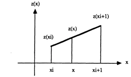
We can also use linear interpolation as shown in Figure 4.5. Let xi be the integer part of x, then x is in between xi and xi+1 and z(x) is interpolated as the point on the line that connects z(xi) and z(xi+1):
z(x) = z(xi) + (x- xi)[z(xi + 1 ) -z(xi)] <4.3a-3>
z(x)
z(xi+l) z(x)
xi+l
Figure 4.5 Two point linear interpolation
Figure 4.6 shows the cost function for least square error estimation computed with the linear interpolation.