HAL Id: hal-01393443
https://hal.archives-ouvertes.fr/hal-01393443
Submitted on 30 Oct 2020
HAL is a multi-disciplinary open access
archive for the deposit and dissemination of
sci-entific research documents, whether they are
pub-lished or not. The documents may come from
teaching and research institutions in France or
abroad, or from public or private research centers.
L’archive ouverte pluridisciplinaire HAL, est
destinée au dépôt et à la diffusion de documents
scientifiques de niveau recherche, publiés ou non,
émanant des établissements d’enseignement et de
recherche français ou étrangers, des laboratoires
publics ou privés.
non-diazotrophic planktonic communities: a
comparative study between Trichodesmium erythraeum,
Crocosphaera watsonii and Cyanothece sp.
Hugo Berthelot, Sophie Bonnet, Olivier Grosso, Veronique Cornet, Aude
Barani
To cite this version:
Hugo Berthelot, Sophie Bonnet, Olivier Grosso, Veronique Cornet, Aude Barani.
Transfer of
diazotroph-derived nitrogen towards non-diazotrophic planktonic communities: a comparative study
between Trichodesmium erythraeum, Crocosphaera watsonii and Cyanothece sp.. Biogeosciences,
Eu-ropean Geosciences Union, 2016, 13 (13), pp.4005-4021. �10.5194/bg-13-4005-2016�. �hal-01393443�
www.biogeosciences.net/13/4005/2016/ doi:10.5194/bg-13-4005-2016
© Author(s) 2016. CC Attribution 3.0 License.
Transfer of diazotroph-derived nitrogen towards non-diazotrophic
planktonic communities: a comparative study between
Trichodesmium erythraeum, Crocosphaera watsonii
and Cyanothece sp.
Hugo Berthelot1, Sophie Bonnet1,2, Olivier Grosso1, Véronique Cornet1, and Aude Barani1
1Aix Marseille Université, CNRS/INSU, Université de Toulon, IRD, Mediterranean Institute of Oceanography
(MIO) UM 110, 13288, Marseille, France
2Institut de Recherche pour le Développement, CNRS/Aix-Marseille Université, Mediterranean Institute
of Oceanography (MIO), 101 Promenade R. Laroque, BPA5, 98848, Noumea cedex, New Caledonia Correspondence to:Hugo Berthelot (hugo.berthelot@gmail.com)
Received: 27 November 2015 – Published in Biogeosciences Discuss.: 15 January 2016 Revised: 17 May 2016 – Accepted: 24 May 2016 – Published: 13 July 2016
Abstract. Biological dinitrogen (N2) fixation is the major
source of new nitrogen (N) for the open ocean, and thus promotes marine productivity, in particular in the vast N-depleted regions of the surface ocean. Yet, the fate of the diazotroph-derived N (DDN) in marine ecosystems is poorly understood, and its transfer to auto- and heterotrophic sur-rounding plankton communities is rarely measured due to technical limitations. Moreover, the different diazotrophs in-volved in N2fixation (Trichodesmium spp. vs. UCYN)
ex-hibit distinct patterns of N2 fixation and inhabit different
ecological niches, thus having potentially different fates in the marine food webs that remain to be explored. Here we used nanometer scale secondary ion mass spectrome-try (nanoSIMS) coupled with 15N2 isotopic labelling and
flow cytometry cell sorting to examine the DDN transfer to specific groups of natural phytoplankton and bacteria dur-ing artificially induced diazotroph blooms in New Caledo-nia (southwestern Pacific). The fate of the DDN was com-pared according to the three diazotrophs: the filamentous and colony-forming Trichodesmium erythraeum (IMS101), and the unicellular strains Crocosphaera watsonii WH8501 and Cyanothece ATCC51142. After 48 h, 7–17 % of the N2
fixed during the experiment was transferred to the dissolved pool and 6–12 % was transferred to non-diazotrophic plank-ton. The transfer was twice as high in the T. erythraeum bloom than in the C. watsonii and Cyanothece blooms, which shows that filamentous diazotrophs blooms are more efficient
at promoting non-diazotrophic production in N-depleted ar-eas. The amount of DDN released in the dissolved pool did not appear to be a good indicator of the DDN transfer ef-ficiency towards the non-diazotrophic plankton. In contrast, the 15N-enrichment of the extracellular ammonium (NH+4) pool was a good indicator of the DDN transfer efficiency: it was significantly higher in the T. erythraeum than in uni-cellular diazotroph blooms, leading to a DDN transfer twice as efficient. This suggests that NH+4 was the main pathway of the DDN transfer from diazotrophs to non-diazotrophs. The three simulated diazotroph blooms led to significant in-creases in non-diazotrophic plankton biomass. This increase in biomass was first associated with heterotrophic bacteria followed by phytoplankton, indicating that heterotrophs took the most advantage of the DDN in this oligotrophic ecosys-tem.
1 Introduction
The availability of nitrogen (N) is one of the key factors con-trolling primary productivity (PP) in the Ocean (Moore et al., 2013). By supplying new N to surface waters, biological N2
-fixation, mediated by some prokaryotes called diazotrophs, plays a critical role in sustaining PP in N-deprived waters such as subtropical gyres (Capone et al., 2005; Karl et al., 2002). The large filamentous bloom-forming cyanobacteria
Trichodesmium spp. and the diatoms-diazotrophs associa-tions (DDAs) were first thought to be the main contributors to oceanic N2-fixation (Capone et al., 1997; LaRoche and
Breitbarth, 2005; Mague et al., 1974). However, the use of molecular tools has demonstrated that the diversity of dia-zotrophs was greater than previously thought, highlighting in particular the role of pico- and nano-planktonic unicel-lular cyanobacteria, termed UCYN (Needoba et al., 2007; Zehr et al., 1998, 2001). The latter are now considered to be of a major importance in the global N2 fixation budget
due to their broad distribution and high abundance in several oceanic basins (Luo et al., 2012; Moisander et al., 2010; Nee-doba et al., 2007). These observations are confirmed by the high contribution of N2fixation rates reported in the < 10 µm
size fraction (Bonnet et al., 2009; Dore et al., 2002; Montoya et al., 2004).
While studies dealing with the diversity and the biogeo-graphical distribution of diazotrophs in the ocean are on the increase, little is known regarding the fate of the fixed N2
by the diazotrophs (hereafter called diazotroph-derived N, DDN) in the ocean. It remains unclear whether the DDN is preferentially directly exported out of the photic zone, recycled by the microbial loop, or transferred into larger organisms, subsequently enhancing indirect particle export. Some studies report low δ15N signatures on zooplankton, ev-idencing the transfer of DDN towards higher trophic levels (Montoya et al., 2002). This transfer can be directly through the ingestion of diazotrophs (O’Neil et al., 1996; Wannicke et al., 2013), or indirect, i.e. mediated through the release of dissolved N by diazotrophs (Capone et al., 1994; Glibert and Bronk, 1994; Mulholland and Capone, 2001; Mulhol-land et al., 2004), which is taken up by heterotrophic and autotrophic plankton (Bonnet et al., 2016c), and is subse-quently consumed by the zooplankton (e.g. O’Neil et al., 1996). Other studies performed in the tropical North Atlantic and Pacific Oceans report low δ15N signatures on particles from sediment traps, suggesting that at least part of the DDN is ultimately exported out of the photic zone (Bourbonnais et al., 2009; Karl et al., 2002; Knapp et al., 2005). However, the export efficiency appears to depend on the diazotrophs involved in N2fixation in surface waters: while it has been
demonstrated that DDAs directly contribute to particle ex-port (Karl et al., 2012; Subramaniam et al., 2008; Yeung et al., 2012), Trichodesmium spp. is rarely found in sediment traps (Walsby, 1992) mainly due to its positive buoyancy, reg-ulated by the production of carbohydrates (Romans et al., 1994). Data on the export efficiency of UCYN are scarce. During the VAHINE mesocom experiment designed to track the fate of DDN in the surface oligotrophic ocean, Berth-elot et al. (2015b) showed that the production sustained by UCYN (mainly related to group C) resulted in a higher rate of particle export compared to the production sustained by DDAs. In this same special issue, Bonnet et al. (2016a) con-firmed that UCYN-C significantly contribute to POC export (up to 22.4 ± 5.5 % at the height of the UCYN-C bloom).
However, most of the particle export associated with UCYN-C was probably mainly indirect through recycling processes and DDN transfer to surrounding planktonic communities (Bonnet et al., 2016a). However, such transfer of DDN to the surrounding planktonic communities and its potential impact on export production is poorly understood and rarely quanti-fied.
The transfer of DDN to surrounding plankton is mediated through the dissolved pool as diazotrophs release a signifi-cant fraction of the fixed N (10–50 %) under the form of am-monium (NH+4) and dissolved organic N (DON; Benavides et al., 2013; Glibert and Bronk, 1994; Konno et al., 2010; Mulholland and Bernhardt, 2005; Mulholland et al., 2004). This release of DDN by diazotrophs has been linked to ex-ogenous processes such as viral lysis (Hewson et al., 2004; Ohki, 1999), copepods sloppy feeding (O’Neil et al., 1996) or programmed cell death (Berman-Frank et al., 2004). Sig-nificant N release was also reported in axenic cultures, sug-gesting that it is also an endogenous process (Mulholland et al., 2004). Once released, fixed N compounds are poten-tially transferred to non-diazotrophic plankton communities, as suggested by massive developments of diatoms (Devassy et al., 1979; Dore et al., 2008; Lee Chen et al., 2011) and di-noflagellates (Lenes and Heil, 2010; Mulholland et al., 2006) during or following blooms of Trichodesmium spp. 15 N-enrichment measured in size-fractioned pico-plankton after
15N
2incubations also supports the idea of a DDN transfer
within the planktonic community; Bryceson and Fay, 1981; Garcia et al., 2007). However, this method probably overes-timates the DDN transfer as it is not possible to discriminate between DDN that has been transferred to pico-plankton and N2fixation by pico-plankton itself. Bonnet et al. (2016c)
re-cently measured the actual transfer of DDN from several Tri-chodesmiumspp. blooms to different groups of autotrophic and heterotrophic plankton using single cell mass spectrom-etry analyses (nanoSIMS) coupled with cell sorting by flow cytometry after 15N2 labelling, and showed that the DDN
was predominantly transferred to diatoms and bacteria, and DDN was mainly converted to diatom biomass. This study was performed during naturally occurring Trichodesmium spp. blooms, but comparative studies on the transfer effi-ciency of DDN from different diazotrophs are lacking. Tri-chodesmiumspp. and UCYN exhibit distinct patterns of N2
fixation (the first fix during the day, while the second fix is during the night, e.g. Bergman et al., 2013; Dron et al., 2012) and inhabit different ecological niches (Luo et al., 2012), thus having potentially different fates in the marine food webs, that remains to be explored.
Here, we compared N2fixation rates, the quantity and the
quality of DDN released in the dissolved pool and the trans-fer of DDN towards non-diazotrophic plankton from three distinct diazotrophic groups: Trichodesmium erythraeum, Crocosphaera watsonii and Cyanothece sp. For this pur-pose, we simulated blooms of these three diazotroph phy-lotypes by inoculating freshly sampled seawater containing
the natural planktonic assemblage with the three diazotrophic strains grown in culture mimicking the natural environment. NanoSIMS was used in combination with flow cytometry cell sorting and15N2labelling to trace the passage of15
N-labelled DDN into several groups of non-diazotrophic phy-toplankton and bacteria to compare the DDN transfer effi-ciency from these three diazotroph groups.
2 Material and methods 2.1 Experimental setup
This experiment was carried in the New Caledonian la-goon (southwestern Pacific), which is a tropical low-nutrient low-chlorophyll (LNLC) system. The specific location at the entrance of the lagoon, 28 km off the coast (166.44◦E, 22.48◦S), was selected as this was the site where the 23 day VAHINE mesocosm experiment presented in this current is-sue was implemented in the austral summer of 2013. The VAHINE experiment was designed to track the fate of DDN in the ecosystem during a diazotroph bloom (Bonnet et al., 2016b). The present experiment performed in microcosms was designed to complement the mesocosm experiment and compare the fate of DDN originating from distinct groups of diazotrophs.
2.1.1 Cultures maintenance
Three unialgal cultures of diazotrophs abundant in the southwestern Pacific (e.g. Bonnet et al., 2015; Turk-Kubo et al., 2015) were used in this study to simulate blooms of the filamentous colony forming Trichodesmium ery-thraeumIMS101, and the UCYN strains Crocosphaera wat-sonii WH8501 and Cyanothece ATTC51142. They were grown in batch cultures under close to lagoon condi-tions, and maintained in exponentially growing phase un-der 120 photons m−2 irradiance on a 12 : 12 light : dark cy-cle at 27◦C. The culture medium was composed of 0.2 µm
filtered and sterilized seawater collected in the New Cale-donian lagoon (166.44◦E, 22.48◦S), at the study site where the DDN transfer experiment described below was per-formed. The collected seawater was characterized by lowF nitrate + nitrite (NOx) concentrations (< 0.1 µmol L−1). It
was amended with phosphate (PO3−4 ) and micronutrients ac-cording to the N-deplete YBCII medium recipe (Chen et al., 1996), except for PO3−4 concentration, which was reduced to 10 instead of 50 µmol L−1 in the original medium. Cul-tures were acclimated to this medium for at least 10 gener-ations before the experiment started. They were not axenic but manipulations under laminar flow hood and sterilization of the lab materials were performed in order to limit bac-terial contamination. Before inoculation into natural seawa-ter, and in order to control the biomass of diazotrophs added, cultures were monitored microscopically every 1–2 days on a Malassez counting cell for UCYN and on a 10 µm
polycar-bonate filter for T. erythraeum, using an epifluorescence mi-croscope (Zeiss Axioplan, Jana, Germany) fitted with a green (510–560 nm) excitation filter.
2.1.2 DDN transfer experiment
Seawater containing the natural planktonic community was collected at the experimental study site on 2 Febru-ary 2014 at 2 m depth, using an air-compressed Teflon pump (AstiPure™) connected to a polyethylene tubing. At the time of the sampling, the seawater temperature was 25.4◦C. Am-biant PO3−4 and NOx concentrations were < 0.2 µmol L−1.
Seawater was transferred into 15 HCl-washed 4.5 L poly-carbonate bottles equipped with septum caps and quickly brought back to the laboratory. Bottles were divided into five sets of three replicates. The first set was immediately amended with Trichodesmium erythraeum (hereafter referred to as “T. erythraeum treatment”), the second with the UCYN Crocosphaera watsonii(hereafter referred to as “C. watsonii treatment”), the third one with the UCYN Cyanothece spp. (hereafter referred to as “Cyanothece treatment”), the fourth set was left unamended and served as a control (hereafter re-ferred to as “Control treatment”), and the last set was imme-diately processed as described below to characterize the ini-tial conditions (T0). To simulate blooms of the different di-azotrophs, we added 5.103trichomes L−1for T. erythraeum treatment and 1.106cells L−1 for the UCYN treatments, to be representative of the diazotroph blooms observed in the southwestern Pacific region (Bonnet et al., 2015; Moisander et al., 2010; Rodier and Le Borgne, 2008; Shiozaki et al., 2014). Care was made to introduce a similar biomass of di-azotrophs in each treatments in order to be able to compare the different treatments. The initial cultures were sufficiently concentrated in cells in such a way that the volume of culture added represented less than 1 % of the 4.5 L bottles volume, so nutrient concentrations, especially PO3−4 concentrations were not significantly influenced by these additions, which represented < 0.05 µmol L−1of added PO3−4 .
Immediately after the diazotrophs inoculation, all 4.5 L bottles were amended with NaH13CO3 (EURISOTOP,
99 atom % 13C, 5 g in 60 mL of deionized water) to ob-tain a ∼ 10 atom % 13C-enrichment (1 mL in each 4.5 L bottles) and 15N2 (98.9 atom % 15N, Cambridge isotopes)
enriched seawater, according to the protocol developed by Mohr et al. (2010) and fully described in Berthelot et al. (2015a). Briefly, 15N2 enriched seawater was
pre-pared by circulating 0.2 µm filtered seawater collected at the same site as described above through a degassing mem-brane (Membrana, Minimodule®, flow rate 450 mL min−1) connected to a vacuum pump (< 850 mbar) for at least 1 h. The degassed seawater was transferred to a 2 L gas tight Tedlar® bag and amended with 1 mL of 15N2 per 100 mL
of seawater. The15N2 bubble was vigorously shaken for 5
to 10 min until its complete dissolution. The incubation bot-tles were then amended with 5 % vol : vol enriched seawater
and closed without headspace with septum caps. The final
15N-enrichment of the N
2pool in the incubation bottles was
measured using a Membrane Inlet Mass Spectrometer (Kana et al., 1994) and was found to be 3.5 ± 0.2 atom % (n = 9). The potential contamination by 15NOx and 15NH3 of the 15N
2bottles, recently highlighted by Dabundo et al. (2014),
was tested on one of our15N2Cambridge Isotope batch.
Ac-cording to the model described in Dabundo et al. (2014), it appeared that the low level of contamination measured (1.4×10⁻⁸mol of15NO3mol−1of15N2and 1.1×10⁻⁸mol
NH+4 mol−1of 15N2) would only contribute to ∼ 0.05 % of
the DD15N measured in our study and was thus neglected. Except for the T0 set of bottles, all bottles were incu-bated for 48 h under in situ-simulated conditions in on-deck incubators at ∼ 26.5◦C with continuous water flowing irra-diances corresponding to the sampling depth using neutral screening. Bottles were gently mixed three times per day dur-ing the experiment to insure homogeneity. After incubation, the four sets of bottles (the three diazotrophs-amended treat-ments and the control treatment) were recovered and sub-sampled to analyze the following parameters: heterotrophic bacteria and phytoplankton abundances, N2 fixation rates,
DDN release, organic and inorganic nutrients concentrations and cellular 15N- and 13C-enrichment on diazotrophs and non-diazotrophic plankton groups (see below for detailed protocols). Unless otherwise stated, samples were taken in-dividually in each bottle of each set, so each parameter was measured in triplicate in every treatment.
2.2 Plankton abundance determination
Samples for micro-phytoplankton were collected from the 4.5 L incubation bottles in 250 mL glass bottles and fixed with lugol (0.5 % final concentration). Diatoms, dinoflagel-lates and micro-zooplankton (ciliates) were identified and enumerated to the lowest possible taxonomic level from a 100 mL subsample following the Utermohl methodology (Hasle, 1978), using a Nikon Eclipse TE2000-E inverted mi-croscope equipped with phase-contrast and a long distance condenser.
Pico-, nano-phytoplankton and bacterial abundances were determined using flow cytometry. For this purpose, samples were collected in 1.8 mL cryotubes, fixed with paraformalde-hyde (final concentration 2 %), left at ambient temperature for 15 min in the dark, flash frozen in liquid N2 and stored
at −80◦C. Analyses were carried out at the PRECYM flow cytometry platform (https://precym.mio.univ-amu.fr/) using standard flow cytometry protocols (Marie et al., 1999) to enumerate phytoplankton and heterotrophe bacteria, using a FACSCalibur analyzer (BD Biosciences, San Jose, CA). Samples were thawed at room temperature and just before analyses, were added to each sample: 2 µm beads (Fluo-resbrite YG, Polysciences), used as internal control (and to discriminate picoplankton < 2 µm < nanoplankton popu-lations), and Trucount beads (BD Biosciences), used to
de-termine the volume analyzed. An estimation of the flow rate was calculated by weighing three tubes of samples before and after a 3 mn run of the cytometer. The cell concentra-tion was determined from both Trucount beads and flow rate measurements. For picoplankton cells, the red fluorescence (670LP, related to chlorophyll a content) was used as trig-ger signal and cells were characterized by three other optical signals: forward scatter (FSC, related to cell size), side scat-ter (SSC, related to cell structure), and the orange fluores-cence (580/30 nm, related to phycoerythrin content). Phyto-plankton communities were clustered as Synechococcus spp. cell like (hereafter called Synechococcus), Prochlorococcus spp. cell like (hereafter called Prochlorococcus) and pico-and nano-eukaryotes (< 20 µm, hereafter called small eu-karyotes). In addition, in the UCYN treatments, C. watsonii and Cyanothece clusters were determined. The resolution of these clusters was realized by comparing the UCYN treat-ments cytograms with the control one. The proportion of di-azotrophic cells in these clusters (i.e. the proportion of the new counts in the UCYN treatments compared to the con-trol treatment) was > 98 and > 90 % for C. watsonii and Cyanothece, respectively. For heterotrophic bacteria (here-after called “bacteria”) samples were stained with SYBR Green II (Molecular Probes, final conc. 0.05 % [v/v], for 15 min at room temperature in the dark), in order to stain nucleic acids; then cells were characterized by two main op-tical signals: side scatter (SSC, related to cell size and struc-ture) and green fluorescence (530/40, related to SYBR Green fluorescence). For the calculation of heterotrophic prokary-otes abundances, phytoplankton cells, Prochlorococcus and Synechococcusparticularly, were gated out on the basis of their chlorophyll a content (red fluorescence; Sieracki et al., 1995). All data were collected in log scale and stored in list mode using the CellQuest software (BD Biosciences). Data analysis was performed a posteriori using SUMMIT v4.3 software (Dako).
The abundance of T. erythraeum added to the natu-ral planktonic assemblage was monitored microscopically: 300 mL from the 4.5 L bottles were filtered on a 10 µm poly-carbonate filter in each triplicate bottle. The cells were fixed with paraformaldehyde (2 % final concentration) for at least 1 h at 4◦C and stored at −20◦C until counting using an epi-fluorescence microscope (Zeiss Axioplan, Jana, Germany) fitted with a green (510–560 nm) excitation filter.
2.3 N2fixation rates determination
For net N2fixation, 2 L from each 4.5 L bottle were filtered
onto precombusted (450◦C, 4 h) GF/F filters. Filters were stored at −20◦C and dried at 60◦C for 24 h before analysis. The particulate organic N (PON) content and PON15N iso-topic enrichment of each filter were measured by continuous-flow isotope ratio mass spectrometry coupled to an elemental analyser (EA-IRMS) using an Integra-CN mass spectrome-ter. The analytical precision associated with the mass
deter-mination averaged 2.8 % for PON. The analytical precision associated with15N was ±0.0010 atom %15N for a measured mass of 0.7 µmol N. The particulate inorganic N contribu-tion was not taken into account. N2fixation rates were
cal-culated according to Montoya et al. (1996). We considered the results to be significant when15N excess enrichment was higher than three times the standard deviation obtained with time zero samples (n = 3).
2.4 DDN released to the dissolved pool
300 mL of the filtrate obtained during N2 fixation
filtra-tions was recovered and stored in 500 mL SCHOTT glass flasks, poisoned with HgCl2(final concentration 10 µg L−1) and stored at 4◦C for further measurement of the 15 N-enrichment of the dissolved pool. This was achieved us-ing the two-step diffusion method extensively described in Berthelot et al. (2015a) and derived from Slawyk and Raim-bault (1995). This method enables the differentiation of the NH+4 and DON pools and measures their respective 15 N-enrichment. It should be noted that in the DON recovery step, NOx were also recovered. However, NOx concentrations
were very low during our experiments (< 0.2 µmol L−1) with respect to DON concentrations (∼ 4.5 µmol L−1). Further-more, they were unlikely to be released by diazotrophs, thus unlikely 15N-enriched. Nitrification, that converts NH+4 to NO−3 at rates rising 5–10 nmolL−1d−1in N-depleted surface waters (e.g. Yool et al., 2007) may have contributed to the underestimation of the transfer of DD15N in the NH+4 pool and to an overestimation of the DD15N in the DON pool. Nevertheless, in surface water, nitrification fluxes are found to be several orders of magnitude lower than NH+4 regenera-tion (Raimbault and Garcia, 2008) and were thus neglected in the interpretation of the results. Net DDN release rates were calculated according to Berthelot et al. (2015a).
2.5 Organic and inorganic nutrient analyses
Samples for NH+4 concentrations determination were col-lected in duplicate in 40 mL SHOTT flasks and NH+4 con-centrations were measured according to Holmes et al. (1999) using a trilogy fluorometer (Turner Design, detection limit =3 nmol L−1). Samples for inorganic nutrients were col-lected in triplicate in 20 mL acid washed scintillation vials, poisoned with HgCl2 (10 µg L−1 final concentration) and stored in the dark at 4◦C until analysis. NO
xand PO3−4
con-centrations were determined by standard colorimetric pro-cedures (Aminot and Kérouel, 2007) on a segmented flow auto-analyzer. The quantification limit was 0.05 µmol L−1. Samples for determination of DON concentrations were col-lected in 40 mL SHOTT flasks after filtration onto combusted GF/F filters (450◦C, 4 h) and stored at −20◦C until analysis. Concentrations were measured by wet oxidation according to Pujo-Pay and Raimbault (1994).
2.6 Cell sorting and sampling for nanoSIMS analyses For flow cytometry cell sorting and subsequent analysis us-ing nanoSIMS, samples were collected as follows to pre-concentrate cells and facilitate cell sorting: for each treat-ment, 300 mL of each triplicate from the 4.5 L bottle were pooled and filtered onto 0.2 µm pore size 47 mm polycarbon-ate filters. Filters were quickly placed in a 5 mL cryotube® filled with 0.2 µm filtered seawater with PFA (2 % final con-centration), for at least 1 h at room temperature in the dark. The cryovials were vortexed, for at least 10 s, in order to de-tach the cells from the filter and were stored at −80◦C un-til analysis. Cell sorting was performed on a Becton Dickin-son Influx™Mariner (BD Biosciences, Franklin Lakes, NJ) high speed cell sorter of the Regional Flow Cytometry Plat-form for Microbiology (PRECYM), hosted by the Mediter-ranean Institute of Oceanography, as described in Bonnet et al. (2016c). Planktonic groups were separated using the same clusters as for the phytoplankton abundance determi-nation as described above. After sorting, the cells were re-covered in Eppendorf tubes and immediately filtered onto a 0.2 µm pore size 25 mm filter. Particular care was taken to drop the cells on the surface as small as possible (∼ 5 mm in diameter) to ensure the highest cell density possible to facil-itate further nanoSIMS analyses. In the UCYN treatments, additional “diazotroph” sort gates were defined. The gates were delimited around the new populations that appeared in the UCYN treatments, compared to the control.
Large phytoplanktonic cells (T. erythraeum and diatoms) were visible and easily recognized on the CCD camera of the nanoSIMS and thus did not require any cell sorting step. Thus, to recover these cells, 300 mL of each triplicate 4.5 L bottle were pooled together and filtered on 10 µm pore size 25 mm polycarbonate filters. The cells were fixed with PFA (2 % final concentration) for at least 1 h at ambient tempera-ture. The filters were then stored at −20◦C until nanoSIMS analyses.
2.7 NanoSIMS analyses and data processing
NanoSIMS analyses were performed using a NanoSIMS N50 at the French National Ion MicroProbe Facility according to Musat et al. (2008) and Bonnet et al. (2016c). Briefly, a ∼ 1.3 pA Cesium (16 KeV) primary beam focused onto ∼100 nm spot diameter was scanned across a 256 × 256 or 512 × 512 pixel raster (depending on the image size) with a counting time of 1 ms per pixel. Samples were pre-sputtered prior to analyses with a current of ∼10 pA for at least 2 min to achieve sputtering equilibrium and insure the analysis to be performed inside the cells by removing cell surface. Negative secondary ions (12C−,13C−,12C14N−,
12C15N− and 28Si−) were collected by electron multiplier
detectors, and secondary electrons were also imaged simul-taneously. A total of 10–50 serial quantitative secondary ion images were generated, that were combined to create the
fi-nal image. Mass resolving power was ∼ 8000 in order to resolve isobaric interferences. From 20 to 100 planes were generated for each cells analyzed. NanoSIMS runs are time-intensive and not designed for routine analysis, but at least 20 cells from each community were analysed to assess the vari-ability in isotopic composition under the same conditions. Thus, for diatoms only the three dominant species present in our experiment and previously counted microscopically were analysed. Data were processed using the LIMAGE and Look@NanoSIMS (Polerecky et al., 2012) software. Briefly, all scans were corrected for any drift of the beam and sample stage during acquisition. Isotope ratio images were created by adding the secondary ion counts for each recorded sec-ondary ion for each pixel over all recorded planes and divid-ing the total counts by the total counts of a selected reference mass. Individual cells were easily identified in nanoSIMS
12C,14N and28Si images that were used to define regions
of interest (ROIs) around individual cells. For each ROI, the
15N- and 13C enrichment were calculated. In total, almost
1000 ROIs were used for this study.
2.8 Cell-specific biomass and DDN transfer calculations
The biomass of the added diazotrophs was measured at T0 by filtering an aliquot of each culture on a precombusted GF/F filter for PON determination as described above. The total biomass was divided by the number of cells determined mi-croscopically to obtain the cell-specific biomass.
For diatoms, the biovolume of the three most abundant di-atom taxa (Chaetoceros spp., Bacteriastrum spp. and Tha-lassionema nitzschioides) was estimated by measuring their cross, apical and transapical sections in order to calculate their biovolume according to Sun and Liu (2003). At least 50 measurements were performed for each diatom taxon. Bio-volume was then converted to N cellular content according to Smayda et al. (1978) and using a C : N ratio of 6.6 : 1 (Redfield, 1934). These three taxa represented ∼ 75 % of the total diatom abundance in this experiment. The remaining 25 % was mainly composed of smaller diatoms (e.g. Pseudo-Nitzschia spp., Cylindrotheca spp. and Leptocylindrus spp.) that probably weakly contributed to the total diatom biomass. For Synechococcus, the C content reported in Buitenhuis et al. (2012) was used (255 fg C cell−1) and converted into N content according to the Redfield ratio of 6.6 : 1 leading to a value of 3.2 ± 0.9 fmol N cell−1. For bacteria, the average N content of 0.15 ± 0.08 fmol N cell−1(Fukuda et al., 1998) was assumed. For the small eukaryotes, the cellular N con-tent of 9.2 ± 2.9 fmol cell−1was used as reported in Gregori et al. (2001). The cellular N content of each group multiplied by their abundances allowed the calculation of the biomasses associated with each plankton group.
The DD15N cell-specific N2 fixation and transfer (in
nmol L−148 h−1) that depict the amount of15N2transferred
from diazotrophs towards the non-diazotrophic plankton was
calculated for each plankton group analysed as follows: DD15N =Rcell
RN2
×Ncon×A, (1)
where Rcell is the mean15N-enrichment of individual cells
(in atom %) after 48 h of incubation, RN2 is the
15
N-enrichment of the 15N2 in the dissolved pool (in atom %),
Nconis the cellular N content (in nmol N cell−1) and A is the
plankton group specific abundance (in cell L−1). 2.9 Statistical analyses
The effect of the diazotrophs treatments on the biomass as-sociated with non-diazotrophs was tested using an Tukey HSD (honest significant difference) test. The differences in the15N-enrichment of cells between the different treatments and the natural abundance were tested using an unpaired non-parametric Mann–Whitney test, as the dispersion of values did not follow a normal distribution pattern. The statistical significance threshold was 5 % (p < 0.05). All the uncer-tainties associated with the parameters measured were taken into account and propagated over the different computations made.
3 Results
3.1 Plankton abundance and biomass
At the start of the experiment (T0), (i.e. ambient waters in which the DDN transfer experiment was performed), diatoms dominated the micro-phytoplanktonic community (89 % of the total abundance), mainly driven by the contribution of Chaetoceros spp. (6130 cells L−1), Thalassionema spp. (5345 cells L−1) and Bacteriastrum spp. (2391 cells L−1), which together represented ∼ 75 % of the total diatom com-munity (Table S1 in the Supplement). Dinoflagellates were an order of magnitude less abundant than diatoms and were mainly composed of Gymnodinium spp. and Gyrodinium spp. Few Trichodesmium spp. filaments were observed in the nat-ural assemblage at abundances lower than 40 trichomes L−1. Ciliate abundance was 430 cells L−1 including 40 to 100 tintinnids cells L−1. The initial abundance of Synechococ-cus, ProchlorococSynechococ-cus, small eukaryotes and bacteria deter-mined by flow cytometry was 5.4 ± 1.1×10⁴, 2.2 ± 0.4×10⁴, 1.4 ± 0.1 × 10³and 5.9 ± 1.5 × 10⁵cells mL−1, respectively (Table S1).
Converted to biomass, Synechococcus dominated to phy-toplanktonic biomass at T0 (120 ± 40 nmol N L−1), fol-lowed by bacteria (90 ± 40 nmol N L−1) and diatoms (40 ± 14 nmol N L−1). The biomass associated with small eukary-otes and Prochlorococcus together represented less than 10 nmol L−1(i.e. 3 % of the total biomass). The dinoflagel-late and ciliate biomass values were 1–2 orders of magnitude lower than the diatom biomass, respectively, and were thus not considered in detail in this study.
Diatom Synechococcus Small eukaryotes Bacteria
*
*
P erce n tage o f in crease (% )Figure 1. Relative increase of biomass associated with non-diazotrophic plankton groups considered in this study in the three diazotrophs-amended treatments relative to the control (%) after 48 h of incubation. Errors bar represent the standard deviations on triplicate incubations of both diazotrophs-amended treatments and
control treatment. ∗ Depict significant increase in biomass
(un-paired Tukey HSD test, at 95 % levels of confidence).
In the control treatment after 48 h of incubation, the abundance of total diatoms and dinoflagellates increased by a factor of 2.3 and 1.9, respectively, while the abun-dances of bacteria remained stable and Synechococcus and Prochlorococcus abundances decreased by a factor of 1.4 and 1.3 respectively (Table S1). In the diazotrophs-amended treatments, the abundance of added diazotrophs decreased slightly in the T. erythraeum treatment (from 5 × 10³ to 3.9 ± 0.5 × 10³trichomes L−1) and remained stable around 1 × 10⁶cells L−1in the UCYN treatments (Table S1).
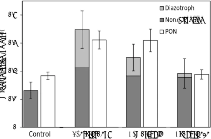
After 48 h of incubation, the biomass associated with non-diazotrophs increased in all the non-diazotrophs-amended treat-ments compared to the control (Fig. 1). The highest increase was observed in the T. erythraeum treatment (62 ± 39 %), mainly driven by a bacterial biomass increase of 90 ± 6 % and to a lesser extent by a Synechococcus (47 ± 22 %) and diatom (37 ± 17 %) biomass increase (Figs. 1 and 2). In the C. watsonii and Cyanothece treatments, the increase of biomass associated with non-diazotrophic plankton was 39 ± 39 and 35 ± 46 %, respectively. It was mainly driven by bacterial (58 ± 12 %), Synechococcus (23 ± 10 %) and di-atom (30 ± 16 %) biomass increase in the C. watsonii treat-ment, and by bacterial biomass increase only (116 ± 16 %) in the Cyanothece treatment. The effect of diazotrophs on the biomass of small eukaryotes was less noticeable.
In all the treatments, the sum of the N biomass associated with every group of plankton was in good agreement with the actual PON concentrations measured by EA-IRMS after
0 0.2 0.4 0.6 0.8
C C TR TR CRO CRO CYA CYA
N con ce n tra tio n (µ m o l L -1) Diazotroph Non-diazotroph PON
Control T. erythraeum C. watsonii Cyanothece
Figure 2. PON concentrations measured by mass spectrometry (EA-IRMS) and biomass associated with each plankton group in
each treatment after 48 h of incubation (µmol L−1). Errors bar
rep-resent the standard deviation on triplicate incubations.
48 h, indicating that the cellular N contents used in this study (described in Sect. 2) are realistic (Fig. 2).
3.2 N2fixation rates and DDN release
Net N2 fixation rates determined by EA-IRMS in the
con-trol treatment were 1.5 ± 0.1 nmol L−148 h−1(Fig. 3). This N2fixation was attributed to the diazotrophs already present
in the natural assemblage (probably Trichodesmium spp. that were found at low abundances in the control, data not shown). In the diazotroph-amended treatments, net N2
fixa-tion rates were 10 to 40 times higher than in the control, indi-cating the that diazotroph blooms artificially induced worked well: ∼ 60 nmol L−148 h−1in the T. erythraeum and C. wat-soniitreatments and 16 nmol L−148 h−1 in the Cyanothece treatment (Fig. 3). The DDN released to the dissolved pool by diazotrophs represented 16.1 ± 6.7 % of the total N2
fix-ation (where total N2 fixation is defined as the sum of N2
fixed recovered in the PON, DON and NH+4 pools) in the T. erythraeum treatment, 13.8 ± 1.9 % in the C. watsonii treatment, 30.5 ± 10.4 % in the Cyanothece treatment and 66.0 ± 21.9 % in the control treatment. In all cases, most of the15N released in the dissolved pool after 48 h of incubation was under the form of DON, which represented 77 to 81 % of the total N release in the diazotrophs-amended treatments without any differences between the treatments. The NH+4 release was below detection limit in the control treatment. 3.3 Cell-specific15N- and13C-enrichments and DD15N
transfer towards the non-diazotrophic plankton NanoSIMS analyses revealed significant15N-enrichment in diazotrophic cells after 48 h of incubation compared to natu-ral15N-enrichment (0.366 atom %; Figs. 4 and 5). Among the three diazotrophs added, C. watsonii and Cyanoth-ece exhibited the highest 15N-enrichments with 1.942 ±
0 20 40 60 80 100 C TR CRO CYA N2 f ixat ion ( n m o l N L -1 48 h -1) NH4 DON PON
Control T. erythraeum C. watsonii Cyanothece
Figure 3. N2fixation rates (dark grey, nmol L−148 h−1), DDN
re-lease (nmol L−148 h−1) as DON (light grey) and NH+4 (white) in
each treatment. Error bars represent the standard deviation of tripli-cate incubations and the propagated analytical error.
0.239 atom % (n = 18) and 2.501 ± 0.300 atom % (n = 46), respectively (Fig. 5). T. erythraeum 15N-enrichment aver-aged 1.147 ± 0.233 atom % (n = 68). The 13C-enrichment was similar for T. erythraeum (3.316 ± 0.634 atom %) and C. watsonii(3.124 ± 0.670 atom %) and higher for Cyanoth-ece (4.612 ± 0.837 atom %). The correlation between13 C-enrichment and 15N-enrichment was significant for T. ery-thraeum (r2=0.50, p < 0.001, n = 68), weaker but still significant for C. watsonii (r2=0.39, p = 0.005, n = 18), and not significant for Cyanothece (r2=0.01, p = 0.500, n =46).
Cell specific N2fixation rates of diazotrophs were 140.8 ±
55.9 fmol N cell−148 h−1(assuming 100 cell per trichomes), 50.3 ± 9.2 fmol N cell−1and 25.0 ± 3.5 fmol N cell−148 h−1 for T. erythraeum, C. watsonii and Cyanothece leading to N2
fixation rates associated with the three groups of 54.8 ± 21.7, 54.5 ± 10.0 and 19.1 ± 2.7 nmol L−148 h−1, respectively.
NanoSIMS analyses performed on non-diazotrophic di-atoms and cell-sorted Synechococcus, small eukaryotes, and bacteria also revealed 15N-enrichments that were at times significantly higher than those measured in the control (Figs. 4 and 6). The 15N-enrichment of non-diazotrophic plankton strongly depended on the treatment considered. When T. erythraeum provided the DD15N, the
15N-enrichment was significantly higher compared to the
control for diatoms (0.468 ± 0.081 atom %, n = 18), Syne-chococcus (0.404 ± 0.090 atom %, n = 105) and bacteria (0.487 ± 0.071 atom %, n = 45; data are not available for small eukaryotes in T. erythraeum treatment). In the C. watsonii treatment, the15N-enrichment of non-diazotrophs was significantly higher compared to the control for Syne-chococcus (0.411 ± 0.079 atom %, n = 134) and bacteria (0.435 ± 0.05 atom %, n = 34) and not significantly differ-ent for diatoms (0.394 ± 0.077 atom %, n = 23) and small eukaryotes (0.383 ± 0.040 atom %, n = 52). In the
Cyan-othece treatment, the 15N-enrichment of non-diazotrophs was significantly higher compared to the control for di-atoms (0.446 ± 0.143 atom %, n = 26) and for Synechococ-cus(0.389 ± 0.080 atom %, n = 25), whereas no significant enrichments were observed for small eukaryotes (0.383 ± 0.030 atom %, n = 88) and bacteria (0.379 ± 0.027 atom %, n =38). It should be noted that in the control, the 15 N-enrichment of all plankton groups (diatoms, Synechococ-cus, small eukaryotes and bacteria) was slightly higher (0.387 ± 0.048 atom %, n = 301) than the natural abundance (0.366 atom %) after 48 h of incubation (Fig. 6).
The amount of DD15N transferred to non-diazotrophs cor-rected from N2fixation detected in the control treatment was
higher in the T. erythraeum treatment (9.5 ± 4.9 nmol N L−1) compared to the C. watsonii and Cyanothece treatments, where it was 4.1 ± 2.3 and 1.2 ± 0.9 nmol N L−1, respec-tively. It represented 11.7 ± 4.4 % of total N2fixation in the
T. erythraeum treatment and was significantly higher than in the C. watsonii (5.8 ± 2.7 %) and Cyanothece treatments (4.9 ± 2.4 %) (Table 1).
4 Discussion
The fate of DDN in the marine food web has been poorly studied, mainly due to technical limitations. Using15N and
13C labeling coupled with cell sorting by flow cytometry and
nanoSIMS analyses at the single cell level, we were able to trace the transfer of DD15N from the diazotrophs to the dis-solved pool and to the non-diazotrophic plankton, and com-pare the DD15N transfer efficiency as a function of the dia-zotroph groups dominating the community.
4.1 Cell-specific photosynthesis and N2fixation
Cell-specific N2fixation rates measured using nanoSIMS are
in the range of previous N2fixation rates measured in
cul-tures using conventional N2 fixation methods for the same
strains of T. erythraeum, C. watsonii and Cyanothece (Berth-elot et al., 2015a). This confirms the ability of nanoSIMS to accurately measure N2fixation rates, as previously shown in
former studies (Finzi-Hart et al., 2009; Foster et al., 2013; Ploug et al., 2010). The high N2 fixation rates induced by
the inoculation of diazotrophs in the natural planktonic com-munity (7–30 nmol N L−1d−1) are representative of those re-ported in the southwestern Pacific region under blooming conditions (Berthelot et al., 2015b; Bonnet et al., 2015; Gar-cia et al., 2007). Thus, the artifiGar-cial diazotroph blooms in-duced for the purpose of this study provided realistic condi-tions to study the DDN transfer to non-diazotrophic plank-ton.
The significant correlation between 13C- and 15 N-enrichments in T. erythraeum cells analyzed after 48 h of in-cubation argue that both PP and N2fixation occur
1.0 2.0 3.0 4.0 5.0 0.3 0.4 0.5 0.6 0.7 0.8 15N (atom %) 13C (atom %) 1.0 2.0 3.0 4.0 5.0 0.3 0.4 0.5 0.6 0.7 0.8 13C (atom %) 15N (atom %) 1.0 2.0 3.0 4.0 5.0 6.0 0.5 1.0 1.5 2.0 2.5 3.0 1.0 2.0 3.0 4.0 5.0 1.0 2.0 3.0 4.0 5.0 2.0 4.0 6.0 8.0 10.0 5.0 10.0 15.0 20.0 1.0 2.0 3.0 4.0 5.0 0.3 0.4 0.5 0.6 0.7 0.8
(a)
(b)
(c)
(d)
(d)
(e)
(f)
(g)
(h)
(i)
(j)
(k)
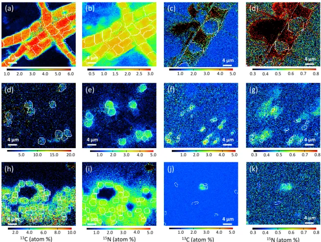
4 µm 4 µm 4 µm 4 µm 4 µm 4 µm 4 µm 4 µm 4 µm 4 µm 4 µm 4 µmFigure 4. NanoSIMS images showing the isotopic enrichment found in cells after 48 h of incubation.13C- (a, d, h, c, f, j) and15N- (b, e, i, d,
g, k) enrichments (atom %) are shown for T. erythraeum (a, b), C. watsonii (d, e), Cyanothece (h, i), Chaetoceros sp. (c, d), Synechococcus (f,
g) and bacteria cells (j, k). The white outlines define the regions of interest (ROIs), which were used to estimate the cells13C- and15
N-enrichments.
with the idea of the cells specialization in N2fixation (called
diazocytes) where high respiration rates and degradation of glycogen and gas vacuoles reduce the O2concentration
en-abling the expression of nif genes allowing daytime N2
fix-ation (Bergman and Carpenter, 1991; Berman-Frank et al., 2001; Sandh et al., 2012). However, it has to be noticed that after 48 h of incubation with the tracers, it is highly prob-able that both 15N and 13C have been exchanged between cells, leading to a homogenization of the cells isotopic en-richments.
More surprisingly, the coupling between 13C- and 15 N-enrichments for individual UCYN cells after 48 h of incu-bation is weaker than for T. erythraeum cells, in particular for Cyanothece. This appears counter-intuitive as UCYN are supposed to perform both N2 fixation and photosynthesis
within the same cell. This uncoupling suggests that UCYN cells might be at least partially specialized in photosynthe-sis or N2fixation, similarly to Trichodesmium spp. These
re-sults confirm the patterns already observed for C. watsonii (Foster et al., 2013). In addition, the weaker correlation be-tween13C- and15N-enrichments in UCYN cells compared to
T. erythraeumalso suggests weaker extracellular fixed N and C exchanges between cells. These differences might be the result of the greater spatial proximity of Trichodesmium spp. cells within colonies and filaments compared to free living UCYN cells in the water column. According to this vision, the high production of extracellular polymeric substances ob-served in different C. watsonii strains (Sohm et al., 2011; Webb et al., 2009) might be a strategy to agglomerate the free living UCYN together to form colonies (Bonnet et al., 2016a; Foster et al., 2013), ensuring a spatial proximity and thus facilitating the exchange of metabolites between cells. 4.2 DDN release to the dissolved pool
The DD15N released to the dissolved pool after 48 h ac-counted from 7 to 17 % of total N2 fixation over the three
diazotroph-amended treatments. These values are at the lower end of values (10–80 %) reported in Trichodesmium spp. blooms in the tropical Atlantic (Glibert and Bronk, 1994; Mulholland et al., 2006), southwestern Pacific (Bon-net et al., 2016c) or in mixed diazotroph assemblages of the North Pacific (Konno et al., 2010) and the Atlantic ocean
Figure 5. 15N-enrichment (atom %) measured in T. erythraeum (red), C. watsonii (green) and Cyanothece (blue) cells relative to
the 13C-enrichment. The coloured line are the linear regressions
for T. erythraeum (red), C. watsonii (green) shown with their re-spective r-squared and p-values. Regression is not significant for
Cyanotheceand thus not shown on the plot. Box plots of13C- and
15N-enrichments are shown, following the same colour code, on
horizontal and vertical axes, respectively.
(Benavides et al., 2013). In contrast, these values of N release are at least 1 order of magnitude higher than those reported in unialgal cultures (< 2 %) for the same strains as those studied here (Berthelot et al., 2015a). It is probable that, in culture, the cells are maintained in optimal growth conditions (ex-ponential growth phase, appropriate light, temperature and nutrient conditions) and optimize the N use, either through a low excretion rate of DDN or through an efficient uptake of DDN (Mulholland et al., 2001). Conversely, in the field, the sampling does not necessarily occur during the exponential growth phase, and exogenous factors may affect the release of DDN, such as viral lysis (Hewson et al., 2004; Ohki, 1999) and sloppy feeding (O’Neil et al., 1996). In this study, the di-azotrophs added to natural seawater were healthy but may have been affected by exogenous factors after inoculation, leading to a moderate proportion (7–17 %) of DDN released in the dissolved pool. These results indicate that the propor-tion of N2fixed released in the dissolved compartment both
depends on the cell status and on exogenous factors more than the type of diazotrophs involved in N2fixation, as
pre-viously stated by Berthelot et al. (2015a).
Figure 6. Box-plot of15N-enrichment measured in diatoms,
Syne-chococcus, small eukaryotes and bacteria in the control (C), T.
ery-thraeum(TR), C. watsonii (CRO) and Cyanothece (CYA)
treat-ments. Red dots indicate the average values. Blue dotted lines
de-pict the natural15N abundance.∗and∗∗depict significant
enrich-ments in diazotrophs treatenrich-ments compared to the control treatment (unpaired Mann–Whitney–Wilcoxon test) at 95 and 99 % levels of confidences, respectively.
4.3 DDN transfer efficiency and pathways
T. erythraeumtransferred ∼ 12 % of DD15N towards the non-diazotrophic plankton. This is in good agreement with pre-vious estimates by Bonnet et al. (2016c) using the same methodology, who report a DD15N transfer of 7 to 12 % in naturally occurring Trichodesmium spp. blooms. These re-sults confirm that Trichodesmium spp. enhances the devel-opment of non-diazotrophic plankton, as already suggested by the frequent observations of co-occurrence or succession of Trichodesmium spp. and non-diazotrophs, particularly in N-depleted environments (Devassy et al., 1979; Dore et al., 2008; Lee Chen et al., 2011; Lenes and Heil, 2010).
The DD15N transfer was half as efficient (4–5 %) in the UCYN treatments compared to the T. erythraeum treatment (Table 1). This is in good agreement with the lower increase in plankton biomass associated with non diazotrophs in the UCYN treatments compared to the T. erythraeum treatment (Fig. 2). The ecology of UCYN is less characterized than that of Trichodesmium spp. and data on their co-occurrence with non-diazotrophic plankton in the ocean are scarce. In this issue, Bonnet et al. (2016a) used the single cell approach described here during a natural occurring bloom of UCYN-C (closely related to UCYN-Cyanothece spp.) in the New UCYN- Caledo-nian lagoon and measured a DD15N transfer of 21 ± 4 % of
Table 1. Synthesis of the distribution of the recently fixed N2(DD15N) in each of the planktonic groups analysed after 48 h of
incuba-tion (nmol L−148 h−1) and their respective proportion relative to the total fixed N2(%). n.a.: not analysed, n.c.: not calculated. Standard
deviations are in parenthesis.
Planktonic group DD15N (nmol L−148 h−1) % of total N2fixed
Trichodesmiumtreatment Trichodesmium 63.05 (21.54) 78.0 (26.7) Dissolved pool 8.30 (3.83) 10.3 (4.7) Sum of non-diazotrophs 9.45 (3.54) 11.7 (4.4) Bacteria 5.79 (3.41) 7.2 (4.2) Diatom 1.99 (0.36) 2.5 (0.5) Synechococcus 1.67 (0.88) 2.1 (1.1)
Small eukaryotes n.a n.c
C. watsoniitreatment C. watsonii 60.92 (10.06) 87.2 (14.4) Dissolved pool 4.90 (14.10) 7.0 (3.0) Sum of non-diazotrophs 4.08 (1.87) 5.8 (2.7) Bacteria 2.36 (1.70) 3.4 (2.4) Diatom 0.06 (0.05) 0.1 (0.1) Synechococcus 1.63 (0.78) 2.3 (1.1) Small eukaryotes 0.03 (0.00) n.c. Cyanothecetreatment Cyanothece 19.41 (5.28) 78.6 (21.4) Dissolved pool 4.10 (9.85) 16.6 (5.0) Sum of non-diazotrophs 1.19 (0.59) 4.9 (2.4) Bacteria −0.05 (0.42) n.c. Diatom 0.93 (0.28) 3.8 (1.1) Synechococcus 0.29 (0.30) 1.2 (1.2) Small eukaryotes 0.02 (0.00) n.c.
the total N2fixation, mainly towards pico-planktonic
com-munities. The DD15N transfer reported in the latter study is ∼ 3 times higher than in the present study. This discrep-ancy may result from the physiological differences between the UCYN-C ecotypes involved in both studied, and/or from the DDN release from diazotrophic cells that is potentially higher is natural communities compared to cultured cells as discussed above. The bloom reported in Bonnet et al. (2016a) study co-occurred with a doubling of Synechococcus and pico-eukaryotes abundances, as well as an increase of di-atoms (Leblanc et al., 2016) and PP (Berthelot et al., 2015b). Crocosphaera-like cells observed in association with the di-atom Climacodium sp. (Carpenter and Janson, 2000) have also been shown to transfer the recently fixed N2towards the
host diatom cell (Foster et al., 2011). All these data confirm that UCYN are able to provide DDN to non-diazotrophic plankton and thus promote marine productivity in N-depleted areas.
The transfer of DDN towards phytoplankton or bacteria requires the release of N in the dissolved pool. Surprisingly, the total amount of DD15N recovered in the dissolved pool was not a good indicator of the DD15N transfer efficiency:
the highest release of DDN was measured in the Cyanoth-ecetreatment (16.6 ± 4.9 % of the total N2fixation) and led
to the lowest DD15N transfer efficiency (4.9 ± 2.4 % of the total N2 fixation). In the T. erythraeum and in C. watsonii
treatments, the proportion of DD15N recovered in the dis-solved pool was lower (10.3 ± 4.7 and 7.0 ± 3.0 % of the total N2fixation, respectively) but led to higher DD15N
trans-fer efficiencies (11.7 ± 4.4 and 5.8 ± 2.7 % of the total N2
fixation, respectively). This suggests that the N compounds released by Cyanothece were less available for the surround-ing plankton communities than the compounds released by T. erythraeumand C. watsonii.
On the opposite, the15NH+4 enrichment appeared to be a relevant indicator of the DDN transfer efficiency: it was twice as high in the T. erythraeum treatment compared to the UCYN treatments (Table 2), leading to a DD15N transfer efficiency twice as high in the T. erythraeum treatments (Ta-ble 1). This coupling between15NH+4 enrichment and trans-fer efficiency suggests that NH+4 is the major form of DD15N that is transferred to non-diazotrophic plankton, and that the DDN released under the form of DON is likely poorly avail-able for the surrounding planktonic communities (Knapp et
Table 2.15N-enrichment (atom %) of diazotrophic cells, PON, NH+4 and DON pools and concentrations (µmol L−1) of NH+4 and DON. In parenthesis are shown the standard deviations on triplicate incubations. n/a: not applicable, n.d.: not detected.
Control T. erythraeum C. watsonii Cyanothece
treatment treatment treatment
15N-enrichment (atom %)
Diazotrophic cells n/a 1.15 (0.23) 1.94 (0.24) 2.50 (0.30)
PON 0.40 (0.01) 0.69 (0.13) 0.67 (0.01) 0.50 (0.01) NH+4 n.d. 2.31 (0.81) 1.20 (0.15) 1.44 (0.44) DON 0.37 (< 0.00) 0.37 (< 0.00) 0.37 (< 0.00) 0.38 (< 0.00) Concentrations (µmol L−1) NH+4 0.010 (0.002) 0.010 (0.003) 0.011 (0.004) 0.009 (0.003) DON 4.05 (0.57) 3.99 (0.17) 4.54 (0.80) 4.18 (0.45)
al., 2005; Bourbonnais et al., 2009). This is in good agree-ment with the known higher bioavailability of NH+4 for phy-toplankton compared to DON (e.g. Bradley et al., 2010; Col-los and Berges, 2002). However, some DON compounds such as urea or amino acids can also be a significant source of N for planktonic communities, e.g. heterotrophic bacteria and mixotrophic plankton (Antia, 1991; Bronk, 2007). Un-fortunately, the methodology used here can not asserts the importance of DON compared to NH+4 in the DDN transfer. It should be noted that the increase of plankton biomass associated with non-diazotrophs in the present study can-not only be explained by the DD15N provided by N2
fixa-tion within the time frame of the incubafixa-tion (48 h). While the DD15N transferred to non-diazotrophic plankton biomass ranged between 1 and 10 nmol N L−1 in the diazotrophs-amended treatments, the non-diazotrophic biomass increased from 90 to 160 nmol N L−1in the diazotrophs-amended treat-ments. This suggests that production was also stimulated by DDN fixed prior the incubations, that was thus not15N la-belled.
4.4 Plankton groups benefiting from the DDN
Bacterial biomass increased from 60 to 120 % after the addi-tion of three diazotrophs; it was the plankton group which responded the most to the diazotrophs inoculations, what-ever the treatment considered (Fig. 1 and Table S1). This is consistent with the high15N-enrichment of bacteria cells in T. erythraeum and C. watsonii treatments compared to the control treatment (Fig. 6). In contrast, the high bacte-rial biomass increase observed in the Cyanothece treatment contrasts with the relatively low 15N-enrichment of bacte-rial individual cells measured in this treatment (Fig. 6). This suggests that, in the T. erythraeum and C. watsonii treat-ments, bacteria took advantage of the DD15N released dur-ing the incubation, while in Cyanothece treatment, bacteria may have mainly relied on DDN fixed prior to the begin-ning of the incubation. This is consistent with the higher
ac-cumulation of DD15N in the DON pool in the Cyanothece treatment compared to the two other treatments, indicating that the DON compounds released by Cyanothece are likely less bio-available for the planktonic community compared to those released by T. erythraeum and C. watsonii.
The presence of bacteria in Trichodesmium spp. colonies has been widely studied (Hewson et al., 2009; Hmelo et al., 2012; Nausch, 1996; Paerl et al., 1989; Rochelle-Newall et al., 2014; Sheridan et al., 2002). Trichodesmium spp. har-bours high heterotrophic bacterial activity (Nausch, 1996; Tseng et al., 2005) and abundance is found to be at least 2 orders of magnitude higher in Trichodesmium spp. colonies than in surrounding waters (Sheridan et al., 2002). Asso-ciations between bacteria and UCYN are less documented. However, similarly to Trichodesmium spp., tight relation-ships may occur between UCYN and bacteria, as sug-gested by the significant increases of bacterial abundances in the UCYN treatments, but further investigations would be needed to understand the nature of their interactions.
Phytoplankton (diatoms, Synechococcus and small eu-karyotes) was also stimulated by the diazotroph blooms, although to a lower extend compared to bacteria in all treatments (Fig. 1). However, the increase in phytoplankton biomass in the T. erythraeum and C. watsonii treatments to-gether with the significant15N-enrichments in diatoms and Synechococcus(Fig. 6) argue that phytoplankton also took advantage of the DDN. This confirms the ability of dia-zotroph to promote non-diadia-zotrophic primary producers as suggested by previous studies (e.g. Bonnet et al., 2016c; Devassy et al., 1979; Lee Chen et al., 2011; Lenes and Heil, 2010). The enhancement of large phytoplanktonic cells such as diatoms by DDN observed within the timespan of this study reveals the tight relationship that may occurs between the new production fuelled by diazotrophy and particle ex-port in oligotrophic areas.
In the present study, the plankton community composi-tion remained relatively stable in comparison to the Bon-net et al. (2016c) study, in which the authors observed
sys-tematic shifts from Trichodesmium spp. biomass towards di-atom biomass. This difference is probably linked to the Tri-chodesmium cells status. In the Bonnet et al. (2016c) study, most of the analyzed Trichodesmium spp. cells were decay-ing, leading to the release of micromolar concentrations of NH+4 accumulating in the dissolved pool, whereas the main form of DDN released in the present study was DON. This rapid increase in NH+4 bio-availability was benefiting to di-atoms, which are known to be highly competitive under high nutrient concentrations (Chavez and Smith, 1995; Kudela and Dugdale, 2000; Miller and Wheeler, 2012; Smetacek, 1998; Wilkerson et al., 2000). This synchronized destruc-tion of the colonies has been shown to be possible within a few hours, mediated by programmed cell death (Berman-Frank et al., 2004). Furthermore, the dense bloom forming behaviour and maintenance of Trichodesmium spp. at the sur-face due to their positive buoyancy (Romans et al., 1994) may be an additional feature that helps the constitution of locally rich N layers promoting the diatom development. In the present study, the T. erythraeum colonies added to the in-cubation bottles were in exponential growing phase and the relative stability of their abundance throughout the experi-ment indicates no substantial cell breakages. The amount of DDN transfer in our study was thus mainly mediated by the release of NH+4 and DON during active N2 fixation rather
than the N release due to cell breakage and was thus much more limited than in the Bonnet et al. (2016c) study, leading to attenuated changes in the planktonic community composi-tion.
5 Conclusions and ecological implications
This study reveals the various short term fates of DDN in the ocean and highlight the complex interactions between diazotrophs and their environment. First, it shows that the DDN released by diazotrophs in the dissolved pool as NH+4 is quickly transferred to non-diazotrophic plankton while the DDN released as DON is mostly accumulated in the dissolved pool. Second, the DDN transfer efficiency to-wards the non-diazotrophic plankton depends on the dia-zotrophs involved in N2fixation: it is twice as much for T.
erythraeum compared to the DDN transfer associated with UCYN strains. This implies that T. erythraeum would be more efficient at promoting non-diazotrophic marine produc-tivity in N-depleted areas than UCYN are. Finally, the re-sults presented here suggest that diazotrophic activity first promotes heterotrophic plankton but also autotrophic plank-ton, albeit to a lower extent. Taken together, theses results show that the fates of DDN are diverse and would need fur-ther investigation, in particular in the vast open-ocean regions where primary productivity extensively depends on diazotro-phy.
The Supplement related to this article is available online at doi:10.5194/bg-13-4005-2016-supplement.
Author contributions. S. Bonnet and H. Berthelot designed the
ex-periments and S. Bonnet and H. Berthelot carried them out. All au-thors analyzed the samples. H. Berthelot prepared the manuscript, which was amended by S. Bonnet.
Acknowledgements. Funding for this research was provided
by the Agence Nationale de la Recherche (ANR starting grant VAHINE ANR-13-JS06-0002). We thank François Robert, Adriana Gonzalez, Smail Mostefaoui and Rémi Duhamel from the French National Ion MicroProbe Facility hosted by the Museum National d’Histoire Naturelle (Paris) for providing nanoSIMS facilities and constant advice. We are grateful to Gerald Gregori from the Regional Flow Cytometry Platform for Microbiology (PRECYM) of the Mediterranean Institute of Oceanography (MIO) for the flow cytometry analyses support.
Edited by: E. Marañón
References
Aminot, A. and Kérouel, R.: Dosage Automatique des Nutriments dans les Eaux Marines, Ifremer, Plouzané, 2007.
Benavides, M., Bronk, D. A., Agawin, N. S. R., Pérez-Hernández, M. D., Hernández-Guerra, A., and Arístegui, J.:
Longitudinal variability of size-fractionated N2 fixation
and DON release rates along 24.5◦N in the subtropical
North Atlantic, J. Geophys. Res.-Oceans, 118, 3406–3415, doi:10.1002/jgrc.20253, 2013.
Bergman, B. and Carpenter, E. J.: Nitrogenase confined to randomly distributed trichomes in the marine cyanobac-terium Trichodesmium thiebautii, J. Phycol., 27, 158–165, doi:10.1111/j.0022-3646.1991.00158.x, 1991.
Bergman, B., Sandh, G., Lin, S., Larsson, J., and Carpenter, E. J.:
Trichodesmium– a widespread marine cyanobacterium with
un-usual nitrogen fixation properties, FEMS Microbiol. Rev., 37, 286–302, doi:10.1111/j.1574-6976.2012.00352.x, 2013. Berman-Frank, I., Lundgren, P., Chen, Y. B., Küpper, H.,
Kol-ber, Z., Bergman, B., and Falkowski, P.: Segregation of nitrogen fixation and oxygenic photosynthesis in the ma-rine cyanobacterium Trichodesmium, Science, 294, 1534–1537, doi:10.1126/science.1064082, 2001.
Berman-Frank, I., Bidle, K. D., Haramaty, L., and Falkowski, P. G.: The demise of the marine cyanobacterium, Trichodesmium spp., via an autocatalyzed cell death pathway, Limnol. Oceanogr., 49, 997–1005, doi:10.4319/lo.2004.49.4.0997, 2004.
Berthelot, H., Bonnet, S., Camps, M., Grosso, O., and Moutin, T.: Assessment of the dinitrogen released as ammonium and dis-solved organic nitrogen by unicellular and filamentous marine diazotrophic cyanobacteria grown in culture, Front. Mar. Sci., 2, 80, doi:10.3389/fmars.2015.00080, 2015a.
Berthelot, H., Moutin, T., L’Helguen, S., Leblanc, K., Hélias, S., Grosso, O., Leblond, N., Charrière, B., and Bonnet, S.: Dinitro-gen fixation and dissolved organic nitroDinitro-gen fueled primary pro-duction and particulate export during the VAHINE mesocosm experiment (New Caledonia lagoon), Biogeosciences, 12, 4099– 4112, doi:10.5194/bg-12-4099-2015, 2015b.
Bonnet, S., Biegala, I. C., Dutrieux, P., Slemons, L. O., and Capone, D. G.: Nitrogen fixation in the western equatorial Pa-cific: rates, diazotrophic cyanobacterial size class distribution, and biogeochemical significance, Global Biogeochem. Cy., 23, 1–13, doi:10.1029/2008GB003439, 2009.
Bonnet, S., Rodier, M., Turk-Kubo, K. A., Germineaud, C., Menkes, C., Ganachaud, A., Cravatte, S., Raimbault, P., Camp-bell, E., Quéroué, F., Sarthou, G., Desnues, A., Maes, C., and
Eldin, G.: Contrasted geographical distribution of N2 fixation
rates and nifH phylotypes in the Coral and Solomon Seas (South-Western Pacific) during austral winter conditions, Global Biogeochem. Cy., 29, 1874–1892, doi:10.1002/2015GB005117, 2015.
Bonnet, S., Berthelot, H., Turk-Kubo, K., Fawcett, S., Rahav, E.,
L’Helguen, S., and Berman-Frank, I.: Dynamics of N2
fixa-tion and fate of diazotroph-derived nitrogen in a low-nutrient, low-chlorophyll ecosystem: results from the VAHINE mesocosm experiment (New Caledonia), Biogeosciences, 13, 2653–2673, doi:10.5194/bg-13-2653-2016, 2016a.
Bonnet, S., Moutin, T., Rodier, M., Grisoni, J.-M., Louis, F., Folcher, E., Bourgeois, B., Boré, J.-M., and Renaud, A.: In-troduction to the project VAHINE: VAriability of vertical and tropHIc transfer of diazotroph derived N in the south wEst Pa-cific, Biogeosciences, 13, 2803–2814, doi:10.5194/bg-13-2803-2016, 2016b.
Bonnet, S., Berthelot, H., Turk-Kubo, K., Cornet-Bartaux, V., Fawcett, S. E., Berman-Frank, I., Barani, A., Dekaezemacker, J., Benavides, M., Charriere, B., and Capone, D. G.: Diazotroph de-rived nitrogen supports diatoms growth in the South West Pa-cific: a quantitative study using nanoSIMS, Limnol. Oceanogr., in press, 2016c.
Bourbonnais, A., Lehmann, M. F, Waniek, J. J., and
Schulz-Bull, D. E.: Nitrate isotope anomalies reflect N2fixation in the
Azores Front region (subtropical NE Atlantic), J. Geophys. Res., 114, C03003, doi:10.1029/2007JC004617, 2009.
Bradley, P. B., Lomas, M. W., and Bronk, D. A.: Inorganic and organic nitrogen use by phytoplankton along Chesapeake Bay, measured using a flow cytometric sorting approach, Estuar. Coast., 33, 971–984, doi:10.1007/s12237-009-9252-y, 2010. Bronk, D. A., See, J. H., Bradley, P., and Killberg, L.: DON as
a source of bioavailable nitrogen for phytoplankton, Biogeo-sciences, 4, 283–296, doi:10.5194/bg-4-283-2007, 2007. Bryceson, I. and Fay, P.: Nitrogen fixation in
Oscillato-ria (Trichodesmium) erythraea in relation to bundle
forma-tion and trichome differentiaforma-tion, Mar. Biol., 61, 159–166, doi:10.1007/BF00386655, 1981.
Buitenhuis, E. T., Li, W. K. W., Vaulot, D., Lomas, M. W., Landry, M. R., Partensky, F., Karl, D. M., Ulloa, O., Camp-bell, L., Jacquet, S., Lantoine, F., Chavez, F., Macias, D., Gos-selin, M., and McManus, G. B.: Picophytoplankton biomass dis-tribution in the global ocean, Earth Syst. Sci. Data, 4, 37–46, doi:10.5194/essd-4-37-2012, 2012.
Capone, D. G., Ferrier, M. D., and Carpenter, E. J.: Amino acid cycling in colonies of the planktonic marine cyanobacterium Tri-chodesmium thiebautii, Appl. Environ. Microb., 60, 3989–3995, 1994.
Capone, D. G., Zehr, J. P., Paerl, H. W., Bergman, B., and Carpenter, E. J.: Trichodesmium, a globally
signif-icant marine cyanobacterium, Science, 276, 1221–1229,
doi:10.1126/science.276.5316.1221, 1997.
Capone, D. G., Burns, J. A., Montoya, J. P., Subramaniam, A., Mahaffey, C., Gunderson, T., Michaels, A. F., and Carpen-ter, E. J.: Nitrogen fixation by Trichodesmium spp.: an im-portant source of new nitrogen to the tropical and subtropical North Atlantic Ocean, Global Biogeochem. Cy., 19, GB2024, doi:10.1029/2004GB002331, 2005.
Carpenter, E. J. and Janson, S.: Intracellular cyanobacterial
symbionts in the marine diatom Climacodium
frauen-feldianum (Bacillariophyceae), J. Phycol., 36, 540–544,
doi:10.1046/j.1529-8817.2000.99163.x, 2000.
Chavez, F. P. and Smith, S. L.: Biological and chemical conse-quences of open ocean upwelling, in: Upwelling in the Ocean: Modern Processes and Ancient Records, edited by: Summer-hayes, C. P., Emeis, K.-C., and Angel, M. V., Wiley, 149–169, 1995.
Chen, Y.-B., Zehr, J. P., and Mellon, M.: Growth and nitro-gen fixation of the diazotrophic filamentous nonheterocystous cyanobacterium Trichodesmium sp. IMS 101 in defined me-dia: evidence for a circadian rhythm, J. Phycol., 32, 916–923, doi:10.1111/j.0022-3646.1996.00916.x, 1996.
Collos, Y. and Berges, J. A.: Nitrogen metabolism in phytoplank-ton, in: Marine Ecology, Encyclopedia of Life Support Systems (EOLSS), 1–18, 2002.
Dabundo, R., Lehmann, M. F., Treibergs, L., Tobias, C. R., Al-tabet, M. A., Moisander, P. H., and Granger, J.: The
Con-tamination of Commercial 15N2 Gas Stocks with 15N
La-beled Nitrate and Ammonium and Consequences for Ni-trogen Fixation Measurements, PLoS ONE, 9, e110335, doi:10.1371/journal.pone.0110335, 2014.
Devassy, V. P., Bhattathiri, P. M. A., and Qasim, S. Z.: Succession of organisms following Trichodesmium phenomenon, Indian J. Mar. Sci., 8, 89–93, 1979.
Dore, J. E., Brum, J. R., Tupas, L. M., and Karl, D. M.: Seasonal and interannual variability in sources of nitrogen supporting ex-port in the oligotrophic subtropical North Pacific Ocean, Lim-nol. Oceanogr., 47, 1595–1607, doi:10.4319/lo.2002.47.6.1595, 2002.
Dore, J. E., Letelier, R. M., Church, M. J., Lukas, R., and Karl, D. M.: Summer phytoplankton blooms in the olig-otrophic North Pacific Subtropical Gyre: historical perspec-tive and recent observations, Prog. Oceanogr., 76, 2–38, doi:10.1016/j.pocean.2007.10.002, 2008.
Dron, A., Rabouille, S., Claquin, P., Le Roy, B., Talec, A., and Sciandra, A.: Light-dark (12 : 12) cycle of carbon and nitrogen metabolism in Crocosphaera watsonii WH8501: re-lation to the cell cycle, Environ. Microbiol., 14, 967–981, doi:10.1111/j.1462-2920.2011.02675.x, 2012.
Finzi-Hart, J. A., Pett-Ridge, J., Weber, P. K., Popa, R., Fal-lon, S. J., Gunderson, T., Hutcheon, I. D., Nealson, K. H., and Capone, D. G.: Fixation and fate of C and N in the cyanobac-terium Trichodesmium using nanometer-scale secondary ion