Advanced Brachytherapy Dosimetric Considerations
ByChristopher S. Melhus
M.Sc. Nuclear Engineering (2002) Massachusetts Institute of Technology
MASSACHUSMTTS INSW
OF TEOHNOLOGYJUN 2 0 2008
LIBRARIES
B.A. Physics (1997) Reed CollegeSubmitted to the Division of Health Science and Technology in Partial Fulfillment of the Requirements for the Degree of
Doctor of Philosophy at the
Massachusetts Institute of Technology
June 2008
© 2008 Christopher S. Melhus. All rights reserved.
The author hereby grants to MIT permission to reproduce and to distribute publicly paper and electronic copies of this thesis document in whole or in part
in any medium now known or hereafter created.
Signature of Author ,
Harvard-MIT Division of Hi1ti.Sdiences andT~iellogy
1, / 7 March 2008
Certified by ,
L Mark J. Rivard, Ph.D.
Associate Professor of Radiation Oncology Tufts University School of Medicine Thesis Supervisor
Accepted by
j
"
Matha L. Gray, Ph.D. Edward Hood Taplin Professor of Medical and Electrical Engineering Director, Harvard-MIT Division of Health Scienc s and TechnologyAdvanced Brachytherapy Dosimetric Considerations by
Christopher S. Melhus
Submitted to the Division of Health Science and Technology and the Department of Nuclear Science and Engineering on
7 March 2008 in Partial Fulfillment of the Requirements
for the Degree of Doctor of Philosophy in Radiological Sciences
ABSTRACT
The practice of brachytherapy and brachytherapy dosimetry was investigated with emphasis on evaluations of dose distributions and shielding considerations for both photon- and neutron-emitting radionuclides. Monte Carlo simulation methods were employed to calculate dose distributions for virtual and commercial brachytherapy sources. Radionuclides studied were 103Pd, 1251, 13 1Cs, 137Cs,
169 b, 192Ir, and 252Cf. 252Cf
sources also emit neutrons from spontaneous fission. The brachytherapy dosimetry protocol recommended by the American Association of Physicists in Medicine was followed and evaluated for conditions of partial scatter (non-infinite media) and material inhomogeneities, both commonly encountered in brachytherapy treatment. Furthermore, energy-dependent characteristics of dosimetry parameters were evaluated and reference calculations performed for virtual photon and neutron sources. These findings were applied to three clinical brachytherapy cases: eye plaques using 10 3Pd,
125I, and 13 1Cs; high-dose rate 252Cf treatment; and, 2 Cf plaques for superficial lesions. For eye plaques, material heterogeneities were significant for each radionuclide with dose reduction at 5 mm of 18%, 11%, and 10% for P03pd, 125I, and 131Cs, respectively. For a proposed
high-dose rate 252Cf source (5mm length), relative brachytherapy dosimetry parameters were found to be similar to those obtained for a low-dose rate Applicator Tube-type source (15 mm length). Considering 252Cf plaque brachytherapy when partial scatter conditions were accounted for, central axis equivalent dose rate decreased by 11 ± 1% and 7 ± 2% for depths of 4 to 50 mm, respectively. The ratio of neutron dose to total physical dose was 70 ± 1% and 57 ± 2% for depths of 4 and 50 mm, respectively, while the fractional dose-equivalent due to neutrons was 93 + 1% and 89 ± 2% at these depths, respectively. Finally, shielding requirements for a clinical high-dose rate 252Cf source were explored for common shielding materials and a linear accelerator vault. Lead, polyethylene, and borated polyethylene were evaluated for neutron, primary photon, and secondary photon attenuation. Half-value layers of 0.70, 0.15, and 0.13 m were obtained for lead, polyethylene, and borated polyethylene, respectively. A linear accelerator vault was found to adequately shield up to a 5 mg 252Cf source for regular clinical use.
Thesis Supervisor: Mark J. Rivard Ph.D.
Title: Associate Professor of Radiation Oncology Tufts University School of Medicine
ACKNOWLEDGEMENTS
Support was provided in part by appointment to the U.S. Department of Energy Nuclear Engineering and Health Physics Fellowship Program, sponsored by the U.S. Department of Energy's Office of Nuclear Energy, Science, and Technology (sections
3.2, 4.1, and 4.2). Computer time for Monte Carlo calculations was provided by the Radiation Saftey Information Computational Center at Oak Ridge National Laboratory (section 4.1). For the use of 252Cf, the author is indebted to the U. S. Department of Energy's Californium University Loan Program as administered by the Office of Nuclear Materials production through the facilities of the Oak Ridge National Laboratory (section 4.2). Additional resources were provided by Tufts-New England Medical Center and Tufts University School of Medicine.
Prof. Ademir da Silva of the Universidade Federal do Rio de Janeiro kindly shared Monte Carlo input files as used in reference 99 for section 4.1. Dr. Sou-Tung Chiu-Tsao provided helpful information for section 2.2. Mrs. Bernadete Kirk was a strong supporter of this work and provided assistance through the Radiation Safety Information Computational Center. The Chair of Radiation Oncology at Tufts University School of Medicine, Dr. David Wazer, generously provided travel support to present this work at national meetings of the American Association of Physicists in Medicine, the American Brachytherapy Society, and the American Nuclear Society.
I am very appreciative of the guidance and support provided by Prof. Jeffrey A. Coderre, the thesis reader. He supported efforts to perform clinical research at Tufts-New England Medical Center and enabled work away from the MIT campus to take place. Prof. Jacquelyn Yanch has supported my clinical research efforts since I matriculated to MIT in 1999 and was on the dissertation committee.
Finally, this dissertation would not have been possible without the mentorship and unyielding support of Prof. Mark J. Rivard, the thesis advisor. At every step, he has encouraged me to be a better scientist and to effectively communicate results to the scientific community. In addition to these academic pursuits, he has supervised my training as a medical physicist which put this work in context with clinical needs and the impact on patient care.
DEDICATION
TABLE OF CONTENTS
ABSTRACT 3 ACKNOWLEDGEMENTS 5 DEDICATION 7 1 INTRODUCTION 11 1.1 BACKGROUND 11 1.1.1 INTRODUCTION TO BRACHYTHERAPY 111.1.2 BRIEF HISTORY OF BRACHYTHERAPY DOSIMETRY 14
1.1.3 CALIFORNIUM BRACHYTHERAPY 17 1.1.4 RADIOLOGICAL PROTECTION 20 1.2 THESIS STATEMENT 21 1.2.1 PHOTON DOSIMETRY 21 1.2.2 NEUTRON DOSIMETRY 22 1.2.3 SHIELDING CONSIDERATIONS 23
1.3 DATA, REFERENCE, AND PUBLICATION HERITAGE 23
2 PHOTON DOSIMETRY 25
2.1 APPROACHES TO CALCULATING AAPM TG-43 BRACHYTHERAPY DOSIMETRY
PARAMETERS FOR 137Cs, 125I, 192Ir, 103Pd, AND 169Yb SOURCES 25
2.1.1 ABSTRACT 25
2.1.2 INTRODUCTION 26
2.1.3 MATERIALS AND METHODS 27
2.1.4 RESULTS AND DISCUSSION 31
2.1.5 CONCLUSIONS 43
2.2 COMS EYE PLAQUE BRACHYTHERAPY DOSIMETRY FOR 103pd, 1251, AND 13 1Cs 43
2.2.1 ABSTRACT 43
2.2.2 INTRODUCTION 44
2.2.3 MATERIALS AND METHODS 44
2.2.4 RESULTS AND DISCUSSION 49
2.2.5 CONCLUSIONS 58
3 NEUTRON DOSIMETRY 59
3.1 APPROACHES TO CALCULATING AAPM TG-43 BRACHYTHERAPY DOSIMETRY
PARAMETERS FOR NEUTRON SOURCES 59
3.1.1 ABSTRACT 59
3.1.2 INTRODUCTION 60
3.1.3 MATERIALS AND METHODS 60
3.1.4 RESULTS AND DISCUSSION 63
3.2 CLINICAL BRACHYTHERAPY DOSIMETRY PARAMETERS AND MIXED-FIELD DOSIMETRY
FOR A HIGH DOSE RATE Cf-252 BRACHYTHERAPY SOURCE. 71
3.2.1 ABSTRACT 71
3.2.2 INTRODUCTION 72
3.2.3 MATERIALS AND METHODS 73
3.2.4 RESULTS AND DISCUSSION 77
3.2.5 CONCLUSIONS 85
3.3 MONTE CARLO VALIDATION OF CLINICAL BRACHYTHERAPY DOSIMETRY UNDER
PARTIAL SCATTER CONDITIONS FOR NEUTRON-EMITTING SOURCES 86
3.3.1 ABSTRACT 86
3.3.2 INTRODUCTION 87
3.3.3 MATERIALS AND METHODS 87
3.3.4 RESULTS 88
3.3.5 CONCLUSIONS 91
4 SHIELDING CONSIDERATIONS 93
4.1 STORAGE SAFE SHIELDING ASSESSMENT FOR A HDR CALIFORNIUM-252
BRACHYTHERAPY SOURCE 93
4.1.1 ABSTRACT 93
4.1.2 INTRODUCTION 94
4.1.3 MATERIALS AND METHODS 94
4.1.4 RESULTS AND DISCUSSION 99
4.1.5 CONCLUSIONS 105
4.1.6 ADDENDUM 106
4.2 SHIELDING EVALUATION OF A MEDICAL LINEAR ACCELERATOR VAULT IN PREPARATION FOR INSTALLING A HIGH-DOSE RATE 252Cf REMOTE AFTERLOADER 109
4.2.1 ABSTRACT 109
4.2.2 INTRODUCTION 109
4.2.3 MATERIALS AND METHODS 111
4.2.4 RESULTS 117 4.2.5 DISCUSSION 121 4.2.6 CONCLUSIONS 125 5 CONCLUSIONS 127 5.1 SUMMARY 127 5.2 FUTURE WORK 128 5.2.1 PHOTON BRACHYTHERAPY 128 5.2.2 NEUTRON BRACHYTHERAPY 129 5.3 SUMMARY STATEMENT 130 6 REFERENCES 131 7 APPENDICES 141
7.1 APPENDIX I: LIST OF ACRONYMS AND ABBREVIATIONS 142
1
INTRODUCTION
1.1 Background
1.1.1 INTRODUCTION To BRA CHYTHERAPY
In the year 2007, the American Cancer Society estimated over 1.4M new cancer diagnoses in the United States.' More than half of these patients are expected to receive radiation therapy towards curing cancerous disease or relieving symptoms. Approximately 0.1M patients will receive brachytherapy for the management of their disease. Brachytherapy is the medical practice of placing material that emits ionizing radiation directly adjacent to or within a tumor or lesion. The placement may be within a body cavity or lumen, in tissue, or superficial. Furthermore, treatment can be delivered over short time periods using mechanical systems to rapidly position and move a radiation source, or over long time periods using permanently implanted radioactive seeds.
Male patients commonly receive brachytherapy treatment for prostate cancer, which accounted for an estimated 0.2M new cancers (29%) and 0.03M (9%) deaths in the year 2007.1 Many anatomical sites and cancer types are treated with brachytherapy or with a combination of brachytherapy and other treatment modalities, such as external beam radiotherapy, surgery, or chemotherapy. For example, ocular melanoma (2,340 estimated 2007 cases) can be treated with superficial plaque brachytherapy; gynecological cancers (78,290 estimated 2007 cancers) can be treated with specialized applicators; and breast cancer (178,480 estimated 2007 cancers) treatments can be augmented with a brachytherapy boost or can be treated with brachytherapy as monotherapy.14
Brachytherapy has been applied for over a century, with the first suggestion of the practice closely following the discovery of radioactivity in 1896.s Early encapsulated clinical brachytherapy sources contained mg quantities of 226Ra, one of the few radionuclides available at the turn of the 19th century. Because the modem quantity "activity" [disintegrations s-1] was not defined at the time, early measurements of 226Ra
source strength were defined using an analytical balance.6 Through the efforts of Marie Curie, the measurement unit "Curie" [Ci] was defined as "the quantity of emanation in equilibrium with one gram of radium" in 1912.7 By the early 1920s, 226Ra standards
were available in Europe to calibrate and certify 226Ra sources.6'7
Brachytherapy treatments during this era comprised needles or capsules loaded with 226Ra (ty = 1600 y) that were implanted throughout a lesion following a loading
system to obtain desired dose distributions. Examples of these loading systems are the Quimby (1932), Manchester (1934), and Paris (1960) systems where the differences arise from uniformity of source strength and/or the geometrical distribution of sources.8 For example, the Paris system employs uniformly loaded sources that are regularly spaced in one or two planes.9 By following source placement rules prescribed by each system, the radiation oncologist (previously known as the radiation therapist) would know the approximate shape and magnitude of dose distributions during treatment. Thus, brachytherapists planned patient treatments while avoiding difficult hand calculations and
in the absence of computers.
Following World War II, nuclear research reactors allowed the production of radionuclides that did not occur naturally, and new radionuclide sources were popularized, such as 60Co,
1251, 137Cs, 19 21r, and 198Au among others. In the 1950s, source strength started being reported in equivalent mg of 226Ra (mg Ra-eq.), where the exposure
rate of the new source was compared to a 226Ra source filtered by a 0.5 mm-thick Pt capsule.6 Thus, the decades of experience with 226Ra sources continued to guide the field
of brachytherapy with new radionuclide-based sources.
The practice of comparing radionuclide sources to 226Ra changed when manufacturing advances allowed the miniaturization of brachytherapy sources and the mass-production of small sources containing radionuclides with high specific activity. These advances led to the introduction of 125I sources (ty = 59.4 d) for use in permanent
implants. With implants containing a higher number of sources, the historic dosimetry systems were no longer adequate for determining dose distributions. The National Cancer Institute commissioned the Interstitial Collaborative Working Group to evaluate methods for calculating brachytherapy dose distributions. This commission was followed in 1988 by the formation of the American Association of Physicists in Medicine (AAPM) Task Group 43 Report (TG-43), which introduced a quantitative brachytherapy dosimetry formalism and recommended brachytherapy dosimetry data for selected brachytherapy sources in 1995.10 A discussion of the AAPM TG-43 brachytherapy dosimetry formalism follows in section 1.1.2.
For much of the 2 0th Century, brachytherapy was administered by hand, which
resulted in high radiation doses to oncologists, physicists, support personnel, and patients in adjacent rooms. Remote afterloading was introduced in the 1960s to reduce these concerns.1 A remote afterloader is a mechanical device that can accurately position a small radiation source attached to a wire. By utilizing mm positioning accuracy and varying the "dwell" time at each position, a remote afterloader can create more conformal dose distributions than low-dose rate (LDR) brachytherapy. Due to the extreme variation in dose rate between LDR and high-dose rate (HDR) brachytherapy, brachytherapists must account for differences in radiobiological effect.12 Most commercial remote afterloaders utilize a HDR 19 2Ir source (ty = 73.8 d) with a maximum activity of 10 Ci, where HDR designates dose rates exceeding 12 Gy h-1 at the prescription point."
In the beginning of the 2 1st Century, novel developments in brachytherapy
technology continue to occur. For example 13 1Cs, a radionuclide not previously used in
brachytherapy, was developed and is in clinical use. A multi-institution phase II clinical trial demonstrated 13 1
Cs to be a viable alternative to seeds containing 125I and 103Pd, which have been in use for 25 and 21 years, respectively.'3'5s Other notable advancements include the introduction of an electronic brachytherapy source, the Xoft Inc., Axxent system.'6 The Axxent is a miniature, single-use x-ray tube allowing a peak accelerating potential of 50 kV with a W target. Photons emitted by the Axxent exhibit similar depth-dose characteristics to 1251; although, the Axxent exhibits increased anisotropy near the connecting cable.'6 In contrast to radionuclide-based sources, the
Xoft Axxent does not provide a serious radiological risk to hospital personnel as a switchable electric current is needed to generate radiation.
1.1.2 BRIEF HISTORY OF BRA CHYTHERAPY DOSIMETRY
Early brachytherapy dosimetry measurements were hindered by the lack of an appropriate exposure calibration standard. While 226Ra standards allowed relative classification of source strength by mass, quantitative measure of the exposure rate from a given source was not possible. By the late 1920s, orthovoltage radiotherapy beams were calibrated in Roentgens [R] using a free-air ionization chamber; however, a comparable method for brachytherapy source calibration was lacking.6 Between 1920 and 1940, investigators attempted to compare treatment by orthovoltage therapy and brachytherapy using biological dosimeters, such as the threshold erythema dose (TED) which is the quantity required to produce erythema in 80% of irradiated subjects (-11 Gy). While biological dosimeters allowed comparison of the two modalities, the need for a directly measurable quantity to benchmark dose distributions limited the development of quantitative brachytherapy dosimetry.
One of the first methods to calculate dose distributions about a linear source was demonstrated by Sievert in 1921.'7 The Sievert Integral was obtained by dividing a line source into short sections that were treated as point sources. By summing the
contributions from each point and multiplying by an estimated exposure rate constant (FRa), the exposure rate at a point was calculated. An example of a Sievert Integral for a source with length L, mass of 226Ra Meq, and exposure rate constant FRa is given in equation 1.1.1.
X(x,y)
= MRaFRa e e-sec()d(1.1.1)
Ly 0
In equation 1.1.1, the encapsulation thickness, t, and average attenuation of the encapsulation, p, contribute to the calculation of exposure at a point (x,y), where x is along the source axis. The Sievert Integral has been shown to accurately simulate dose distributions for 226Ra and 1921r sources in regions parallel to the source, but the technique does not compare favorably to Monte Carlo (MC) calculations near the source long
axis." These differences are likely due to the impact of attenuation in source internal components (e.g., Ag or Pb markers), which was not included in the Sievert Integral.
In addition to the semi-empirical Sievert Integral, individual source dosimetry in the latter-half of the 20th Century was reported using along-and-away tables. These tables listed the dose rate as a function of position in a two-dimensional grid along and away from the source long axis. For multiple source implants, the brachytherapist employed superposition to determine the total dose at any point in the implant. However, this process became tedious as the number of sources in an implant increased. Furthermore, it is necessary to interpolate along-and-away data, and steep dose gradients near the source can introduce interpolation errors.
The development of Bragg-Gray cavity theory in 1957 allowed calibration of
226Ra sources in terms of exposure [R], which was the calibration standard for
orthovoltage x-rays during that era.6 Thus, brachytherapists could compare clinical findings to outcomes from external-beam treatment. The National Institute of Standards and Technology (NIST; formerly the National Bureau of Standards) introduced exposure standards for 137Cs, 60Co, 192Ir, and 1251 between 1970 and 1984 based on the Ritz
Free-Air Chamber, which is the national primary calibration standard for W x-ray sources between 20 and 100 kVp.18 19
As described in section 1.1.1, the 1995 AAPM TG-43 Report defined a quantitative dosimetry formalism that replaced Sievert integrals and along-and-away tables.1o A summary of the TG-43 brachytherapy dosimetry parameters is included in Appendix II, section 7.2. Generally, the TG-43 formalism is designed to reproduce dose distributions in a semi-infinite liquid water phantom. Brachytherapy dosimetry investigators are advised to perform simulations and measurements in a phantom large enough to minimize losses due to lack of backscattering material.20 Because the medium
is water, no considerations are given to differences in material or tissue heterogeneities. Both of these effects have been successfully addressed for treatment planning of external photon beams, but still have not been incorporated into TPS for brachy.2123 For external photon beam TPS, heterogeneity corrections in the mediastinum were demonstrated to range from 5% to 16% for photon energeies between 60Co and 25 MV, with correction factors as high as 21% for 25 MV in lung tissue.24
Since this landmark publication, brachytherapy dose distributions have largely been reported using the AAPM TG-43 formalism. To be included in consensus treatment data approved by the AAPM, a brachytherapy source must have MC-calculated and experimentally-measured brachytherapy dosimetry parameters published in a peer-reviewed journal.25 The AAPM revised the dosimetry formalism in a 2004 update to the
TG-43 report (TG-43U1), but limited the scope to 10 3Pd and 1251 sources.20 The AAPM
TG-43U1 formalism, which calculates the dose rate in spherical coordinates about a
source aligned along 0 = 00, is given in equation 1.1.2.
GL (r,9)
6)(r, 9)= SK A gL (r)F(r,O) (1.1.2)
GL (ro, 0o)
In equation 1.1.2, SK is the air kerma strength [cGy cm2 h-], L is the dose rate constant [cm2], GL(r,0) is the geometry function that corrects for inverse square effects, gL(r) is the radial dose function that corrects for attenuation and scatter along 0 = 900, and F(r,0) is the two-dimensional anisotropy function that corrects for attenuation and scatter at (r,0) relative to (r, 900).
The radionuclide limitation was imposed by the physical dimensions of the Wide Angle Free Air Chamber (WAFAC) air kerma strength primary calibration standard at NIST in 1999.18 The WAFAC was developed because deficiencies were identified in the previous standard, the Ritz Free-Air-Chamber, and the introduction of new radionuclides, such as '03Pd, warranted a new standard.1s The WAFAC was not physically large enough to measure air kerma strength for high-energy sources such as 192Ir due to the increased pathlength of secondary charged particles, i.e., electrons. Nonetheless, dose distributions in water using the TG-43 brachytherapy dosimetry formalism are published for several high- and low-energy brachytherapy sources, such as '3 1Cs, 13 7Cs, 16 9yb, 192Ir, and 2 5 2Cf.2 6 30
To be compatible with the AAPM TG-43U1 formalism, SK and A for high-energy radionuclides would be defined for primary calibration standards other than the
NIST WAFAC air kerma standard.
Contemporary treatment planning systems implement the TG-43 dosimetry formalism; however, not all systems are capable of calculating the two-dimensional dosimetry formalism. These systems employ the one-dimensional version of the formula
where the geometry function is replaced with a point source representation (r2); and
F(r,0) is replaced with
.n(r),
the one-dimensional anisotropy function. When multiple
sources are employed, superposition determines the total dose distribution. Thus, the
limitations and assumptions of the AAPM TG-43 brachytherapy dosimetry formalism
discussed above are also present in contemporary brachytherapy treatment planning
systems. Even for planning systems that display three-dimensional patient data, such as
computed tomography, the material heterogeneities and volume limitations are not
included in dose calculation.
Both of the recently developed novel brachytherapy sources discussed in section
1.1.1,
the
131Cs seed and electronic Xoft Axxent, can utilize the AAPM TG-43
brachytherapy dosimetry formalism and be implemented in contemporary brachytherapy
treatment planning systems. However, the formalism is not easily applied to other novel,
developing brachytherapy techniques. For example,
90Y-loaded microspheres (glass
beads with diameter
25
±
10
gm)
have been delivered to liver tumors through the hepatic
artery using interventional radiology techniques.
31Microsphere implants result in large
numbers of small sources in a highly non-uniform manner that would not be readily
applied to the AAPM TG-43 formalism. Similarly, a technique for treating brain tumors
has been developed where a balloon is inserted into a resection cavity and filled with a
liquid
125Isolution.
3 2Additional brachytherapy dosimetry research is needed to
determine if the AAPM TG-43 formalism can be modified to include these sources or if a
new formalism is needed.
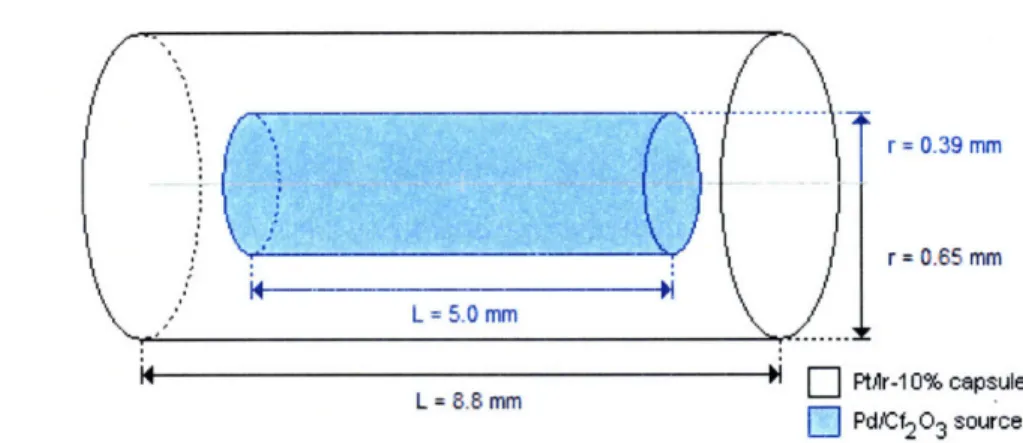
1.1.3 CALIFORNIUM BRACHYTHERAPY
252
Cf is a man-made trans-plutonium radionuclide that largely undergoes alpha
decay; however, 3.09% of nuclear transitions follow spontaneous fission.
33The half-life
of
252Cf is
2.645
y. Oak Ridge National Laboratory (ORNL) produced Cf isotopes from
approximately 100 g highly-enriched heavy Cm targets (
244Cm,
246Cm, and
248Cm)
irradiated in the High-Flux Isotope Reactor for
-7
months.
34Combinations of neutron
activation and beta decay leads to successively higher atomic numbers. For example,
neutron activation of 248Cm produces 249Cm (ty, = 64 m) that beta decays to 24 9Bk (ty, =
330 d) that beta decays
to 249Cfrequired to produce Cf isotopes with atomic numbers 249-254.33 Due to the amount of time required to produce 252Cf and the extensive radiochemistry needed to separate Cf
isotopes from the Cm target, 252Cf is both extremely rare and expensive.
Clinical use of the neutron-emitting radionuclide 252Cf was first proposed in 1965 by Schlea and Stoddard.35 Schlea and Stoddard noted that early radiobiological studies of external neutron beam therapy demonstrated effectiveness in anoxic tumors that were typically resistant to conventional photon therapies. They also described the advantage of intracavitary and interstitial brachytherapy treatment over external neutron beam therapy, which includes higher radiation doses to healthy tissue.3s The first medical source containing 252Cf was described in 1967 and was modeled after a radium needle.36
Thousands of patients worldwide have been treated over the past 40 years with 252Cf-based brachytherapy.36 The mixed neutron and photon radiation has been shown to be particularly effective for large tumors and traditionally radio-resistant tumors, such as melanomas and late-stage cervical carcinomas. For example, Tacev et al. have reported increased survival rates (+16%, p < 0.002) and decreased relapse rates (-17%, p <
0.0002) using 252Cf for the treatment of advanced cervical carcinoma with twelve years post treatment follow-up in comparison to conventional treatment.37 The increased effectiveness is attributed to a number of radiological characteristics of neutrons, such as increased relative biological effectiveness (RBE), decreased oxygen effect, and decreased cell-cycle dependence. In the United States, Yosh Maruyama M.D. treated many disease sites and published nearly one hundred clinical papers on the subject before his death in 1995. Consider ref. 36 for a detailed overview of medical applications of 252Cf brachytherapy and summary of clinical results for head and neck, gynecological, skin, rectal, esophageal, prostate, and brain tumors, among others.
Brachytherapy dosimetry for 252Cf sources was aggressively pursued in the early 1970s once medical sources were available.38-4 Calculations by Krishnaswamy used MC
methods to determine a point source model for four different source types, including the Applicator Tube (AT) source with source length L = 1.5 cm.38 Each point source was subsequently used in a numerical integration to calculate the dose to points up to 5 cm along and away from the source. Neutron kerma factors were not reported, and the photon cross-sections of Storm and Israel41 were employed. Colvett et al. followed with
measurements using paired ion chambers (tissue equivalent and Al) in a large volume of tissue equivalent liquid to simulate infinite scatter conditions. Measurements from 1 to 5 cm on the transverse plane agreed within 0.94 ± 0.02 to Krishnaswamy's calculations; however, the measurements did not agree favorably with other reported measurements.3 9
Differences between measurements were attributed to differences in phantom size, which resulted in varying scatter conditions, and to differences in chamber or 252Cf source calibration.39,40 Windham et al. calculated dose distributions for 252Cf sources using a one-dimensional discrete ordinates code, 21 energy groups, and neutron kerma factors published by Ritz et al.40,42 Calculations by Windham et al. were in good agreement with the Krishnaswamy calculations (1.01 ± 0.03) and Colvett et al. measurements (0.97 ± 0.02) for 0.5 < r < 5.0 cm on the transverse plane. Anderson reviewed these and six other publications (3 calculations and 3 measurements) in a 1973 review paper on 252Cf
dosimetry.43 The measurements of Colvett et al. were recommended for 252Cf, although, the Colvett et al. photon data were only recommended for AT-type sources. The calculations of Krishnaswamy for non-AT-type sources needle sources were the recommended alternative.43 These data were reported as along-and-away tables up to 5 cm from the source center.
The most recent determinations of 252Cf neutron dose distribution about a low-dose rate source were published by Yanch and Zamenhof in 199244 and by Rivard et al. in 2000.30 Yanch and Zamenhof calculated along-and-away tables using MC methods for AT-type sources.44 Rivard adapted the TG-43 formalism for a generalized source of variable active length and provided brachytherapy dosimetry parameters for the neutron dose component.30
To date, 252Cf brachytherapy in the United States is applied using manually loaded, LDR sources delivering below 2 Gy h-1 to the prescription point during patient treatment.36 Both manual loading and LDR treatment increases radiological risk to the brachytherapist and support personnel who must attend to patient needs during long treatment sessions. Considering these issues, there is an interest in employing 252Cf brachytherapy in the HDR regime. In addition, remote afterloading technology, which is well-documented and successful for HDR 192Ir brachytherapy, is being considered for 252Cf.45
has not been attempted because sources comparable in size and geometry to conventional HDR 192Ir sources have only recently become possible due to advances in radiochemistry.4s Furthermore, while radiochemistry now allows an HDR source containing > 1 mg/mm3 of 252Cf, one has not been constructed.
1.1.4 RADIOLOGICAL PROTECTION
Brachytherapy presents unique radiological protection concerns for the brachytherapist. Marie Curie, one of the first people to regularly handle encapsulated radioactive sources, experienced radiation burns on her fingers after extended handling periods.' Henri Becquerel similarly experienced skin erythema and desquamation after carrying a few tenths of a gram of radium chloride in a glass tube in his pocket for six hours. After noting a less intense reaction from carrying the source for one hour, he repeated the experiment by placing the source in a 5 mm-thick lead capsule and noting that the shielded source needed significantly more time to produce a biological effect.46 Many of the early experimenters and clinicians died as a result of their repeated radiation exposures.
Until the development of remote afterloading technology, brachytherapy procedures required manual loading or placement of sources into treatment position. These placed the brachytherapist at risk for repeated radiobiological harm. Thus, tools were developed to increase the distance or to incorporate shielding between clinicians and brachytherapy sources. Furthermore, LDR treatments could have durations exceeding one week that posed a radiological risk to ancillary clinical staff, such as nurses, who were attending to patient needs. Generally, these concerns were addressed by using patient rooms in relatively remote locations with portable, rolling shields to reduce radiation levels outside the room.
However, the methods that were effective for traditional photon-based brachytherapy would not provide adequate protection for neutron-based brachytherapy using 252Cf. While Pb shields would be effective against the photon emissions from a
252Cf source, Pb does not notably attenuate or moderate neutrons. Maruyama et al. noted
that early 252Cf treatment centers utilized a treatment vault that was originally designed for megavoltage energy photon beams.36 At one facility in the Czech Republic, a vault designed for a 60Co treatment unit was augmented with a 24-cm thick layer of borated
polyethylene before being used for 252Cf therapy.47 Borated polyethylene is an effective neutron shield because the hydrogen component in polyethylene moderates the neutrons, increasing their probability of absorption by boron, which has a high neutron-capture cross section.
During HDR 2 5 2Cf therapy, treatment vaults will be subjected to extremely high neutron fluence. One mg of 2 5 2Cf emits 2.314 x 10⁹ (± 2%) neutrons s-1 and 1.3 x 10¹⁰
(± 4%) photons s- 1, which correlate to approximately 0.28 Gy h-1 and 0.14 Gy h-1 at 1 m from an unshielded source.45s'4 As such, it is possible for the instantaneous dose rate outside of the treatment vault to exceed regulatory limits. Current regulations specifiy maximum permissible radiation doses to a member of the public at 0.02 mSv in one hour and 0.1 mSv in one year.
Shielding calculations for 2 5 2Cf emissions were published in technical reports of the Savannah River Laboratory by Hootman (1970) and by Hootman and Stoddard (1971).48,49 These shielding analyses solved a one-dimensional Boltzman equation in a slab, sphere or cylindrical geometry using discrete ordinates and anisotropic scattering.4 8 In addition, the calculations included RBE factors [sic] to convert absorbed dose (rad or Gy) to equivalent dose (rem or Sv). Historical RBE values (1957) were employed because radiation weighting factors were not available at the time. The International Council for Radiation Protection (ICRP) recommended radiation weighting factors for dose equivalent calculations in Report 60 (1990).50
1.2 Thesis statement
1.2.1 PHOTON DOSIMETRY
As noted in section 1.1.2, the AAPM TG-43 brachytherapy dosimetry formalism determines dose distributions in a semi-infinite volume of liquid water, i.e., dose calculations assume negligible losses due to energy escape from the phantom. Unfortunately, this assumption rarely simulates patient geometry. Similarly, differences between energy absorption in water and other media have been studied with corrections implemented for external beam treatments. However, corrections are not used for brachytherapy.
These concepts are explored in Chapter 2, where dose distributions from virtual and commercial photon-emitting brachytherapy sources were calculated using MC methods and compared to the current brachytherapy dosimetry formalism in AAPM TG-43U1 Report. While the TG-TG-43U1 report describes commercial low-energy photon-emitting 1251 and 103pd sources, the formalism was applied to a number of radionuclide
sources spanning a wide energy range. Furthermore, mono-energetic sources were employed to identify generalizable characteristics in photon-based brachytherapy dosimetry parameters.
These observations were subsequently applied to eye plaque brachytherapy using commercial 10 3Pd, 125I, and 1 31Cs brachytherapy sources. Gold-alloy plaques provide
significant attenuation of emitted radiation, but existing treatment planning systems exclude these effects. Furthermore, material inhomogeneities in plaque components have been shown to perturb single-seed 125I dose distributions by as much as 10% at clinically relevant distances.5 1 MC-based treatment simulations described in section 2.2
demonstrate changes in dose distributions for fully-loaded eye plaques using common low-energy radionuclide sources. Correction factors accounting for these effects were derived for conventional treatment plans.
1.2.2 NEUTRON DOSIMETRY
Chapter 3 extends the photon-specific effects described in Chapter 2 to
neutron-emitting sources. Thus, generalizable characteristics of neutron brachytherapy were derived and compared to the dosimetry formalism of AAPM TG-43U1 Report in section
3.1. Distinctions between dose distributions obtained for photon and neutron brachytherapy were established towards a better understanding of the photon contribution in neutron-based brachytherapy. Furthermore, recommendations to future dosimetry investigators were presented.
These analyses were broadened to a theoretical HDR 252Cf source in section 3.2.
As discussed in section 1.1.3, HDR 252Cf sources are still in development. However, comparisons were made between LDR AT-type sources and a theoretical HDR source in preparation for neutron-based HDR treatment.
The clinical impact of partial scatter conditions during surface plaque brachytherapy was examined for comparison to the semi-infinite water phantom assumed
by the AAPM TG-43U1 dosimetry formalism in section 3.3. MC calculations were
performed for comparison to treatment plans calculated using conventional brachytherapy treatment planning software in support of a 252Cf brachytherapy clinical
trial.
1.2.3 SHIELDING CONSIDERATIONS
In further support of neutron-based brachytherapy using HDR 252Cf, the shielding requirements for mg quantities of 2 52Cf were evaluated using MC methods in Chapter 4. Shielding properties of concrete, barite concrete, lead, water, polyethelyene, and borated polyethylene were evaluated for thicknesses up to 1 m in section 4.1. MC calculations employed three dimensional radiation transport modeling, modem cross-section libraries, and contemporary radiation weighting factors.
Finally, a linear accelerator vault at Tufts-New England Medical Center in Boston, MA was assessed for shielding an HDR-equivalent 2 5 2Cf source in section 4.2. MC calculations were performed in conjunction with ionization chamber, proportional counter, and track-etch detector measurements of a 252Cf neutron radiography source (> 1 mg) positioned where treatment would occur within the accelerator vault. The resulting measurements were utilized to estimate limitations on patient throughput due to radiation exposure to personnel and to the public during HDR 2 52Cf treatment assuming
conventional permissible exposures.
1.3 Data, reference, and publication heritage
Segments of this dissertation are reprinted or reproduced from previously published materials. These materials include two reviewed publications, one peer-reviewed abstract, two peer-peer-reviewed papers from a conference proceeding, and one
manuscript submitted for publication.
Section 2.1 is reprinted from: Melhus and Rivard "Approaches to calculating AAPM TG-43 brachytherapy dosimetry parameters for 137Cs,
1251, 192Ir, 103Pd, and 169Yb
sources" in Medical Physics Vol. 33, pgs. 1729-1737 (© 2006 by the AAPM).s2
Section 2.2 is reproduced from the manuscript "COMS eye plaque brachytherapy dosimetry for 103Pd, 125I, and 13 1Cs" by Melhus and Rivard, submitted (07-853) to
Section 3.2 is from Melhus et al. "Clinical brachytherapy dosimetry parameters
and mixed-field dosimetry for a high dose rate Cf-252 brachytherapy source" in The Monte Carlo method: Versatility unbounded in a dynamic computing world published by the American Nuclear Society (ANS; © 2005 by the ANS, La Grange Park, IL).5 4
Section 3.3.1 is from Melhus and Rivard "Monte Carlo validation of clinical
brachytherapy dosimetry under partial scatter conditions for neutron-emitting sources" in
Medical Physics Vol. 34, pg. 2330 (C 2007 by the AAPM).55
Section 4.1, "Storage safe shielding assessment for a HDR californium-252
brachytherapy source," by Melhus et al. is also reprinted from The Monte Carlo method: Versatility unbounded in a dynamic computing world (© 2005 by the ANS, La Grange Park, IL).56
Section 4.2 by Melhus et al. titled "Shielding evaluation of a medical linear
accelerator vault in preparation for installing a high dose rate 252Cf remote afterloader" was published in Radiation Protection Dosimetry Vol. 113, pgs. 428-437 (© 2004 by C.S. Melhus).5 7
2
PHOTON DOSIMETRY
2.1 Approaches to calculating AAPM TG-43 brachytherapy dosimetry
parameters for
137Cs,
125I, 192Ir, 103Pd, and
169Yb sourcest
2.1.1 ABSTRACT
Underlying characteristics in brachytherapy dosimetry parameters for medical radionuclides 137Cs, 125I, 192Ir, 103pd, and 169yb were examined using Monte Carlo methods. Sources were modeled as un-encapsulated point or line sources in liquid water to negate variations due to materials and construction. Importance of phantom size, mode of radiation transport physics - i.e., photon transport only (MODE P) or coupled photon:electron transport (MODE PE), phantom material, volume averaging, and Monte Carlo tally type were studied. For non-infinite media, g(r) was found to degrade as r approached R, the phantom radius. MCNP5 results were in agreement with those published using GEANT4. Brachytherapy dosimetry parameters calculated using coupled photon:electron radiation transport simulations did not differ significantly from those using photon transport only. Radial dose from low-energy photon-emitting radionuclides 125I and 10 3Pd were sensitive to phantom material by upto a factor of 1.4 and 2.0, respectively, between tissue-equivalent materials and water at r = 9 cm. In
comparison, high-energy photons from 137Cs, 19 2
Ir, and 16 9Yb demonstrated ± 5% differences in radial dose between water and tissue-substitutes at r = 20 cm. Similarly, volume-averaging effects were found to be more significant for low-energy
t Reproduced from: C. S. Melhus and M. J. Rivard, "Approaches to calculating AAPM TG-43 brachytherapy dosimetry parameters for 13 7Cs, 1251, 192Ir, 10 3Pd, and 169yb sources," Med. Phys. 33,
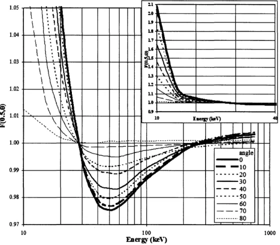
radionuclides. When modeling line sources with L 5 0.5 cm, the 2-D anisotropy function was largely within ± 0.5% of unity for 137Cs, 1251, and 192Ir. However, an energy and
geometry effect was noted for 10 3Pd and 69Yb, with Pd-103F(0.5,00) = 1.05 and
Yb-169F(0.5,00) = 0.98 for L = 0.5 cm. Simulations of monoenergetic photons for L = 0.5 cm produced energy-dependent variations in F(r,0) having a maximum value at 10 keV, minimum at 50 keV, and - 1.0 for higher-energy photons up to 750 keV. Both the F6
cell heating and *F4 track-length estimators were employed to determine brachytherapy dosimetry parameters. F6 was found to be necessary for g(r), while both tallies provided equivalent results for F(r,0).
2.1.2 INTRODUCTION
For brachytherapy dosimetry calculations using radiation transport codes, TG-43U1 makes a number of "good practice" recommendations.20 These nine recommendations include allowing for adequate backscatter material, utilizing modem photon cross-sections, and maintaining volume-averaging effects below 1%, among others. Regardless of the TG-43U1 guidance, the dosimetry investigator is allowed considerable flexibility in designing and conducting both simulations and measurements, resulting in notable methodology variations between authors. These variations are clearly evidenced in Appendix A of TG-43U1, which includes a discussion of the number of authors and publications dedicated to an individual seed design (manufacturer and model), and each publication contains investigator-specific methodologies and approaches. Additional complexity is added as the scope extends to multiple seed models, each with a unique subset of contributing dosimetry investigators and related methodologies that change over time in response to new discoveries, other publications, and AAPM recommendations.
Furthermore, specific dosimetry parameters have garnered considerable interest in the brachytherapy dosimetry community during recent years.58ss 63 For example, dosimetry investigators have suggested both adding complexity" and simplifying the geometry function, G(r,0),60 towards improving brachytherapy dosimetry. The AAPM TG-43UI currently recommends either a line- or point-source approach to approximate all commercially available brachytherapy seeds. As much of the recent interest relates to
derivation of G(r,0), the AAPM published a clarification stressing the need for consistent application of a geometry function, rather than accurate representation of particle streaming behavior.63
Unlike G(r,0), which is not a function of radionuclide energy spectrum and is purely mathematical, other AAPM TG-43UI brachytherapy dosimetry parameters are determined by comparing absorbed dose measurements or calculations within a phantom. The brachytherapy dosimetry investigator must carefully model and sample the phantom to avoid introducing bias related to the radiation transport model or technique. Errors incurred through inappropriate technique usage can be magnified when dose distribution results are processed using the dosimetry formalism because of the relative nature of the calculations.
Assuming further advances in computer processing capabilities, the current dosimetry formalism standard may eventually be abandoned in favor of direct MC-based brachytherapy dosimetry simulations,64,'6 though careful measurements will be needed to commission such a system. At present, the brachytherapy dosimetry formalism presented in the AAPM TG-43U1 report represents the current standard. Therefore, this publication provides reference brachytherapy dosimetry parameters for a variety of un-encapsulated medical radionuclides using MCNP5. This goal will not only elucidate inherent characteristics of each individual source photon spectrum, but also provide reference data towards comparing with other MC-based brachytherapy dosimetry calculations. At the same time, the impact of phantom geometry, including backscatter and sampling volume effects, and phantom material will be explored for MCNP5.
2.1.3 MATERIALS AND METHODS
Dose distributions from five radionuclides (137Cs, 1251, 192Ir, 10 3Pd, and 169Yb) used for brachytherapy were modeled. The 2004 update to the AAPM TG-43 report included only low-energy photon emitting brachytherapy sources containing 1251 or 103Pd.20 This study employed the same formalism and extended it to high-energy photon sources (EAVG
> 50 keV) with a more energetic population of secondary particles. All photon energies
were included in spectral characterization with the exception of those below 9 keV, because the average pathlength is < 0.2 cm in water. Photon and electron spectra for each
radionuclide were taken from the National Nuclear Data Center (NNDC).33 Furthermore, AAPM TG-43U1 recommended photon energy and emission frequencies for 1251 and
103Pd and the 192Ir spectrum of Glasgow and Dillman66 were used in radiation transport simulations for comparison to results obtained using NNDC-published values.
Simulations were performed using version 5 of the MCNP Monte Carlo code (MCNP5) developed by Los Alamos National Laboratory.6 7 The default MCNP5 photon cross-section library, p04, was applied, which is based upon Release 8 of ENDF/B-VI.68 Computed results of cell particle fluence, energy fluence, and heating were obtained using the MCNP F4, *F4, and F6 tallies, respectively. These three tallies correspond to track length estimates in a cell of photon flux, product of photon flux with photon energy, and energy deposition, respectively. Source encapsulation was not included in any of the simulations in order to ascertain underlying characteristics of each radiation spectrum in materials of dosimetric interest and to eliminate manufacturer-specific effects due to source construction. Water was modeled using an atomic ratio of 2:1 for H:O and a mass density of 0.998 g cm3 at standard temperature and pressure, 220C and 101.325 kPa,
respectively. Due to their low-energy photon emissions and subsequent rapid dose falloff, 103Pd and 125I results were excluded beyond 9 cm and 15 cm, respectively.69
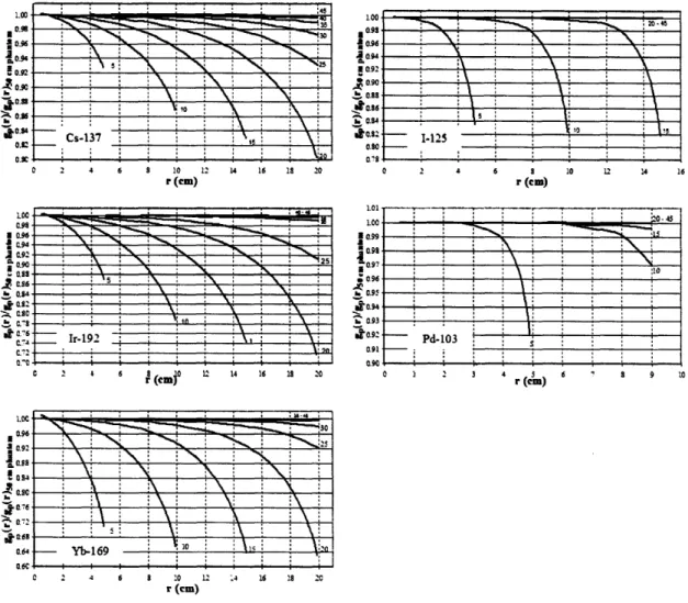
2.1.3.1 Radial dose function
The radial dose function, g(r), was assessed for each radionuclide photon spectrum using an un-encapsulated point source in a spherical water phantom with R = 50 cm in radius, i.e., X-Ag(r)R. where the prefix subscript denotes the radionuclide and the suffix subscript denotes the phantom radius. Concentric spheres were designed to create spherical shells in which MCNP5 tallies were computed, allowing calculation of g(r) at radial distances between 0.1 and 20 cm. Values of R were 5, 10, 15, 20, 25, 30, 35, 40, and 45 cm were used to assess full scatter conditions. While a smaller radius is generally representative of human anatomy, e.g., the R = 15 cm used by Daskalov et. al. for HDR 192Ir calculations29, g(r)
50 data is presented here. Varying the radial width of the sampling volume, i.e., the thickness of the spherical shell, assessed the impact of volumetric averaging. Data were collected for 0.01, 0.02, 0.05, 0.1, 0.2, 0.3, 0.5, 1, and 2 cm thick shells, as well as for a variable thickness of 5% or 10% of the radial distance. The inner
and outer radii defining the sampling volume were equally displaced from the reported point of calculation. Volume averaging errors caused by non-zero voxel width were evaluated for ± 0.5% variation in g(r) by comparing results to the 0.01 cm-thick voxel. Simulations were also performed in breast tissue, muscle, soft tissue, and four-component soft tissue, as defined in ICRU 44,70 and presented by NIST,71 to compare with the in-water calculations required by TG-43U1.
Source emissions were modeled as photon and electron spectra for all five radionuclides. To determine the impact of simplifying radiation transport calculations for the five photon source spectra, photon-only (MODE P) and coupled photon:electron (MODE PE) calculations were performed. Furthermore, simulations of electrons emitted following P-decay or electron capture, including auger electrons, were performed using the default MCNP5 electron transport mode (par = 3) to evaluate whether electron-only transport (MODE E) or secondary photons generated by electrons (MODE PE) contributed to total radial dose deposition. The algorithm used for default electron transport calculation is subject to non-physical energy effects, see for example Ref. 72, and may not accurately reproduce electron dose distributions. As such, comparisons of electron and photon transport are made on a relative basis to show order-of-magnitude results.
The F6 energy deposition tally was evaluated by transporting 0.001 to 10 MeV photons from a point source through a sphere of liquid water(R = 15 cm) and sampling within a thin sampling volume (spherical shell) 0.01 cm from the source. Using the tally energy card (En), energy bins were established with an average energy bin width of 7% ±
6% (± 1 s.d.) of the bin energy. A ratio of F6 and *F4 results for each energy bin was taken to evaluate energy absorption coefficients (!len p-1) employed when using MCNP5. The dose rate per mCi was calculated from F6 results at (ro,00) to allow comparison of radial dose data in a non-normalized manner. The normalized and balkanized nature of TG-43U1 dosimetry parameters, such as g(r), precludes clear understanding of absolute dose rates. Thus, the dosimetry investigator may use dose rate values for additional comparison and benchmarking. It is important to note, however, that the AAPM discourages use of apparent activity or exposure rate constants that describe dose rate as a
The statistical uncertainties for 107 starting particles were < 0.05% at r = 1 cm for
all radionuclides for a spherical shell with radial thickness of 0.01 cm. At r = 10 cm, statistical uncertainties were 5 0.06% for high-energy sources and 0.2% for 125I. For 103Pd, statistical uncertainties were 0.7% and 0.1% for 107 and 5x10⁸ starting particles, respectively. At r = 20 cm, statistical uncertainties were 5 0.1% for 13 7Cs, 192Ir, and
169yb"
2.1.3.2 MC F(r, 9) analysis
The 2-D anisotropy function F(r,0) was calculated for six active lengths (0, 0.1, 0.2, 0.3, 0.4, and 0.5 cm) in a liquid water phantom of R = 25 cm for low-energy and R = 40 cm for high-energy radionuclides. Monoenergetic photons were generated for active lengths of 0.1, 0.3, and 0.5 cm at 10, 20, 30, 50, 75, 100, 200, 300, 500, and 750 keV for comparison to the radionuclide energy spectra. Line sources were approximated using a cylinder with radius 10-6 cm. A series of intersecting concentric spheres and cones were
designed to provide 10 angular resolution about the long axis of the source. Data were calculated at 11 radial distances between 0.5 and 12 cm, and two voxel thickness models were examined - a fixed radial width of 0.02 cm and variable width determined using
10% of the radial distance at the point of calculation.
Monte Carlo simulations were performed using MODE P transport. A total of 5 x 10⁷ starting particles were utilized for all radionuclides, with the exception of l03Pd that required 2.5 x 10⁸ starting particle histories for improved statistics. The concentric cones result in sampling volumes that are cylindrically located about the long axis of the source. As a result the minimum statistical uncertainty occurs on the transverse plane (0 = 900) and the maximum uncertainty is located on the long axis of the source (0 = 00 and 1800), where the sampling volume is approximately 450 times smaller. At a radial distance of 1 cm from a point source and a bin-width of 10% of the radial distance, the statistical uncertainties at 50 and 85' were 0.5% and 0.2% for 137Cs; 0.5% and 0.1% for 125I; 0.5% and 0.2% for 19 2Ir; 0.3% and 0.1% for
10 3Pd; and, 0.5% and 0.2% for 169Yb, respectively. At a radial distance of 10 cm from a point source and a bin-width of 10% of the radial distance, the statistical uncertainties at 50 and 850 were 0.5% and 0.1% for 13 7Cs; 1.4%
and 0.4% for 125I; 0.4% and 0.1% for 19 2Ir; 3.3% and 1.0% for 103Pd; and, 0.4% and 0.1%
for 169Yb, respectively.
In addition, data from the transverse plane were taken to determine gL(r) for each active length and radionuclide, and a comparison of the point source (L = 0 cm) result
was made to the values calculated using concentric spheres. In this way, results obtained using 4t geometry were compared to those employing the relatively small sampling volumes introduced using concentric cones as described above.
2.1.4 RESULTS AND DISCUSSION
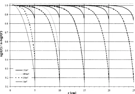
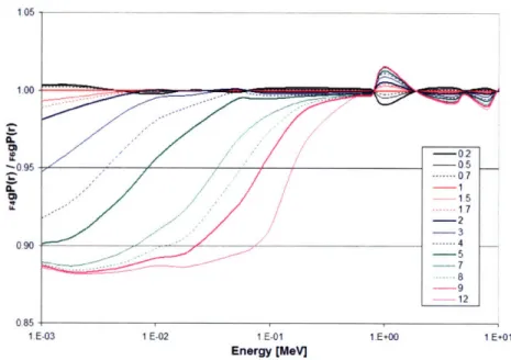
2.1.4.1 Phantom size
The effect of phantom size is shown in Figure 2.1.1 as the ratio of gp(r) calculated for a given phantom thickness and gp(r) for a 50 cm radius phantom. For all radionuclides at any distance, g(r) for R < 50 cm was less than or equal to g(r)50. Furthermore, as r approached R, the ratio of g(r)/g(r)50o rapidly diminished from unity.
This decrease became more pronounced as r increased due to the increased proportion of scatter dose compared to primary dose. Similarly, this increase was more pronounced and gradual for sources having higher average photon energy (137Cs, 192Ir, 169Yb) in comparison to lower average energy sources (125I and 103Pd).
*~ ~ \> ~ K'o Cs-137 ! ! ! ! , 1.00 II 099 0.96 S0.92 1~0.900 ,. 0.90 b,0.23 .0.340o.$ 0.80 07S 0 2 4 6 0 10 12 14 16 18 20 (Cm) N. N 2 5 i Ir- 1921 a r 0 1 L Ij 14 16 is 20 U .• &0.84 80.92 0.90 to.So 0.92 .00 .09, 20-45 1-125 o , . I 2 4 6 8 10 12 14 10 r (em) i _ _______ 20,4is ~ 094 h 005 .1092 Pd-103 0.91 o090 0 2 3 4 r(c) 6 9 10 S.900 - 0.94 ~o.-i • -, ^ • - Yb-169 0 2 4 6 8 10 12 .4 16 18 2C r (cm)
Figure 2.1.1: Comparison of g(r) values calculated for a given phantom size, R,
compared to R = 50 cm, which is assumed to provide full scatter conditions. Data for 7Cs, 125I, 192Ir, 103Pd, and 169yb include curves for R = 5, 10, 15, 20, 25, 30,
35, 40, and 45 cm shown from left to right. Curves for large phantom sizes within statistical uncertainty of unity, e.g., R = 45 and 40 cm, are not discernable. Note the variation in the x-ordinate for the low-energy radionuclides 125I and '03Pd.
Similarly, P6rez-Calatayud et al. determined phantom sizes needed to provide full scatter conditions for 137Cs, 125I, 192Ir, and 103Pd using the GEANT4 Monte Carlo code.73
Although they did not report the photon cross-sections utilized in their study, reasonable comparisons may be made between results using the GEANT4 and MCNP5 Monte Carlo codes. P6rez-Calatayud et al. observed full scatter conditions within 0.5% for I-125g(10)15
and Pd-103g(10)15. Using MCNP5 in this study, 1-125g(10)1s/I.125g(10)so = 0.997
±
0.001 andPd-103g(l0)15/Pd-103g(10)5o = 0.989 ± 0.002. This study obtained Cs-137g(20)40/Cs-137g(20)50o =
0.996 ± 0.001 and Ir-192g(20)40/lr-192g(20)50 = 0.997 ± 0.001 which is in agreement with
1.00 1.01 LO0 S.099 0.t97 A--. S0.90-.c.09 ot oz S004 - .10.02-at 0-! C.2 10 A V: -- 'I Il I I I I I I ' '•
Sr~
IC_ i __t
n-cu 0.6c -FC -ý --. -.--- M38--~ ' I l L .... I :'• • :•',• • • : : i 0.6Perez-Calatayud et al. who noted full scatter conditions within 0.5% for g(20)40 for both radionuclides. Excellent agreement was also obtained for 137Cs and 192Ir at all r and R, with differences < 0.3% between our results and those obtained by Perez-Calatayud et al. We recommended R > 40 cm for 16 9yb to provide full scatter conditions (within 0.2%)
for r < 20 cm.
2.1.4.2 MCNP5 F6 tally
The dosimetry investigator using MCNP5 has several cell or surface-based tally options to choose from. Volume-based tallies are preferred as they reduce the statistical uncertainties and improve the accuracy of MCNP5 simulations, though, the sampling volume must be sufficiently small to minimize differences in particle fluence and average photon path length within the cell. The *F4 tally, providing energy flux results in MeV cm-2, can be converted to absorbed dose through application of appropriate Pen P-1 coefficeints. However, the investigator is required to segment results into energy bins compatible with published pen p-I coefficeints, or include a tally multiplier (FMn) card to convert results from MeV cm-2 to MeV g-1 in a direct fashion. Using F6 tally results and
a radial voxel thickness of 0.01 cm, un-encapsulated g(r) in water for a point source is shown in Table 2.1.1. These reference data may be used to benchmark other radiation transport codes and ensure accurate derivation of brachytherapy dosimetry parameters between other sampling geometries and radiation transport codes.
Table 2.1.1: Radial dose functions for un-encapsulated point sources in a 50
cm-radius liquid water phantom using the MCNP5 F6 cell-heating tally. Based upon the number of histories simulated and the MCNP5 coefficient of variation in the tally result, all results have a maximum statistical uncertainty of 0.1%. The tally volume at each distance was a spherical shell of radial thickness 0.01 cm centered on the specified r. Italicized data for
attributed to substantive attenuation.
1251 r [cm] 0.1 0.2 0.3 0.5 0.8 1.0 1.5 2.0 2.5 3.0 3.5 4.0 4.5 5.0 6.0 7.0 8.0 9.0 10.0 11.0 12.0 13.0 14.0 15.0 16.0 17.0 18.0 19.0 20.0 1.006 1.006 1.005 1.004 1.002 1.000 0.995 0.990 0.985 0.979 0.972 0.966 0.958 0.951 0.936 0.920 0.903 0.885 0.866 0.847 0.826 0.805 0.784 0.763 0.740 0.717 0.695 0.672 0.649 1.004 1.018 1.025 1.030 1.017 1.000 0.939 0.862 0.780 0.699 0.622 0.549 0.482 0.422 0.319 0.239 0.179 0.132 0.0972 0.0719 0.0527 0.0386 0.0283 0.0207 0.0152 0.0112 0.0082 0.0061 0.0044
'ZI and '0 3Pd indicate larger uncertainties
gp(r)
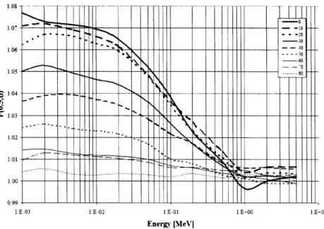
192Ir 0.988 0.990 0.992 0.994 0.998 1.000 1.005 1.009 1.012 1.014 1.015 1.015 1.014 1.013 1.006 0.997 0.984 0.968 0.949 0.927 0.903 0.876 0.848 0.817 0.785 0.751 0.715 0.677 0.638 1.469 1.429 1.381 1.274 1.107 1.000 0.761 0.571 0.424 0.313 0.229 0.168 0.123 0.0902 0.0483 0.0261 0.0142 0.0080 0.0046 0.0029 0.0019 0.0013 0.0010 0.0008 0.000 7 0.0006 0.0006 0.0005 0.0005 0.874 0.890 0.905 0.934 0.974 1.000 1.056 1.103 1.142 1.172 1.197 1.213 1.224 1.229 1.226 1.206 1.175 1.135 1.088 1.035 0.983 0.927 0.871 0.816 0.763 0.712 0.6616 0.6152 0.5705Towards determining these parameters, the F6 tally with appropriate units of MeV
g-1 is recommended for simulating radiation transport using MCNP because it directly
correlates with absorbed dose rate without using potentially inconsistent g p-1
_ __
![Figure 2.2.3: Dose [Gy] for a 16 mm COMS eye plaque with SK per seed to deliver DR, (5 mm) = 85 Gy](https://thumb-eu.123doks.com/thumbv2/123doknet/13878115.446578/57.918.165.765.244.594/figure-dose-gy-coms-eye-plaque-seed-deliver.webp)