HAL Id: hal-01785372
https://hal-amu.archives-ouvertes.fr/hal-01785372v2
Submitted on 4 May 2018
HAL is a multi-disciplinary open access
archive for the deposit and dissemination of
sci-entific research documents, whether they are
pub-lished or not. The documents may come from
teaching and research institutions in France or
abroad, or from public or private research centers.
L’archive ouverte pluridisciplinaire HAL, est
destinée au dépôt et à la diffusion de documents
scientifiques de niveau recherche, publiés ou non,
émanant des établissements d’enseignement et de
recherche français ou étrangers, des laboratoires
publics ou privés.
Comet assay on thawed embryos: An optimized
technique to evaluate DNA damage in mouse embryos
L. Rolland, B. Courbiere, V. Tassistro, A. Sansoni, T. Orsiere, W. Liu, C. Di
Giorgio, J. Perrin
To cite this version:
L. Rolland, B. Courbiere, V. Tassistro, A. Sansoni, T. Orsiere, et al.. Comet assay on thawed embryos:
An optimized technique to evaluate DNA damage in mouse embryos. Toxicology in Vitro, Elsevier,
2017, 44, pp.266-272. �10.1016/j.tiv.2017.07.010�. �hal-01785372v2�
Contents lists available atScienceDirect
Toxicology in Vitro
journal homepage:www.elsevier.com/locate/toxinvit
Comet assay on thawed embryos: An optimized technique to evaluate DNA
damage in mouse embryos
L. Rolland
a,b, B. Courbiere
a,b, V. Tassistro
b, A. Sansoni
c, T. Orsière
b, W. Liu
d, C. Di Giorgio
e,
J. Perrin
a,b,f,⁎aDepartment of Gynecology, Obstetrics and Reproductive Medicine, AP-HM La Conception, Pôle femmes parents enfants, 147 bd Baille, 13005 Marseille, France bInstitut Méditerranéen de Biodiversité et d'Ecologie marine et continentale (IMBE), Aix Marseille Univ, CNRS, IRD, Univ Avignon, Marseille, France cCentre d'Immunophénomique - CIPHE, PHENOMIN, INSERM US012, CNRS UMS3367, UM2 Aix-Marseille Université Marseille, France dCNRS, Aix Marseille Univ, IRD, CEREGE UM34, UMR 7330, 13545 Aix en Provence, France
eLaboratoire de mutagagénèse environnementale, Aix Marseille Univ, Univ Avignon, CNRS, IRD, IMBE, Marseille, France
fCECOS, Laboratory of Reproductive Biology, Department of Gynecology, Obstetric and Reproductive Medicine, Pôle femmes parents enfants, AP-HM La Conception, 147 bd Baille, 13005 Marseille, France
A R T I C L E I N F O
Keywords: DNA damage embryo cryopreservation Single-Cell Gel Electrophoresis technical issue
literature review
A B S T R A C T
Our objective was to optimize the CA technique on mammal embryos.
Materials and methods: 1000 frozen 2-cell embryos from B6CBA mice were used. Based on a literature review, and after checking post-thaw embryo viability, the main outcome measures included: 1) comparison of the embryo recovery rate between 2 CA protocols (2 agarose layers and 3 agarose layers); 2) comparison of DNA damage by the CA on embryos with (ZP +) and without (ZP−) zona pellucida; and 3) comparison of DNA damage in embryos exposed to 2 genotoxic agents (H2O2and simulated sunlight irradiation (SSI)). DNA damage was quantified by the % tail DNA.
Results: 1) The recovery rate was 3,3% (n = 5/150) with the 2 agarose layers protocol and 71,3% (n = 266/ 371) with the 3 agarose layers protocol. 2) DNA damage did not differ statistically significantly between ZP− and ZP+ embryos (12.60 ± 2.53% Tail DNA vs 11.04 ± 1.50 (p = 0.583) for the control group and 49.23 ± 4.16 vs 41.13 ± 4.31 (p = 0.182) for the H2O2group); 3) H2O2and SSI induced a statistically sig-nificant increase in DNA damage compared with the control group (41.13 ± 4.31% Tail DNA, 36.33 ± 3.02 and 11.04 ± 1.50 (p < 0.0001)).
The CA on mammal embryos was optimized by using thawed embryos, by avoiding ZP removal and by the adjunction of a third agarose layer.
1. Introduction
The exposure of parents to environmental toxicants, such as poly-cyclic aromatic hydrocarbons (Einaudi et al., 2014; Perrin et al., 2011), Bisphenol A (Goldstone et al., 2015), solvents (Kolstad et al., 1999), metals (Thompson and Bannigan, 2008; Zhou et al., 2016) or various therapies (Bujan et al., 2014; Esquerré-Lamare et al., 2015; Pecou et al., 2009; Roti Roti et al., 2012) may induce DNA damage in male and female germ cells. DNA damage in parental germ cells can lead to re-productive issues, such as reduced fertilization, impaired early em-bryonic development, a decreased pregnancy rate and increased mis-carriage rate (Simon et al., 2014; Zhao et al., 2014). The transmission of paternal germ cell DNA damage to preimplantation embryos has been
demonstrated in humans (Zenzes et al., 1999), although DNA repair occurs in the zygote (Ménézo et al., 2010). This observation raises the question of evaluating DNA damage in preimplantation embryos be-cause, currently, no genotoxicity test on the embryo has been validated. The comet assay is a simple and rapid test for evaluating DNA da-mage in eukaryotic cells (Speit et al., 2009). It allows for the visuali-zation of denatured DNA fragments after ex-nucleus migration by electrophoresis. After staining, the obtained shape mimics a “comet” with the head containing intact DNA and the remaining parts of the chromosome and the tail containing relaxed DNA loops or broken DNA fragments (Horváthová et al., 2004). This test is validated by tox-icological regulatory agencies for the assessment of DNA damage in somatic cells (OECD, 2014). The comet assay is also used by researchers
http://dx.doi.org/10.1016/j.tiv.2017.07.010
Received 15 February 2017; Received in revised form 7 July 2017; Accepted 12 July 2017 ⁎Corresponding author at: IMBE, Faculty of Medicine, 27 bd Jean Moulin, 13005 Marseille, France.
E-mail addresses:[email protected](B. Courbiere),[email protected](V. Tassistro),[email protected](A. Sansoni),
[email protected](T. Orsière),[email protected](W. Liu),[email protected](C. Di Giorgio),[email protected](J. Perrin).
Available online 14 July 2017
0887-2333/ © 2017 Elsevier Ltd. All rights reserved.
for the assessment of DNA damage in sperm and male germ cells (Baumgartner et al., 2009; Perrin et al., 2007; Preaubert et al., 2016) and in oocytes (Berthelot-Ricou et al., 2013, 2011a, 2011b; Courbiere et al., 2013; Einaudi et al., 2014). Studies using the comet assay on animal embryos are scarce and use heterogeneous protocols and species (Blerkom et al., 2001; Fabian et al., 2003; Harrouk et al., 2000; Hwang et al., 2013; Ju et al., 2010; Kitagawa et al., 2004; Müller et al., 1996; Natarajan et al., 2010; Rajesh et al., 2010; Sturmey et al., 2009; Takahashi et al., 1999, 2000; Thiyagarajan and Valivittan, 2009a, 2009b; Tranguch et al., 2003; Tsuda et al., 1998; Webster et al., 2000). As comet assay is a very sensitive test, the analysis of fresh embryos by comet assay requires the proximity between animal facilities and la-boratory. Moreover, the analysis of fresh embryos does not allow con-trol or additional analyses if required, notably after in vivo exposure or for an hypothetical use for regulatory issues.
The aim of our study was to optimize the comet assay for applica-tion on frozen mouse embryos, in order to simplify the handling and allow subsequent analysis.
2. Material and methods
All of the chemicals were purchased from Sigma-Aldrich (Saint-Quentin Fallavier, France) unless otherwise stated.
2.1. Institutional Review Board
Institutional Review Board approval (C2EA-14) was obtained after submission to the National Ethics Committee on Animal Experimentation. All of the experimental protocols and animal handling procedures were approved by The National Ethics Committee on Animal Experimentation.
2.2. Source of embryos
In total, 1000 frozen 2-cell embryos were purchased from the ImmunoPHEnomique Center (CIPHE, Luminy, Marseille, France). Briefly, the embryos were obtained by natural mating on superovulated B6CBA females aged 5 weeks. 48 h after mating, the oviduct was flu-shed and the two-cells embryo obtained were cryopreserved. We used a slow freezing protocol with a controlled rate freezing machine and 1.5 M propanediol as a cryoprotectant, according to the procedure de-scribed by the European Mouse Mutant Archive (EMMA) (EMMA—The
European mouse mutant archive, 2013a; Hagn et al., 2007).
2.3. Embryo thawing
The embryos were thawed according to the procedure described by the European Mouse Mutant Archive (EMMA) (EMMA—The European
mouse mutant archive, 2013b; Hagn et al., 2007). Briefly, the straw was
thawed at room temperature (RT), and then the embryos were rinsed in 4 successive M2 medium drops and placed in KSOM medium (Embry-oMax®, Merck Millipore, Darmstadt, Germany).
The number of thawed embryos used for each experiment per-formed in this study is presented inTable 1.
2.4. Embryo viability and culture conditions
To validate the use of frozen embryos instead of fresh embryos for comet assay, we assessed viability and blastulation rates in thawed embryos. After 1 h of recovery in KSOM, the 2-cells embryos were ex-amined under a microscope and were classified as lysed or intact. The survival rate was defined as the ratio of the intact embryos to the total thawed embryos.
The embryos were then cultured for 24 h in a humidified chamber at 37 °C (95% air/5% CO2) to determine viability. The viability rate was
defined by the number of 2-cell embryos moving to the 4–8 cell stage to the total intact embryos. The embryos were cultured for 24 h more to determine the evolution rate to the blastocyst stage, assessing the number of blastocysts compared to the total intact embryos. For each separate experiment, 20 embryos were used (5 experiments = 100 embryos) (Table 1).
Literature review of the comet assay protocols used for mammalian embryos.
A literature review using PubMed with the key words“comet assay” and“embryo” was conducted. We excluded studies using non-mam-malian species, post-implantation embryos, studies with incomplete data about the comet assay technique and studies applying the comet assay on other cells than embryos. We collected the following in-formation from the protocols: species, embryo production, zona pellu-cida (ZP), embryo mixing technique in agarose, number of layers, lysis solution/duration and temperature used, incubation/electrophoresis duration, number and stage of embryos deposited per slide and tech-nique used for the quantification of DNA damage.
The main technical details of the published protocols are presented in Table 2. We selected 14 studies using the comet assay on animal embryos matching our criteria, out of 118 studies retrieved. In these studies, fresh embryos were all obtained by natural mating (Fabian et al., 2003; Harrouk et al., 2000; Müller et al., 1996; Takahashi et al., 1999; Tranguch et al., 2003; Webster et al., 2000) or IVF (Hwang et al., 2013; Ju et al., 2010; Kitagawa et al., 2004; Natarajan et al., 2010; Rajesh et al., 2010; Sturmey et al., 2009; Takahashi et al., 2000; Thiyagarajan and Valivittan, 2009a, 2009b). This review of the litera-ture shows that there is no standardization of the protocol: Seven dif-ferent animal species were used; The ZP was removed in 4 studies; The protocols for the transfer of the embryos in the LMP agarose layer were heterogeneous: a minimum of half a blastocyst and a maximum of 20 cleavage embryos were transferred, and the volume of LMP varied from 4μL to 50 μL; The lysis and electrophoresis protocols were also het-erogeneous. No publication described the recovery rate of the embryos in the agarose layer after lysis and electrophoresis.
Thefindings of this literature review were used in the following paragraphs and tests to optimize comet assay.
Table 1
Experiments performed in the study and number of thawed embryos used for each one.
Experiments performed Embryo stage used Protocol used after embryo thawing Nb of separate experiments Total nb of 2-cells embryos used Assessment of embryo viability and blastulation
rates
2-Cell 2-Cells embryo in vitro culture to blastocyst stage
5 20 × 5 = 100 Impact of the nb of agarose layers on embryo
recovery rate
2-Cell CA Protocol 1 5 30 × 5 = 150 2-Cell CA Protocol 2 5 30 × 5 = 150 Impact of ZP on CA results 2-Cell CA Protocol 2 5 60 × 5 = 300 Impact of genotoxic agents exposure on DNA
damage
2-Cell CA Protocol 2 5 60 × 5 = 300
Total 1000
CA: comet assay (Protocol 1 uses 2 agarose layers; Protocol 2 uses 3 agarose layers), ZP: zona pellucida.
L. Rolland et al. Toxicology in Vitro 44 (2017) 266–272
Table 2 Published protocols of comet assay on preimplantation embryos. Reference Species Embryo production Zona pellucida % agar in NMP/LMP layers Vol of LMP agar in which embryos are placed/vol of mix deposited to form the 2nd agar layer Number of embryos per slide/embryo stage Third layer agarose
Lysis
solution
composition
Lysis duration (min) Lysis temperature (°C) Incubation/ electrophorese duration
(min) Quanti fi cation of DNA damages Müller et al. (1996) Mouse Mating Removed 0.1/0.8 500 μ L ?/C No LS + SLS + SDS 15 ? ?/5 Tail/head ratio Takahashi et al. (1999) Bovine IVF Intact 1/1 200 μ L/10 μ L 10 to 20/C No LS + SLS + PK 180 RT 20/20 Tail length Takahashi et al. (1999) Hamster Mating Intact 1/1 200 μ L/10 μ L 4 to 5/Z and C No LS + SLS 120 RT 20/20 Tail length Harrouk et al. (2000) Rat Mating Removed ?/0,25 Mixed with 5 μ L of PBP recovered with LMP ?/Z No LS + SLS + SDS + DMSO 120 4 °C 40/5 Tail/head ratio Tail length Fabian et al. (2003) Mouse Mating Intact 1/1 10 μ L 2 to 6/B No LS + SDS + PK 180 RT 20/20 Undamaged, necrotic, apoptic Tranguch et al. (2003) Mouse Mating Intact ? 75 μ L 10/C No LS + SLS 60 4 °C 60/10 Tail: yes/no Kitagawa et al. (2004) Porcine IVF Intact 1/1 50 μ L 10/C No LS 120 ? 20/20 Tail length Thiyagarajan and Valivittan (2009a) Bu ff alo IVF Intact 1/1 200 μ L/10 μ L 10 to 20/C No LS + SLS + PK 180 RT 20/20 Tail length Thiyagarajan and Valivittan (2009a) Bu ff alo IVF Intact 1/1 200 μ L/10 μ L 10 to 20/C No LS + SLS + PK 180 RT 20/20 Tail length Sturmey et al. (2009)
Porcine, Human, Bovine
IVF Removed 1/0,8 4 μ L 1/B No LS + DMSO Overnight 4 °C 40/20 % DNA migration Ju et al. (2010) Porcine IVF Removed 1/1 50 μ L 10 to 15/C and M NMP LS + SDS 60 4 °C 15/15 Undamaged, apoptic, Natarajan et al. (2010) Sheep IVF Intact 1/1 200 μ L/10 μ L 10/C No LS + SLS + PK 180 RT 20/20 Tail length Rajesh et al. (2010) Sheep IVF Intact 1/1 200 μ L/10 μ L 10 to 20/C No LS + SLS + PK 180 RT 20/20 Tail length Hwang et al. (2013) Bovine IVF Removed ? 75 μ L 10/Z No LS + DMSO 180 4 °C 30/20 Tail length IVF: in vitro fertilization; LMP: low melting point; NMP: normal melting point; LS: lysis solution (2.5 M NaCl; 100 mM Na2EDTA; 10 mM Tris-HCl, pH 10; 1% Triton X-100); PK: proteinase K; DMSO: dimethyl sulfoxide; SLS: sodium lauroyl sarcosinate; SDS: sodium dodecyl sulfate; RT: room temperature; OTM: Olive Tail Moment; Z: zygote (one cell); C: cleavage stage embryo; M: morula sta ge embryo; B: blastocyst stage embryo.
2.5. Impact of the number of agarose layers on the embryo recovery rate All of the published comet assay protocols on mice embryos (Fabian et al., 2003; Harrouk et al., 2000; Hwang et al., 2013; Kitagawa et al., 2004; Müller et al., 1996; Natarajan et al., 2010; Rajesh et al., 2010; Sturmey et al., 2009; Takahashi et al., 1999, 2000; Thiyagarajan and Valivittan, 2009a, 2009b; Tranguch et al., 2003; Webster et al., 2000) used 2 layers of agarose except one (Ju et al., 2010). A glass microscope slide is initially coated with 1% high or normal-melting point (HMP, NMP) agarose (thefirst agarose layer), and then, the embryos are mixed with a drop of low-melting point (LMP) agarose and placed on thefirst layer (the second agarose layer). We compared the impact of two protocols on the rate of cell loss. In Protocol 1, as described by
Berthelot-Ricou et al. (2011a)we used 2 layers. In protocol 2, we used 3 layers (as described in the “Alkaline comet assay” section). For each
protocol, the recovery rate was defined by the number of embryos found and analyzed on a slide at the end of the protocol compared to the total embryos initially deposited on the slide. For each of the 5 separate experiments, 30 embryos were used with protocol 1 and 30 embryos with protocol 2 (300 embryos) (Table 1).
2.6. Zona pellucida impact on comet assay.
To determine the impact of the ZP on the comet assay, we tested normal embryos (ZP +) and zona-free embryos (ZP−). In the ZP- em-bryos, the ZP was digested by an acidic Tyrode's solution at RT and was washed three times in M2 medium before use as described by Nicolson et al. (Nicolson et al., 1975). The comet assay (Protocol 2) was per-formed on both control ZP + and ZP− embryos and on ZP+ and ZP− embryos exposed to H2O2. For each of the 5 separate experiments, 30
embryos were used for control (15 ZP +, 15 ZP−) and 30 for exposed embryos (15 ZP +, 15 ZP−) (300 embryos) (Table 1).
2.7. Exposure conditions
The subsequent experiments were performed on embryos with an intact zona pellucida (ZP +). To validate the sensitivity of the assay, we used two positive controls of well-known chemical and physical geno-toxic agents (Berthelot-Ricou et al., 2011a; Kumaravel and Jha, 2006; Miranda-Vilela et al., 2010): H2O2and Sun Simulated Irradiation (SSI).
For the H2O2group, the embryos were placed in a 220μM H2O2
solu-tion for 6 min at 4 °C in the dark. SSI induces DNA damage by irra-diation to light. For the SSI group, light irrairra-diation was carried out with a Suntest CPS + solar simulator (Atlas Material Testing Technology BV, Moussy le Neuf, France) equipped with a xenon arc lamp (1100 W) and special glassfilters restricting transmission of light below 290 nm and near IR-blockingfilter. The addition of glass filters limited the spectral emission to the 320–800 nm band (UVA–Visible light). The irradiation dose was 120 kJ/m2. The temperature of the samples was kept at 4 °C
by using a regulated cooling system during the irradiation experiments. For each set of experiments, the control group consisted of embryos incubated for 1 h in KSOM medium. For each of the 5 separate ex-periments, 20 embryos were analyzed for each condition (control, H2O2
or SSI) (300 embryos) (Table 1).
2.8. Alkaline comet assay
Protocol 2 for the alkaline comet assay was based on the protocol described by Berthelot-Ricou et al. (2011a) for oocytes with some modifications to adapt the method for embryos. We first covered glass microscope slides with HMP agarose (1.6% in PBS) and then dried these slides overnight at 20 °C. A 50μL drop of 1% LMP agarose in PBS at 37 °C was placed on thefirst agarose layer, and the embryos were im-mediately transferred from the culture medium into this drop (under a stereo microscope to visualize the embryos). The resulting suspension was then gently mixed with the 1% LMP agarose. After a few seconds of
solidification, the agarose drop with the embryos was surrounded by an 80μL ring of 1% LMP agarose, covered with a coverslip (24 × 32 mm) and then kept 20 min at 4 °C for solidification (the second layer). After removing the coverslip, a third layer of 80μL of 1% LMP agarose was deposited and was covered with a coverslip (24 × 32 mm) before so-lidification at RT. After removing the coverslips, slides were lysed in a freshly prepared lysis solution (LS) (2.5 M NaCl, 100 mMol Na2EDTA, 10 mMol Tris-HCl, pH 10, 1% Triton X-100, and 10% DMSO in ultra-pure water) for 90 min at 4 °C. DNA unwinding was performed with an alkaline solution (1 mMol Na2EDTA and 300 mMol NaOH) for 20 min at 20 °C in a 30.5 × 22 cm horizontal electrophoresis unit. Next, elec-trophoresis was conducted for 30 min (25 V, 300 mA) in the same buffer solution at RT. After the electrophoretic run, the slides were neutralized 3 times with 0.4 M Tris-HCl (pH 7.5), rinsed with ultrapure water, dipped into 100% methanol (HPLC grade purity solvent), and dried overnight at RT. Staining was performed with a propidium iodide solution (0.1 mg/mL), and the slides were examined with a fluores-cence microscope at a 400 ×final magnification (Carl ZEISS Axio Im-ager A2, Bayern, Germany). The digital pictures were analyzed, and the determination of the % tail DNA was performed with a CCD Nikon camera (DS-Fi2, Nikon, Champigny-sur Marne, France) and Komet software (version 7.0, Nottingham, UK).
2.9. Statistical analysis
The % tail DNA was used to determine the DNA damage in each embryo. The experiments were replicated 5 times for each group. A t-test or Pearson's Chi-squared t-test as appropriate for statistical analysis was performed using StatView software. The % tail DNA is expressed as the means ± standard error of the mean (S.E.M.). A probability of p < 0.05 was considered significant.
3. Results
3.1. Embryo survival and culture
After thawing and recovery in KSOM, the overall survival rate of the 5 separate experiments was 85% (85/100: 85 intact embryos and 15 lysed embryos). After a 24 h culture, the viability rate was 99% (84 2-cell embryos moving to the 4–8 2-cell stage out of 85 intact embryos). After a 48 h culture, the blastocyst rate was 82% (70 blastocysts ob-tained from in vitro culture of 85 intact 2-cell embryos).
3.2. Impact of the number of agarose layers on the embryos recovery rate The overall recovery rate in the 5 different experiments using Protocol 1 (two agarose layers) was 3,3% (n = 5/150). With Protocol 2 (three agarose layers), the overall recovery rate was significantly in-creased to 71,3% (n = 107/150) (p < 0.0001).
3.3. Impact of the zona pellucida on the comet assay
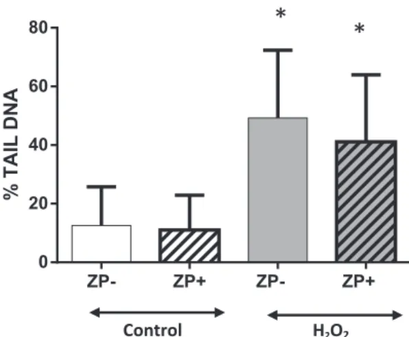
The impact of the ZP on the Comet Assay is illustrated inFig. 1. No statistically significant difference was observed in DNA damage for the ZP- and ZP + control groups (12.60 ± 2.53% tail DNA vs 11.04 ± 1.50, p = 0.5837, respectively). The ZP− and ZP+ groups after H2O2 exposure were also not statistically significant different
(49.23 ± 4.16% Tail DNA vs 41.13 ± 4.31, p = 0.1820, respec-tively).
3.4. Alkaline comet assay
Fig. 2shows the aspects of the comets observed in the control groups (A) and in the groups exposed to the genotoxic agents (B, C). The control embryos showed a low basal level of DNA damage.Fig. 3
shows that H2O2and SSI induced a statistically significant increase in
L. Rolland et al. Toxicology in Vitro 44 (2017) 266–272
DNA damage compared with the control group (41.13 ± 4.31% Tail DNA, 36.33 ± 3.02 and 11.04 ± 1.50, p < 0.0001, respectively).
4. Discussion
4.1. Thawed embryos can be used for comet assay
We describe, for thefirst time, the use of the alkaline comet assay on thawed mouse embryos by optimizing protocols that were previously published in the literature. Control mouse thawed embryos showed a low basal level of DNA damage, comparable to the level of DNA damage observed in control sperm (Preaubert et al., 2016) and control oocytes (Berthelot-Ricou et al., 2011a) in our hands. In the literature, we se-lected 14 studies using the comet assay on mammal embryos matching with our criteria, and no study was performed on frozen embryos. Among the 3 studies in mice (Fabian et al., 2003; Müller et al., 1996; Tranguch et al., 2003), only Fabian et al. evaluated the evolution rate to the blastocyst stage in embryos obtained by natural fertilization, and it was estimated to be 88%. This result suggests that the freezing/thawing step in our protocol did not affect the embryo quality. The use of frozen embryos simplifies the handling by allowing the researcher to dissociate the location and the timing of the embryo collection from the embryo analysis, which facilitates collaboration between laboratories. The use of frozen embryos also allows subsequent analysis if required.
Deleting the removal of zona pellucida improves feasibility. Many authors remove the ZP before the comet assay, in order to study individual blastomeres and to allow DNA migration (Harrouk et al., 2000; Hwang et al., 2013; Ju et al., 2010; Müller et al., 1996; Sturmey et al., 2009; Webster et al., 2000); nevertheless, they did not study the impact the ZP on the downstream results. Moreover, this step requires the use of a chemical and physical stress that could un-necessarily damage the embryo and induce DNA damage. In a previous study of our group (Berthelot-Ricou et al., 2011a), we performed the
comet assay on mouse oocytes with and without ZP, and no significant difference was found in the DNA damage in both of the groups. In the present study, we did not observe significant differences in DNA da-mage in the ZP- and ZP + embryos, indicating that the presence of the ZP did not affect the embryo lysis and/or DNA migration. Deleting the ZP removal step simplifies the protocol, saving time and improving feasibility.
4.2. Adding a third layer of agarose decreases the embryo loss
One of the main issues of our study was to reduce embryo loss during lysis and electrophoresis. As the main difficulty of embryos studies is the low number of available samples, this issue was very important. The significant embryo loss we experienced with Protocol 1 (two agarose layers) was surprising. Indeed, we never observed such a significant loss during the comet assay with Protocol 1 on mouse oo-cytes (Berthelot-Ricou et al., 2011b, 2011a, 2013; Courbiere et al., 2013; Einaudi et al., 2014; Greco et al., 2015), which sizes are com-parable to the embryo size. In the studies using the comet assay on embryos, selected by our literature review, the authors used protocols with 2 agarose layers and deposited between 1 and 20 embryos per glass slide (Table 2), which is comparable with Protocol 1 (20 embryos, 2 agarose layers). None of these studies described the ratio between the number of embryos deposited and the number of recovered embryos after lysis and electrophoresis. Nevertheless, we can assume that em-bryo loss was not as high as in the present study. Our hypothesis is that the important embryo loss we observed with Protocol 1 could be related to the alteration of ZP due to the freezing-thawing protocol: dehy-dratation due to cryprotectant could induce hardening and thinning of the ZP (Cavusoglu et al., 2016; Trounson and Mohr, 1983). These al-terations could modify the interaction between the ZP and the agarose, and increase the embryo loss during the steps of coverslip removal and/ or lysis. The use of a third agarose layer dramatically decreased the rate of embryo loss.
Fig. 1. No impact of zona pellucida for 2-cell embryo comet assay. ZP−: zona-free em-bryo; ZP +: intact emem-bryo; H2O2: exposure to hydrogen peroxide (220μM H2O2). No statistical difference was observed on DNA damage between ZP− et ZP+ embryo on control group and on H2O2exposure group. *: p < 0.01 between DNA damage in Control and in H2O2exposed embryos. t-Test.
A B C
Fig. 2. Examples of different aspects of DNA damage asdetected by the comet assay on mouse 2-cell embryos with zona pellucida. A: Control group: intact 2-cell embryo after comet assay process. B: 2-cell embryo exposed in vitro for 5 min to 220μMol H2O2. C: 2-Cell embryo exposed in vitro to Simulated Sun Irradiation. The direction of electro-phoresis was from left to right, and the comet tail con-taining the DNA fragments was stained by iodide propi-dium 0.1 mg/mL. When 2-cell embryo DNA is intact, the two nuclei remain separated (A). When 2-cell embryo DNA is damaged, the two nuclei overlap and appear merged. (B, C). Arrows show the polar bodies. Scale bar, 100μm. Magnification ×400.Fig. 3. % Tail DNA of mouse 2-cells embryo with zona pellucida exposed to two genotoxic agents and measured by comet assay. Control; H2O2, exposure to hydrogen peroxide (220μM); SSI, exposure to simulated sunlight irradiation (120 J/cm2); ***statistical significance (p < 0.001). t-Test.
4.3. Limitations
In our study, we measured DNA damage by the % Tail DNA, as recently recommended by the OECD (OECD, 2014). The use of the % Tail DNA gives a clear indication of the appearance of the comets, and it is also linearly related to the DNA break frequency over a wide range of levels of damage (Ersson and Möller, 2011; Hartmann et al., 2003; OECD, 2014; Uno et al., 2015). Therefore, the amount of DNA damage we observed could not be easily compared with the DNA damage in the other studies we selected. Indeed, the tail moment and/or the tail length measurements are also reported herein (Table 2), as these studies were published before the OECD recommendations. To better evaluate the sensitivity of our optimized protocol, despite the inability to com-pare these factors, we used two well-known genotoxic agents as positive controls. Nevertheless, % Tail DNA should be used for DNA damage quantification. Another limitation is that we performed the Comet Assay experiments on thawed embryos after an in vitro exposure to two well-known genotoxic agents; our protocol should also be tested to detect embryo DNA damage induced after in vivo mice exposure as-sociated to DNA damage in gametes (Einaudi et al., 2014; Singh et al., 2015).
5. Conclusion
Our optimized protocol of the comet assay on thawed embryos al-lows the rapid detection of primary DNA damage in the mouse embryo. We simplified the protocol by showing that the lack of ZP removal does not alter DNA lysis and migration. In addition, we demonstrated that the adjunction of a third agarose layer decreases embryo loss and allows for the study of a small amount of embryos. This new optimized tool could be used to analyze DNA damage in mice embryos after in vivo exposure of the mothers to environmental agents.
Transparency document
The http://dx.doi.org./10.1016/j.tiv.2017.07.010 associated with this article can be found, in online version.
Funding
This work was conducted thanks to the support of the A*MIDEX project“CREER” (n ANR-11-IDEX-0001-02), which was funded by the “Investissements d'Avenir” French Government program and managed by the French National Research Agency (ANR). The study received additional funding from“Association Laurette Fugain” (no. 2014/01), the ARC Foundation (no. PJA 20141201596), and the Aix Marseille University Foundation (no. 16/07/01).
Conflicts of interest None declared. Acknowledgments
The authors thank technicians from CECOS Laboratory (AP-HM La Conception) for technical help.
References
Baumgartner, A., Cemeli, E., Anderson, D., 2009. The comet assay in male reproductive toxicology. Cell Biol. Toxicol. 25, 81–98.
Berthelot-Ricou, A., Perrin, J., Di Giorgio, C., De Meo, M., Botta, A., Courbiere, B., 2011a. Comet assay on mouse oocytes: an improved technique to evaluate genotoxic risk on female germ cells. Fertil. Steril. 95, 1452–1457.
Berthelot-Ricou, A., Perrin, J., di Giorgio, C., de Meo, M., Botta, A., Courbiere, B., 2011b. Assessment of 1,2-propanediol (PrOH) genotoxicity on mouse oocytes by comet assay. Fertil. Steril. 96, 1002–1007.
Berthelot-Ricou, A., Perrin, J., di Giorgio, C., de Meo, M., Botta, A., Courbiere, B., 2013. Genotoxicity assessment of mouse oocytes by comet assay before vitrification and after warming with three vitrification protocols. Fertil. Steril. 100, 882–888.
Blerkom, J.V., Davis, P., Alexander, S., 2001. A microscopic and biochemical study of fragmentation phenotypes in stage-appropriate human embryos. Hum. Reprod. 16, 719–729.
Bujan, L., Walschaerts, M., Brugnon, F., Daudin, M., Berthaut, I., Auger, J., Saias, J., Szerman, E., Moinard, N., Rives, N., Hennebicq, S., 2014. Impact of lymphoma treatments on spermatogenesis and sperm deoxyribonucleic acid: a multicenter prospective study from the CECOS network. Fertil. Steril. 102, 667–674.e3.
Cavusoglu, T., Popken, J., Guengoer, T., Yilmaz, O., Uyanikgil, Y., Ates, U., Baka, M., Oztas, E., Zakhartchenko, V., 2016. Ultra-structural alterations in in vitro produced four-cell bovine embryos following controlled slow freezing or vitrification. Anat. Histol. Embryol. 45, 291–307.
Courbiere, B., Auffan, M., Rollais, R., Tassistro, V., Bonnefoy, A., Botta, A., Rose, J., Orsière, T., Perrin, J., 2013. Ultrastructural interactions and genotoxicity assay of cerium dioxide nanoparticles on mouse oocytes. Int. J. Mol. Sci. 14, 21613–21628.
Einaudi, L., Courbiere, B., Tassistro, V., Prevot, C., Sari-Minodier, I., Orsiere, T., Perrin, J., 2014. In vivo exposure to benzo(a)pyrene induces significant DNA damage in mouse oocytes and cumulus cells. Hum. Reprod. 29, 548–554.
EMMA—The European mouse mutant archive, 2013a. Harvest and Cryopreservation of In Vivo Derived Mouse Embryos.
EMMA—The European mouse mutant archive, 2013b. Thawing and Use of Cryopreserved Mouse Embryos.
Ersson, C., Möller, L., 2011. The effects on DNA migration of altering parameters in the comet assay protocol such as agarose density, electrophoresis conditions and dura-tions of the enzyme or the alkaline treatments. Mutagenesis 26, 689–695.
Esquerré-Lamare, C., Isus, F., Moinard, N., Bujan, L., 2015. Sperm DNA fragmentation after radioiodine treatment for differentiated thyroid cancer. Basic Clin. Androl. 25, 8.
Fabian, D., Rehák, P., Czikková, S., Il’ková, G., Baran, V., Koppel, J., 2003. Induced cell death of preimplantation mouse embryos cultured in vitro evaluated by comet assay. Theriogenology 60, 691–706.
Goldstone, A.E., Chen, Z., Perry, M.J., Kannan, K., Louis, G.M.B., 2015. Urinary bisphenol A and semen quality, the LIFE study. Reprod. Toxicol. Elmsford N 51, 7–13.
Greco, F., Perrin, J., Auffan, M., Tassistro, V., Orsière, T., Courbiere, B., 2015. A new approach for the oocyte genotoxicity assay: adaptation of comet assay on mouse cumulus-oocyte complexes. Lab. Anim. 49, 251–254.
Hagn, M., Marschall, S., de Angelis, M.H., 2007. EMMA—the European mouse mutant archive. Brief. Funct. Genomic. Proteomic. 6, 186–192.
Harrouk, W., Codrington, A., Vinson, R., Robaire, B., Hales, B.F., 2000. Paternal exposure to cyclophosphamide induces DNA damage and alters the expression of DNA repair genes in the rat preimplantation embryo. Mutat. Res. Repair 461, 229–241.
Hartmann, A., Agurell, E., Beevers, C., Brendler-Schwaab, S., Burlinson, B., Clay, P., Collins, A., Smith, A., Speit, G., Thybaud, V., Tice, R.R., 4th International Comet Assay Workshop, 2003. Recommendations for conducting the in vivo alkaline Comet assay. 4th International Comet Assay Workshop. Mutagenesis 18, 45–51.
Horváthová, E., Dusinská, M., Shaposhnikov, S., Collins, A.R., 2004. DNA damage and repair measured in different genomic regions using the comet assay with fluorescent in situ hybridization. Mutagenesis 19, 269–276.
Hwang, I.-S., Bae, H.-K., Cheong, H.-T., 2013. Mitochondrial and DNA damage in bovine somatic cell nuclear transfer embryos. J. Vet. Sci. 14, 235–240.
Ju, S., Rui, R., Lu, Q., Lin, P., Guo, H., 2010. Analysis of apoptosis and methyltransferase mRNA expression in porcine cloned embryos cultured in vitro. J. Assist. Reprod. Genet. 27, 49–59.
Kitagawa, Y., Suzuki, K., Yoneda, A., Watanabe, T., 2004. Effects of oxygen concentration and antioxidants on the in vitro developmental ability, production of reactive oxygen species (ROS), and DNA fragmentation in porcine embryos. Theriogenology 62, 1186–1197.
Kolstad, H.A., Bonde, J.P., Spano, M., Giwercman, A., Zschiesche, W., Kaae, D., Larsen, S.B., Roeleveld, N., 1999. Change in semen quality and sperm chromatin structure following occupational styrene exposure. ASCLEPIOS. Int. Arch. Occup. Environ. Health 72, 135–141.
Kumaravel, T.S., Jha, A.N., 2006. Reliable Comet assay measurements for detecting DNA damage induced by ionising radiation and chemicals. Mutat. Res. Toxicol. Environ. Mutagen. 605, 7–16.
Ménézo, Y., Dale, B., Cohen, M., 2010. DNA damage and repair in human oocytes and embryos: a review. Zygote Camb. Engl. 18, 357–365.
Miranda-Vilela, A.L., Alves, P.C., Akimoto, A.K., Lordelo, G.S., Gonçalves, C.A., Grisolia, C.K., Klautau-Guimarães, M.N., 2010. Gene polymorphisms against DNA damage induced by hydrogen peroxide in leukocytes of healthy humans through comet assay: a quasi-experimental study. Environ. Health Glob. Access Sci. Source 9, 21.
Müller, W.-ü, Bauch, T., Wojcik, A., Böcker, W., Streffer, C., 1996. Comet assay studies indicate that caffeine-mediated increase in radiation risk of embryos is due to in-hibition of DNA repair. Mutagenesis 11, 57–60.
Natarajan, R., Shankar, M.B., Munuswamy, D., 2010. Effect of α-tocopherol supple-mentation on in vitro maturation of sheep oocytes and in vitro development of preimplantation sheep embryos to the blastocyst stage. J. Assist. Reprod. Genet. 27, 483–490.
Nicolson, G.L., Yanagimachi, R., Yanagimachi, H., 1975. Ultrastructural localization of lectin-binding sites on the zonae pellucidae and plasma membranes of mammalian eggs. J. Cell Biol. 66, 263–274.
OECD, 2014. Test No. 489: In Vivo Mammalian Alkaline Comet Assay. Organisation for Economic Co-operation and Development, Paris.
Pecou, S., Moinard, N., Walschaerts, M., Pasquier, C., Daudin, M., Bujan, L., 2009. Ribavirin and pegylated interferon treatment for hepatitis C was associated not only
L. Rolland et al. Toxicology in Vitro 44 (2017) 266–272
with semen alterations but also with sperm deoxyribonucleic acid fragmentation in humans. Fertil. Steril. 91 (933.e17-22).
Perrin, J., Lussato, D., De Méo, M., Durand, P., Grillo, J.-M., Guichaoua, M.-R., Botta, A., Bergé-Lefranc, J.-L., 2007. Evolution of DNA strand-breaks in cultured spermato-cytes: the comet assay reveals differences in normal and γ-irradiated germ cells. Toxicol. in Vitro 21, 81–89.
Perrin, J., Tassistro, V., Mandon, M., Grillo, J.-M., Botta, A., Sari-Minodier, I., 2011. Tobacco consumption and benzo(a)pyrene-diol-epoxide-DNA adducts in sperma-tozoa: in smokers, swim-up procedure selects spermatozoa with decreased DNA da-mage. Fertil. Steril. 95, 2013–2017.
Preaubert, L., Courbiere, B., Achard, V., Tassistro, V., Greco, F., Orsiere, T., Bottero, J.-Y., Rose, J., Auffan, M., Perrin, J., 2016. Cerium dioxide nanoparticles affect in vitro fertilization in mice. Nanotoxicology 10, 111–117.
Rajesh, N., Shankar, M.B., Deecaraman, M., 2010. Effect of vitamin A supplementation at different gaseous environments on in vitro development of pre-implantation sheep embryos to the blastocyst stage. Animal 4, 1884–1890.
Roti Roti, E.C., Leisman, S.K., Abbott, D.H., Salih, S.M., 2012. Acute doxorubicin insult in the mouse ovary is cell- and follicle-type dependent. PLoS One 7, e42293.
Simon, L., Murphy, K., Shamsi, M.B., Liu, L., Emery, B., Aston, K.I., Hotaling, J., Carrell, D.T., 2014. Paternal influence of sperm DNA integrity on early embryonic develop-ment. Hum. Reprod. Oxf. Engl. 29, 2402–2412.
Singh, S., Giri, A., Giri, S., 2015. The antimalarial agent artesunate causes sperm DNA damage and hepatic antioxidant defense in mice. Mutat. Res. Toxicol. Environ. Mutagen. 777, 1–6.
Speit, G., Vasquez, M., Hartmann, A., 2009. The comet assay as an indicator test for germ cell genotoxicity. Mutat. Res. 681, 3–12.
Sturmey, R.G., Hawkhead, J.A., Barker, E.A., Leese, H.J., 2009. DNA damage and me-tabolic activity in the preimplantation embryo. Hum. Reprod. 24, 81–91.http://dx. doi.org/10.1093/humrep/den346.
Takahashi, M., Saka, N., Takahashi, H., Kanai, Y., Schultz, R.M., Okano, A., 1999. Assessment of DNA damage in individual hamster embryos by comet assay. Mol. Reprod. Dev. 54, 1–7.
Takahashi, M., Keicho, K., Takahashi, H., Ogawa, H., Schulte, R.M., Okano, A., 2000. Effect of oxidative stress on development and DNA damage in in-vitro cultured bo-vine embryos by comet assay. Theriogenology 54, 137–145.
Thiyagarajan, B., Valivittan, K., 2009a. Effect of all-trans retinol on in vitro development of preimplantation buffalo embryos. Animal 3, 385.
Thiyagarajan, B., Valivittan, K., 2009b. Ameliorating effect of vitamin E on in vitro de-velopment of preimplantation buffalo embryos. J. Assist. Reprod. Genet. 26, 217–225.
Thompson, J., Bannigan, J., 2008. Cadmium: toxic effects on the reproductive system and the embryo. Reprod. Toxicol. Elmsford N 25, 304–315.
Tranguch, S., Steuerwald, N., Huet-Hudson, Y.M., 2003. Nitric oxide synthase production and nitric oxide regulation of preimplantation embryo development. Biol. Reprod. 68, 1538–1544.
Trounson, A., Mohr, L., 1983. Human pregnancy following cryopreservation, thawing and transfer of an eight-cell embryo. Nature 305, 707–709.
Tsuda, S., Kosaka, Y., Matsusaka, N., Sasaki, Y.F., 1998. Detection of pyrimethamine-induced DNA damage in mouse embryo and maternal organs by the modified alkaline single cell gel electrophoresis assay. Mutat. Res. Toxicol. Environ. Mutagen. 415, 69–77.
Uno, Y., Kojima, H., Omori, T., Corvi, R., Honma, M., Schechtman, L.M., Tice, R.R., Burlinson, B., Escobar, P.A., Kraynak, A.R., Nakagawa, Y., Nakajima, M., Pant, K., Asano, N., Lovell, D., Morita, T., Ohno, Y., Hayashi, M., 2015. JaCVAM-organized international validation study of the in vivo rodent alkaline comet assay for the de-tection of genotoxic carcinogens: I. Summary of pre-validation study results. Mutat. Res. Genet. Toxicol. Environ. Mutagen. 786–788, 3–13.
Webster, W.S., Vaghef, H., Ryan, B., Dencker, L., Hellman, B., 2000. Measurement of DNA damage by the comet assay in rat embryos grown in media containing high con-centrations of vitamin K1. Toxicol. in Vitro 14, 95–99.
Zenzes, M.T., Bielecki, R., Reed, T.E., 1999. Detection of benzo(a)pyrene diol epoxide-DNA adducts in sperm of men exposed to cigarette smoke. Fertil. Steril. 72, 330–335.
Zhao, J., Zhang, Q., Wang, Y., Li, Y., 2014. Whether sperm deoxyribonucleic acid frag-mentation has an effect on pregnancy and miscarriage after in vitro fertilization/ intracytoplasmic sperm injection: a systematic review and meta-analysis. Fertil. Steril. 102, 998–1005.e8.
Zhou, Y., Fu, X.-M., He, D.-L., Zou, X.-M., Wu, C.-Q., Guo, W.-Z., Feng, W., 2016. Evaluation of urinary metal concentrations and sperm DNA damage in infertile men from an infertility clinic. Environ. Toxicol. Pharmacol. 45, 68–73.