HAL Id: hal-02961328
https://hal.umontpellier.fr/hal-02961328
Submitted on 28 Oct 2020
HAL is a multi-disciplinary open access
archive for the deposit and dissemination of sci-entific research documents, whether they are pub-lished or not. The documents may come from teaching and research institutions in France or abroad, or from public or private research centers.
L’archive ouverte pluridisciplinaire HAL, est destinée au dépôt et à la diffusion de documents scientifiques de niveau recherche, publiés ou non, émanant des établissements d’enseignement et de recherche français ou étrangers, des laboratoires publics ou privés.
Biobased pH-responsive and self-healing hydrogels
prepared from O-carboxymethyl chitosan and a
3-dimensional dynamer as cartilage engineering scaffold
Rui Yu, Yan Zhang, Mihail Barboiu, Marie Maumus, Danièle Noël, Christian
Jorgensen, Suming Li
To cite this version:
Rui Yu, Yan Zhang, Mihail Barboiu, Marie Maumus, Danièle Noël, et al.. Biobased pH-responsive and self-healing hydrogels prepared from O-carboxymethyl chitosan and a 3-dimensional dynamer as cartilage engineering scaffold. Carbohydrate Polymers, Elsevier, 2020, 244, pp.116471. �10.1016/j.carbpol.2020.116471�. �hal-02961328�
Biobased pH-responsive and self-healing hydrogels prepared from O-carboxymethyl chitosan 1
and a 3-dimensional dynamer as cartilage engineering scaffold 2
3
Rui Yu,a Yan Zhang,d Mihail Barboiu,a* Marie Maumus,b Danièle Noël,b,c* Christian 4
Jorgensen,b,c Suming Lia* 5
6
a
Institut Européen des Membranes, IEM UMR 5635, Univ Montpellier, CNRS, ENSCM,
7
Montpellier, Franc
8
b
IRMB, Univ Montpellier, INSERM,Montpellier, France;
9
c
Clinical Immunology and Osteoarticular Diseases Therapeutic Unit, Hôpital Lapeyronie,
10
Montpellier, France
11
d
Key Laboratory of Carbohydrate Chemistry and Biotechnology, Ministry of Education, School
12
of Pharmaceutical Sciences, Jiangnan University, 1800 Lihu Avenue, Wuxi, 214122, China
13 14
* Corresponding authors: suming.li@umontpellier.fr (S. Li) 15
mihail-dumitru.barboiu@umontpellier.fr (M. Barboiu), and daniele.noel@inserm.fr (D. Noel) 16
Abstract: 18
Novel dynamic hydrogels were prepared from O-carboxymethyl chitosan (CMCS) and a water 19
soluble dynamer Dy via crosslinking by imine bond formation using an environmentally friendly 20
method. Dy was synthesized by reaction of Benzene-1,3,5-tricarbaldehyde with Jeffamine. The 21
resulting soft hydrogels exhibit a porous and interconnected morphology, storage modulus up to 22
1400 Pa, and excellent pH-sensitive swelling properties. The swelling ratio is relatively low at 23
acidic pH due to electrostatic attraction, and becomes exceptionally high up to 7000% at pH 8 24
due to electrostatic repulsion. Moreover, hydrogels present outstanding self-healing properties as 25
evidenced by closure of split pieces and rheological measurements. This study opens up a new 26
horizon in the preparation of dynamic hydrogels with great potential for applications in drug 27
delivery, wound dressing, and in particular in tissue engineering as the hydrogels present 28
excellent cytocompatibility. 29
30
Keywords: O-carboxymethyl chitosan; Imine chemistry; Dynamic hydrogel; Self-healing; pH 31 responsive; Cytocompatibility 32 33 1. Introduction 34
In the past decades, hydrogels have been widely studied as biomaterials for various 35
applications in drug delivery, wound dressing and tissue engineering due to their outstanding 36
properties such as biocompatibility (Qu et al., 2018), biodegradability (Ghobril & Grinstaff, 37
2015), mechanical properties (Van Vlierberghe, Dubruel, & Schacht, 2011), and stimuli-38
responsive properties (Zhang, Tao, Li, & Wei, 2011). Hydrogels consist of a cross-linked 39
network (Lv et al., 2018; Su et al., 2015) which can absorb large amounts of water (Yang, Wang, 40
Yang, Wang, & Wu, 2018) or physiological fluids (Dimatteo, Darling, & Segura, 2018) while 41
maintaining their three-dimensional structural integrity. Hydrogels can be classified into two 42
categories according to the preparation approach: chemical gels or irreversible gels, and physical 43
gels or reversible gels (Iftime, Morariu, & Marin, 2017; Stewart et al., 2017). Chemical gels are 44
formed by irreversible covalent bonding, whereas in physical gels, the polymeric chains are held 45
together by chain entanglements and/or supramolecular hydrophobic, ion-pair or hydrogen 46
bonding interactions. 47
Dynamic hydrogels or dynagels are a dynamic system on both the molecular and 48
supramolecular levels (Burdick & Murphy, 2012; Marin et al., 2014). They are able to reversibly 49
exchange their components (Sreenivasachary & Lehn, 2005), responding to external stimuli such 50
as pH (Zeng et al., 2017), ions (Arnal Hérault, Banu, Barboiu, Michau, & van der Lee, 2007; 51
Rotaru et al., 2017), and temperature (Zhang, Jin, Li, Zhang, & Wu, 2018). Among the various 52
dynamic reactions (Zhang & Barboiu, 2015a), imine bond formation is considered as the most 53
promising strategy to generate dynamic materials with modulable properties. In fact, imine 54
chemistry allows to implement reversible rearrangements of the components in a multivalent 55
material which can bind bioactive molecules, cells or present self-healing properties (Chao, 56
Negulescu, & Zhang, 2016). This dynamic constitutional framework is composed of linear 57
and/or multi-armed components reversibly interconnected via imine bonds and containing 58
stimuli-responsive functional groups (Zhang & Barboiu, 2015b). 59
Chitosan (CS), a polysaccharide obtained from alkaline hydrolysis of chitin found in the 60
exoskeleton of crustaceans, presents remarkable properties such as biocompatibility, 61
biodegradability, low toxicity, low cost or immune-stimulatory activity (Ali & Ahmed, 2018). 62
Chitosan is a good candidate for in-situ dynamic reversible crosslinking via its amino groups 63
present along the polymer chain with aldehydes (Marin, Simionescu, & Barboiu, 2012), alginate 64
(Qin et al., 2019), or gelatin (Qiao, Ma, Zhang, & Yao, 2017), resulting in the formation of pH-65
responsive and biodegradable hydrogels. The water solubility of chitosan depends on many 66
factors, in particular pH of the medium, chain length and degree of acetylation (Varum, Ottoy, & 67
Smidsrod, 1994). The carboxymethylation of the D-glucosamine moieties of chitosan generates 68
O-carboxymethyl chitosan (CMCS) which is readily soluble in water at neutral pH, thus allowing 69
uses in tissue engineering, drug delivery, wound dressing and food industry. 70
Crosslinking of CS or CMCS with aldehydes via imine bond formation along polymeric chains 71
proceeds with very low yield in aqueous medium, but is significantly improved in hydrogels or 72
in solid state films with dynamic properties (Marin et al., 2012). It is well known that no 73
continuous cross-linked networks are formed when monoaldehydes (Iftime et al., 2017; Marin, et 74
al., 2012) or dialdehydes (Yu et al., 2017) are used for cross-linking. The resulting hydrogels 75
exhibit good swelling behaviors, but disordered micro-structure and weak mechanical strength. 76
In contrast, dynamic hydrogels exhibiting pH and temperature-responsive swelling behaviors, 77
strong mechanical performance, and self-healing behavior have been obtained by using 3-armed 78
(Deng et al., 2015) or 4-armed aldehydes (Huang et al., 2016). Nevertheless, the toxicity of 79
aldehydes, and especially of glutaraldehyde (Bhatia, 2010; Ghobril & Grinstaff, 2015) restricts 80
their use for biomedical applications, and imposes the necessity of finding new biocompatible 81
crosslinking agents. 82
Jeffamine is a polyetheramine composed of poly(propylene oxide) (PPO) and/or poly(ethylene 83
oxide) (PEO) blocks with primary amino groups attached to the chain ends. It is widely used as a 84
macromonomer to prepare PEO-based hydrogels as its water solubility facilitates reaction in 85
aqueous medium (Zimmermann, Bittner, Stark, & Mülhaupt, 2002). In this work, bifunctional 86
Jeffamine ED-2003 was linked to benzene-1,3,5-tricarbaldehyde via imine formation, yielding a 87
constitutional dynamer Dy that can be used as a water soluble cross-linking component. CMCS 88
based hydrogels were then prepared by mixing CMCS and Dy aqueous solutions through a 89
“green” synthetic route. The chemical structures of the hydrogels, their morphological, 90
rheological and swelling properties, as well as their self-healing behaviors were evaluated and 91
discussed. The cytocompatibility of hydrogels was assessed by co-culture in the presence of 92
human mesenchymal stromal cells (MSCs) to evaluate their potential as scaffold in cartilage 93 engineering. 94 95 2. Experimental section 96
2.1 Materials: Benzene-1,3,5-tricarbaldehyde (BTA) from Manchester Organics and O,O’-Bis(2-97
aminopropyl) PPO-b-PEO-b-PPO (Jeffamine® ED-2003, Mn 1.9 × 10³) from Sigma Aldrich 98
were used without purification. CMCS (Mn 2 × 10⁵ Da, degree of deacetylation 90 %, degree of 99
carboxymethylation 80 %) was purchased from Golden-shell Biochemical Co., Ltd. Methanol 100
(96%), citric acid (≥99.5%), disodium hydrogen phosphate dodecahydrate (≥99%), boric acid 101
(≥99.5%), borax(≥99%) were of analytical grade, and obtained from Sigma Aldrich. 102
2.2 Synthesis of dynamer Dy: Typically, BTA (162 mg, 1 mmol), Jeffamine (1.90 g, 1 mmol) are 103
added in 30 mL methanol, and the reaction mixture was stirred at 70 °C for 4 h. After 104
evaporation of the solvent, 20 mL Milli Q water was added, yielding a homogeneous dynamer 105
solution of 5 × 10⁻² M as calculated from the remaining aldehyde groups. 106
2.3 Preparation of CMCS-Dy hydrogels: CMCS (1.06 g, 5 mmol calculated from D-glucosamine 107
units) was dissolved in 50 mL Milli-Q water at room temperature, yielding a transparent solution 108
of 1× 10⁻¹ M. CMCS and dynamer solutions were mixed at different ratios to a total volume of 109
12 mL, followed by ultra-sonication for 1 min to remove trapped bubbles. Gelation then 110
proceeded at 37 °C for 24 h, yielding a CMCS-based hydrogel. 111
Freeze-drying was performed as follows so as to conserve the original structure. As-prepared 112
hydrogels were placed in small vials and immersed in liquid nitrogen (-196 °C) for instantaneous 113
freezing. The vials were then placed in a 500 mL round-bottomed flask which was fixed on 114
LABCONCO® freeze dryer. The hydrogels were freeze-dried for 24 h before analyses. 115
2.4 Characterization: 1H NMR spectroscopy was carried out using Bruker NMR spectrometer 116
(AMX500) of 300 MHz. CDCl3 or D2O was used as the solvent. 5 mg of sample were dissolved
117
in 0.5 mL of solvent for each analysis. Chemical shifts were recorded in ppm using 118
tetramethylsilane (TMS) as internal reference. The morphology of freeze dried hydrogels was 119
examined using scanning electron microscopy (SEM, Hitachi S4800). The samples were 120
subjected to gold coating prior to analysis. Fourier-transform infrared spectroscopy (FT-IR) was 121
performed with Nicolet Nexus FT-IR spectrometer, equipped with ATR diamant Golden Gate. 122
2.5 Structural stability of Dy: The stability of the dynamer Dy was evaluated in D2O under
123
neutral and acidic conditions since imine bond formation is reversible at low pH. D2O solutions
124
at pH of 1, 3, and 5 were prepared by addition of trifluoroacetic acid. NMR spectra were 125
registered just after dynamer dissolution and after 7 days. 126
2.6 Rheology: The rheological properties of hydrogels were examined with Physical MCR 301 127
Rheometer (Anton Paar). Hydrogels prepared in Milli-Q water were placed on a cone plate 128
(diameter of 4 cm, apex angle of 2 °, and clearance 56 µm). Measurements were made in the 129
linear visco-elastic range as a function of time, strain, or frequency. 130
2.7 Swelling: The swelling behavior of hydrogels was evaluated in buffer solutions at various pH 131
values. Solutions from pH 1 to pH 7 were prepared using 0.1 × 10⁻³ M citric acid solution and 132
0.2 × 10⁻³ M disodium hydrogen phosphate solution, whereas solutions of pH 8 and 9 were 133
prepared using 0.2 × 10⁻³ M boric acid solution and 0.5 × 10⁻⁴ M borax solution. Freeze-dried 134
gels were immersed in a buffer, and taken out at different time intervals. The swollen hydrogels 135
were weighed after wiping surface water with filter paper, freeze-dried for 24 h, and weighed 136
again. The swelling ratio and mass loss ratio of hydrogel were calculated according to equation 137
(1) and equation (2), respectively: 138 Swelling ratio % =(𝑀𝑠−𝑀𝑑) 𝑀𝑑 × 100 (1) 139 Loss ratio % =(𝑀0−𝑀𝑑) 𝑀0 × 100 (2) 140
Where M0 is the initial mass of xerogel, Ms is the wet mass of the swollen hydrogel, and Md is
141
the dried mass of the swollen hydrogel after lyophilization. 142
2.8 Self-healing experiments: Various hydrogel samples were prepared in Milli-Q water, and in 143
pH = 7 and pH = 8 buffers. Some of them were dyed yellow with 5 µL of lucigenin, or dyed red 144
with Rhodamine. 3 different approaches were applied to examine the self-healing behavior of 145
hydrogels: 1) a hole with diameter around 3 mm was punched at the circle center of the sample; 146
2) samples were split into two pieces, and then a yellow piece was put together with a transparent 147
piece immediately at 37 °C ; 3) injection of transparent and red samples on the surface of a Petri 148
dish to observe color changes. 149
2.9 Cell cultures and Cytotoxicity: 2 mL of CMCS at 100 mmol and 1 mL of dynamer at 50 150
mmol were mixed. Human MSCs isolated from adipose tissue (AT-MSCs) or from bone marrow 151
(BM-MSCs) were added at a concentration of 1 x 10⁶ cells / mL. As control, AT-MSCs or BM-152
MSCs were seeded on 96-wells TCPS (Tissue Culture Polystyrene System) plates at 5 x 10³ cells 153
per well, or were embedded in 3 mg / mL rat collagen type I hydrogel (Corning) at 1 × 10⁶ cells / 154
mL. 50 µL of cell laden solution were loaded in each well of 96 wells ultra-low adhesion plates 155
(Corning). After 2 h gelation in an atmosphere at 37 °C, 5 % CO2 and 95 % of humidity, 100 µL
156
of proliferative medium (αMEM containing 10% fetal calf serum, 100 µg/mL 157
penicillin/streptomycin, 2 × 10⁻³ M glutamine, 1 ng / mL of basic fibroblast growth factor) were 158
added on the top of the hydrogel. MSCs were cultured for 7 days with medium change at day 3. 159
After 1 or 7 days culture, the cell viability was analyzed by confocal microscopy (Leica) after 160
staining the live cells in green and the dead cells in red using the live/dead assay kit (Invitrogen). 161
162
3. Results and discussion 163
3.1 Synthesis of dynamer: A water soluble dynamer Dy was first synthesized by reaction of BTA 164
as the core structure and bifunctional diamine, Jeffamine® ED-2003 (Mn=1900) as the water-165
soluble linker at a molar ratio of 1:1 via reversible imine bond formation, as shown in Scheme 1. 166
Thus, it remains in average one aldehyde group per molecule of BTA for further cross-linking 167
reaction with the amine groups of CMCS. 168
1H NMR spectroscopy was used to monitor the formation and stability of imine bonds during the
169
synthesis of the dynamer. Fig. 1 presents the 1
H NMR spectrum of the dynamer mixture obtained 170
after 4 h reaction at 70 °C. Three signals of aldehyde groups are observed in the 10.1-10.3 ppm 171
range, corresponding to different degrees of substitution in trialdehyde. Signals a at 10.21 ppm, b 172
at 10.15 ppm, and c at 10.09 ppm belong to non-substituted, mono-substituted, and di-substituted 173
trialdehydes, respectively. The molar ratio of signals a, b and c is 1:6.6:11.5, as determined from 174
the peak integrations. These findings indicate formation of a dynamer with various free aldehyde 175
groups, which is beneficial for subsequent crosslinking with amino groups of CMCS by imine 176
formation. Signal d in the range of 8.0-8.7 ppm is assigned to the imine and aromatic protons, 177
signal e around 3.7 ppm to the methylene and methine protons, and signal f around 1.2 ppm to 178
the methyl protons of Jeffamine, respectively (Catana et al., 2015). The presence of residual 179
CHCl3 and H2O is detected at 7.3 and 1.8 ppm, respectively.
180 181
182
Scheme 1. Synthesis route of CMCS-based dynamic hydrogel: a) chemical structures of BTA,
183
CMCS and Jeffamine; b) synthesis of the dynamer Dy by reaction of equimolar BTA and 184
Jeffamine, and synthesis of hydrogel by imine formation between Dy and CMCS; c) images of as 185
prepared hydrogel and freeze dried hydrogel; and d) SEM images of freeze dried hydrogel. 186
188
Fig. 1. 1H NMR spectrum of the dynamer Dy obtained by reaction of BTA and Jeffamine in 189
CDCl3.
190 191
The effect of reaction time on the formation of dynamer was investigated. No difference was 192
observed on the 1H NMR spectra of samples up to 72 h reaction (Fig. S1, Supporting 193
Information), thus implying that equilibrium was reached after 4 h reaction. Therefore, the 194
dynamer obtained after 4 h reaction was selected for further studies. Moreover, the dynamer 195
apparently remained unchanged for a week in pure D2O and in acidic D2O at pH 3 and 5, while
196
became highly hydrolyzed in strongly acidic medium at pH 1 (Fig. 2). The spectra obtained in 197
D2O and at pH 3 and 5 remain unchanged even after 7 days (Fig. S2, Supporting Information). In
198
contrast, major changes are observed on the spectrum of dynamer at pH 1. The signal a (10.21 199
ppm) belonging to free aldehydes becomes much more intense, indicating that imine bonds are 200
hydrolyzed back to aldehydes. Therefore, the dynamer Dy seems stable at neutral and slightly 201
acidic pH, but unstable at strongly acidic pH. As previously observed for PEGylated networks, 202
Jeffamine chains could have a protecting effect against the hydrolysis of imine bonds, favoring 203
the imine formation in slightly acidic or neutral media (Catana et al., 2015). 204
206
Fig. 2. 1H NMR spectra of the dynamer Dy in pure D2O and acidic D2O at pH = 1, 3, and 5
207 208
3.2 Synthesis of CMCS-Dy hydrogels: CMCS based hydrogels were prepared via a ‘green’ and 209
environmentally friendly method, as shown in Scheme 1. The free aldehyde groups of Dy react 210
with the amine groups of CMCS to form imine bonds in water, leading to a three-dimensional 211
network of CMCS based hydrogel. A series of hydrogels were obtained by mixing 1 × 10⁻¹ M 212
CMCS and 5 × 10⁻² M dynamer aqueous solutions to a total volume of 12 mL. Gelation 213
proceeded at 37 °C for 24 h. The D-glucosamine to dynamer molar ratio varied from 1:1 to 8:1, 214
as shown in Table 1. 215
216
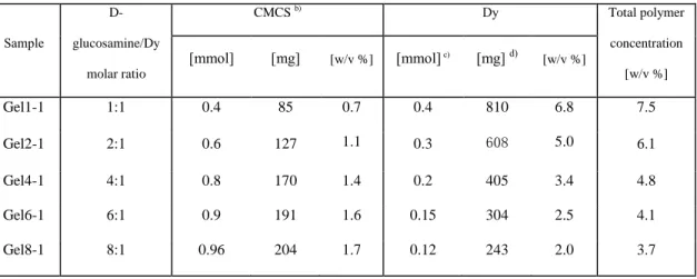
Table 1. Molar and mass composition of CMCS-Dy hydrogels a)
217 Sample D-glucosamine/Dy molar ratio CMCS b) Dy Total polymer concentration [w/v %] [mmol] [mg] [w/v %] [mmol] c) [mg] d) [w/v %] Gel1-1 1:1 0.4 85 0.7 0.4 810 6.8 7.5 Gel2-1 2:1 0.6 127 1.1 0.3 608 5.0 6.1 Gel4-1 4:1 0.8 170 1.4 0.2 405 3.4 4.8 Gel6-1 6:1 0.9 191 1.6 0.15 304 2.5 4.1 Gel8-1 8:1 0.96 204 1.7 0.12 243 2.0 3.7
a)
Hydrogels are prepared by mixing CMCS and dynamer solutions at different ratios to a total volume of 12 mL; 218
b)
The concentration of CMCS solution is 100 mM. Calculations are made on the basis of the average molar mass of 212 g/mol 219
obtained for D-glucosamine, taking into account the degree of deacetylation of 90 % and the degree of carboxymethylation of 220
80 %; 221
c) The concentration of Dy solution is 50 mM calculated from the remaining aldehyde groups. In a typical reaction, 1 mmol BTA 222
(162 mg) reacts with 1 mmol Jeffamine (1900 mg) to form a dynamer. As BTA has 3 aldehydes and Jeffamine 2 amines, there 223
remains theoretically 1 mmol of aldehydes in the dried dynamer. Addition of 20 mL water yields a dynamer solution of 50 mM. 224
d) The amount of Dy in solution is obtained from the initial quantities of BTA and Jeffamine. 225
226
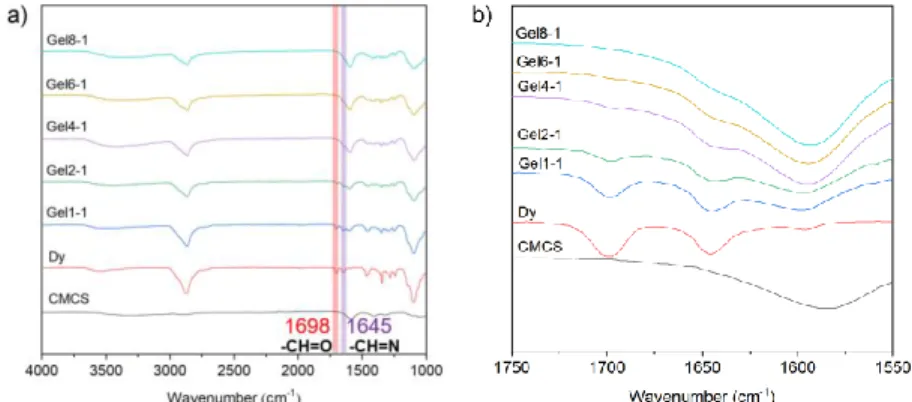
FTIR was used to confirm the formation of imine bonds during the synthesis of the dynamer 227
Dy and CMCS based hydrogel. As shown in Fig. 3, the characteristic bands of aldehyde and 228
imine bonds are observed at 1698 and 1645 cm-1 on the spectra of Dy, respectively. The dried 229
gels present all the characteristic bands of CMCS and dynamer: a large band in the 3200-3500 230
cm-1 assigned to free OH and NH2 groups, an intense band at 1595 cm-1 assigned to carboxyl
231
groups of CMCS, and two strong signals at 2850 and 1100 cm-1 attributed to C-H and C-O 232
stretching in the dynamer, respectively. With increasing D-glucosamine to dynamer molar ratio, 233
the aldehyde band progressively disappears at ratios above 4:1, while the imine band merges 234
with that of carboxyl groups at 1595 cm-1 which turns more intense. These findings confirm that 235
hydrogels are formed because of imine formation between aldehyde and amine groups. 236
237
238
Fig. 3. a) FT-IR spectra and b) enlarged view of the 1550-1750 cm-1 wavelength range of 239
CMCS, dynamer Dy and freeze-dried CMCS-Dy hydrogels. 240
241
3.3 Rheological studies. The rheological properties of hydrogels were investigated under various 242
conditions. CMCS and Dy aqueous solutions were mixed in situ on the plate of rheometer, and 243
changes of the storage modulus (G) and loss modulus (G) were followed as a function of time 244
at 37°C (Fig. 4a). For all samples, at the beginning of experiment the storage modulus is lower 245
than the loss modulus (G < G), which illustrates a liquid-like behavior of the starting mixture. 246
After an induction time, both G and G begin to increase, G increasing faster than G. A cross-247
over point between G and G is detected, indicating sol-gel transition. As shown in Figure 4a, 248
the gelation time decreases from 600 s for Gel1-1 to a minimum of 360 s for Gel4-1, and then 249
increases to 660 s for Gel8-1. In fact, gelation occurs by crosslinking via imine bonds formation 250
and is thus dependent on the ratio between amine groups of CMCS and aldehyde groups of the 251
dynamer. In Gel1-1, there are less amine groups than aldehyde ones, as calculated by taking into 252
account the degree of deacetylation of 90 %. Thus, gelation is relatively slow. Gelation is 253
progressively improved for Gel2-1 and Gel4-1, as the concentration of amine groups increases. 254
Gelation is not optimal for Gel2-1, since unreacted aldehyde groups are detected by FTIR after 255
24 h at 37 °C (Fig. 3). In contrast, optimal imine bond formation is achieved for Gel4-1 as 256
aldehyde groups are no longer detectable. Nevertheless, with further increase of D-257
glucosamine/dynamer ratio to 6:1 and 8:1, the gelation becomes longer as there are less aldehyde 258
groups available for imine formation. 259
261
262
Fig. 4. a) Storage modulus (G) and loss modulus (G) changes as a function of time after mixing 263
CMCS and Dy aqueous solutions at various ratios at 37 °C, strain of 1%, and frequency of 1 Hz; 264
b) G changes as a function of applied strain for all hydrogels at 25 °C, and frequency of 1 Hz; 265
and c) G and G changes of Gel4-1 as a function of frequency at 25 °C, and strain of 1%. All 266
hydrogels are prepared in Milli-Q water. 267
268
Rheological measurements performed at 25 °C illustrate the viscoelastic behaviors of as prepared 269
hydrogels. The storage modulus of all gels slightly decreases (less than 20 % of the initial value) 270
when increasing the strain up to 20 % (Fig. 4b), indicating that the hydrogels are stable in this 271
strain range with viscoelastic behavior. On the other hand, the modulus increases with increasing 272
D-glucosamine to dynamer molar ratio from 1:1 to 4:1, reaching a maximum value of c.a. 1200 273
Pa at 4:1. In contrast, higher D-glucosamine to dynamer ratios of 6:1 and 8:1 result in decrease in 274
modulus because there are less aldehydes available for crosslinking in Gel6-1 and Gel8-1 275
compared to Gel4-1. These findings well agree with storage modulus (G) and loss modulus (G) 276
changes versus time in Fig. 4a, confirming that optimal crosslinking is achieved with Gel4-1. In 277
order to investigate the stability of the hydrogels, a frequency sweep over a range from 0.01 to 278
50 Hz was carried out at a fixed strain of 1 %. Taking Gel4-1 as an example (Fig. 4c), the storage 279
modulus G is always much higher than the loss modulus G. G remains nearly unchanged, 280
whereas G exhibits some fluctuations with increasing frequency. The other hydrogels exhibit 281
similar behaviors (Fig. S3, Supporting Information). These rheological results well corroborate 282
with the formation of highly stable covalent networks, in contrast to physical hydrogels whose 283
storage and loss moduli are dependent on the frequency (Li, El Ghzaoui, & Dewinck, 2005; 284
Zhang et al., 2010). It is generally admitted that hydrogels with G’ below 2000 Pa are ‘soft’ 285
materials suitable for specific tissue engineering applications (brain, cartilage, muscle, etc). 286
287
3.4 Morphology and swelling studies: Scanning electron microscopy (SEM) was used to 288
qualitatively assess the microstructure of the freeze-dried hydrogels. As shown in Fig. 5, all 289
samples exhibit a sponge-like structure with open and interconnected pores. Gel4-1 apparently 290
exhibits the most uniform porous structure with mean pore size around 150 μm and mean wall 291
thickness of c.a 3 µm, whereas the other samples, in particular Gel1-1 and Gel8-1, present larger 292
and irregular pore size and larger wall thickness. These findings well agree with the optimal 293
imine formation or crosslinking of Gel4-1 since higher crosslinking leads to smaller pore size 294
and wall thickness. 295
297
298
299
Fig. 5. SEM images of freeze dried hydrogels: (a, b) Gel1-1; (c, d) Gel2-1; (e, f) Gel4-1; (g, h)
302
Gel6-1; (i, j) Gel8-1. 303
304
The swelling behaviors of hydrogels are of major importance for the applications as drug 305
carrier or as tissue engineering scaffold. The five samples exhibit similar swelling behaviors at a 306
given pH value in the pH range from 3 to 9. The highly pH-sensitive swelling ratios are below 307
1000 % for acidic media (gray bar, pH 3/4), between 1000 % and 2000 % for neutral media 308
(violet bar, pH 5/6/7), above 2000 % and up to 6000 % for alkaline media (orange bar, pink bar, 309
reddish orange bar, magenta bar, and red bar, pH 8/9) as shown in Fig. 6a. Interestingly, when 310
immersed in slightly alkaline medium at pH 8, the swelling ratio of Gel4-1 dramatically depends 311
on the immersion time (Fig. 6c). It increases from 3130 % (green bar) after 1 h to 7050 % (red 312
bar) after 48 h immersion. In more alkaline medium at pH 9, the variation of the swelling ratio is 313
attenuated, from 2500 % (blue bar) after 1 h to 3500 % (green bar) after 48 h immersion (Fig. 6c). 314
In contrast, this exceptional time dependent swelling behavior of Gel4-1 rapidly reaches an 315
equilibrium at 1 h for pH 3-7. 316
Mass loss could occur after swelling of hydrogels at various pH values, resulting from the 317
diffusion and washing away of non-crosslinked species, including those initially present or 318
formed by hydrolysis of imine bonds under acidic conditions. Thus the mass loss ratio reflects 319
the crosslinking degree and the stability of hydrogels. Obviously, when immersed in acidic 320
media at pH ≤ 5 for all hydrogels or in the whole pH range for Gel1-1 and Gel2-1 with low 321
CMCS and high Dy contents (Fig. 6b), the mass loss ratio is above 60 % (green bar). Loss ratios 322
below 60 % (violet and gray bars) are obtained only in neutral or alkaline media (pH ≥6) for 323
Gel4-1 with optimal crosslinking, and Gel6-1 and Gel8-1 with decreasing Dy content (Table 1). 324
These findings indicate that higher Dy content and acidic medium are conducive to the mass loss 325
of hydrogels during swelling. It is thus supposed that unconnected or incompletely connected Dy 326
is predominant in the soluble fraction. Dy rings or homopolymers could be formed during 327
reaction of BTA and Jeffamine (Scheme 1). These species may escape coupling with CMCS in 328
the hydrogel preparation procedure. This assumption is consistent with IR analysis showing the 329
presence of the aldehyde band for Gel1-1 and Gel2-1, and its absence for Gel4-1, Gel6-1 and 330
Gel8-1 samples (Fig. 3). The reaction conditions could be improved by reducing the time and/or 331
lowering the temperature to minimize the formation of these species. 332
333
334
336
Fig. 6. a) Equilibrium swelling ratios, and b) Mass loss ratios of Gel1-1, Gel2-1, Gel4-1, Gel6-1,
337
and Gel8-1 at various pH values for 24 h, c) Swelling ratios, and d) Mass loss ratios of Gel4-1 at 338
different pH values as a function of immersion time, e) Schematic presentation of the swelling 339
behavior of freeze-dried hydrogels immersed in buffers at various pH values. 340
341
The mass loss ratio of Gel4-1 also varies with immersion time at different pH values: in the 342
range of 20-60 % (milk white bar) up to 48 h in neutral / alkaline media at pH ≥ 6, and above 60% 343
(dusty blue bar) in acidic media at pH=3-5 probably because of the partial hydrolysis of imine 344
bonds (Fig. 6d). There results indicate that freeze dried hydrogels could be interesting for uses in 345
physiological environment owing to higher swelling and better stability. 346
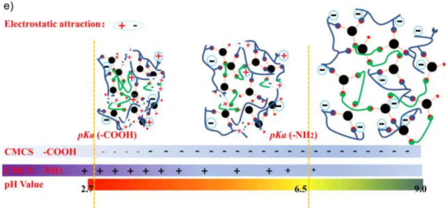
The pH dependent swelling behaviors of hydrogels could be explained by the electrostatic 347
interactions due to the presence of amino and carboxyl groups along CMCS chains. In fact, the 348
pKa of amino and carboxyl groups is 6.5 and 2.7 (Lv et al., 2018), respectively. Thus, at acidic
349
pH 3 and 4, there is strong electrostatic attraction between negatively charged -COO- and 350
positively charged -NH3+ groups, which results in shrinkage or low swelling ratio of hydrogels
351
(Fig. 6e). With increasing pH up to 7, there are less protonated NH3+ and ionized -COO- groups,
352
leading to lower electrostatic attraction and higher swelling. In contrast, at pH 8, the NH2 groups
353
are not charged, while the electrostatic repulsion between the charged -COO- groups along 354
CMCS chains leads to strong swelling. However, at pH 9, the electrostatic repulsion between the 355
-COO- groups is counterbalanced by the OH- ions in solution. Consequently, the swelling is 356
attenuated as compared to that observed at pH 8. 357
Changes of the micro-structure of hydrogels were observed by using SEM after 24 h swelling 358
at two pH values. At pH 4, all freeze-dried hydrogels strongly shrink with reduced pore size and 359
pore number (Fig. S4, Supporting Information). Noticeably, the pore size of Gel4-1 decreases 360
from c.a 150 to 100 μm, and the wall thickness increases from c.a 3.5 μm to 15 μm, reminiscent 361
with the contraction of hydrogels due to electrostatic attraction at acidic pH (Figure 6e). In 362
contrast, expansion of the porous structure is observed at pH 8 (Fig. S5, Supporting Information). 363
The pores wall shows a cracked structure, with the thickness strongly decreasing from c.a 3.5 μm 364
to 200-600 nm due to strong swelling of hydrogels provoked by electrostatic repulsion at basic 365
pH (Fig. 6e). 366
367
3.5 Self-healing: CMCS-based hydrogels present interesting self-healing behaviors as evidenced 368
by rheological recovery tests at fixed frequency of 1 Hz and at 37°C. Gel4-1 hydrogels were 369
prepared in Milli-Q water, and in pH 7 and pH 8 buffers in order to examine the self-healing 370
behavior under different swollen conditions. Gelation was realized at 37°C for 24 h. As shown in 371
Fig. 7a, both the storage modulus (G) and loss modulus (G) of Gel4-1 in Milli-Q water slightly 372
decreases until a strain of 20%. Beyond, G dramatically decreases, whereas G rapidly increases. 373
A crossover point of G and G values is observed at a strain of 60%. Similar profiles are 374
observed for Gel4-1 at pH 7 with a crossover point at 35% (Fig. S6a, Supporting Information). In 375
contrast, Gel4-1 at pH 8 exhibits lower storage modulus because of its highly swollen state as 376
shown in Figure 6a. A crossover point of G and G is detected at 55% at pH 8 (Fig. S6c, 377 Supporting Information). 378 379 380 381 382 383
384
Fig. 7. a) Modulus changes as a function of strain of Gel4-1 prepared in Milli-Q water; b)
385
Modulus changes of Gel4-1 prepared in Milli-Q water with alternatively applied high and low 386
oscillatory shear strains at 37°C; c-f) Self-healing macroscopic approaches using hydrogel 387
samples prepared in Milli-Q water (c-d), at pH 7 (e) and at pH 8 (f) , see text for details. 388
389
Based on the strain amplitude sweep results, continuous step strain measurements were 390
performed to examine the rheological recovery behavior of Gel4-1. At 1 %, Gel4-1 in Milli-Q 391
water behaves as a hydrogel since G is largely superior to G. As the oscillatory shear strain 392
increases from 1% to 60% and is maintained at 60% for 105 s (Fig. 7b), G becomes lower than 393
G, indicating the destruction of hydrogel structure. Both G and G immediately recover their 394
initial values when the strain is back to 1%. Modulus recovery is observed when larger strains 395
(100, 200, and 300%) and small strain (1%) are alternatively applied. Similar phenomena are 396
also observed for Gel4-1 prepared in pH 7 and 8 buffers (Fig. S6b, S6d, Supporting Information). 397
Therefore, it could be concluded that dynamic hydrogels exhibit rapid recovery (self-healing) 398
behavior probably due to the reconstruction of reversible imine bond linkage when they are 399
subjected to alternatively applied high and low oscillatory shear strains. 400
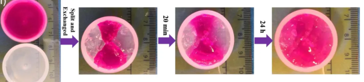
The self-healing behavior of Gel4-1 was further evidenced with four different macroscopic 401
approaches using one transparent hydrogel sample and another one incorporating yellow 402
lucigenin or red Rhodamine B dyes. First, a hole with diameter of 3 mm was punched at the 403
center of a hydrogel sample prepared in Milli-Q water, and the hole disappeared after 20 min at 404
37°C (Fig. 7c). In a second approach, transparent and yellow hydrogel samples prepared in Milli-405
Q water were cut into two semicircular pieces. They became integrated after only 20 min contact 406
at 37°C. The merged piece could be then taken off and support its own weight (Fig. 7d). 407
In a third approach, one transparent hydrogel and another one containing red Rhodamine B dye 408
prepared in pH 7 buffer were crushed via injection onto a Petri dish using a syringe, and became 409
integrated 20 min later (Fig. 7e). Almost the whole hydrogel was dyed red after 24 h. Similar 410
phenomena were observed in a fourth approach for transparent and dyed red hydrogels prepared 411
in pH 8 buffer (Fig. 7f), demonstrating that the color exchange may be observed via diffusion at 412
the restored self-healed interfaces between different dynagels at pH 7 or pH 8. 413
These tests strongly demonstrate the outstanding self-healing properties of the dynamic 414
hydrogels - dynagels via reconstruction of reversible imine bond crosslinking, and migration of 415
components or constituent exchanges between different hydrogels. Importantly, the use of these 416
hydrogels with distinct and interchangeable states at different pH conditions would be 417
advantageous for biomedical applications such as drug delivery and tissue engineering. 418
419
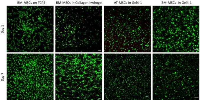
3.6 Cytocompatibility of hydrogels: Human mesenchymal stromal cells (MSCs) isolated from 420
subcutaneous adipose tissue (AT-MSCs) or from bone-marrow (BM-MSCs) were encapsulated 421
inside Gel4-1 (1 x 10⁶ cells/mL), and cultured up to 7 days in proliferative medium at 37 °C to 422
evaluate the cytocompatibility. Compared to control conditions in 2D on TCPS plate or 423
encapsulated in a type I collagen hydrogel, AT-MSCs and BM-MSCs exhibited a round shape in 424
Gel4-1 and not a fibroblastic phenotype (Fig. 8). One day after inclusion in the hydrogel, the 425
large majority of AT-MSCs (95%) and BM-MSCs (99%) were alive as indicated by the green 426
color in confocal microscopy using live/dead assay, whereas only 66% of viability was observed 427
in type I collagen hydrogel (Fig. 8). The two cell types survived for at least 7 days, as only 1 and 428
4% of dead cells were quantified for AT-MSCs and BM-MSCs, respectively (Fig. 8). These 429
findings demonstrate the excellent cytocompatibility of the hydrogel. It is noteworthy that the 430
size of AT-MSCs and BM-MSCs after 7 days was smaller than that at day 1, suggesting that the 431
pore size was reduced without affecting cell viability. Three-D reconstruction clearly shows the 432
homogeneous distribution of MSCs in the whole hydrogel volume, indicating that the gelation 433
time was compatible with homogenous distribution of the cells without sedimentation (Vid. S7, 434
Supporting Information). 435
436
437
Fig. 8. Cell viability of human AT-MSCs or MSCs in Gel4-1, in comparison with
BM-438
MSCs in collagen hydrogel or plated on TCPS as control. Cells were labelled using the 439
Live/Dead assay after 1 or 7 days in culture and imaged using confocal microscopy. Viable cells 440
were stained in green and dead cells in red. Images are maximal projections of z-axis and scale 441
bars represent 100 µm (TCPS: Tissue Culture Polystyrene Surface; AT-MSCs: Adipose Tissue-442 MSCs; BM-MSCs: Bone Marrow-MSCs). 443 444 4. Conclusions 445
Multistate pH-sensitive hydrogels were synthesized via dynamic covalent imine bonding from 446
two water soluble polymers, i.e. O-carboxymethyl chitosan (CMCS) and a cross-linking dynamer 447
obtained by reaction of amine terminated Jeffamine as connector and Benzene-1,3,5-448
tricarbaldehyde as core center. The hydrogel Gel4-1 with D-glucosamine to dynamer molar ratio 449
of 4:1 exhibits the shortest gelation time and the highest storage modulus, in agreement with 450
optimal cross-linking or imine bond formation. Freeze-dried gels exhibit interconnected porous 451
structures and pH-dependent swelling behavior. The swelling ratio is relatively low at acidic pH 452
3-5 due to electrostatic attraction, while became very high, up to 7000 % at pH 8 due to 453
electrostatic repulsion. Moreover, hydrogels present outstanding self-healing properties as 454
evidenced by closure of split pieces and rheological studies. Self-healing occurs autonomously 455
for different pH-dependent states, being able to reshape or to regenerate a strong chemical gel 456
from various situations. Last but not least, MSCs encapsulated in hydrogels are all alive after 7 457
days, in agreement with the excellent cytocompatibility of hydrogels. 458
This concept, exploiting different physical swelling states depending on pH values, results in the 459
definition of stimuli-responsive dynagels which self-adapt their structure in response to 460
environmental conditions. These ‘two-in-one’ dynagels may find potential uses in biomedical 461
applications in particular as scaffold in tissue engineering. 462
463
ASSOCIATED CONTENT
Supporting Information.
465
The following files are available free of charge. 466
NMR spectra of daynamers; rheology data of as-prepared hydrogels; SEM images of freeze dried 467
as-prepared hydrogels and hydrogels after swelling in buffers (pH4, pH8); rheology data of self-468
healing hydrogels made in buffers (pH7, pH8), 3-D reconstruction of MSCs after 7 days culture 469 in Gel4-1 (AVI). 470 471 ABBREVIATIONS 472
CMCS, O-carboxymethyl chitosan; Dy, Dynamer; BTA, Benzene-1,3,5-tricarbaldehyde; PBS, 473
phosphate buffered saline; TMS, tetramethylsilane; MSCs, human mesenchymal stromal cells. 474
475
ACKNOWLEDGMENT 476
This work is supported by the scholarship from China Scholarship Council (CSC) under the 477
Grant CSC N° 201706240281, and the Institut Européen des Membranes (Exploratory project 478
“Biostent - Health” of the Internal IEM Call 2017). Authors acknowledge funding support from 479
the Inserm Institute and the University of Montpellier. 480
481
REFERENCES 482
Ali, A., & Ahmed, S. (2018). A review on chitosan and its nanocomposites in drug delivery. 483
International Journal of Biological Macromolecules, 109, 273-286.
484
Arnal-Hérault, C., Banu, A., Barboiu, M., Michau, M., & van der Lee, A. (2007). Amplification 485
and transcription of the dynamic supramolecular chirality of the guanine quadruplex. 486
Angewandte Chemie International Edition, 46(23), 4268-4272.
487
Bhatia, S. K. (2010). Traumatic injuries. In Biomaterials for clinical applications (pp. 213-258): 488
Springer 489
Burdick, J. A., & Murphy, W. L. (2012). Moving from static to dynamic complexity in hydrogel 490
design. Nature Communications, 3(1), 1-8. 491
Catana, R., Barboiu, M., Moleavin, I., Clima, L., Rotaru, A., Ursu, E.-L., & Pinteala, M. (2015). 492
Dynamic constitutional frameworks for DNA biomimetic recognition. Chemical 493
Communications, 51(11), 2021-2024.
494
Chao, A., Negulescu, I., & Zhang, D. (2016). Dynamic covalent polymer networks based on 495
degenerative imine bond exchange: tuning the malleability and self-healing properties by 496
solvent. Macromolecules, 49(17), 6277-6284. 497
Deng, G., Ma, Q., Yu, H., Zhang, Y., Yan, Z., Liu, F., et al. (2015). Macroscopic organohydrogel 498
hybrid from rapid adhesion between dynamic covalent hydrogel and organogel. ACS Macro 499
Letters, 4(4), 467-471.
500
Dimatteo, R., Darling, N. J., & Segura, T. (2018). In situ forming injectable hydrogels for drug 501
delivery and wound repair. Advanced Drug Delivery Reviews, 127, 167-184. 502
Ghobril, C., & Grinstaff, M. (2015). The chemistry and engineering of polymeric hydrogel 503
adhesives for wound closure: a tutorial. Chemical Society Reviews, 44(7), 1820-1835. 504
Huang, W., Wang, Y., Chen, Y., Zhao, Y., Zhang, Q., Zheng, X., et al. (2016). Strong and 505
rapidly self-Healing hydrogels: potential hemostatic materials. Advanced Healthcare 506
Materials, 5(21), 2813-2822.
507
Iftime, M. M., Morariu, S., & Marin, L. (2017). Salicyl-imine-chitosan hydrogels: 508
Supramolecular architecturing as a crosslinking method toward multifunctional 509
hydrogels. Carbohydrate Polymers, 165, 39-50. 510
Li, S., El Ghzaoui, A., & Dewinck, E. (2005). Rheology and drug release properties of 511
bioresorbable hydrogels prepared from polylactide/poly (ethylene glycol) block 512
copolymers. Macromolecular Symposia (Vol. 222, pp. 23-36): Wiley Online Library. 513
Lv, X., Zhang, W., Liu, Y., Zhao, Y., Zhang, J., & Hou, M. (2018). Hygroscopicity modulation 514
of hydrogels based on carboxymethyl chitosan/Alginate polyelectrolyte complexes and its 515
application as pH-sensitive delivery system. Carbohydrate Polymers, 198, 86-93. 516
Marin, L., Moraru, S., Popescu, M. C., Nicolescu, A., Zgardan, C., Simionescu, B. C., et al. 517
(2014). Out-of-water constitutional self-organization of chitosan-cinnamaldehyde dynagels. 518
Chemistry-A European Journal, 20(16), 4814-4821.
519
Marin, L., Simionescu, B., & Barboiu, M. (2012). Imino-chitosan biodynamers. Chemical 520
Communications, 48(70), 8778-8780.
521
Qiao, C., Ma, X., Zhang, J., & Yao, J. (2017). Molecular interactions in gelatin/chitosan 522
composite films. Food Chemistry, 235, 45-50. 523
Qin, C., Zhou, J., Zhang, Z., Chen, W., Hu, Q., & Wang, Y. (2019). Convenient one-step 524
approach based on stimuli-responsive sol-gel transition properties to directly build chitosan-525
alginate core-shell beads. Food Hydrocolloids, 87, 253-259. 526
Qu, J., Zhao, X., Liang, Y., Zhang, T., Ma, P. X., & Guo, B. (2018). Antibacterial adhesive 527
injectable hydrogels with rapid self-healing, extensibility and compressibility as wound 528
dressing for joints skin wound healing. Biomaterials, 183, 185-199. 529
Rotaru, A., Pricope, G., Plank, T. N., Clima, L., Ursu, E. L., Pinteala, M., et al. (2017). G-530
Quartet hydrogels for effective cell growth applications. Chemical Communications, 53(94), 531
12668-12671. 532
Sreenivasachary, N., & Lehn, J.-M. (2005). Gelation-driven component selection in the 533
generation of constitutional dynamic hydrogels based on guanine-quartet formation. 534
Proceedings of the National Academy of Sciences of the United States of America, 102(17),
535
5938-5943. 536
Stewart, D., Antypov, D., Dyer, M. S., Pitcher, M. J., Katsoulidis, A. P., Chater, P. A., et al. 537
(2017). Stable and ordered amide frameworks synthesised under reversible conditions 538
which facilitate error checking. Nature Communications, 8(1), 1102. 539
Su, F., Wang, J., Zhu, S., Liu, S., Yu, X., & Li, S. (2015). Synthesis and characterization of 540
novel carboxymethyl chitosan grafted polylactide hydrogels for controlled drug delivery. 541
Polymers for Advanced Technologies, 26(8), 924-931.
542
Van Vlierberghe, S., Dubruel, P., & Schacht, E. (2011). Biopolymer-based hydrogels as 543
scaffolds for tissue engineering applications: a review. Biomacromolecules, 12(5), 1387-544
1408. 545
Varum, K. M., Ottoy, M. H., & Smidsrod, O. (1994). Water-solubility of partially N-acetylated 546
chitosans as a function of pH: effect of chemical composition and depolymerisation. 547
Carbohydrate Polymers, 25(2), 65-70.
548
Yang, Y., Wang, X., Yang, F., Wang, L., & Wu, D. (2018). Highly elastic and ultratough hybrid 549
ionic-covalent hydrogels with tunable structures and mechanics. Advanced Materials, 550
30(18), 1707071. 551
Yu, S., Zhang, X., Tan, G., Tian, L., Liu, D., Liu, Y., et al. (2017). A novel pH-induced 552
thermosensitive hydrogel composed of carboxymethyl chitosan and poloxamer cross-linked 553
by glutaraldehyde for ophthalmic drug delivery. Carbohydrate Polymers, 155, 208-217. 554
Zeng, X., Liu, G., Tao, W., Ma, Y., Zhang, X., He, F., et al. (2017). A drug-self-gated 555
mesoporous antitumor nanoplatform based on pH-sensitive dynamic covalent bond. 556
Advanced Functional Materials, 27(11), 1605985.
557
Zhang, W., Jin, X., Li, H., Zhang, R., & Wu, C. (2018). Injectable and body temperature 558
sensitive hydrogels based on chitosan and hyaluronic acid for pH sensitive drug release. 559
Carbohydrate Polymers, 186, 82-90.
560
Zhang, Y., & Barboiu, M. (2015a). Dynameric asymmetric membranes for directional water 561
transport. Chemical Communications, 51(88), 15925-15927. 562
Zhang, Y., & Barboiu, M. (2015b). Constitutional dynamic materials toward natural selection of 563
function. Chemical Reviews, 116(3), 809-834. 564
Zhang, Y., Tao, L., Li, S., & Wei, Y. (2011). Synthesis of multiresponsive and dynamic 565
chitosan-based hydrogels for controlled release of bioactive molecules. Biomacromolecules, 566
12(8), 2894-2901. 567
Zhang, Y., Wu, X., Han, Y., Mo, F., Duan, Y., & Li, S. (2010). Novel thymopentin release 568
systems prepared from bioresorbable PLA-PEG-PLA hydrogels. International Journal of 569
Pharmaceutics, 386(1-2), 15-22.
570
Zimmermann, J., Bittner, K., Stark, B., & Mülhaupt, R. (2002). Novel hydrogels as supports for 571
in vitro cell growth: poly (ethylene glycol) and gelatin-based (meth) acrylamidopeptide 572
macromonomers. Biomaterials, 23(10), 2127-2134. 573