Publisher’s version / Version de l'éditeur:
Journal of Applied Physiology, 113, 4, pp. 574-583, 2012-08-15
READ THESE TERMS AND CONDITIONS CAREFULLY BEFORE USING THIS WEBSITE. https://nrc-publications.canada.ca/eng/copyright
Vous avez des questions? Nous pouvons vous aider. Pour communiquer directement avec un auteur, consultez la première page de la revue dans laquelle son article a été publié afin de trouver ses coordonnées. Si vous n’arrivez pas à les repérer, communiquez avec nous à PublicationsArchive-ArchivesPublications@nrc-cnrc.gc.ca.
Questions? Contact the NRC Publications Archive team at
PublicationsArchive-ArchivesPublications@nrc-cnrc.gc.ca. If you wish to email the authors directly, please see the first page of the publication for their contact information.
NRC Publications Archive
Archives des publications du CNRC
This publication could be one of several versions: author’s original, accepted manuscript or the publisher’s version. / La version de cette publication peut être l’une des suivantes : la version prépublication de l’auteur, la version acceptée du manuscrit ou la version de l’éditeur.
For the publisher’s version, please access the DOI link below./ Pour consulter la version de l’éditeur, utilisez le lien DOI ci-dessous.
https://doi.org/10.1152/japplphysiol.00523.2012
Access and use of this website and the material on it are subject to the Terms and Conditions set forth at
Influence of circulating cytokines on prolactin during slow vs. fast
exertional heat stress followed by active or passive recovery
Wright, Heather E.; Mclellan, Tom M.; Friesen, Brian J.; Casa, Douglas J.;
Kenny, Glen P.
https://publications-cnrc.canada.ca/fra/droits
L’accès à ce site Web et l’utilisation de son contenu sont assujettis aux conditions présentées dans le site
LISEZ CES CONDITIONS ATTENTIVEMENT AVANT D’UTILISER CE SITE WEB.
NRC Publications Record / Notice d'Archives des publications de CNRC:
https://nrc-publications.canada.ca/eng/view/object/?id=aac8e376-676b-4dcf-b742-1efc65ba60aa https://publications-cnrc.canada.ca/fra/voir/objet/?id=aac8e376-676b-4dcf-b742-1efc65ba60aa
Influence of circulating cytokines on prolactin during slow vs. fast exertional
heat stress followed by active or passive recovery
Heather E. Wright,1Tom M. McLellan,2Brian J. Friesen,1Douglas J. Casa,3and Glen P. Kenny1
1Faculty of Health Sciences, Human and Environmental Physiology Research Unit, University of Ottawa, Ottawa, Ontario,
Canada;2TM McLellan Research Inc., Stouffville, Ontario, Canada; and3Korey Stringer Institute, Department of
Kinesiology, University of Connecticut, Storrs, Connecticut
Submitted 27 April 2012; accepted in final form 15 June 2012
Wright HE, McLellan TM, Friesen BJ, Casa DJ, Kenny GP.
Influence of circulating cytokines on prolactin during slow vs. fast exertional heat stress followed by active or passive recovery. J
Appl Physiol113: 574 –583, 2012. First published June 21, 2012; doi:10.1152/japplphysiol.00523.2012.—Prolactin (PRL) has been suggested as an indicator of fatigue during exertional heat stress (EHS), given its strong relationship with body core temperature (Tc); however, the strength of this relationship during different rates of Tc increase and subsequent recovery is unknown. In addition, given the influence that systemic cytokines, such as interleukin (IL)-6 and tumor necrosis factor (TNF)-␣, have on the pituitary gland, it would be of interest to determine the relationship between PRL, IL-6, and TNF-␣ during EHS. The purpose was to examine the PRL, IL-6, and TNF-␣ heat stress responses during slow and fast heating and subsequent resting or cold water immersion recovery. On 4 days, nine individuals walked at ⬃45% (slow heating) or ran at ⬃65% (fast heating) maximal oxygen consumption on a treadmill in the heat (40°C, 30% relative humidity) until rectal temperature (Tre) reached 39.5°C (esophageal temperature; fast ⫽ 39.41 ⫾ 0.04°C, slow ⫽ 39.82 ⫾ 0.09°C). Post-EHS, subjects were either immersed in 2°C water or rested seated until Trereturned to 38.0°C. Venous blood, analyzed for PRL, IL-6, and TNF-␣, was obtained at rest, during exercise (Tre 38.0, 39.0, 39.5°C), the start of recovery (⬃5 min after 39.5°C), and subsequent recovery (Tre39.0, 38.0°C). IL-6 exhibited myokine properties, given the greater increases with slow heating and lack of increase in TNF-␣. A strong temperature-dependent PRL response during slow and fast heating provides additional support for the use of PRL as a peripheral marker of impending fatigue, which is independent of IL-6 and TNF-␣ cytokine responses.
neuroimmune response; core temperature; peripheral fatigue marker; serotonin and dopamine interplay
DURING EXERTIONAL HEAT STRESS (EHS), the development of
central and peripheral fatigue leads to physical and cognitive performance impairments (18, 22), which can increase the risk of injury in recreational and occupational settings. Currently, several mechanisms have been proposed to explain the devel-opment of fatigue, or heat intolerance, during exercise in the heat, including the interplay between increased serotonin and decreased dopamine in the brain, or the Central Fatigue Hy-pothesis (7, 16, 23, 35–36, 63), increased endotoxin leakage at the gut further aggravating hyperthermia (49, 51), the critical core temperature hypothesis (19), and the Central Governor Model of the sensory representation of neural integrative pro-cesses (56).
High core temperatures are believed to alter motor center control in the brain through the interplay of neurotransmitter and hormonal release. The Central Fatigue Hypothesis has been proposed to explain the development of fatigue during EHS (25, 48, 55), given that increased serotonin and decreased dopamine concentrations in the brain have been linked to depressed mood and motivation (3, 29, 33, 55), lethargy, potentially reduced central nervous system drive (66), and altered heat loss responses (i.e., vasodilation) (8).
Direct quantification of the serotonergic and dopaminergic activity within the human brain is not possible. However, changes in circulating prolactin (PRL) concentrations may reflect changes in the release of these neurotransmitters and serve as a peripheral indicator of impending fatigue (5, 29, 32, 65), given that serotonergic and dopaminergic neurons in the brain stem stimulate and inhibit pituitary gland PRL secretion, respectively (4, 29, 62). Previous research has shown that PRL concentrations peak at the point of peak heat strain, during EHS where thermoregulatory strain is intolerable (29). In addition, PRL secretion has been shown to be strongly corre-lated with perceived exertion (4), mean skin temperature (28), increases in core temperature (i.e., r ⱖ 0.55) during prolonged exercise in the heat (44), and uncompensable heat stress, irrespective of fitness level (65). Although these studies pro-vide support for the use of PRL as an indicator of maximal heat tolerance and impending fatigue, circulating PRL levels at a given core temperature have been shown to be variable among studies, with exercise in trained and untrained individuals to a rectal temperature (Tre) of 39.0 –39.7°C at exhaustion [⬃1°C/h,
peak PRL (PRLpeak) ⬃2,350 –2,500 pmol/l] (65), during active
and passive heating to a Tre of 38.8°C (⬃1.6°C/h, PRLpeak
⬃1,000 –1,150 pmol/l) (29), and following 45 min of cycling in 30°C with a ⫹1.19°C change in Tre(⬃1.6°C/h, PRLpeak⬃750
pmol/l) (28), indicating that PRL responses may be influenced by factors in addition to absolute thermal strain. It is unknown whether a faster rate of change in core temperature, associated with greater exercise intensities (50), and/or exposure to higher ambient temperatures (17), together with the concomitant in-crease in perceived exertion (20), cardiovascular strain, neu-roendocrine and immune changes (27), and reduced cognitive performance (22), may help to explain such differences in the PRL responses during EHS. Interestingly, Buckler (6) has shown that the growth hormone response during exercise is directly related to the rate of increase in core temperature. One approach to test whether the rate of change in core temperature affects PRL release is to have participants exercise at different exercise intensities while using core temperature as the inde-pendent variable for blood sampling. In addition, examining the relationship between circulating PRL and core temperature,
Address for reprint requests and other correspondence: G. P. Kenny, Univ. of Ottawa, School of Human Kinetics, 125 Univ. Ave., Montpetit Hall, Rm. 367, Ottawa, ON, Canada K1N 6N5 (e-mail: gkenny@uottawa.ca).
J Appl Physiol113: 574–583, 2012.
First published June 21, 2012; doi:10.1152/japplphysiol.00523.2012.
8750-7587/12 Copyright©2012 the American Physiological Society http://www.jappl.org 574
by 10.220.32.247 on January 6, 2017
http://jap.physiology.org/
and changes in PRL, during fast and slow decreases in core temperature (i.e., active and passive cooling, respectively) could be used to further test the premise that PRL can be used as a surrogate marker of the effects of thermal strain on the interplay between brain serotonin and dopamine. Although cold water immersion recovery from EHS results in the quick reestablishment of cardiovascular (i.e., heart rate) and thermal parameters (45, 59), it is unclear whether PRL would exhibit a similar recovery response.
The release of PRL from the pituitary gland is also positively stimulated by cytokines, such as interleukin (IL)-6 and Tumor Necrosis Factor (TNF)-␣ (61). In response to a single bout of exercise, the immune system exhibits an acute phase response due to the associated local muscle damage, metabolic changes, and hyperthermia (34, 38, 51). Experimental designs where the normal rise in core temperature during exercise has been reduced demonstrate the importance of thermal strain for the release of pro- and anti-inflammatory cytokines, such as IL-6, IL-1ra, and TNF-␣, into the circulation (46). Increases in IL-6 and TNF-␣ during exercise are dependent on the exercise intensity and duration, the muscle mass being utilized, and the endurance capacity of the individual (14, 39, 40, 42). Although PRL secretion is strongly stimulated by increases in core temperature, the strength of the cytokine influence on PRL secretion from the pituitary gland during EHS is unknown. Thus, examining the cytokine and PRL responses simultane-ously during different rates of increase in, and recovery of, core temperature will help elucidate both the strength of the influ-ence of cytokines on pituitary PRL secretion and the stimulus for the variability in PRL responses reported in the literature at a given core temperature.
The purpose of this study was, therefore, to examine the influence of different rates of increase (i.e., slow vs. fast heating) and decrease (active vs. passive cooling) in core temperature on PRL and circulating cytokine responses (i.e., IL-6, TNF-␣) and, subsequently, the strength of the relation-ship between cytokine stimuli on the pituitary and potential PRL secretion. Although PRL has been reported to be strongly related to the increase in core temperature, variability of the PRL responses at a given core temperature in the literature may be partially explained by differences in the rate of increase core temperature. Given that cytokine responses are partly depen-dent on exercise intensity, it was hypothesized that the faster rate of core temperature increase associated with higher exer-cise intensities would stimulate greater cytokine and PRL responses compared with slow heating. Subsequent to the EHS, it was hypothesized that active, compared with passive, cooling would result in greater reductions in circulating PRL and cytokines, corresponding to the quicker reestablishment of baseline thermal and cardiovascular responses.
EXPERIMENTAL PROCEDURES
Subjects
On receiving approval from the University of Ottawa Health Sciences and Science Research Ethics Board, nine healthy (no history of respiratory, metabolic, or cardiovascular conditions), physically active individuals (7 men, 2 women), aged 20 – 46 yr, were recruited from the university population and general community. Participants were informed of all experimental procedures, associated risks, and discomforts before providing written, informed consent. Mean ⫾ SE age, height, mass, body surface area, percent body fatness, and
maximal aerobic power of the participants were as follows: 26.0 ⫾ 2.5 yr, 174.2 ⫾ 3.8 cm, 74.6 ⫾ 4.3 kg, 17.9 ⫾ 2.8%, and 56.5 ⫾ 2.2 ml O2·kg⫺1·min⫺1, respectively.
Experimental Design
Testing was conducted in the Human and Environmental Physiol-ogy Research Unit at the University of Ottawa. During the preliminary session, participants received an orientation to the instrumentation and experimental protocols, completed the American Heart Association/ American College of Sports Medicine Pre-Participation Screening (1) and the Canadian Society for Exercise Physiology Physical Activity Readiness (10) questionnaires to ensure their safety for participation, and performed fitness and body composition assessments. Aerobic fitness was assessed by measuring peak O2 consumption (V˙O2peak) using a treadmill protocol and open-circuit spirometry. Participants began running at a self-selected pace, where the treadmill incline was increased 1% each minute to a maximum of 10%, after which speed and incline were increased alternatively each minute by 0.8 km/h and 1% incline, respectively, until the participant could no longer con-tinue. V˙O2peak was defined as the highest oxygen uptake during the incremental test, taken as the mean of the three highest consecutive 15-s recordings at the end of the test. In addition, hydrostatic weighing was used to measure body density, and percent body fatness was estimated based upon the Siri equation (54).
Following the preliminary session, all participants completed four EHS exposures, which were separated by a minimum of 5 days. Participants were asked to refrain from strenuous exercise (running, swimming, cycling, weight lifting, etc.), alcohol, and the use of over-the-counter medications for 24 h, and caffeine for 12 h, before each EHS session. During the experimental EHS sessions, participants either walked (4.0 – 4.5 km/h, 2% incline) or jogged (⬃7.0 km/h, 2% incline) on a treadmill in a hot-dry environment (40°C, 20 –30% relative humidity) for a slow or fast rate of Treincrease, respectively, while wearing a nonpermeable rain poncho. On reaching a Tre of 39.5°C, participants recovered either passively (resting ambient envi-ronment, ⬃29°C) or actively (2°C cold water immersion) until re-turning to a Treof 38.0°C. Thus the four EHS sessions, which were performed in a randomized order, included the following: 1) slow heating with passive recovery (SP); 2) slow heating with active recovery (SA); 3) fast heating with passive recovery (FP); and 4) fast heating with active recovery (FA).
Physiological Measurements
Core temperature. Tre and esophageal temperature (Tes) were measured continuously using a pediatric thermocouple probe (Mon-a-therm Nasopharyngeal Temperature Probe, Mallinckrodt Medical, St. Louis, MO). The rectal probe was inserted ⬃15 cm beyond the anal sphincter. Teswas measured by inserting the probe, of ⬃2 mm in diameter, through the nostril to the level of the heart.
Heart rate.Heart rate was continuously monitored on a receiver using a transmitter (Polar Electro, Oy, Finland) fitted around the chest.
Ratings of thermal comfort and perceived exertion. Ratings of perceived exertion (RPE; scale “6 ⫽ no exertion at all” to “20 ⫽ maximal exertion”) and thermal comfort (TC; scale “1 ⫽ so cold I am helpless” to “13 ⫽ so hot I am sick and nauseated”) were recorded at baseline resting (PRE) and at each increment change in Treof 0.5°C. The perceptual strain index (PeSI) was calculated using TC and RPE, according to the following equation;
PeSI ⫽
关
5 ·共
TCt ⫺ 7兲
⁄ 6兴
⫹关
5 ·共
RPEt ⫺ 6兲
⁄ 14兴
where t represents the Tre at which the rating was recorded, the denominators of 6 and 14 represent the number of values on the scale above rest for TC and RPE, respectively, and 6 represents the RPE at rest (60).
Blood sampling and measurements. Venous blood was collected via an indwelling intravenous catheter (Jelco I.V. 18G Catheter,
by 10.220.32.247 on January 6, 2017
http://jap.physiology.org/
Smiths Medical International, Rossendale, Lancashire, UK), inserted into an antecubital vein, with a 21-in. extension line. Blood was collected at rest (PRE), at a Treof 38.0°C, 39.0°C, and 39.5°C during EHS, at the beginning of recovery, and at a Treof 39.0°C and 38.0°C during recovery. Blood was drawn into sterile plastic syringes and transferred immediately into serum with no additive, 5.4 mg plasma K2EDTA, and plasma sodium heparin (NH, 30 USP) BD Vacutainer tubes (BD, Franklin Lakes, NJ). Nonadditive blood sat for 20 min to clot before centrifugation at ⬃3,300 rpm for 10 min, whereas the EDTA blood was mixed by inversion, used for hematological analyses (Beckman Coulter, Miami, FL) and the measurement of plasma protein concentration (Reichert TS 400 total solids refractometer, Reichert, Depew, NY), and centrifuged immediately with the sodium heparin plasma. Serum and plasma aliquots were transferred into polypropylene Eppendorf tubes, frozen at ⫺20°C, and stored at ⫺70°C until analyzed.
Enzyme immunoassay techniques were used to determine circulat-ing serum PRL (ALPCO Immunoassays, ALPCO Diagnostics, Salem, NH), and serum IL-6 and plasma TNF-␣ (Quantikine High Sensitivity R&D Systems, Minneapolis, MN) concentrations. The PRL, IL-6, and TNF-␣ assays’ minimum detectable concentrations were 2 ng/ml, 0.016 – 0.110 pg/ml, and 0.038 – 0.191 pg/ml, respectively. All sam-ples were analyzed in duplicate following manufacturer instructions, and concentrations were corrected for plasma volume changes, based on the Dill and Costill method using blood hemoglobin and hemato-crit changes (11).
Statistical Analyses
A two-way ANOVA with repeated measures of heating (slow vs. fast heating) and temperature (PRE, Treof 38.0, 39.0, and 39.5°C) during exercise, and cooling (Active vs. Passive) and temperature (the start of recovery, and Treof 39.0 and 38.0°C) during recovery was performed on the dependent measures (i.e., heart rate, PeSI, percent plasma volume change, plasma protein, PRL, PRL rate of change relative to Treand Tes, IL-6, TNF-␣) during the experimental sessions. A one-way ANOVA was used to compare dependent measures at PRE, end of exercise, start of recovery, and end of recovery, the exercise and recovery times and rates of change in Treand Tes, and the
PRL response at a given Trein heating vs. cooling. When a significant
Fratio was obtained, a Newman-Keuls post hoc analysis was used to isolate differences among treatment means. All ANOVAs were com-pleted using Statistica data analysis software (StatSoft 2007, version 8.0, Tulsa, OK, www.statsoft.com). Results are reported as means ⫾ SE. Statistically significant differences were identified for P ⬍ 0.05, and trends were identified for 0.05 ⬍ P ⬍ 0.1. An outlier IL-6 response for one participant was identified by calculating the Chau-venet’s criterion [Q ⫽ (potential outlier ⫺ maximum value)/range], where n ⫽ 8 for IL-6 based on the data being rejected for a Q greater than the 95% confidence level (for n ⫽ 9, Q 95% ⫽ 0.493).
RESULTS
Physiological Measures
Exercise. By design, the exercise time to reach a Tre of
39.5°C was greater for the slow (SP ⫽ 76.2 ⫾ 5.1 and SA ⫽ 86.1 ⫾ 6.8 min) compared with fast (FP ⫽ 34.8 ⫾ 2.6 and FA ⫽ 39.8 ⫾ 4.1 min) (P ⬍ 0.000) heating, but there was no difference between SP and SA or between FP and FA. These rates of increase correspond to ⬃1.8 and 4.2°C/h for the slow and fast heating protocols, respectively. There was no differ-ence among the protocols for PRE Tre(P ⫽ 0.785) or Tes(P ⫽
0.986) or at the end of exercise for Tre(SP ⫽ 39.50 ⫾ 0.01, SA
⫽ 39.51 ⫾ 0.00, FP ⫽ 39.5 ⫾ 0.03, and FA ⫽ 39.51 ⫾ 0.00°C, P ⫽ 0.840). However, Teswas significantly higher at
the end of exercise following the fast (FP ⫽ 39.84 ⫾ 0.17 and FA ⫽ 39.81 ⫾ 0.11°C) compared with slow (SP ⫽ 39.40 ⫾ 0.08 and SA ⫽ 39.41 ⫾ 0.05°C) (P ⫽ 0.005) heating. There were no differences between the two slow and the two fast heating conditions for heart rate, percent change in plasma volume, plasma protein concentration, or PeSI (Table 1). No differences were observed for heart rate or plasma protein concentration at PRE (P ⫽ 0.600 and 0.964, respectively) or the end of exercise (P ⫽ 0.133 and 0.847, respectively) among the heating protocols.
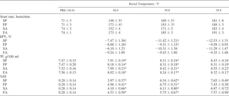
Table 1. Physiological measures relative to increases in rectal temperature during exercise-induced hyperthermia for the
slow and fast heating conditions, followed by active and passive cooling recovery
Rectal Temperature, °C
PRE (36.9) 38.0 39.0 39.5
Heart rate, beats/min
SP 71 ⫾ 3 148 ⫾ 5† 169 ⫾ 5† 181 ⫾ 8 FP 71 ⫾ 3 173 ⫾ 4† 183 ⫾ 3† 188 ⫾ 3 SA 74 ⫾ 3 152 ⫾ 4 171 ⫾ 5 183 ⫾ 4 FA 74 ⫾ 1 173 ⫾ 4 185 ⫾ 3 191 ⫾ 3 ⌬PV, % SP ⫺7.47 ⫾ 1.36† ⫺11.42 ⫾ 1.21† ⫺12.53 ⫾ 1.31 FP ⫺8.88 ⫾ 1.26† ⫺9.31 ⫾ 1.15† ⫺9.58 ⫾ 0.95 SA ⫺6.10 ⫾ 1.23 ⫺10.31 ⫾ 1.38 ⫺11.29 ⫾ 1.47 FA ⫺9.24 ⫾ 1.89 ⫺9.43 ⫾ 1.80 ⫺9.35 ⫾ 1.88 PP, g/100 ml SP 7.47 ⫾ 0.15 7.91 ⫾ 0.19* 8.31 ⫾ 0.18* 8.43 ⫾ 0.18* FP 7.47 ⫾ 0.20 8.18 ⫾ 0.14* 8.31 ⫾ 0.18* 8.31 ⫾ 0.19* SA 7.52 ⫾ 0.16 7.99 ⫾ 0.23* 8.42 ⫾ 0.21* 8.55 ⫾ 0.22* FA 7.56 ⫾ 0.13 8.02 ⫾ 0.18* 8.24 ⫾ 0.17* 8.32 ⫾ 0.17* PeSI SP 0.28 ⫾ 0.14 3.97 ⫾ 0.37* 6.34 ⫾ 0.62* 7.65 ⫾ 0.49* FP 0.28 ⫾ 0.14 4.96 ⫾ 0.41* 6.75 ⫾ 0.31* 7.43 ⫾ 0.38* SA 0.28 ⫾ 0.14 4.10 ⫾ 0.66* 6.11 ⫾ 0.80* 6.87 ⫾ 0.72* FA 0.28 ⫾ 0.14 4.53 ⫾ 0.50* 5.75 ⫾ 0.67* 7.57 ⫾ 0.98* Values are means ⫾ SE. PRE, baseline resting temperature; ⌬PV, change in plasma volume, PP, plasma protein; PeSI, perceptual strain index; S and F, slow and fast heating conditions, respectively; A and P, active and passive cooling recovery, respectively. *Main effect of temperature, P ⬍ 0.0001. †Condition ⫻ temperature interaction, P ⬍ 0.005.
576 Prolactin, Cytokines, Fatigue, and Exertional Heat Stress • Wright HE et al.
J Appl Physiol•doi:10.1152/japplphysiol.00523.2012•www.jappl.org
by 10.220.32.247 on January 6, 2017
http://jap.physiology.org/
Heart rate increased at a greater rate during the fast com-pared with slow heating conditions (P ⫽ 0.000) from PRE to a Treof 39.5°C. Plasma volume exhibited a heating ⫻
temper-ature interaction (P ⫽ 0.000), where the percent plasma vol-ume change was significantly greater at a Tre of 38.5 and
39.0°C compared with the change at 38.0°C for the slow heating conditions protocols, but the decrease in plasma vol-ume was constant throughout the fast heating protocols. A trend was observed for greater decreases in percent change of plasma volume following the slow compared with fast heating (P ⫽ 0.091). Plasma protein and PeSI increased significantly throughout exercise, but changes were not different at any given Treamong the heating protocols.
Recovery.Cooling recovery time to a Treof 38.0°C tended to
be longer following fast compared with slow heating (11.1 ⫾ 2.4 vs. 6.6 ⫾ 0.7 min) (P ⫽ 0.091), whereas passive cooling recovery time was significantly longer following the fast (76.1 ⫾ 8.0 min) compared with the slow (53.8 ⫾ 5.3 min) (P ⫽ 0.033) heating protocols. A main effect of condition was observed at the end of recovery for Tes (P ⫽ 0.000) between the two passive (SP ⫽
37.78 ⫾ 0.06 and FP ⫽ 37.78 ⫾ 0.09°C) and two active (SA ⫽ 36.27 ⫾ 0.29 and FA ⫽ 35.93 ⫾ 0.44°C) cooling conditions.
Heart rate, plasma volume changes, plasma protein, and PeSI responses during recovery are shown in Table 2. Regard-less of the rate of increase in Tre during the exercise phase,
heart rates at any Tre were similar during recovery for all
conditions. However, heart rate did significantly decrease as Tre decreased. Percent change in plasma volume was not
different between the two active nor two passive cooling conditions (P ⫽ 0.978 and 0.402, respectively), yet decreased as Tredecreased during the passive cooling (P ⫽ 0.000), but
not the active cooling (P ⫽ 0.233). A temperature ⫻ condition interaction was observed for the four recovery conditions (P ⫽ 0.001) and the active vs. passive cooling following slow (P ⫽ 0.030) and fast (P ⫽ 0.012) heating. Plasma protein concen-tration was not different between the two active cooling con-ditions (P ⫽ 0.468), with decreases in Tre during the active
cooling (P ⫽ 0.673), or between the two passive cooling
conditions (P ⫽ 0.593); however, decreases were observed with decreases in Treduring passive cooling (P ⫽ 0.000).
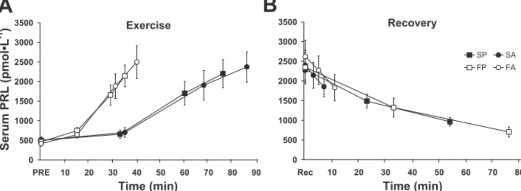
PRL
PRL exhibited a strong temperature-dependent response throughout exercise from PRE to the end of exercise (Tre ⫽
39.5°C, Tesfast ⫽ 39.8 ⫾ 0.1°C, Tes slow ⫽ 39.4 ⫾ 0.0°C)
(P ⫽ 0.000), with no differences between the fast and slow heating protocols (P ⫽ 0.962) (Fig. 1, A and C). Regardless of the prior heating protocol, the PRL response during recovery was similar during the two passive and the two active cooling sessions (Fig. 1, B and D). However, PRL values were signifi-cantly higher during the active vs. passive cooling protocols at a Treof 39.0 and 38.0°C. The rate of change of PRL relative to Tes
(pmol/l/°C) was increased during passive recovery following both slow (P ⫽ 0.038) and fast (P ⫽ 0.000) heating; however, it was reduced during active recovery following slow and fast heating for both Tre(P ⫽ 0.006 and 0.014, respectively) and
Tes (P ⫽ 0.000 and 0.001, respectively) (Fig. 2).
Cytokines
IL-6 was not different during EHS between the two slow (P ⫽ 0.328) or the two fast (P ⫽ 0.907) heating conditions from PRE to a Tre of 39.5°C (Fig. 3, A and C, n ⫽ 8). A
temperature ⫻ condition interaction (P ⫽ 0.024) was observed for IL-6, where increases were observed at a Treof 38.0, 39.0,
and 39.5°C compared with PRE for all conditions, with greater increases at 39.5°C following the slow compared with fast heating conditions. During active and passive recovery, IL-6 tended to be elevated following slow vs. fast heating (P ⫽ 0.076); however, no effect of temperature was observed (P ⫽ 0.598) (Fig. 3, B and D). Although no differences were ob-served between the active and passive recovery conditions (P ⫽ 0.905) or with decreases in temperature (P ⫽ 0.465) following slow heating, a temperature ⫻ condition interaction (P ⫽ 0.013) was observed between the active and passive recovery conditions following fast heating, where IL-6 contin-ued to rise during the passive recovery as Trebegan to fall.
TNF-␣ was not different between the two slow (P ⫽ 0.500) or the two fast (P ⫽ 0.936) heating conditions from PRE to a Tre of 39.5°C (Fig. 4, A and C). An increase in TNF-␣ was
observed at a Treof 39.0 and 39.5°C compared with PRE (P ⫽
0.038) during the fast heating conditions only while a trend was observed for a greater TNF-␣ response at a Treof 39.5°C
for the fast compared with slow heating conditions (P ⫽ 0.092). TNF-␣ was not different between the active and pas-sive recovery conditions (P ⫽ 0.519) or with decreases in temperature (P ⫽ 0.349) (Fig. 4, B and D); however, when comparing the two passive recovery conditions, TNF-␣ was higher at a Treof 38.0°C compared with the start of recovery
following the slow heating (P ⫽ 0.018). Following slow heating, TNF-␣ was not different between the active and passive recovery conditions (P ⫽ 0.282); however, TNF-␣ was elevated at the end of recovery (Tre⫽ 38.0°C) compared with
the start of recovery (P ⫽ 0.027).
DISCUSSION
This study examined the influence of different rates of increase and decrease of core temperature on circulating PRL and cytokine responses, and the influence of circulating cyto-Table 2. Physiological measures relative to rectal
temperature during active and passive cooling recovery following slow and fast heating
Rectal Temperature, °C
Rec (39.7) 39.0 38.0
Heart rate, beats/min
SP 133 ⫾ 4 106 ⫾ 6* 89 ⫾ 3* FP 137 ⫾ 6 103 ⫾ 5* 86 ⫾ 3* SA 143 ⫾ 8 101 ⫾ 9* 94 ⫾ 5* FA 139 ⫾ 8 107 ⫾ 5* 104 ⫾ 9* ⌬PV†, % SP ⫺12.13 ⫾ 1.54 ⫺8.75 ⫾ 1.95* ⫺6.45 ⫾ 2.22* FP ⫺9.31 ⫾ 1.23 ⫺6.67 ⫾ 1.54* ⫺5.46 ⫾ 1.64* SA ⫺11.28 ⫾ 1.64 ⫺9.78 ⫾ 1.78 ⫺8.82 ⫾ 1.97 FA ⫺9.75 ⫾ 1.46 ⫺10.00 ⫾ 1.62 ⫺10.33 ⫾ 2.20 PP, g/100 ml SP 8.70 ⫾ 0.26 8.41 ⫾ 0.21* 8.26 ⫾ 0.23* FP 8.48 ⫾ 0.16 8.31 ⫾ 0.15* 8.13 ⫾ 0.20* SA 8.72 ⫾ 0.23 8.59 ⫾ 0.23 8.59 ⫾ 0.28 FA 8.36 ⫾ 0.16 8.39 ⫾ 0.21 8.46 ⫾ 0.22 Values are means ⫾ SE. Change in PV is relative to PRE. Rec, start of recovery. *Main effect of temperature, P ⬍ 0.0001. †Condition ⫻ temperature interaction, P ⬍ 0.005.
by 10.220.32.247 on January 6, 2017
http://jap.physiology.org/
kines on PRL responses during EHS and recovery. Further-more, this study used a slow vs. fast EHS ramping protocol to verify the use of PRL as an indicator of changes within the brain that reflect impending fatigue. Unique to this study was the observation that, regardless of the rate of hyperthermia development, PRL exhibited a strong temperature-dependent response during EHS. However, during recovery, the relation-ship between PRL and Tre was dependent on the rate of
cooling. During EHS, increased metabolic demands with long-duration exercise exaggerated the IL-6 response during the slow compared with fast heating protocols, whereas TNF-␣ responded to the greater exercise intensity during the fast
compared with slow heating. However, the different responses of these circulating cytokines during the heating protocols did not influence the PRL response. In addition, neither recovery method, of active vs. passive cooling, was superior in signifi-cantly reducing the circulating cytokines (i.e., IL-6 and TNF-␣) following EHS.
PRL and Central Fatigue
Circulating PRL has been implicated as an indicator of central fatigue during heat stress, given that increased values reflect the relationship of increased serotonin and decreased
Fig. 1. Circulating serum prolactin (PRL) concentrations during slow (S) vs. fast (F) exertional heat stress (A and C), followed by active (A) vs. passive (P) cooling (B and D), relative to rectal (Tre; A and B) and esophageal temperature (Tes; C and D). Values are means ⫾ SE. Main effect of temperature (*P ⬍ 0.0001)
and temperature ⫻ condition interaction (†P ⬍ 0.005) is shown.
Fig. 2. The rate of change in circulating serum PRL concentrations relative to the change in Treand Tes during S vs. F
exer-tional heat stress (A) followed by A vs. P cooling (B). Values are means ⫾ SE. Signif-icantly different during passive (‡P ⬍ 0.05) and active (§P ⬍ 0.05) recovery compared with exercise.
578 Prolactin, Cytokines, Fatigue, and Exertional Heat Stress • Wright HE et al.
J Appl Physiol•doi:10.1152/japplphysiol.00523.2012•www.jappl.org
by 10.220.32.247 on January 6, 2017
http://jap.physiology.org/
dopamine levels in the brain (4, 29, 62) and are correlated with increases in core temperature (44, 65). The present study showed a strong temperature-dependent PRL response during EHS from PRE (Tre ⫽ 36.96 ⫾ 0.05°C) to 39.5°C, which
supports the use of PRL as a marker of increased and decreased brain serotonin and dopamine, respectively, also known as the central fatigue hypothesis (31). This temperature-dependent PRL response is consistent with previous studies reporting increases in PRL during EHS in trained and untrained men (65), following 5 h of cycling in trained individuals (57), and in hypohydrated, heat-acclimated men during EHS (7). The greater rate of increase in PRL beyond a Treof 38.0°C is also
consistent with our laboratory’s earlier work (65) and the other studies reporting increased circulating hormone and immune markers above a Treof 38.0°C (9, 46). Furthermore, this is the
first study to report a strong temperature-dependent response for PRL that is independent of different rates of increase in Tre
[i.e., slow (⬃2°C/h) vs. fast (⬃4°C/h) hyperthermia develop-ment] associated with greater exercise intensities (Fig. 5). Although it was hypothesized that different rates of increase in core temperature would contribute to the variable PRL re-sponses at a given core temperature reported in the literature, the present study refutes this possibility, given that the PRL response was independent of the rates of increase in Tre.
This is further supported by Fig. 2, which shows no differ-ences in the rate of change in PRL relative to Tre and Tes
(pmol/l/°C) between the slow and fast heating and our labora-tory’s previous work, which showed similar PRL responses between trained and untrained individuals at a slower rate of Treincrease of ⬃1°C/h, with very similar PRL values at a Tre
of 39.5°C for the trained group (2,353 ⫾ 220 pmol/l) (65) compared with the present study with individuals of similar fitness levels (2,303 ⫾ 360 pmol/l). Thus, during EHS, the present study provides further support for the use of PRL as a peripheral marker of impending fatigue at high core tempera-tures.
Despite the present study and previous research showing a strong relationship between PRL and core temperature (44, 65), the divergent PRL response at a given core temperature between the two recovery methods in the present study indi-cates that thermal strain alone cannot solely explain the PRL responses and may provide some explanation for the variable PRL responses reported in the literature. Regardless of the rate of increase in core temperature during EHS, the manipulation of the rate of change in core temperature during recovery resulted in PRL concentrations decreasing at a slower rate during the active compared with passive cooling recovery. Since it is believed that Tes better reflects brain temperature
compared with Tre(53), representing PRL responses relative to
Tes may be more indicative of the serotonergic and
dopami-nergic changes within the brain and thus circulating PRL concentrations (Figs. 1B and 2B). However, PRL responses in
Fig. 3. Circulating serum interleukin (IL)-6 concentrations during S vs. F exertional heat stress (A and C), followed by A vs. P cooling (B and D), relative to Tre(A and B) and Tes(C and D). Values are means ⫾ SE. Temperature ⫻ condition interaction (†P ⬍ 0.05) is shown.
by 10.220.32.247 on January 6, 2017
http://jap.physiology.org/
the present study were more strongly correlated with Trethan
Tesduring slow (r ⫽ 0.67 and 0.61, respectively) and fast (r ⫽
0.72 and 0.64, respectively) heating, and during passive (r ⫽ 0.58 and 0.48, respectively) but not active recovery (r ⫽ 0.24 and 0.31, respectively). Furthermore, a greater divergence in PRL response is observed during recovery when represented relative to Tes. Despite similar PRL concentrations at the start
of recovery in all conditions, PRL concentrations at a given Tre
during passive recovery reflected EHS concentrations better than the PRL responses observed during active recovery. For instance, at the end of recovery, at a Treof 38.0°C, PRL values
were not different than PRL values at a Treof 38.0°C during
EHS (P ⫽ 0.120), although they were still different at 39°C (P ⫽ 0.045) and were better correlated with Treand Tes during
passive (r ⫽ 0.58 and 0.48, respectively) but not active (r ⫽ 0.24 and 0.31, respectively) recovery. Although active cooling reestab-lishes heart rate and thermal parameters (i.e., Tre, Tes) more
quickly than passive cooling (45, 59), the present study shows that circulating PRL concentrations lag behind the changes in Tre
during active cooling, indicating that factors other than this mea-sure of thermal strain are contributing to this response.
The divergent PRL response during the active cooling re-covery may be attributed to the likely reduced clearance capacity with the decreased peripheral blood flow from cold
Fig. 4. Circulating plasma tumor necrosis factor (TNF)-␣ concentrations during S vs. F exertional heat stress (A and C), followed by A vs. P cooling (B and D), relative to Tre(A and B) and Tes(C and D). Values are means ⫾ SE. Temperature ⫻ condition interaction (†P ⬍ 0.05) is shown.
Fig. 5. Circulating serum PRL concentrations during S vs. F exertional heat stress (A), followed by A vs. P cooling (B), relative to time. Values are means ⫾ SE.
580 Prolactin, Cytokines, Fatigue, and Exertional Heat Stress • Wright HE et al.
J Appl Physiol•doi:10.1152/japplphysiol.00523.2012•www.jappl.org
by 10.220.32.247 on January 6, 2017
http://jap.physiology.org/
water vasoconstriction and/or a reduced inhibition of PRL secretion by dopamine in the brain. Circulating PRL concen-trations are predominantly affected by inhibitory and stimula-tory actions of dopamine and thyrotropin-releasing hormone, respectively; however, clearance by the kidneys also influences concentrations (i.e., reduced renal function reduces PRL clear-ance) (12, 52). During the active recovery, not only is cold water diuresis, an increase in urine production due to periph-eral vasoconstriction, likely reduced in the present study due to the extent of dehydration from the EHS, but reduced renal function can also occur during cold water immersion (30). Together, the reduced cold water diuresis and renal function may have resulted in a reduced clearance of PRL from circu-lation, leading to the sustained elevation in PRL during active recovery. Furthermore, PRL secretion is inhibited by dopamine in the brain (2, 62), and thus the reduced peripheral blood circulation may be indicative of reduced circulation within the brain, blunting the rate of dopamine-induced inhibition of PRL, and delaying the decrease in PRL at a given core temperature during active cooling recovery. In addition, although chronic elevations in PRL are indicative of disease (i.e., hypothyroid-ism, kidney disease, tumors), PRL secretion may have actually continued during the cooling recovery, given that the cold stress may have further stimulated PRL release (15, 26). The divergence between the active and passive recovery may have been further exacerbated by an increased clearance during passive recovery. Given that the rate of change in PRL relative to the change in Tes (pmol/l/°C) is greater during passive
recovery compared with slow and fast heating (Fig. 2), and that blood is redistributed away from the gut splanchnic area with increasing core temperatures with hyperthermia (49, 51), clear-ance of PRL during exercise may have been progressively decreasing and potentially reversed during passive recovery, such that clearance was increased. In addition, circulating norepinephrine levels have been shown to be strongly related to core temperature (64) and could be used as an indicator of sympathetic response and the extent of splanchnic bed vaso-constriction in the present study. For instance, heart rates at a Tre of 39.0°C during exercise were ⬃170 –180 and ⬃105
beats/min during passive recovery, with similar differences at a Tre of 38.0°C (Tables 1 and 2), suggesting that lower
norepinephrine levels, as reflected by the lower heart rates, may be promoting stronger renal blood flow and increased clearance of PRL during passive recovery.
PRL-Cytokine Interplay
Although the immune system acute phase response observed during exercise and heat stress has been well documented (43, 51, 58), the role of the immune system in the development of fatigue is not fully understood. Given the importance of ther-mal strain on the release of pro- and anti-inflammatory cyto-kines (i.e., IL-6, TNF-␣) into circulation during exercise (46), and that PRL secretion is not only strongly stimulated by increases in core temperature, but also positively stimulated by such cytokines, this study was unique in examining the rela-tionship and the extent of the influence of cytokines on the PRL secretion from the pituitary during different rates of increase in, and recovery of, Tre. During EHS, the present study showed
divergent increases in cytokine responses between the slow and fast heating conditions, whereby IL-6 was elevated at higher
core temperatures of 39.5°C (i.e., end of exercise) for the slow compared with the fast heating, yet TNF-␣ showed a trend for the reverse (i.e., elevated for fast compared with slow heating). Although cytokine (i.e., IL-6, TNF-␣) secretion is partially dependent on exercise intensity (40, 42), the elevated IL-6 responses at higher core temperatures during the slow heating, or lower intensity exercise, are consistent with an increased IL-6 secretion observed when muscle glycogen is low follow-ing long-duration exercise (13, 41). The addition of heat stress to exercise also accelerates the utilization of muscle glycogen, which stimulates the release of IL-6 to a greater extent (24). Thus it appears that IL-6 may be driven by metabolic demands, acting as a myokine to mobilize substrates and possibly en-hance lipolysis (37). The trend for a reversed response for TNF-␣ between the slow and fast heating protocols is likely due to the fact that intense exercise is a strong stimulant for TNF-␣ secretion (41, 43), as would be the case during the fast rather than during the slow heating. In addition, given that IL-6 exhibits anti-inflammatory properties by inhibiting the produc-tion of the pro-inflammatory cytokine TNF-␣ (43), the elevated IL-6 during the slow heating protocol may have contributed to the lack of increase in TNF-␣ during these sessions.
Neither active nor passive cooling influenced the cytokine responses, although there were trends for elevated IL-6 con-centrations throughout both recovery methods following the slow compared with the fast heating and an increase in TNF-␣ at 38.0°C compared with the start of passive recovery follow-ing slow heatfollow-ing. The lack of change in circulatfollow-ing IL-6 with active or passive cooling is consistent with Halson et al. (21), who also observed no effect of cold water immersion on circulating IL-6 concentrations and other neuroendocrine and immune (i.e., cortisol, C-reactive protein) markers following cycling in the heat (⬃34°C), despite subjective reports of enhanced recovery following cold water immersion compared with passive recovery. Similarly, although no formal assess-ment was conducted, most participants in the present study verbally commented that they felt an enhanced recovery fol-lowing active compared with passive cooling recovery.
Given that IL-6 and TNF-␣ are partially dependent on exercise intensity, it was hypothesized that these cytokines would exhibit a greater increase during the fast compared with slow heating conditions (i.e., higher and lower exercise inten-sities, respectively). Furthermore, although speculative, IL-6 and TNF-␣ are believed to positively stimulate the secretion of PRL from the pituitary gland (61) and thus may enhance the secretion of PRL during the fast heating conditions. However, contrary to our hypothesis, PRL remained tightly correlated to Treand similar during both fast and slow heating, despite the
observed changes in IL-6 and TNF-␣. Given that IL-6 has been reported to be positively correlated with increased sensations of fatigue (47), the elevated IL-6 concentrations during the slow heating may show evidence for the role of IL-6 as a myokine, with the sustained IL-6 elevation during both cooling protocols following slow heating potentially reflecting a greater level of fatigue following prolonged EHS and thus a delayed recovery (47). Thus it appears that PRL was influenced by continued thermal stimulation (i.e., cold stress) and/or reduced clearance from circulation rather than IL-6 and/or TNF-␣.
This was the first study to systematically examine the effects of different rates of increase, and recovery, of core temperature
by 10.220.32.247 on January 6, 2017
http://jap.physiology.org/
on PRL as a peripheral marker of central fatigue and the influence of circulating cytokines on this relationship. PRL exhibited a strong temperature-dependent response during ex-ercise, despite a twofold variation in the rate of increase in thermal strain and during subsequent passive cooling recovery, which required ⬃60 min to return Tre to 38.0°C. However,
during active cooling recovery, which rapidly decreased core temperature, much higher PRL values were observed at any given core temperature compared with passive cooling. IL-6 exhibited myokine and anti-inflammatory properties during exercise, as shown with the increases in IL-6 and lack of increase in TNF-␣ during the slow heating, while being ele-vated during recovery following slow heating as an indicator of low muscle glycogen and increased fatigue. Thus it appears that IL-6 and TNF-␣ acted independently of PRL during EHS and recovery, in that these cytokines did not influence the release of PRL from the pituitary gland. Rather, the predomi-nant stimulus affecting the interplay between serotonin and dopamine control of PRL release appears to be the level of thermal strain.
ACKNOWLEDGMENTS
The authors acknowledge the contribution of Dr. Francois Haman for the use of laboratory and analytic equipment and thank the participants who volunteered for the present study and the laboratory members of the Human and Environmental Physiology Research Unit for assistance during data collection.
GRANTS
The present work was supported by the National Sciences and Engineering Research Council (RGPIN-298159-2009, grant held by Glen Kenny) and Leaders Opportunity Fund from the Canada Foundation for Innovation (22529, grant held by G. P. Kenny). G. P. Kenny is supported by a University of Ottawa Research Chair in Environmental Physiology.
DISCLOSURES
No conflicts of interest, financial or otherwise, are declared by the author(s).
AUTHOR CONTRIBUTIONS
H.E.W., T.M.M., D.J.C., and G.P.K. conception and design of research; H.E.W. and B.J.F. performed experiments; H.E.W. analyzed data; H.E.W., T.M.M., and G.P.K. interpreted results of experiments; H.E.W. prepared figures; H.E.W. drafted manuscript; H.E.W., T.M.M., B.J.F., D.J.C., and G.P.K. edited and revised manuscript; H.E.W., T.M.M., B.J.F., D.J.C., and G.P.K. approved final version of manuscript.
REFERENCES
1. Balady GJ, Chaitman B, Driscoll D, Foster C, Froelicher E, Gordon
N, Pate R, Rippe J, Bazzarre T. Recommendations for cardiovascular
screening, staffing, and emergency policies at health/fitness facilities.
Circulation97: 2283–2293, 1998.
2. Besses GS, Burrow GN, Spaulding SW, Donabedian RK. Dopamine infusion acutely inhibits the TSH and prolactin response to TRH. J Clin
Endocrinol Metab41: 985–988, 1975.
3. Blomstrand E, Celsing F, Newsholme EA. Changes in plasma concen-trations of aromatic and branched-chain amino acids during sustained exercise in man and their possible role in fatigue. Acta Physiol Scand 133: 115–121, 1988.
4. Bridge MW, Weller AS, Rayson M, Jones DA. Responses to exercise in the heat related to measures of hypothalamic serotonergic and dopaminer-gic function. Eur J Appl Physiol 89: 451–459, 2003.
5. Brisson GR, Audet A, Ledoux M, Matton P, Pellerin-Massicotte J,
Peronnet F. Exercise-induced blood prolactin variations in trained adult
males: a thermic stress more than an osmotic stress. Horm Res 23: 200 –206, 1986.
6. Buckler JM. The relationship between changes in plasma growth hor-mone levels and body temperature occurring with exercise in man.
Biomedicine19: 193–197, 1973.
7. Cheuvront SN, Carter R 3rd, Kolka MA, Lieberman HR, Kellogg
MD, Sawka MN. Branched-chain amino acid supplementation and human
performance when hypohydrated in the heat. J Appl Physiol 97: 1275– 1282, 2004.
8. Cox B, Kerwin RW, Lee TF, Pycock CJ. A dopamine-5-hydroxytryp-tamine link in the hypothalamic pathways which mediate heat loss in the rat. J Physiol 303: 9 –21, 1980.
9. Cross MC, Radomski MW, VanHelder WP, Rhind SG, Shephard RJ. Endurance exercise with and without a thermal clamp: effects on leuko-cytes and leukocyte subsets. J Appl Physiol 81: 822–829, 1996. 10. CSEP. Physical Activity Readiness-Questionnaire Canadian Society for
Exercise Physiology. Ottawa, Ontario, Canada: CSEP, 2002.
11. Dill DB, Costill DL. Calculation of percentage changes in volumes of blood, plasma, and red cells in dehydration. J Appl Physiol 37: 247–248, 1974.
12. Emmanouel DS, Fang VS, Katz AI. Prolactin metabolism in the rat: role of the kidney in degradation of the hormone. Am J Physiol Renal Fluid
Electrolyte Physiol240: F437–F445, 1981.
13. Febbraio MA, Hiscock N, Sacchetti M, Fischer CP, Pedersen BK. Interleukin-6 is a novel factor mediating glucose homeostasis during skeletal muscle contraction. Diabetes 53: 1643–1648, 2004.
14. Febbraio MA, Pedersen BK. Muscle-derived interleukin-6: mechanisms for activation and possible biological roles. FASEB J 16: 1335–1347, 2002.
15. Freeman ME, Kanyicska B, Lerant A, Nagy G. Prolactin: structure, function, and regulation of secretion. Physiol Rev 80: 1523–1631, 2000. 16. Fuller A, Carter RN, Mitchell D. Brain and abdominal temperatures at fatigue in rats exercising in the heat. J Appl Physiol 84: 877–883, 1998. 17. Gagge AP, Stolwijk JA, Hardy JD. Comfort and thermal sensations and associated physiological responses at various ambient temperatures.
En-viron Res1: 1–20, 1967.
18. Gaoua N, Racinais S, Grantham J, El Massioui F. Alterations in cognitive performance during passive hyperthermia are task dependent. Int
J Hyperthermia27: 1–9, 2011.
19. Gonzalez-Alonso J, Teller C, Andersen SL, Jensen FB, Hyldig T,
Nielsen B. Influence of body temperature on the development of fatigue
during prolonged exercise in the heat. J Appl Physiol 86: 1032–1039, 1999.
20. Green JM, Laurent CM, Bacon NT, Oneal EK, Davis JK, Bishop PA. Crossmodal session rating of perceived exertion response at low and moderate intensities. J Strength Cond Res 25: 1598 –1604, 2011. 21. Halson SL, Quod MJ, Martin DT, Gardner AS, Ebert TR, Laursen
PB. Physiological responses to cold water immersion following cycling in
the heat. Int J Sports Physiol Perform 3: 331–346, 2008.
22. Hancock PA, Ross JM, Szalma JL. A meta-analysis of performance response under thermal stressors. Hum Factors 49: 851–877, 2007. 23. Hargreaves M. Physiological limits to exercise performance in the heat.
J Sci Med Sport11: 66 –71, 2008.
24. Jentjens RL, Wagenmakers AJ, Jeukendrup AE. Heat stress increases muscle glycogen use but reduces the oxidation of ingested carbohydrates during exercise. J Appl Physiol 92: 1562–1572, 2002.
25. Leite LH, Rodrigues AG, Soares DD, Marubayashi U, Coimbra CC. Central fatigue induced by losartan involves brain serotonin and dopamine content. Med Sci Sports Exerc 42: 1469 –1476, 2010.
26. Lenox RH, Kant GJ, Sessions GR, Pennington LL, Mougey EH,
Meyerhoff JL. Specific hormonal and neurochemical responses to
differ-ent stressors. Neuroendocrinology 30: 300 –308, 1980.
27. Leuenberger U, Sinoway L, Gubin S, Gaul L, Davis D, Zelis R. Effects of exercise intensity and duration on norepinephrine spillover and clear-ance in humans. J Appl Physiol 75: 668 –674, 1993.
28. Low D, Cable T, Purvis A. Exercise thermoregulation and hyperprolac-tinaemia. Ergonomics 48: 1547–1557, 2005.
29. Low D, Purvis A, Reilly T, Cable NT. The prolactin responses to active and passive heating in man. Exp Physiol 90: 909 –917, 2005.
30. Marx J, Hockberger R, Walls R. Rosen’s Emergency Medicine:
Con-cepts and Clinical Practice. St. Louis, MO: Mosby, 2010.
31. Meeusen R, Watson P, Hasegawa H, Roelands B, Piacentini MF. Central fatigue: the serotonin hypothesis and beyond. Sports Med 36: 881–909, 2006.
32. Melin B, Cure M, Pequignot JM, Bittel J. Body temperature and plasma prolactin and norepinephrine relationships during exercise in a warm
582 Prolactin, Cytokines, Fatigue, and Exertional Heat Stress • Wright HE et al.
J Appl Physiol•doi:10.1152/japplphysiol.00523.2012•www.jappl.org
by 10.220.32.247 on January 6, 2017
http://jap.physiology.org/
environment: effect of dehydration. Eur J Appl Physiol Occup Physiol 58: 146 –151, 1988.
33. Newsholme EA, Acworth IN, Blomstrand E. Advances In
Myochemis-try. London: John Libbey, 1987, p. 127–133.
34. Nieman DC. Immune response to heavy exertion. J Appl Physiol 82: 1385–1394, 1997.
35. Nybo L, Nielsen B. Hyperthermia and central fatigue during prolonged exercise in humans. J Appl Physiol 91: 1055–1060, 2001.
36. Nybo L, Secher NH, Nielsen B. Inadequate heat release from the human brain during prolonged exercise with hyperthermia. J Physiol 545: 697– 704, 2002.
37. Pedersen BK, Akerstrom TC, Nielsen AR, Fischer CP. Role of myo-kines in exercise and metabolism. J Appl Physiol 103: 1093–1098, 2007. 38. Pedersen BK, Bruunsgaard H, Klokker M, Kappel M, MacLean DA,
Nielsen HB, Rohde T, Ullum H, Zacho M. Exercise-induced
immuno-modulation–possible roles of neuroendocrine and metabolic factors. Int J
Sports Med18, Suppl 1: S2–S7, 1997.
39. Pedersen BK, Hoffman-Goetz L. Exercise and the immune system: regulation, integration, and adaptation. Physiol Rev 80: 1055–1081, 2000. 40. Pedersen BK, Steensberg A, Fischer C, Keller C, Keller P, Plomgaard
P, Febbraio M, Saltin B. Searching for the exercise factor: is IL-6 a
candidate? J Muscle Res Cell Motil 24: 113–119, 2003.
41. Pedersen BK, Steensberg A, Fischer C, Keller C, Keller P, Plomgaard P,
Wolsk-Petersen E, Febbraio M. The metabolic role of IL-6 produced during
exercise: is IL-6 an exercise factor? Proc Nutr Soc 63: 263–267, 2004. 42. Pedersen BK, Steensberg A, Schjerling P. Muscle-derived interleukin-6:
possible biological effects. J Physiol 536: 329 –337, 2001.
43. Petersen AM, Pedersen BK. The anti-inflammatory effect of exercise. J
Appl Physiol98: 1154 –1162, 2005.
44. Pitsiladis YP, Strachan AT, Davidson I, Maughan RJ. Hyperprolacti-naemia during prolonged exercise in the heat: evidence for a centrally mediated component of fatigue in trained cyclists. Exp Physiol 87: 215– 226, 2002.
45. Proulx CI, Ducharme MB, Kenny GP. Effect of water temperature on cooling efficiency during hyperthermia in humans. J Appl Physiol 94: 1317–1323, 2003.
46. Rhind SG, Gannon GA, Shephard RJ, Buguet A, Shek PN, Radomski
MW. Cytokine induction during exertional hyperthermia is abolished by
core temperature clamping: neuroendocrine regulatory mechanisms. Int J
Hyperthermia20: 503–516, 2004.
47. Robson-Ansley PJ, de Milander L, Collins M, Noakes TD. Acute interleukin-6 administration impairs athletic performance in healthy, trained male runners. Can J Appl Physiol 29: 411–418, 2004.
48. Rodrigues AG, Soares DD, Marubayashi U, Coimbra CC. Heat loss during exercise is related to serotonin activity in the preoptic area.
Neuroreport20: 804 –808, 2009.
49. Sakurada S, Hales JR. A role for gastrointestinal endotoxins in enhance-ment of heat tolerance by physical fitness. J Appl Physiol 84: 207–214, 1998.
50. Schlader ZJ, Raman A, Morton RH, Stannard SR, Mundel T. Exercise modality modulates body temperature regulation during exercise in un-compensable heat stress. Eur J Appl Physiol 111: 757–766, 2011. 51. Selkirk GA, McLellan TM, Wright HE, Rhind SG. Mild endotoxemia,
NF-kappaB translocation, and cytokine increase during exertional heat stress in trained and untrained individuals. Am J Physiol Regul Integr
Comp Physiol295: R611–R623, 2008.
52. Serri O, Chik CL, Ur E, Ezzat S. Diagnosis and management of hyperprolactinemia. CMAJ 169: 575–581, 2003.
53. Shiraki K, Konda N, Sagawa S. Esophageal and tympanic temperature responses to core blood temperature changes during hyperthermia. J Appl
Physiol61: 98 –102, 1986.
54. Siri WE. Gross Composition of the Body. New York: Academic, 1956, p. 239 –280.
55. Soares DD, Coimbra CC, Marubayashi U. Tryptophan-induced central fatigue in exercising rats is related to serotonin content in preoptic area.
Neurosci Lett415: 274 –278, 2007.
56. St. Clair Gibson A, Noakes TD. Evidence for complex system integra-tion and dynamic neural regulaintegra-tion of skeletal muscle recruitment during exercise in humans. Br J Sports Med 38: 797–806, 2004.
57. Struder HK, Hollmann W, Platen P, Wostmann R, Ferrauti A, Weber
K. Effect of exercise intensity on free tryptophan to branched-chain amino
acids ratio and plasma prolactin during endurance exercise. Can J Appl
Physiol22: 280 –291, 1997.
58. Suzuki K, Nakaji S, Yamada M, Totsuka M, Sato K, Sugawara K. Systemic inflammatory response to exhaustive exercise. Cytokine kinetics
Exerc Immunol Rev8: 6 –48, 2002.
59. Taylor NA, Caldwell JN, Van den Heuvel AM, Patterson MJ. To cool, but not too cool: that is the question–immersion cooling for hyperthermia.
Med Sci Sports Exerc40: 1962–1969, 2008.
60. Tikuisis P, McLellan TM, Selkirk G. Perceptual versus physiological heat strain during exercise-heat stress. Med Sci Sports Exerc 34: 1454 – 1461, 2002.
61. Turnbull AV, Rivier CL. Regulation of the hypothalamic-pituitary-adrenal axis by cytokines: actions and mechanisms of action. Physiol Rev 79: 1–71, 1999.
62. Van de Kar LD, Rittenhouse PA, Li Q, Levy AD. Serotonergic regu-lation of renin and prolactin secretion. Behav Brain Res 73: 203–208, 1996.
63. Watson P, Shirreffs SM, Maughan RJ. The effect of acute branched-chain amino acid supplementation on prolonged exercise capacity in a warm environment. Eur J Appl Physiol 93: 306 –314, 2004.
64. Wright HE, Selkirk GA, McLellan TM. HPA and SAS responses to increasing core temperature during uncompensable exertional heat stress in trained and untrained males. Eur J Appl Physiol 108: 987–997, 2010. 65. Wright HE, Selkirk GA, Rhind SG, McLellan TM. Peripheral markers of central fatigue in trained and untrained during uncompensable heat stress. Eur J Appl Physiol 112: 1047–1057, 2012.
66. Young SN. Nutrition and the Brain. New York: Raven, 1986.
by 10.220.32.247 on January 6, 2017
http://jap.physiology.org/