Primary angiosarcoma of the abdominal aorta:
multi-row computed tomography
Leopold Winter,
1,3Jan Langrehr,
2Enrique Lopez Ha¨nninen
11
Department of Radiology, Charite Medical University Center, Campus Virchow Clinic, Augustenburger Platz 1, 13353 Berlin, Germany
2
Department of General, Visceral and Transplant Surgery, Charite Medical University Center, Campus Virchow Clinic, Augustenburger Platz 1, 13353 Berlin, Germany
3
University Hospital Basel, Diagnostic Imaging, Petersgraben 4, 4031 Basel, Switzerland
Abstract
Primary angiosarcoma of the aorta is a rare tumor. The symptoms resemble those of atherosclerotic occlusive disease, and the radiomorphologic pattern is often non-specific. In most published cases, the malignant vascular obstruction was diagnosed histopathologically after surgical vascular reconstruction. We report on interven-tional and CT-angiographic features of an abdominal aortic angiosarcoma, observed in a 71-year-old patient. The polyploid intimal alteration is clearly depicted on CT images. Morphology and the segmental obstruction of the aortic lumen without aneurysmal or extensive atherosclerotic mural changes should lead to the differ-ential diagnosis of an intravascular malignancy.
Key words: Angiosarcoma—Aorta—CT
Primary angiosarcoma of the aorta is an unusual tumor. It arises from intimal endothelial cells (intima type) and myofibroblastic cells (mural type). Only a few cases have been reported so far [1–8].
Case report
A 71-year-old female patient presented with increasing crampy pain in the buttocks, hip, and both legs over a period of 2 months. The claudication regularly started after walking less than 200 m. The patient denied risk factors for atherosclerotic disease such as smoking or diabetes mellitus, and her body mass index was normal. The physical examination revealed weak pulses of the femoral, popliteal, anterior and posterior tibial arteries on both sides. The tibio-brachial index was significantly
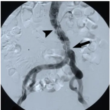
reduced for both legs, and color-coded duplex sonogra-phy of the external iliac arteries showed a monophasic pulse profile on either side, indicating a proximal aorto-iliac stenosis. Angiography of the aorta and aorto-iliac arteries under fluoroscopic control (injection of 90 mL Xenetix through a 4 French pigtail catheter placed in the external femoral artery) demonstrate multiple confluent stenotic mural changes in conjunction with a few atheromatous calcifications over 3–4 cm, causing a stenosis just above the aortic bifurcation. Curiously, the iliac and distal arterial run-offs revealed a regular shape (Fig.1).
A partially thrombosed aortic aneurysm was ruled out by CT angiography (Lightspeed 16, slice thickness 1.25 mm, pitch 0.98:1 mm/rot, 0.5 s rotation time; 120 mL Imeron 400 i.v. at a speed of 4 mL/s, Smart-Prep).
The CT images showed multiple dissociated endolu-minal thickenings, which appeared fuzzy on axial slices and polypoid in the multiplanar reconstruction (Figs.2
and 3). No enhancement of contrast material was seen within the thickened wall itself, and the periaortic fat showed no inflammatory or infiltrative changes. Neither enlarged lymph nodes nor metastases to parenchymatous organs were identified.
The patient underwent surgical resection of the infrarenal aorta and implantation of a Y-endograft. No signs of malignancy were apparent intraoperatively. Histopathology revealed slight mural calcifications within the adventitia and an intraluminal thrombus, containing numerous vital polymorphous malignant cells with an eosinophilic cytoplasm and several apoptotic cells (Fig.4). The malignant cells showed positive for pancytoceratine, which, in conjunction with the positive vascular markers CD 31 and CD 34, led to the diagnosis of an intravascular angiosarcoma. The resection margins were found to be tumor free. The patient recovered well
Correspondence to:Leopold Winter; email: [email protected]
ª Springer Science+Business Media, LLC 2009 Published online: 22 May 2009
A
bdominal
I
maging
Abdom Imaging (2010) 35:485–487 DOI: 10.1007/s00261-009-9537-4
and was discharged from hospital 10 days after surgical resection. Eight weeks later, osseous and pulmonary le-sions were detected, shown by biopsy to be metastases of the resected angiosarcoma.
Discussion
An angiosarcoma as a primary malignant tumor of the aorta (PMTA) is rarely diagnosed. The first case report was given by Brodowski in 1973 [1]. A metaanalysis of 87 cases until 1998 reported 28 female patients and 59 male patients with a median age of 60 years (range 3 months to 82 years) [2]. Thirty-nine tumors (45%) were located in the thoracic aorta, 39 (45%) in the abdominal portion and nine (10%) in the thoracoabdominal aorta. In most cases, the disease is rapidly progressive and lethal within a few months.
Macroscopically, the intimal type forms plaque-like lesions along the intimal vascular layer. More often, however, a polypoid endoluminal growth pattern has been described. The molecular background of intimal angiosarcomas is still unclear.
Fig. 1. Angiographic image: Confluent mural changes with a few atheromatous calcifications over 3–4 cm (arrowhead), causing a stenosis just above the aortic bifurcation (arrow). The iliac arteries appear to have a regular shape.
Fig. 2. Angulated reformatted CT image: Multiple polypoid, centrally dissociated (arrowhead) and distally confluent (arrow) mural alterations.
Fig. 3. Enlarged axial CT image of the aorta: Fuzzy polypoid appearance of the endoluminal wall (arrow).
Fig. 4. HE stained histologic sample: Numerous vital poly-morphous malignant cells with an eosinophilic cytoplasm and apoptotic bodies (arrow).
Mural vascular sarcomas behave less aggressively than the intimal type, despite the nearly similar overall and average survival (median 7 months) [4].
The clinical signs of primary aortic angiosarcoma are nonspecific. Embolic occlusion of peripheral or mesen-teric arteries occurs as well as intermittent claudication, abdominal pain, back pain, or metastatic complications [4, 6–8]. Aortic angiosarcoma may even coincide with pathology and clinical symptoms of abdominal aortic aneurysm [7].
Imaging findings are also non-specific, and the malignant stenosis can hardly be differentiated from aorto-iliac atherosclerosis. CT angiographic images may show a filling defect in the lumen of the aorta, iliac arteries or mesenteric artery origin [8].
An extension of the process beyond the wall of the aorta and the periaortic fat was not seen on CT images in previous reports.
Secondary signs of tumorous obstruction are ischemic infarction of parenchymatous organs, and metastasis to liver, spleen, lung, and skeletal system.
Two aspects in our case may have led to the differ-ential diagnosis of a malignant tumor. First, there was a rapid development of clinical symptoms of peripheral vascular occlusive disease, despite missing individual risk factors and very few atherosclerotic changes in periphe-ral arteries. Second, there were focally exuberant polyp-oid mural changes without aneurysmal dilatation of the vessel, which were seen only on the CT images. A mag-netic resonance angiography might have shown an atypical mural enhancement in the region of the tumor. Since the rate of osteolytic bone metastases is very high, a bone scintigram should have been carried out. Once
bone metastases are diagnosed, a major surgical inter-vention may not be indicated, because the situation turns out to be palliative.
In conclusion, intimal angiosarcoma is rare, but should be included in the differential diagnosis in case of polypoid mural alterations without aneurysm and cor-responding atherosclerotic changes in other parts of the vascular system [7,8].
Nevertheless, since most patients with these tumors are older than 60 years, most cases will be coincidental with generalized atherosclerotic, or even aneurysmal vascular changes and will become apparent through metastasis or will be diagnosed postoperatively [6,7]. References
1. Brodowski W (1973) Prima¨res Sarkom der Aorta thoracica mit Verbreitung des Neugebildes in der unteren Ko¨rperha¨lfte. Jahresb Leistung Fortschr Ges Med 8:243–246
2. Seelig MH, Oldenburg WJ, Blackshear JL (1998) Angiosarcoma of the aorta: report of a case and review of literature. J Vasc Surg 28:732–737
3. Burke AP, Virmani R (1993) Sarcomas of the great vessels: a clinicopathologic study. Cancer 71:1761–1773
4. Thalheimer A, Fein M, Geissinger E, et al. (2004) Intimal angio-sarcoma of the aorta: report of a case and review of the literature. J Vasc Surg 40(3):548–553
5. Patel KR, Griffiths AP, et al. (1997) Massive osteolytic bone metastasis from a primary aortic sarcoma: a case report. Hum Pathol 28:1306–1310
6. Majeski J, Majeski EI, Duttenhaver JR (1998) Primary aortic intimal sarcoma of the endothelial cell type with long term survival. J Vasc Surg 27:555–558
7. Defawe CD, Thry A, Lapiere CM, et al. (2006) Primary sarcoma of an abdominal aortic aneurysm. Abdom Imaging 31:117–118 8. Hagspiel KD, Hunter YR, Ahmed HK, et al. (2004) Primary
sar-coma of the distal abdominal aorta: CT angiography findings. Abdom Imaging 29:507–510