REVIEW
Glioblastoma stem cells
Ghazaleh Tabatabai&Michael WellerReceived: 17 November 2010 / Accepted: 15 December 2010 / Published online: 21 January 2011 # Springer-Verlag 2011
Abstract Glioblastomas are highly malignant primary brain tumors with one of the worst survival rates among all human cancers. With a more profound understanding of the cellular and molecular mechanisms of tumor initiation and acquired resistance to conventional radio- and chemo-therapy, novel therapeutic targets might be discovered to optimize therapeutic approaches. In this regard, the identi-fication of a small cellular subpopulation, called glioblas-toma stem cell or stem-like cells or glioma-initiating cells or brain tumor propagating cells, has gained attention. In this article, we briefly summarize the current state of knowledge about this tumor cell population and discuss future directions for basic and clinical research.
Keywords Neurooncology . Glioblastoma . Tumor intitiation . Resistance to therapy . Targeted therapy
Stem cell and cancer biology
A histological key feature of glioblastomas is the cellular and morphological heterogeneity and the parallel detection of cell populations of apparently different grades of differentiation. Moreover, glioblastomas harbor a variety of genetic abnormalities. This tumor heterogeneity may pose a therapeutic challenge because different cell popula-tions within the tumor tissue might respond differently to therapies. One strategy to overcome these problems is to identify and characterize distinct subpopulations that drive
specific disease pathologies, e.g. tumor initiation or tumor recurrence or both.
In this regard, overlaps have been defined in stem cell biology and oncology, particularly because of the growing evidence demonstrating that genes with important roles in stem cell biology also play a role for cancer biology. Consequently, the existence of a stem-like tumor cell was hypothesized, i.e. a relatively small percentage of cells that drive tumorigenesis and maintain tumor viability despite multimodal anticancer therapy. Candidate cancer stem cells should display several features that signify stemness including the unique ability to morphologically recapitulate the tumor of origin in xenografts.
The first experimental evidence for the actual existence of cancer stem cells came from hematological malignan-cies. Studies of acute myeloid leukemia (AML) revealed that a single cell initiated the disease after transplantation in mice. Cell isolation depending on cell surface marker expression revealed that CD34+CD38+ cells initiated AML, whereas CD34+CD38− did not. After development of AML in mice, the frequency of these leukemia-initiating cells in peripheral blood was about 1,000-fold lower than the frequency of other leukemic cells. These findings suggested that a hierarchy of leukemic stem cells existed in human AML (Lapidot et al.1994).
This hierarchical model implies that the majority of cancer cells within a tumor are derived from a few cancer stem cells. Thus, the tumor consists in a small subset of cancer stem cells and a large population of bulk cells that cannot per se form new tumors and might represent a mix of partially and terminally differentiated cancer cells with limited proliferative capacity.
This concept was also investigated in brain tumors. The seminal study in the field of neurooncology demonstrated that as few as 100 glioma stem-like cells, defined as
G. Tabatabai (*)
:
M. WellerLaboratory of Molecular Neurooncology, Department of Neurology, University Hospital Zurich and University of Zurich, Zurich, Switzerland
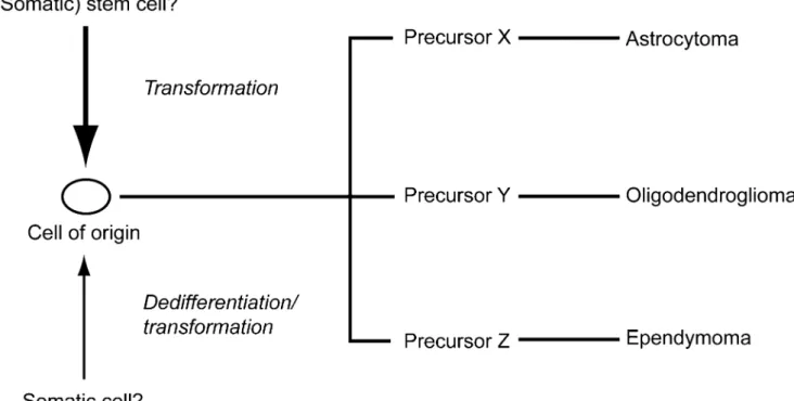
CD133+cells, were able to recapitulate the heterogeneity of glioblastomas in immunocompromised mice whereas CD133−tumor cells could not. Notably, 100 CD133+ cells sufficed to recapitulate the original tumor whereas 10,000 CD133− cells were not tumorigenic (Singh et al. 2004). Therefore, a hierarchy was postulated in gliomas, i.e. a tumor cell of origin that upon differentiation might lead to the establishment of astrocytomas, oligodendrogliomas or ependymomas (Fig.1).
Several other groups have also isolated brain tumor stem cells from primary glioblastomas, based on cell surface marker expression or the ability to form neurospheres (also called gliomaspheres) as do normal neural stem cells (Table1).
The identification of somatic mutations of the isocitrate dehydrogenase 1 gene (IDH1) most frequently found in glioblastomas which has evolved from lower-grade glio-mas, so-called secondary glioblastoglio-mas, might be of relevance for further characterization of a putative cell of origin in gliomas, i.e. a cell of origin in gliomas that upon differentiation might lead to the establishment of astrocy-tomas or oligodendrogliomas (Fig.1). Low-grade gliomas (astrocytomas and oligodendrogliomas) may carry IDH mutations in the absence of any other genetic aberration, suggesting that IDH mutations occur early, e.g. in a stem or precursor cell that can give rise to both, astrocytes and oligodendrocytes (Yan et al. 2009). Interestingly, IDH mutations never occur in ependymomas.
The nomenclature
The nomenclature of stem cells in glioblastoma has remained controversial. This is due to the lack of standardized criteria for identifying them. The terms glioblastoma stem cell, glioblastoma stem-like cell or glioblastoma-initiating cell are commonly used inter-changeably but whether cells identified by the methods defined below are the only cells that can initiate tumors has remained unclear. This terminology reflects some shared characteristics with normal stem cells, especially adult somatic stem cells, including the capacities for self-renewal, differentiation and maintained proliferation. Yet, whether the cell of origin in glioblastomas is sometimes or always a stem cell (Fig.1), as the terminology glioblastoma stem cell implies, is unclear at the moment. The term tumor-propagating cell has been alternatively proposed, because the defining bioassay involves tumor propagation in secondary hosts, e.g. mice (Kelly et al.2007). In this article, we use the term glioblastoma stem-like cell (GSC) and are fully aware of controversies regarding its limitations.
Isolating and culturing GSC
GSC have been identified based on different cell surface markers, culture conditions or functional criteria. Currently used approaches are summarized in Table 1. The most
Fig. 1 Glioma stem-like cells might serve as a“cell of origin” either derived from a transformed stem cell or a transformed somatic cell. Further differentiation of this cell of origin might then lead to different
cellular lineages resulting in the development of astrocytomas, oligodendrogliomas or ependymomas
mentioned and controversial cell surface marker for GSC isolation is CD133, a cholesterol-bindig membrane protein of unknown biological function.
CD133+ progenitor cells promoted the healing of ischemic ulcers through stimulating angiogenesis and activating the Wnt pathway (Barcelos et al. 2009). Recently, CD133 expression was demonstrated to increase in response to hypoxia and to mitochondrial dysfunction. This questions CD133 as a reliable marker for GSC.
One reason explaining these inconsistencies between various groups could be the lack of standardization regarding cell sorting methods and assays to confirm “stemness” across laboratories.
Recently, a marker-independent approach has been demonstrated exploiting cellular phenotypes. Combining morphological aspects and autofluorescence emission were used to enrich a subpopulation with self-renewal abilityin vitro, tumor-initiating and propagating capacity in vivo and expression of stem cell-related genes (Clement et al.2010). Another ongoing debate exists on how to best culture GSC to facilitate their study in vitro. The first studies applied techniques used for culturing normal neural stem cells. In neurobasal medium supplemented with growth factors such as epidermal growth factor and basic fibroblast growth factor, neural stem cells can be propagated and expanded indefinitely, whereas most differentiating or differentiated cells rapidly die (Reynolds and Weiss1996).
This approach allowed the prospective GSC isolation from fresh glioblastoma specimens, i.e. a subpopulation of cells capable of proliferation, self-renewal and differentiation (Singh et al.2004). Some studies, on the other hand, have proposed protocols for adherent cultures of GSC in serum-free medium using attachment factors (Al-Mayhani et al. 2009; Pollard et al. 2009). The latter studies claim that adherent monolayer cultures are superior to non-adherent sphere cultures because of the possibility to ensure uniform exposure to growth factors, oxygen and nutrients leading to a more homogenous cell population.
The high relevance and the potential impact of in vitro culturing conditions on cellular characteristics are underscored by a study describing the emergence of cancer stem-like cells fromin vitro cultured adult rat subventricular zone stem cells. After expansion in vitro, neural stem cells transformed into tumorigenic cell lines that acquired multiple chromosomal aberrations. They maintained characteristic stem cell expres-sion markers like Nestin, Musashi-1 and CD133 but continued to proliferate upon differentiation induction and displayed tumorigenicityin vivo (Siebzehnrubl et al.2009).
Functional evaluation of GSCin vitro and in vivo The gold standard assay for investigating self-renewal and tumor initiation of GSC is the ability to form tumors in
Table 1 Cell surface markers and functional assays for the isolation of glioma stem-like cells
Parameter Assessment Functional correlate Key reference CD 133 Magnetic sorting or flow cytometry sorting
for CD133 expression
Self renewal Singh et al.2004
Multipotency Tumorigenicityin vivo Alpha 6 integrin Sorting for alpha 6 integrin expression alone
or in combination with CD133 expression by flow cytometry
High enrichment for cells with self-renewal
Lathia et al.2010
Sphere formation Tumorigenicityin vivo Culture condition Culture condition: serum-free medium
supplemented with EGF, FGF
Self renewal Galli et al.2004
Multipotency Günther et al.2008
Tumorigenicityin vivo Side population Flow cytometry sorting based on exclusion of
Hoechst 33342 (side population)
Tumorigenicityin vivo only in side population
Kondo et al.2004
A2B5 Flow cytometry sorting for A2B5 and CD133 expression
Tumorigenicity Ogden et al.2008
ALDH1 activity Flow cytometry sorting for ALDH1 activity High ALDH1 levels keep glioma cells in an undifferentiated state
Rasper et al.2010
High ALDH1-expressing glioma cells grow in neurospheres
ALDH1 inhibition induces differentiation and reduces clonogenicity
Autofluorescence Intrinsic autofluorescence and morphology Self renewal Clement et al.2010
Multipotency Tumorigenicityin vivo
animals after orthotopic transplantation even of very low cell numbers. These tumors should resemble the morpho-logical and immunohistochemical phenotype of the original tumor. Further, serial dilutions should be performed, i.e. the definition of the smallest cell number capable of generating tumorsin vivo. Finally, serial passaging, i.e. harvesting of an in vivo established tumor and implantation into a secondary mouse, should occur. Regarding these criteria, GSC should display much greater tumorigenic potential than corresponding non-stem tumor cellsin vivo (Hemmati et al.2003; Galli et al.2004; Singh et al.2004).
Of note, the role of GSC beside tumor initiation has also been studied in vivo: many key malignant features of glioblastomas have been attributed to GSC, e.g. elevated levels of vascular endothelial growth factor (VEGF) to promote angiogenesis (Bao et al. 2006a, b), expression of stromal-derived factor-1 (SDF-1) (Salmaggi et al. 2006), and resistance to radiation therapy (Bao et al.2006a,b).
In vitro, GSC can be induced to differentiate into astrocytes, oligodendrocytes and neurons by addition of serum and withdrawal of the culture conditions outlined above (Hemmati et al.2003; Galli et al.2004; Singh et al. 2004). Given these standard functional assessments, it remains to be clarified, however, how stable a GSC phenotype is. This is of particular interest in the face of studies suggesting the isolation of stem-like cells from
established glioma cell lines, e.g. GL-261 or C6 (Pellegatta et al.2006; Kondo et al.2004).
Resistance of GSC to therapy
CD133 cell surface expression has been examined in glioblastoma specimens from ten patients who had under-gone surgical resection before and after high-dose irradia-tion with gamma knife surgery and external beam radiairradia-tion therapy. Comparative analysis of histological sections before and after radiation revealed that CD133+ cells were very infrequent in primary tumor sections. In post-irradiated tumors, however, the percentage of CD133+ cells was significantly higher. The authors concluded that this confirms the hypothesis that GSC are capable of escaping high doses of radiation (Tamura et al. 2010). However, studies showing that cellular stress induces CD133 must be taken into account in interpreting these data. Probably, CD133 increases after radiation therapy were due to CD133 induction on previously CD133− tumor cells, because—as demonstrated by Griguer and colleagues—CD133 can be induced by hypoxia and cellular stress (Griguer et al.2008). Preclinical evidence for resistance to therapy were provided by Bao et al. (2006a,b). They performed colony formations assays using untreated or irradiated CD133+and
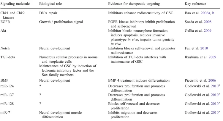
Table 2 Signaling pathways and molecules in glioma stem-like cells
Signaling molecule Biological role Evidence for therapeutic targeting Key reference Chk1 and Chk2
kinases
DNA repair Inhibitors enhance radiosensitivity of GSC Bao et al.2006a,b
EGFR Growth / proliferation signal EGFR kinase inhibitors inhibit proliferation and self-renewal
Soeda et al.2008
Akt Inhibitor blocks neurosphere formation, induces apoptosis, reduces invasive phenotypein vivo, impairs tumorigenicity in vivo
Gallia et al.2009
Notch Neural development Inhibition blocks self-renewal and promotes radioresistance
Fan et al.2010
TGF-beta Numerous cellular processes in normal and neoplastic cells
Inhibition of TGF-beta interferes with maintenance of GSC
Ikushima et al.2009
Maintenance of GSC by induction of leukemia inhibitory factor and the Sox family members
BMP Neural development BMP 4 treatment induces differentiation Piccirillo et al.2006
miR-124 ? Decreases proliferation and promotes differentiation
Godlewski et al.2010a
miR-137 ? Decreases proliferation and promotes differentiation
Godlewski et al.2010a
miR-128 ? Blocks self renewal and decreases proliferation
Godlewski et al.2010a
miR-7 Neural development muscle differentiation
Inhibits migration and decreases proliferation
Godlewski et al.2010a
Chk Checkpoint kinase, EGFR epidermal growth factor receptor, TGF transforming growth factor, BMP bone morphogenetic protein
a
CD133− cells derived either from a biopsy specimen or from glioma xenografts. These assays revealed a relative radioresistance of CD133+ cells. Beier et al. (2008) demonstrated on the other hand that temozolomide prefer-entially depleted CD133+cells in glioblastoma cancer stem cell lines.
Therapeutic targets in GSC
Cell surface molecules differentially expressed in glioma stem-like cells and functionally associated with the main-tenance of glioma stem-like cells may be ideal targets for therapeutic intervention. Several molecules including CD133 and A2B5 (Singh et al. 2004; Son et al. 2009; Tchoghandijan et al.2010) have been discovered on the cell surface of GSC (Table 1). A2B5 monoclonal antibodies recognize GT3 gangliosides, which characterize a small fraction of cells in the subcortical white matter with neural stem cell properties. In glioblastomas, A2B5+ cells display
important features including migration and differentiation into oligodendroglia and astroglial cells. Moreover, A2B5+ cells but not A2B5− cells form tumors in nude mice. Interestingly, combining A2B5 and CD133 for cell isolation revealed that both A2B5+/CD133+ and A2B5+/CD133− cells formed tumors in nude mice (Tchoghandijan et al. 2010).
Further, identification of specific signaling pathways involved in the maintenance and functions of GSC may also identify useful therapeutic targets for novel strategies to improve the treatment for glioblastoma. The signaling pathways associated with GSC are summarized in Table2. Disruption of the Notch pathway by Notch shRNA sensitized GSC to radiation therapy (Wang et al. 2009). Differentiation therapy forcing GSC to differentiate might be another promising and notably non-cytotoxic strategy for GSC targeting. In this regard, bone morphogenetic protein (BMP) 4 has been reported to activate BMP receptors, triggering Smad signaling cascade in GSC and leading to a reduction in GSC proliferation and increased
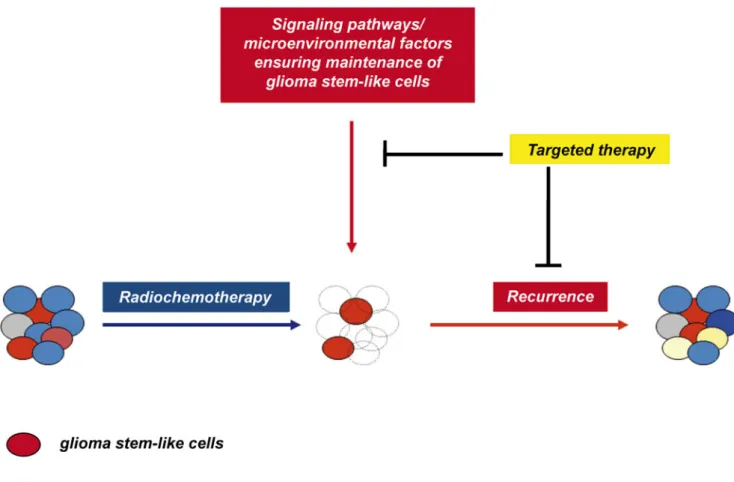
Fig. 2 Conventional radiotherapy might only hit glioma bulk cells while glioma stem-like cells survive and are maintained by key components of the tumor microenvironment. For succesful therapeutic targeting, it will be important to design targeted therapies to interfere
with the signaling pathways maintaining glioma stem-like cells. Combining radiotherapy with targeted anti-GSC therapy might then diminish tumor recurrence
expression of differentiated neural markers in CD133+cells (Piccirillo et al.2006).
Novel important players include microRNAs, which have a central role in gene expression. A role for microRNAs has recently been established for cancers of different types, including glioblastomas. A very comprehensive and detailed overview on the role of miRNA in glioblastomas has recently been published (Godleswski et al.2010) (Table2).
The preferential distribution of GSC in the perivas-cular area has led to the identification of a GSC niche (Calabrese et al. 2007). Disrupting the niche could therefore be another therapeutic strategy. Thus, it is important to identify specific elements in these regions that maintain GSC. A role for endothelial cells as one niche component seems to be established: Co-implantation of GSC with endothelial cells accelerated tumor initiation and progression (Calabrese et al. 2007). Apart from the obvious interaction with endothelial cells, GSC are in close contact with extracellular matrix components that are preferentially expressed in the perivascular niche. Integrin alpha 6 has been recently identified as an important molecule for GSC in the perivascular niche (Lathia et al. 2010). It ensures interaction with laminin-expressing endothelial cells in the microenvironment. Sorting by flow cytometry based on alpha 6 expression alone or alpha 6 integrin/CD133 double-expression led to enrichment of cells with a self renewal capacity in vitro and tumorige-nicity in vivo. Targeted depletion of alpha 6 integrin by lentiviral short hairpin RNA inhibited GSC growth and sphere formation. Moreover, treatment with an alpha 6 integrin-blocking antibody reduced tumor formationin vivo. Targeting integrin alpha 6 might be a promising strategy to interfere with the niche and to impair self-renewal, prolifer-ation and tumor initiprolifer-ation capacity (Lathia et al.2010).
Further, it will be desirable to identify candidate targets, e.g. for immunotherapy. It should be taken into account, for example, that the human AC133 gene, encoding CD133, has at least seven alternatively spliced 5′-UTR isoforms of AC133 mRNA, which are expressed in a tissue-dependent manner. The transcrip-tion of these AC133 isoforms is driven by five different promoters, P1–P5, depending on their location relative to different exons. Finding the brain-specific isoform of AC133 and targeting this specific isoform (Shmelkov et al. 2004), e.g. by immunotherapy, will also increase the specifity of these targeted approaches and decrease potential toxicity.
Future perspectives
Selective GSC targeting will be challenging, even if highly specific therapeutic targets are presently available, because
GSC may change and differently adapt in situ during the course of the disease within a patient. Because of many overlapping pathways and cell surface markers shared by neural stem cells and GSC, however, one challenge will be to identify therapeutic approaches that avoid potential toxicities to normal neural stem cells.
Taken together, the identification of GSC has increased the complexitiy of glioblastoma biology.
It remains to be determined to what extent the cells defined by the current methodology give rise to gliomas initially, maintain tumor viability during treatment and promote clinical recurrence. Successful future therapeutic approaches against glioblastoma will require a combination of standard radiochemotherapy with tailored personalized therapies targeting the unique molecular profile and GSC profile of every patient (Fig.2).
Acknowledgements This study was supported by DFG (SFB 773, project A6) and by NCCR Neural Plasticity and Repair (P4).
References
Al-Mayhani TMF, Ball SLR, Zhao JW, Fawcett J, Ichimura J, Colins PV, Watts C (2009) An efficient method for derivation and propagation of glioblastoma cell lines that conserves the molecular profile of their original tumors. J Neuorsci Methods 176:192–199
Bao S, Wu Q, Sathornsumetee S, Hao Y, Li Z, Hjelmeland AB, Shi Q, Mc Lendon RE et al (2006a) Stem cell-like glioma cells promote tumor angiogenesis through vascular endothelial growth factor. Cancer Res 66:7843–7848
Bao S, Wu Q, McLendon RE, Hao Y, Shi Q, Hjelmeland AB, Dewhirst MW, Bigner DD, Rich JN (2006b) Glioma stem cells promote radioresistance by preferential activation of the DNA damage response. Nature 444:756–760
Barcelos LS, Duplaa C, Krankel N et al (2009) Human CD133+ progenitor cells promote the healing of diabetic ischemic ulcers by paracrine stimulation of angiogenesis and activation of Wnt signaling. Circ Res 104:1095–1102
Beier D, Röhrl S, Pillai DR, Schwarz S, Kunz-Schughart LA, Leukel P et al (2008) Temozolomide preferentially depletes cancer stem cells in glioblastoma. Cancer Res 68:5706–5715
Calabrese C, Poppleton H, Kocak M, Hongg TL, Fuller C, Hamner B et al (2007) A perivascular niche for brain tumor stem cells. Cancer Cell 11:69–82
Clement V, Marino D, Cudalbu C, Hamou MF, Mlynarik MF, Tribolet N, Dietrich PY et al (2010) Marker-independent identification of glioma-initiating cells. Nat Meth 7:224–228
Fan X, Matsui W, Khaki L, Stearns D, Chun J, Li YM et al (2010) Notch pathway inhibition depletes CD133-positive glioblastoma cells and inhibits growth of tumor neurospheres and xenografts. Stem Cells 28:5–16
Galli R, Binda E, Orfanelli U, Cipeletti B, Gritti A, De Vitis S et al (2004) Isolation and characterization of tumorigenic, stem-like neural precursors from human glioblastomas. Cancer Res 64:7011–7021
Gallia GL, Tyler BM, Hann CL, Siu UM, Giranda VL, Vescovi AL et al (2009) Inhibition of Akt inhibits growth of glioblastoma and glioblastoma stem-like cells. Mol Cancer Ther 8:386–393
Godlewski J, Newton HB, Chiocca EA, Lawler SE (2010) Micro-RNAs and glioblastomas: the stem cell connection. Cell Death Differ 17:221–228
Griguer CE, Oliva CR, Gobin E, Marcorelles P, Benos DJ, Lancaster JR et al (2008) CD133 is a marker of bioenergetic stress in human glioma. PLoS ONE 3:e3655
Günther HS, Schmidt NO, Philipps HS, Kemming D, Kharbanda S, Soriano R et al (2008) Gliolastom-derived stem cell-enriched cultures from distint subgroups according to molecular and phenotypic criteria. Oncogene 27:2897–2909
Hemmati HD, Nakano I, Lazareff JA, Masterman-Smith M, Geschwind DH, Bonner-Fraser M et al (2003) Cancerous stem cells can arise from pediatric brain tumors. Proc Natl Acad Sci USA 100:15178– 15183
Ikushima H, Todo T, Ino Y, Takahashi M, Miyazawa K, Miyazono K (2009) Autocrine TGF-beta signaling maintains tumorigenicity of glioma-initiating cells through Sry-related HMB-box factors. Cell Stem Cell 5:504–514
Kelly PN, Dakic A, Adams JM, Nutt SL, STrasser A (2007) Tumor growth need not be driven by rare cancer stem cells. Science 317:337 Kondo T, Setoguchi T, Taga T (2004) Persistance of a small
subpopulation of cancer stem-like cells in the C6 glioma cell line. Proc Natl Acad Sci USA 101:781–786
Lapidot T, Sirard C, Vormoor J et al (1994) A cell initiating human acute myeloid leukaemia after transplantation into SCID mice. Nature 367:645–648
Lathia JD, Gallagher J, Heddleston JM, Wang J, Eyler CE, Macwords J, Wu Q et al (2010) Integrin alpha 6 regulates glioblastoma stem cells. Cell Stem Cell 6:421–432
Ogden AT, Waziri AE, Lochhead RA, Fusco D, Lopez K, Ellis JA et al (2008) Identification of A2B5+CD133- tumor-initiating cells in adult human gliomas. Neurosurgery 62:505–514
Pellegatta S, Poliani PL, Corno D, Menghi F, Ghielmetti F, Suarez-Merino B, Caldera V et al (2006) Neurospheres enriched in cancer stem-like cellsa re highly effective in eliciting a dendritic cell-mediated response against malignant gliomas. Cancer Res 66:10247–10252 Piccirillo SG, Reynolds BA, Zanetti N, Lamorte G, Binda E, Broggi
G, Brem H, Olivi A, Dimeco F, Vescovi AL (2006) Bone morphogenetic proteins inhibit the tumorigenic potential of human brain tumor-inititaing cells. Nature 444:761–765 Pollard SM, Yoshikawa K, Clarke ID, Danovi D, Stricker S, Russell
R, Bayani J, Head R, Lee M, Bernstein M et al (2009) Brain cancer stem cells: a level playing field. Cell Stem Cell 5:468–469
Rasper M, Schafer A, Piontek G, Teufel J, Brockhoff G, Ringel F et al (2010) Aldehyde dehydrogenase 1 positive glioblastoma cells show brain tumor stem cell capacitiy. Neuro Oncol 12:1024– 1033
Reynolds BA, Weiss S (1996) Clonal and population analyses demonstrate that an EGF-responsive mammalian embryonic CNS precursor is a stem cell. Dev Biol 175:1–13
Salmaggi A, Boiardi A, Gelati M, Russo A, Calatozzolo C, Ciusani E, Sciacca FL et al (2006) Glioblastoma-derived tumorospheres identify a population of tumor stem-like cells with angiogenic potential and enhanced multidrug resistance phenotype. Glia 54:850–860
Shmelkov SV, Jun L, Clair D, McGarrigle D, Derderian JK et al (2004) Alternative promoters regulate transcription of the gene that encodes stem cell surface protein AC133. Blood 103:2055– 2061
Siebzehnrubl FA, Jeske I, Muller D, Buslei R, Coras R, Hahnen E et al (2009) Spontaneous in vitro transformation of adult neural precursors into stem-like cancer cells. Brain Pathol 19:399–408
Singh SK, Hawkins C, Clarke ID, Squire JA, Bayani J, Hide T et al (2004) Identification of human brain tumor initiating cells. Nature 432:396–401
Soeda A, Inagaki A, Oka N, Ikegame Y, Aoki H, Yoshimura S et al (2008) Epidermal growth factor plays a crucial role in mitogenic regulation of human brain tumor stem cells. J Biol Chem 283:10958–10966
Son MJ, Woolard K, Nam DH, Lee J, Fine H (2009) SSEA-1 is an enrichment marker for tumor-initiating cells in human glioblas-toma. Cell Stem Cell 4:440–452
Tamura K, Aoyagi M, Wakimoto H, Ando N, Nariai T, Yamamoto M, Ohno K (2010) Accumulation of CD133-positive glioma cells after high-dose irradiation by gamma knife surgery plus external beam radiation. J Neurosurg (in press)
Tchoghandijan A, Baeza N, Colin C, Cayre M, Metellus P, Beclin C et al (2010) A2B5 cells from human glioblastoma have cancer stem cell properties. Brain Pathol 20:211–221
Wang J, Wakeman TP, Lathia JD, Hjemeland AB, Wang XF, White RR et al (2009) Notch promotes radioresistance of glioma stem cells. Stem Cells 28:17–28
Yan H, Parsons W, GEnglin J, McLendon R, Rasheed A, Weishi Y, Kos I, Batinic-Haberle I et al (2009) IDH1 and IDH2 mutations in gliomas. N Engl J Med 360:765–773