HAL Id: inserm-00391173
https://www.hal.inserm.fr/inserm-00391173
Submitted on 29 Jul 2011HAL is a multi-disciplinary open access archive for the deposit and dissemination of sci-entific research documents, whether they are pub-lished or not. The documents may come from teaching and research institutions in France or abroad, or from public or private research centers.
L’archive ouverte pluridisciplinaire HAL, est destinée au dépôt et à la diffusion de documents scientifiques de niveau recherche, publiés ou non, émanant des établissements d’enseignement et de recherche français ou étrangers, des laboratoires publics ou privés.
Involvement of right piriform cortex in olfactory
familiarity judgments.
Jane Plailly, Moustafa Bensafi, Mathilde Pachot-Clouard, Chantal
Delon-Martin, David Kareken, Catherine Rouby, Christoph Segebarth, Jean
Royet
To cite this version:
Jane Plailly, Moustafa Bensafi, Mathilde Pachot-Clouard, Chantal Delon-Martin, David Kareken, et al.. Involvement of right piriform cortex in olfactory familiarity judgments.: Familiarity judgment in olfaction. NeuroImage, Elsevier, 2005, 24 (4), pp.1032-41. �10.1016/j.neuroimage.2004.10.028�. �inserm-00391173�
Involvement of the right piriform cortex in the familiarity
judgment task of odors
Jane Plailly,* Moustafa Bensafi,* Mathilde Pachot-Clouard,† Chantal Delon-Martin,†
David A. Kareken,‡ Catherine Rouby, Christoph Segebarth,† and Jean-P. Royet*§
* Neurosciences et Systèmes Sensoriels, Université Claude Bernard Lyon1, UMR CNRS 5020,
IFR 19, Institut Fédératif des Neurosciences de Lyon, 69366 Lyon cedex 07, France.
† Unité mixte INSERM/Université Joseph Fourier U594, LRC-CEA, Hôpital Michallon, 38043 Grenoble, France.
‡ Neuropsychology Section, Department of Neurology, Indiana University School of Medicine, Indianapolis, Indiana 46202, USA.
§CERMEP, 69003 Lyon, France.
Running Title: Familiarity judgment in olfaction
Number of words in the abstract: 174 Number of words in the introduction: 504 Number of words in the discussion: 1970
Number of figures : 4
Number of tables: 3
Author to whom correspondence should be sent: Jane PLAILLY
Neurosciences et Systèmes Sensoriels UMR CNRS 5020
Université Claude Bernard Lyon1 50, Av. Tony Garnier
69366 Lyon cedex 07, France
Abstract
Previous studies have shown activation of right orbitofrontal cortex during judgments of odor familiarity. In the present study, we sought to extend our knowledge about neural circuits involved in such a task. Fourteen right-handed male subjects were tested in fMRI in a single functional run of two olfactory conditions (detection and familiarity judgment). Within each condition, three epochs were performed. During the familiarity condition, subjects rated whether odors were familiar or unfamiliar, whereas during the detection condition, participants decided whether the odor was present or not. Contrasting familiarity with detection conditions, odor-activated areas were found mainly including the piriform cortex (PC), and hippocampus in the right hemisphere, and the inferior frontal gyrus and amygdala in the left hemisphere. Right PC activation was functionally connected with right hippocampus and left PC during that same task. By showing that PC was activated during a familiarity judgment task, the present study gives supports to the notion that PC participates in the processing of odor memory. It further showed preferential involvement of the right hemisphere in familiarity judgments.
INTRODUCTION
Hemispheric asymmetry is well-established for high-level brain functions such as language and spatial attention (Broca, 1863; Weintraub and Mesulam, 1987). Hemispheric predominance also exists in sensory functions such as hand somatosensory representation (Soros et al., 1999), and temporal and spectral auditory resolution (Zatorre et al., 2002). Studies in olfaction lead towards similar conclusions. Early cerebral imaging studies showed a functional lateralization of olfactory processes in the right hemisphere, especially in orbitofrontal cortex (OFC) (Zatorre et al., 1992), and most subsequent studies confirmed this result (Yousem et al., 1997; Dade et al., 1998; Sobel et
al., 1998). Zald and colleagues however reported predominant activations in the left OFC and
amygdala for very aversive odors, as if hedonic quality of odorant was determining in olfactory processing (Zald and Pardo, 1997; Zald et al., 1998). In positron emission tomography (PET) studies, Royet et al. (1999, 2001) found functional lateralization with specific olfactory judgments. Whereas there was bilateral OFC activation with judgments of odor familiarity and hedonicity olfactory judgment tasks, the familiarity judgment preferentially activated the right OFC, while the hedonic judgment mainly activated the left OFC.
Beyond OFC, more recent cerebral imaging data have extended these observations to the piriform cortex (PC). In a functional magnetic resonance imaging (fMRI) study, we showed left piriform-amygdala involvement in hedonic intensity rating (Royet et al., 2003), a result further consistent with Gottfried et al. (2002a) and Anderson et al. (2003)’s findings. Convergent findings from Dade
et al. (2002)’s lesion and PET studies further show the role of primary olfactory cortex (piriform
region) in olfactory long-term recognition, reinforcing the idea that this area is considerably more than primary sensory cortex (e.g., Schoenbaum and Eichenbaum, 1995). Specifically, Dade et al. (2002) found that the extend of piriform activity corresponded with different cognitive demands. The activation in PC thus appeared to follow a continuum between the mnemonic encoding, with
suggested that piriform activity could be in relation with odor familiarity. Familiarity judgments indeed require subjects to compare odors with previously stored olfactory representations, and thus represent a type of long-term olfactory reference-memory.
In the present study, we set out to explore that question by specifically asking whether PC is implied in the processing of odor familiarity, and if so, whether familiarity-evoked activations were lateralized. We further addressed the possibility that other brain areas, not revealed in our previous experiments, may be implied in the processing of odor familiarity. We studied familiarity judgments and their relation to PC activation with fMRI using a classical block paradigm design. Familiar and unfamiliar odors were presented in a same epoch allowing subjects to rate familiarity in a binary fashion using two key-press buttons. A control condition was also performed in which subjects had simply to judge the presence or absence of an odor. The contrast between both conditions allowed us to identify the areas specifically involved in the familiarity judgment of odors.
MATERIALS AND METHODS
Subjects
Fourteen right-handed men (18-32 years old) participated in the study. They were selected on the basis of their olfactory ability with a forced-choice suprathreshold detection test (at least 87%
correct) and of the mean duration of their breathing cycle (3.88 s ± 0.72). Subjects with rhinal
disorders (colds, active allergies, history of nasal-sinus surgery, or asthma), neurologic disease, ferrous implants (e.g., pacemakers, cochlear implants), or claustrophobia were excluded. Participation required a medical screening and written informed consent. The study was approved by the local Institutional Review Board and conducted according to French regulations on biomedical experiments on healthy volunteers.
Odorous stimuli
One hundred and eight stimuli were used for training (27) and fMRI experiment (81). For fMRI 54 odorants were used for the familiarity (F) condition and 27 odorants for the detection (D) condition (Table 1). For F conditions, 3 sets (Fa, Fb, Fc) contained 9 familiar and 9 unfamiliar odorants selected so as to respectively provide high and low familiarity scores from data obtained in a previous work (Royet et al., 1999). An analysis of variance (ANOVA) indicated that familiarity scores were significantly higher for familiar than for unfamiliar odorants [F(1,48) = 129.173, p < 0.0001]. For D conditions, 3 sets (Da, Db, Dc) contained 9 odorants with low familiarity and 9 bottles with odorless air For training, 3 sets of 9 odorants with low familiarity scores and 9 bottles with odorless air were used. In each set, the presentation order of stimuli was pseudorandomized, but identical for all subjects. Odorants were diluted to a concentration of 10% using mineral oil (Sigma Aldrich, France). For presentation, 5 ml of this solution was absorbed by compressed polypropylene filaments inside of 100 ml white polyethylene squeeze-bottles with a dropper (Osi, France).
Stimulating and recording materials
Odors were presented with an airflow olfactometer, which allowed synchronization of stimulation with breathing. The stimulation equipment was essentially the one used in a previous PET study (Royet et al., 1999), but adapted so as to avoid interference with the static magnetic field of the scanner (Royet et al., 2003). Briefly, the apparatus was split into two modules: the electronic part of the olfactometer positioned outside the magnet room (shielded with a Faraday cage), and the non-ferrous (Duralumin®) air-dilution injection head placed in the stray-field of the magnet. Compressed air (10 l / min) was pumped into the olfactometer, and delivered continuously through a standard anesthesia mask. At the beginning of each inspiration, one stimulus was injected into the olfactometer, which carried it to the subject’s anesthesia mask. Breathing was recorded with the aid of a PVC foot bellows (Herga Electric Limited, Suffolk, UK) held on the stomach with a judo belt. An operator monitored breathing and squeezed the odor (or odorless) bottle so as to flush the stimulus into the injection head during subject inspiration.
Subjects rated familiarity or judged odor presence by using the two key-press buttons. Response signal was transmitted outside the magnetically shielded room by means of optical fibers to analog-to-digital converters powered by nickel-cadmium batteries. Behavioral data were recorded on line (100 Hz sampling rate) using a NEC PC computer equipped with a digital acquisition board DAQCard-500 (National Instruments, USA). LabView 5.0 software (National Instruments, USA) was used to acquire, store, and read data. Data analysis was performed with the WinDaq Waveform Browser 1.91 software (DataQ Instruments, USA).
Experimental procedure
A single functional run was used, presented in blocks, consisting of 2 olfactory conditions (F and D) alternating with odorless rest (R) epochs (Figure 1). Each epoch lasted 60 s. Both F and D conditions were presented three times each, either 3 F followed by 3 D conditions or vice versa. In a same condition, the presentation order of the 3 sets (a, b, c) was modified between subjects. The order of conditions and odor sets were counterbalanced so as to permit a balanced experimental
design (Latin square). For olfactory conditions, subjects were asked to rate whether they smelled an odor or not (D condition) or whether the odor was familiar or unfamiliar (F condition). Subjects were then asked to make a ‘yes’ or ‘no’ rating using the two key-press buttons with their dominant hand. For half of the subjects, ‘yes’ and ‘no’ responses were obtained with the index and the middle fingers, respectively. For the other half of the subjects, the meaning of the two key-press buttons was reversed. For R, no stimulation was done and the subjects were instructed not to respond.
Figure 1
General instructions were provided to subjects before the functional run. During the run, and 3 s before each experimental condition (F, D or R), subjects were instructed orally by means of specific key words (‘familiarity,’ ‘detection,’ and ‘rest’) which task was to be performed next. Subjects wore earplugs to protect from excessive scanner noise and kept their eyes closed during scanning. The day before the fMRI examination, subjects were trained outside the MR facility to breathe regularly, to detect odorants without sniffing during normal inspiration, and to give the most rapid possible response (odor vs. no odor) using the two key-press buttons.
Imaging parameters
Functional MR imaging was performed on a 1.5 Tesla MR imager (Philips NT). Twenty-five adjacent 5-mm-thick axial slices were imaged. The imaging volume covered the subjects' whole brain and was oriented parallel to the bicommissural plane. The image planes were positioned from scout images acquired in the sagittal plane. A 3D three-shot PRESTO MR imaging sequence (Liu
et al., 1993) was used with the following parameters: TR = 26 ms, TE = 38 ms, flip angle = 14°,
field-of-view = 256 x 205 mm2, imaging matrix = 64 x 51 (pixel size of 4 x 4 x 5 mm3). This
sequence is less prone to the magnetic susceptibility artifacts than is the usual echoplanar imaging sequence. During the functional run, the volume of interest was scanned 144 times successively. The signal was averaged three times, leading to an acquisition time per volume of 5 s. A high-resolution anatomical 3D T1-weighted MR scan was acquired before the functional run.
Data processing and statistical analyses
Functional images were analyzed using SMP99 (Wellcome Department of Cognitive Neurology, London, UK). Image processing included interscan realignment, spatial normalization to stereotactic space as defined by one of the Montreal Neurological Institute (MNI) templates, and image smoothing with a three-dimensional Gaussian kernel (FWMH: 8 x 8 x 10 mm) to overcome residual anatomical variability during group analysis, to increase the signal-to-noise ratio and to conform to the hypotheses underlying the statistical analysis (Friston et al., 1995a). A boxcar reference function was convolved with SPM99’s ‘canonical hemodynamic’ response function. A low-pass filter (cut-off period of 720 s) was used to eliminate instrumental and physiological very low frequency signal variations. Global differences in BOLD signal were covaried out of all voxels, and comparisons across conditions were effected with t tests. The significance of signal differences was assessed through Z scores in an omnibus sense, using an uncorrected height threshold (p < 0.001). Only clusters of more than 20 adjacent activated voxels were taken into account as a significant hemodynamic response (Z > 3.50 at voxel level). Duvernoy’s (1991) and Mai et al.’s (1997) anatomic atlases were used to localize and describe anatomic regions of activation since the currently used Talairach’s atlas does not describe accurately enough the areas involved in this kind of study. The MNI coordinates of activated regions expressed in the form of three-dimensional coordinate system defined by Talairach and Tournoux (1988) were however provided for sake of homogeneity with the neuroimaging literature.
Specific effects for the familiarity judgment task were calculated by comparing F and D conditions using the general linear model (Friston et al., 1995b). Intrasubject analyses were first performed, followed by a random effects analysis which extend statistical inference into the healthy population. This two-stage analysis accounted first for intrasubject variance (scan-to-scan), and second for intersubject variance. During the first step, scan-to-scan variance was separately modeled for each subject by creating a summary contrast image from weighted parameter estimates that represented each scan condition. During the second step, these contrast images were then
analyzed using a basic model one sample t tests to assess the difference F-D condition versus null hypothesis.
A cluster analysis was further performed to detect areas functionally connected with those activated in the familiarity judgment using Marsbar SPM toolbox (Brett et al., 2002). A ROI corresponding to the right PC was functionally defined by thresholding at p = 0.0001 the F-D contrast image obtained with our group of subjects. This threshold was chosen to dissociate the right PC activation voxel cluster from its bordering activation clusters. For each subject, the
activity level within this right PC ROI (β value in SPM) was computed, providing an activity
level vector for all the subjects. A basic model ‘simple regression (correlation)’ analysis was then performed to extract among the brain, areas that presented values correlated with the activity level vector obtained for the right PC. A covariance matrix of activation values deduced from functionally connected areas was obtained and correlations between these values were further calculated.
RESULTS
Behavioral data
Response accuracy was determined for the detection task only, since the familiarity judgment depends on personal experience. Mean response accuracies for the 3 odor sets (Da, Db, Dc) of the detection task rose to 0.924 ± 0.107, 0.823 ± 0.130, and 0.906 ± 0.118, respectively. A one-way ANOVA with repeated measurements showed a significant main effect of set factor [F(2,13) = 8.732, p = 0.002] indicating that odors of Da and Dc sets were better detected than in those of the Db set.
For the F condition, the quantity of odorants personally judged as familiar and unfamiliar by the subjects was determined for the 3 odor sets (Fa, Fb, and Fc). For each subject, data were normalized with respect to the number of stimulations delivered per epoch. One subject not systematically responding across the 3 epochs was eliminated. The mean ratios of familiar odorants for the 3 sets were 0.512 ± 0.236, 0.503 ± 0.197, and 0.508 ± 0.172, respectively. A one-way ANOVA with repeated measurements performed on these data showed no significant effect of set factor [F(2,13) = 0.022, p = 0.978], indicating that the same proportion of familiar odorants was found in the 3 odor sets.
Reaction times were also analyzed. Since the number of stimulation delivered per epoch was dependent on a given subject's breathing rhythm, and could influence the reaction times, data were normalized with respect to the number of stimulations. They are represented as a function of task and set factors (Figure 2). A two-way ANOVA with repeated measurements showed a significant effect of the judgment task with reaction times higher in the familiarity judgment than detection task [F(1,12) = 20.219, p = 0.0007], but no significant effect of set factor (F(2,24) =, p = 0.22), and no significant task x set interaction (F(2,24) =, p = 0.36). This suggests a higher complexity of the familiarity judgment task than detection task, that is consistent with findings shown in our previous studies (Royet et al., 1999, 2001).
fMRI data
Contrast of F-D condition. When images obtained in the detection task were subtracted from
those obtained in the familiarity judgment task, odor-specific responses were detected in the right temporal piriform cortex (30, 2, -16; Z = 3.88) spanning the cortico-amygdaloid transition area, the preamygdalar claustrum, the periamygdalar area, and the lateral amygdaloid nucleus (22, -14, -8; Z = 3.66) (Table 2 and Figure 3A). PC activation was not apparent in the left hemisphere (even using a lower height threshold), but a strong foci was observed 8 mm more posteriorly in the amygdala (-24, -6, -12; Z = 4.79) activating the basomedial, basolateral, central, lateral and medial amygdaloid nuclei, and the anterior amygdaloid area. Right PC familiarity-related activation extended into the right hippocampal region (Figure 3B), from its anterior part in the CA1 field (24, -8, -10; Z = 3.74) towards its posterior part with the subiculum and the CA3 field of the hippocampus (22, -22, -10; Z = 3.56). Note that hippocampus activation was also observed on the left side (-30, -28, -8), but this did not reach statistical significance (Z = 3.24). The familiarity judgment task also activated the left cingulate gyrus (-8, 16, 40; Z = 4.77), and the left inferior frontal gyrus in its opercular part (-50, 28, 2; Z = 3.74), as depicted in Figure 3C. We finally noted a significant activation in the middle frontal gyrus (-20, 38, 28; Z = 3.72) and bilateral mid-fusiform gyrus (40, -42, -16; Z = 4.06 and -42, -56, -12; Z = 3.69).
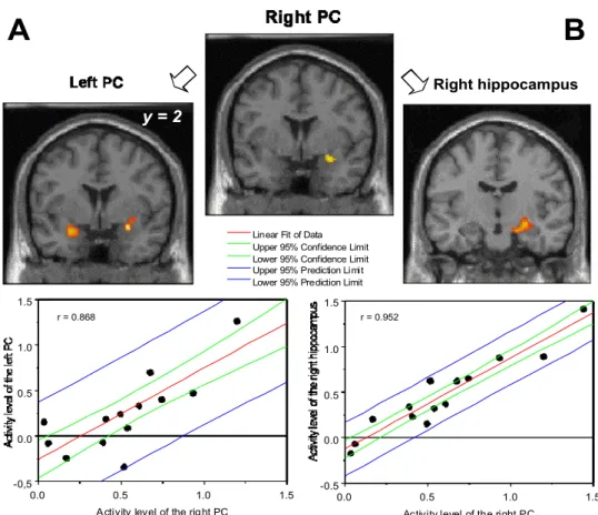
Areas functionally connected with the right PC. Table 3 shows the areas functionally connected
with the right PC in the F-D contrast. The temporal and frontal portions of the left PC, and a large cluster in the right preamygdalar claustrum, spreading in its anterior part from the frontal piriform cortex to, in its posterior part, the dentate gyrus, the CA1 and the CA3 portions of the hippocampus, were strongly functionally connected (r = 0.868 for the left PC and r = 0.952 for the right hippocampus, respectively) with the right PC (Figure 4).
DISCUSSION
The aim of this study was to characterize the neural mechanisms underlying familiarity judgment of odors using fMRI. With such an approach, we identified odor-evoked neural responses in several putative olfactory regions, including piriform cortex, amygdala, hippocampus, the opercular part of the inferior frontal gyrus and the mid-fusiform gyrus.
Activation of the mesial temporal region
The presence of activations in mesial temporal regions using neuroimaging methods is still a debate of controversy. Whereas some authors found activation in PC in PET studies (e.g., Zatorre
et al., 1992; Small et al., 1997; Savic et al., 2000; Kareken et al., 2001, 2003), we did not find any
activation in areas of the mesial temporal region in our previous studies (Royet et al., 1999, 2001). Activation in piriform cortex is also inconsistently reported in fMRI studies (e.g., Sobel et al., 1998; Yousem et al., 1999). With the EPI sequence, a well-known problem is the magnetic susceptibility artifact that induces a signal loss in this region, which can be compensated for by the use of ad hoc methods (Zald and Pardo, 2000). The PRESTO sequence used in the current study appears also to be adapted to reduce artifacts and reveal signal in this region. One may say also that olfactory habituation could explain in part absence of signal in these ventral regions. Olfactory habituation phenomenon has been reported with constant odor presentation (60 s) in studies using block design paradigm (Sobel et al., 2000; Poellinger et al., 2001). One way to circumvent these problems may be to use shorter stimulation epoch (Gottfried et al., 2002a,b). Another solution may be found in the use of event-related design in which large inter-stimulation interval (~ 30 s) are applied (Anderson et al., 2003). In the present study, we attempted to minimize these effects by using a different odorant on each breath cycle (from 12 to 20 different odorants per 60 s-epoch, depending on the duration of breathing cycle of the subject). In addition to the fact that this procedure of stimulation avoids self-adaptation phenomenon (Cain and Engen, 1969; Engen, 1982), it has also been described as reducing the sensory habituation phenomenon (Démonet et al., 1993). As in our previous fMRI study (Royet et al., 2003), the present data thus prove that, in the
framework of our experiments, the fMRI technique is well adapted in activation in primary olfactory areas.
Involvement of the piriform cortex in memory processes
Induction of memories by environmental information implies two different processes that are known as familiarity and recollection (Mandler, 1980; Rajaram, 1998; Bogacz et al., 2001). According to the ‘dual process theory’, processes underlying familiarity are perceptual in nature, and those subserving recollection include the retrieval of contextual information. Lehrner et al. (1999) demonstrated that these two forms of recognition memory processes also exist in olfaction. In other words, familiarity judgments are made on the basis of a feeling, without specific information about the encoding episode, and thus relate to implicit or unconscious memory, whereas recollection is seen as a form of elaborate or conceptually driven process, and thus relates to explicit or conscious memory. To illustrate these concepts, “personal experience indicates that it is not uncommon to be able to recognize that a person is familiar to us even though we cannot immediately recollect anything more about the person or our previous encounters with them” (Bogacz et al., 2001). Since the familiarity judgment is involved in recognition memory processes, the present results are consistent with previous findings in humans indicating that PC is involved in a long term recognition memory task of odors (Dade et al., 2002), and in appetitive and aversive olfactory learning (Gottfried et al., 2002b). These findings are further coherent with a large body of research using animal models and lending support to the theory that the piriform cortex is involved in learning- and memory-related processes (e.g., Schoenbaum and Eichenbaum, 1995; Datiche et
al., 2001). For instance, synaptic potentiation has been shown to occur in rat PC in vitro (e.g., Jung et al., 1990; Saar et al., 2002) and in vivo at the conclusion of learning (Roman et al., 1993;
Litaudon et al., 1997).These findings are finally in agreement with the theoretical works that indicate that the primary olfactory cortex presents a parallel-distributed architecture characteristic
Lateralization of familiarity judgment process
The current study indicates a unilateral activation of the PC involving only the right hemisphere. This is in line with a large body of researches. In a monorhinal odor recognition task, Savic et al. (2000) noted significantly right but not left piriform activity. Although Dade et al. (2002) did not explicitly reported hemispheric asymmetry for long-term olfactory memory, their results distinctly indicated a strong activation in the right OFC, and more activation in the right than left PC. Using behavioral measures, Broman et al. (2001) observed that odors presented to the right nostril were rated as more familiar than odors presented to the left nostril. They also reported that episodic recognition via the right nostril tended to have more ‘know’ responses and fewer ‘remember’ responses nominally than did odors recognized via the left nostril, which is in keeping with the right-nostril advantage for the familiarity ratings. Taken together, these findings are consistent with the notion that the left temporal lobe mediates distinctiveness processing, that is a processing that makes an individual item clearly distinct from another one, whereas the right temporal lobe structures subserve processes underlying perceptual fluency, that is a processing which involves perceptual analysis of surface features of each item (Blaxton and Theodore, 1997; Rajaram, 1998). Such a perceptual analysis of surface features is especially observed for odors that intrinsically are nameable with difficulty.
Findings with brain-damaged patients and in neuroimagery are convergent with these data. For instance, findings on recognition of abstract visuospatial designs in unilateral temporal lobe epilepsy patients indicate that left lesioned patients give more ‘know’ than ‘remember’ responses, whereas right lesioned patients show the opposite pattern (Blaxton and Theodore, 1997). Along the same line, Henson and his colleagues (1999) explored word recognition processes with fMRI and showed a dissociation whereby a ‘know’ judgment induces a right frontal activation, whereas a ‘remember’ judgment induces a left frontal activation.
In conclusion, the present data are consistent with our previous findings in PET studies indicating a preferential involvement of the right hemisphere in the familiarity judgment (Royet et
al., 1999, 2001). Although not significantly activated in the F-D contrast, the left PC was found to
be functionally connected with the right PC in the current study, thus highlighting its effective involvement in odor recognition memory process. However, this result does not invalidate the fact that the right PC participated more strongly than the left PC in the familiarity judgment task. An intriguing result is the lack of activation found in the right OFC in the present study. Examining specifically activation resulting from contrasts between either familiarity and rest, or detection and rest, we however found an activation in the right OFC in both cases (44, 32, -16; Z = 3.62 and 46, 30, -12; Z = 2.79, respectively). These results could explain the lack of activation observed when we compared images obtained in the familiarity judgment task with those obtained in detection task.
Participating of the hippocampal region, inferior frontal and mid-fusiform gyri in modality-independent memory process and semantic processing
Since few authors previously reported hippocampal activations independently of type of investigated olfactory task (e.g., Suzuki et al., 2001; Kareken et al., 2003), the right hippocampal activation observed in the familiarity judgment task was not expected in the present study. Inconsistent activation of the hippocampal formation is not a specific result of olfaction. Whereas hippocampal activation is thought to be associated with the process of conscious recollection (Schacter et al., 1996; Brown and Aggleton, 2001), several PET experiments have thus failed to find hippocampal activation in association with explicit retrieval (Shallice et al., 1994; Andreasen
et al., 1995; Tulving et al., 1996).
Lesion studies recently examined whether the brain structures that comprise the medial temporal lobe memory system differ in how they support its recollective and familiarity components. Manns
et al. (2003) found that patients with bilateral damage to the hippocampal region had severe
parahippocampal region) supports familiarity-based memory discrimination. Our data do not allow to distinguish both of these aspects of memory, but appears however consistent with the idea that the hippocampal region probably participates to recognition memory processes. This assumption is reinforced by the finding that the right PC and hippocampal region were functionally connected, thus suggesting their simultaneous involvement in the recognition memory process. Taken together, these results further reveal an activation in the right side only, a result consistent with our hypothesis of a preferential involvement of the right hemisphere in familiarity processing (Royet et
al., 2001).
Gottfried and Dolan (2003) recently showed that odor detection was facilitated when odors appeared in the context of semantically congruent visual cues. Congruent-specific activity was found in the anterior hippocampus and was interpreted as mediating retrieval or reactivation of semantic associations between odors and pictures. The authors indeed emphasized that using a low-level odor detection task, subjects were not asked to make explicit semantic judgments. Although no explicit visual stimuli were delivered in the present study, automatic visual associations are however inevitable and were probably used by subjects to identify odors (Royet et al., 1999). Activation of the left inferior frontal gyrus, in its opercular part, during the familiarity judgment task further supports the fact that subjects gather evidences from all modalities to identify the odor. Homae et al. (2002) thus showed that this region is involved in the selection and integration of semantic information, in a modality-independent manner. The mid-fusiform gyrus activation evidenced in the present study similarly supports these observations, since it has further been associated with visual, tactile and auditory recognition and categorization of objects (Adams and Janata, 2002; Joseph and Gathers, 2003; Stoeckel et al, 2003). Its involvement in olfactory object recognition therefore reinforces the idea of the polymodal nature of this area (Adams and Janata, 2002) and its implication in semantic processing (Wagner et al., 1998; Price, 2000).
Conclusion
In addition to our previous PET studies indicating that the right OFC is involved in the familiarity judgment task, the results of the present fMRI study shows that the right PC is also activated during this task and participates on this account to memory processes of odors. In previous fMRI and PET studies, we demonstrated that a neural network in the left hemisphere, composed among others of the OFC and primary olfactory areas, participated in perception of hedonic response (Royet et al., 2000, 2001, 2003). It thus appears that odor processing activates a large neural network in both hemispheres, but is however lateralized depending on the type of olfactory task. The present data also provide evidence that the hippocampal region, left inferior frontal gyrus and mid-fusiform gyrus take part in recognition memory processes, probably to help the subject to gather semantic cues allowing odor identification.
LEGENDS
Fig. 1. Experimental procedure showing the functional run including 12 epochs of 60 s each. Two olfactory conditions were performed: one detection condition with 3 epochs (Da, Db, Dc), and one familiarity condition with 3 epochs (Fa, Fb, Fc). Example of an epoch (Da) for which 15 stimuli (from S1 to S15) were delivered. R, rest.
Fig. 2. Reaction times represented as a function of olfactory task (Detection and Familiarity), and sets (Da, Db, Dc and Fa, Fb, Fc). The vertical bars show the standard errors of the means.
Fig. 3. Localization of task-specific activations in the F-D contrasts. A. Piriform cortex. Left, coronal view. Right, the region bounded by the rectangle on the left view is shown with higher magnification. B, hippocampus and C, inferior frontal gyrus. Neural responses are depicted on coronal and horizontal sections from a subject’s normalized T1-weighted brain. Clusters were thresholded at t = 3.10.
Fig. 4. Coronal views showing the right PC activity in the F-D contrast (cluster thresholded at t = 5.11) functionally correlated with (A) the left PC, and (B) the right hippocampus activations (both clusters thresholded at t = 3.10). Bottom: graphs depicting correlations between activity levels of the right and left PC (on left) and between the right PC and right hippocampus (on right). r, correlation coefficient.
Acknowledgments
We thank the technical team (M. Vigouroux, B. Bertrand and V. Farget) for designing and building the stimulation and recording materials, and J.P. Lomberget and M.B. Sanglerat for medical examinations of subjects participating in the study. We are grateful to the companies Givaudan, International Flavors and Fragrances, Lenoir, Davenne, and Perlarom for supplying the odorants used in this study. This work was supported by research grants from the ‘Région Rhône-Alpes’ and the ‘GIS Sciences de la Cognition’, the ‘Centre National de la Recherche Scientifique’, and the ‘Université Claude-Bernard de Lyon’.
REFERENCES
Adams, R.B., Janata, P. 2002. Comparison of neural circuits underlying auditory and visual object categorization. NeuroImage 16, 361-377.
Anderson, A.K., Christoff, K., Stappen, I., Panitz, D., Ghahremani, D.G., Glover, G., Gabrieli, J.D., Sobel, N. 2003. Dissociated neural representations of intensity and valence in human olfaction. Nat. Neurosci. 6, 196-202.
Andreasen, N.C., O'Leary, D.S., Cizadlo, T., Arndt, S., Rezai, K., Watkins, G.L., Ponto, L.L., Hichwa, R.D. 1995. Remembering the past: two facets of episodic memory explored with positron emission tomography. Am. J. Psychiatry 152, 1576-1585.
Blaxton, T.A., Theodore, W.H. 1997. The role of the temporal lobes in recognizing visuospatial materials: remembering versus knowing. Brain Cogn. 35, 5-25.
Bogacz, R., Brown, M.W., Giraud-Carrier, C. 2001. Model of familiarity discrimination in the perirhinal cortex. J. Comput. Neurosci. 10, 5-23.
Bower, J.M. 1991. Piriform cortex and olfactory recognition, in Davis J.D., Eichenbaum, H. (Eds.), Olfaction: A Model System for Computational Neuroscience, MIT Press, Cambridge, MA, pp. 266-285.
Brett, M., Anton, J.L., Valabregue, R., Poline J.B. 2002. Region of interest analysis using an SPM toolbox [abstract]. The 8th International Conference on Functional Mapping of the Human Brain, June 2-6, 2002, Sendai, Japan. Available on CD-ROM in NeuroImage, Vol 16, No 2.
Broca, P. 1863. Localisation des fonctions cérébrales. Siège de la faculté du langage articulé. Bull. Soc. Anthropol. Paris 4, 200-204.
Broman, D.A., Olsson, M.J., Nordin, S. 2001. Lateralization of olfactory cognitive functions: Effects of rhinal side of stimulation. Chem. Senses 26, 1187-1192.
Brown, M.W., Aggleton, J.P. 2001. Recognition memory: What are the roles of the perirhinal cortex and hippocampus? Natl. Rev. Neurosci. 2, 51-61.
(Ed.), Olfaction and Taste III, Rockfeller University Press, New York, pp. 127-141.
Dade, L.A., Jones-Gotman, M., Zatorre, R.J., Evans, A.C. 1998. Human brain function during odor encoding and recognition. A PET activation study. Ann. N Y Acad. Sci. 855, 572-574.
Dade, L.A., Zatorre, R.J., Jones-Gotman, M. 2002. Olfactory learning: convergent findings from lesion and brain imaging studies in humans. Brain 125, 86-101
Datiche, F., Roullet, F., Cattarelli, M. 2001. Expression of Fos in the piriform cortex after acquisition of olfactory learning: an immunohistochemical study in the rat. Brain Res. Bull. 55, 95-99.
Démonet, J.F., Wise, R., Frackowiak, R.S.J. 1993. Les fonctions linguistiques explorées en tomographie par émission de positons. Méd. /Sci. 9, 934-942.
Duvernoy, H.M. 1991. The Human Brain - Surface, Three Dimensional Sectional Anatomy and MRI. Springer, Wien.
Engen, T. 1982. The Perception of Odors. Academic Press, New York.
Friston, K.J., Ashburner, J., Frith, C.D., Poline, J.B., Healther, J.D. Frackowiak, R.S.J. 1995a. Spatial registration and normalisation of images. Hum. Brain Map. 3, 165-189.
Friston, K.J., Holmes, A.P., Worsley, K.J., Poline, J.B., Frith, C.D. Frackowiak, R.S.J. 1995b. Statistical parametric maps in functional imaging: A general linear approach. Hum. Brain Mapp. 2, 189-210.
Gottfried, J.A., Deichmann, R., Winston, J.S., Dolan, R.J. 2002a. Functional heterogeneity in human olfactory cortex: An event-related functional magnetic resonance imaging study. J. Neurosci. 22, 10819-10828.
Gottfried, J.A., Dolan, R.J. 2003. The nose smells what the eye sees: crossmodal visual facilitation of human olfactory perception. Neuron 39, 375-386.
Gottfried, J.A., O'Doherty, J., Dolan, R.J. 2002b. Appetitive and aversive olfactory learning in humans studied using event-related functional magnetic resonance imaging. J. Neurosci. 22,
Haberly, L.B. 2001. Parallel-distributed processing in olfactory cortex: new insights from morphological and physiological analysis of neuronal circuitry. Chem. Senses 26, 551-576. Haberly, L.B., Bower, J.M. 1989. Olfactory cortex: Model circuit for study of associative memory.
TINS 12, 258-264.
Henson, R.N.A., Rugg, M.D., Shallice, T., Josephs, O., Dolan, R.J. 1999. Recollection and familiarity in recognition memory: An event-related functional magnetic resonance imaging study. J. Neurosci., 19, 3962-3972.
Homae, F., Hashimoto, R., Nakajima, K., Miyashita, Y., Sakai, K.L. 2002. From perception to sentence comprehension: The convergence of auditory and visual information of language in the left inferior frontal cortex. NeuroImage 16, 883-900.
Joseph, J.E., Gathers, A.D. 2003. Effects of structural similarity on neural substrates for object recognition. Cogn. Affect Behav. Neurosci. 3, 1-16.
Jung, M.W., Larson, J., Lynch, G. 1990. Long-term potentiation of monosynaptic EPSPs in rat piriform cortex in vitro. Synapse 6, 279-283.
Kareken, D.A., Doty, R.L., Moberg, P.J., Mosnik, D., Hsing Chen, S., Farlow, M.R., Hutchins, G.D. 2001. Olfactory-Evoked regional Cerebral Blood Flow in Alzheimer’s Disease. Neuropsychology 15, 18-29.
Kareken, D.A., Mosnik, D.M., Doty, R.L., Dzemidzic, M., Hutchins, G.D. 2003. Functional anatomy of human odor sensation, discrimination, and identification in health and aging. Neuropsycholy 17, 482-495.
Lehrner, J.P., Walla, P., Laska, M., Deecke, L. 1999. Different forms of human odor memory: a developmental study. Neurosci. Lett. 272, 17-20.
Litaudon, P., Mouly, A.M., Sullivan, R., Gervais, R., Cattarelli, M. 1997. Learning-induced changes in rat piriform cortex activity mapped using multisite recording with voltage sensitive dye. Eur. J. Neurosci. 9, 1593-1602.
principles of echo-shifting with a train of observations (PRESTO). Magn. Reson. Med. 30, 764-768.
Mai, J.K., Assheuer, J., Paxinos, G. 1997. Atlas of the Human Brain, Academic Press, San Diego. Mandler, G. 1980. Recognizing: The judgment of previous occurrence. Psychol. Rev. 87, 252-271. Manns, J.R., Hopkins, R.O., Reed, J.M., Kitchener, E.G., Squire, L.R. 2003. Recognition memory
and the human hippocampus. Neuron 37, 171-180.
Poellinger, A., Thomas, R., Lio, P., Lee, A., Makris, N., Rose, B.R., Kwong, K.K. 2001. Activation
and habituation in olfaction An fMRI study. NeuroImage 13, 547-560.
Price, C.J. 2000. The anatomy of language: contributions from functional neuroimaging. J. Anat. 3, 335-359.
Rajaram, S. 1998. The effects of conceptual salience and perceptual distinctiveness on conscious recollection. Psychon. Bull. Rev. 5, 71-78.
Roman, F.S., Chaillan, F.A., Soumireu-Mourat, B. 1993. Long-term potentiation in rat piriform cortex following discrimination learning. Brain Res. 601, 265-272.
Royet, J.P., Hudry, J., Zald, D.H., Godinot, D., Grégoire, M.C., Lavenne, F., Costes, N., Holley, A. 2001. Functional neuroanatomy of different olfactory judgments. NeuroImage 13, 506-519. Royet, J.P., Koenig, O., Gregoire, M.C., Cinotti, L., Lavenne, F., Le Bars, D., Costes, N.,
Vigouroux, M., Farget, V., Sicard, G., Holley, A., Mauguière, F., Comar, D., Froment, J.C. 1999. Functional anatomy of perceptual and semantic processing for odours. J. Cogn. Neurosci. 11, 94-109.
Royet, J.P., Plailly, J., Delon-Martin, C., Kareken, D.A., Segebarth, C. 2003. Functional anatomy of the emotional responses to odors : Influence of hedonic valence, hedonic judgment, handedness, and gender. NeuroImage, 20, 713-728.
Saar, D., Grossman, Y., Barkai, E. 2002. Learning-induced enhancement of postsynaptic potentials in pyramidal neurons. J. Neurophysiol. 87, 2358-2363.
Savic, I., Gulyas, B., Larsson, M., Roland, P. 2000. Olfactory functions are mediated by parallel and hierarchical processing. Neuron 26, 735-745.
Schacter, D.L., Alpert, N.M., Savage, C.R., Rauch, S.L., Albert, M.S. 1996. Conscious recollection and the human hippocampal formation: evidence from positron emission tomography. Proc. Natl Acad. Sci. USA 93, 321-325.
Schoenbaum, G., Eichenbaum, H. 1995. Information coding in the rodent prefrontal cortex. I. Single-neuron activity in orbitofrontal cortex compared with that in pyriform cortex.. J. Neurophysiol. 74, 733-750.
Shallice, T., Fletcher, P., Frith, C.D., Grasby, P., Frackowiak, R.S., Dolan, R.J. 1994. Brain regions associated with acquisition and retrieval of verbal episodic memory. Nature 368, 633-635.
Small, D.N., Jones-Gotman, M., Zatorre, R.J., Petrides, M., Evan, A.C. 1997. Flavor processing : More than the sum of its parts. NeuroReport 8, 3913-3917.
Sobel, N., Prabhakaran, V., Desmond, J.E., Glover, G.H., Goode, R.L., Sullivan, E.V., Gabrieli, J.D.E. 1998. Sniffing and smelling: separate subsystems in the human olfactory cortex. Nature, 392, 282-286.
Sobel, N., Prabhakaran, V., Zhao, Z., Desmond, J.E., Glover, G.H., Sullivan, E.V., Gabrieli, J.D.E. 2000. Time-course of odorant-induced activation in the human primary olfactory cortex. J. Neurophysiol. 82, 537-551.
Soros, P., Knecht, S., Imai, T., Gurtler, S., Lutkenhoner, B., Ringelstein, E.B., Henningsen, H. 1999. Cortical asymmetries of the human somatosensory hand representation in right- and left-handers. Neurosci. Lett. 271, 89-92
Stoeckel, M.C., Weder, B., Binkofski, F., Buccino, G., Shah, N.J., Seitz, R.J. 2003. A fronto-parietal circuit for tactile object discrimination: an event-related fMRI study. NeuroImage 19, 1103-1114.
Suzuki, Y., Critchley, H.D., Suckling, J., Fukuda, R., Williams, S.C.R., Andrew, C., Howard, R., Ouldred, E., Bryant, C., Chir, B., Swift, C.G., Jackson, S.H.D. 2001. Functional magnetic resonance imaging of odour identification: The effect of aging. J. Gerontol. 56, 756-760.
Tulving, E., Markowitsch, H.J., Craik, F.I.M., Habib, R., Houle, S. 1996. Novelty and familiarity activations in PET studies of memory encoding and retrieval. Cereb. Cortex 6, 71-79.
Wagner, A.D., Schacter, D.L., Rotte, M., Koutstaal, W., Maril, A., Dale, A.M., Rosen, B.R., Buckner, R.L. 1998. Building memories: remembering and forgetting of verbal experiences as predicted by brain activity. Science 281, 1188-1191.
Weintraub, S., Mesulam, M.M. 1987. Right cerebral dominance in spatial attention. Further evidence based on ipsilateral neglect. Arch. Neurol. 44, 621-625.
Yonelinas, A.P., Kroll, N.E., Quamme, J.R., Lazzara, M.M., Sauve, M.J., Widaman, K.F., Knight, R.T. 2002. Effects of extensive temporal lobe damage or mild hypoxia on recollection and familiarity. Nat. Neurosci. 5, 1236-1241.
Yousem, D.M., Maldjian, J.A., Siddiqi, F., Hummel, T., Alsop, D.C., Geckle, R.J., Bilker, W.B., Doty, R.L. 1999. Gender effects on odor-stimulated functional magnetic resonance imaging. Brain Res. 818, 480-487.
Yousem, D.M., Williams, S.C.R., Howard, R.O., Andrew, C., Simmons, A., Allin, M., Geckle, R.J., Suskind, D., Bullmore, E.T., Brammer, M.J., Doty, R.L. 1997. Functional MR imaging during odour stimulation: Preliminary data. Neuroradiology 204, 833-838.
Zald, D.H., Donndelinger, M.J., Pardo, J.V. 1998. Elucidating dynamic brain interactions with across-subjects correlational analyses of positron emission tomographic data: The functional connectivity of the amygdala and orbitofrontal cortex during olfactory tasks. J. Cereb. Blood Flow. Metab. 18, 896-905.
Zald, D.H., Pardo, J.V. 1997. Emotion, olfaction, and the human amygdala: Amygdala activation during aversive olfactory stimulation. Proc. Natl Acad. Sci. USA 94, 4119-4124.
Psychophysiol. 36, 165-181.
Zatorre, R.J., Belin, P., Penhune, V.B. 2002. Structure and function of auditory cortex: music and speech. Trends Cogn. Sci. 6, 37-46.
Zatorre, R.J., Jones-Gotman, M., Evans, A.C., Meyer, E. 1992. Functional localization and lateralization of human olfactory cortex. Nature 360, 339-340.
Table 1
List of odorants selected for the Da, Db, Dc, Fa, Fb, and Fc epochs.
Da Db Dc Fa Fb Fc
1 Plum Sage Acacia Apricot Raspberry Citronella
2 Turpentine Bergamot
Orange
Tetralin Trans-2-Hexenal
3 Diethyl ether Mint Cypress
4 Tarragon Orange Pine Honey Geranium
5 Parsley Acetophenone Bornyl acetate Patchouli Vienna bread
6 Guaiacol Caramel Jasmine Anise
7 Jonquil Camphor Grass Basil Iris
8 Neroli Strawberry Toluene Incense
9 Musk Pepper Green lily Lavender
10 Carrot Pine needle Oyster Banana Gingerbread
11 Ethyl benzoyl
acetate
Gardenia Vanilla Apple
12
1,4-Dichlorobutane
Butanol Celery Lily Garlic
13 Eglantine Hazelnut Bitter almond Phenyl
propionaldehyde
14 Tar Caprylic
aldehyde
Rose
15 Cherry 2-Bromophenol Methyl acetate Passion fruit Thyme
16 Acetol Liqueur wine Tangerine Biscuit Eucalyptus Lime
17 2-Octanol Tobacco Coconut 1-Octen-3-ol
18 Liquorice Blackcurrant 3-Methyl anisol Clove Camomile
Familiarity
Mean score (SD) 4.00 (0.72) 4.11 (0.87) 4.29 (0.92) 5.55 (1.15) 5.33 (1.11) 5.30 (1.06) Range score 3.14 – 5.11 3.19 – 5.16 3.09 – 5.17 4.03 – 7.27 3.41 – 7.24 3.89 – 6.92
Table 2
Areas activated in F-D contrast.
Brain region L/R k Z value MNI coordinates
x y z
Amygdala L 146 4.79 -24 -6 -12
Cingulate gyrus L 85 4.77 -8 16 40
Mid-fusiform gyrus R 59 4.06 40 -42 -16
Temporal piriform cortex R 275 3.88 30 2 -16
Amygdala R 3.66 22 -14 -8
Hippocampus (CA1) R 3.56 22 -22 -10
Hippocampus (CA3, subiculum) R 3.50 24 -8 -10
Inferior frontal gyrus, opercular part L 104 3.74 -50 28 2
Middle frontal gyrus L 36 3.72 -20 38 28
Mid-fusiform gyrus L 50 3.69 -42 -56 -12
Note. F, familiarity; D, detection; k, size of the cluster in number of connected voxels; x, y, z, MNI coordinates of the maximum in the Montreal Neurological Institute Brain template.
Table 3
Areas functionally connected with the right PC (30, 2, -16) in the F-D contrast.
Brain region L/R k Z value MNI coordinates
x y z
Preamygdalar claustrum R 419 5.65 28 0 16
Hippocampus (CA1, dentate gyrus) R 4.40 28 -12 -12
Hippocampus (CA1, dentate gyrus) R 4.24 20 -12 -18
Hippocampus (CA3) R 3.64 26 -20 -6
Frontal piriform cortex R 3.55 32 10 -20
Temporal claustrum R 3.54 34 0 -8
Temporal piriform cortex L 179 4.29 -24 2 -20
Frontal piriform cortex L 3.86 -30 10 -18
Frontal piriform cortex L 3.82 -30 14 -20
Figure 1.
S1 S2
S14
S15
R
Da
Db
Dc
Fa
Fb
Fc
Figure 4. Right hippocampus
A
B
0.0 0.5 1.0 1.5 -0,5 0.0 0.5 1.0 1.5Linear Fit of Data Upper 95% Confidence Limit Lower 95% Confidence Limit Upper 95% Prediction Limit Lower 95% Prediction Limit
Activity level of the right PC
0.0 0.5 1.0 1.5 -0.5 0.0 0.5 1.0 1.5
Activity level of the right PC
r = 0.868 r = 0.952