HAL Id: hal-01138973
https://hal.archives-ouvertes.fr/hal-01138973
Submitted on 8 Apr 2015
HAL is a multi-disciplinary open access
archive for the deposit and dissemination of
sci-entific research documents, whether they are
pub-lished or not. The documents may come from
teaching and research institutions in France or
abroad, or from public or private research centers.
L’archive ouverte pluridisciplinaire HAL, est
destinée au dépôt et à la diffusion de documents
scientifiques de niveau recherche, publiés ou non,
émanant des établissements d’enseignement et de
recherche français ou étrangers, des laboratoires
publics ou privés.
Stress clamp experiments on multicellular tumor
spheroids
Fabien Montel, Morgan Delarue, Jens Elgeti, Laurent Malaquin, Markus
Basan, Thomas Risler, Bernard Cabane, Danijela Vignjevic, Jacques Prost,
Giovanni Cappello, et al.
To cite this version:
Fabien Montel, Morgan Delarue, Jens Elgeti, Laurent Malaquin, Markus Basan, et al.. Stress clamp
experiments on multicellular tumor spheroids. Physical Review Letters, American Physical Society,
2011, 107 (18), pp.188102. �10.1103/PhysRevLett.107.188102�. �hal-01138973�
Stress clamp experiments on multicellular tumor spheroids
Fabien Montel1, Morgan Delarue1, Jens Elgeti1, Laurent Malaquin1, Markus Basan1, Thomas Risler1,
Bernard Cabane3, Danijela Vignjevic2, Jacques Prost1,3, Giovanni Cappello1,∗ and Jean-Fran¸cois Joanny1
1UMR 168, Institut Curie, Centre de Recherche, 26 rue d’Ulm 75005 Paris France.
2UMR 144, Institut Curie, Centre de Recherche, 26 rue d’Ulm 75005 Paris France. and
3
ESPCI, 10 rue Vauquelin, 75005 Paris, France. (Dated: November 28, 2011)
The precise role of the microenvironment on tumor growth is poorly understood. Whereas the tumor is in constant competition with the surrounding tissue, little is known about the mechanics of this interaction. Using a novel experimental procedure, we study quantitatively the effect of an applied mechanical stress on the long-term growth of a spheroid cell aggregate. We observe that a stress of 10kPa is sufficient to drastically reduce growth by inhibition of cell proliferation mainly in the core of the spheroid. We compare the results to a simple numerical model developed to describe the role of mechanics in cancer progression.
PACS numbers: 87.19.xj,87.19.R-,87.55.Gh
Cancer progression occurs in several stages. In the
case of carcinomas, which are cancers of epithelial cells, the primary tumor grows locally, until some cells invade the neighboring tissue called the stroma which is essen-tially made of extracellular matrix, fibroblast cells, im-mune cells and capillary vessels. Three key elements con-trol proliferation of the primary tumor: the accumulation of gene mutations and the tumor biochemical and me-chanical micro-environments. It is difficult to isolate in vivo one of these factors to measure accurately its im-portance. Several recent works suggest that mechanical stress plays a role in tumor progression. A mechanical stress applied to genetically predisposed tissues or tumor spheroids grown in vitro induces signaling pathways that are characteristic of cancer invasion [1, 2]. It has also been shown that an increase of mechanical stress leads to a reduction in cancer cell proliferation in vitro, and drives apoptosis through the mitochondrial pathway [3– 5]. In spite of these experimental evidences, the precise role of the micro-environment, and its interaction with the tumor, are poorly understood. Our group has devel-oped a theoretical framework [6–8] to describe the influ-ence of the balance between cell division and apoptosis on tumor growth under stress. The theory is based on the existence of a homeostatic state of a tissue. This is the steady state of the tissues where cell division bal-ances cell death. The homeostatic stress is a function of the biochemical state of the tissue and depends on the local concentrations of nutrients, oxygen and growth factors as well as on the environment of the tissue. Sig-naling induced by the stroma can for example modify the homeostatic state. In the simple case where the biochem-ical state of the tissue can be maintained constant, the homeostatic stress is the stress that the tissue can exert at steady state on the walls of a confining chamber. It is a measure of mechanical forces that cells can sustain in this state. Indeed to grow against the surrounding tissue, cells have to exert mechanical stress on the neighboring
cells.
In this paper we test experimentally the relevance of the homeostatic stress concept. We measure the effect of a known external stress on the growth of a cellular aggregate mimicking a tumor over timescales longer than the typical time scales of cell division or apoptosis. We use a new experimental strategy to exert a well defined mechanical stress on multicellular tumor spheroids for a period of time exceeding 20 days.
We prepare colon carcinoma cell spheroids derived from mouse CT26 cell lines (ATCC CRL-2638) using a classical agarose cushion protocol [9]. The wells of a 48 wells plate are covered with agarose gel (Ultrapure agarose, Invitrogen Co, Carlsbad, CA) and cell suspen-sions are seeded on the gels at concentration of 20000 cells per well. Cells self-assemble into spheroids in less than
24h. Cells are cultured under 95% air/ 5% CO2
atmo-sphere in DMEM enriched with 10% calf serum (culture medium). Using confocal microscopy, we check that the shape of the spheroid is indeed close to a sphere. A con-stant stress is applied on the tissue over long time scales by imposing the osmotic pressure of a solution of the
bio-compatible polymer Dextran (Mw= 100kDa,
Sigma-Aldrich Co, St Louis, Mo). This polymer is known to be neutral and is not metabolized by mammalians cells. We also confirmed that it is neither a growth or a death factor by plating cells for 3 days with Dextran and measuring cell concentration and viability.
We first perform indirect stress measurements. A grow-ing spheroid is positioned inside a closed dialysis bag (di-ameter 10 mm, Sigma-Aldrich) which is then placed in
an external medium with added Dextran. The
dialy-sis membrane was chosen so that its molecular weight cut-off (10 kDa) impedes the diffusion of Dextran. The osmotic stress induces a force on the dialysis membrane, which is transmitted in a quasi-static equilibrium to the spheroid and calibrated as in [10, 11]. The stress ex-erted on the cellular system can be seen as a network
2 stress that tends to reduce the volume occupied by the
spheroid. It acts directly on the cells, and not on the
in-terstitial fluid. The volume V (t)/V0, normalized by the
initial volume of the spheroid V0, is measured at
succes-sive times from a top view using differential interference contrast microscopy (Axiovert 100, Zeiss). In the absence of any applied stress, the spheroid reaches a steady state with a typical diameter 900 µm. When Dextran is added
to the medium, a decrease of the growth rate dVdt and of
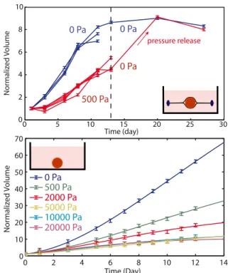
the steady state volume are observed (Fig.1-Top). Inter-estingly, after a stress release, the growth of the spheroid resumes until it reaches the same steady state volume as in the absence of external pressure. This indicates that the effect of stress is fully reversible. Altogether these results show that an external applied stress modulates the growth of tumor spheroids.
We have also performed direct experiments where the osmotic stress is applied onto the spheroid in the absence of the dialysis membrane. In order to verify that Dextran cannot diffuse inside the spheroid, we have placed it in a medium supplemented with fluorescent FITC-Dextran at an osmotic stress Π = 1000Pa. After 4 days of in-cubation, the first 70 µm of the spheroid were imaged using spinning disc microscopy. We measured that the amount of Dextran able to penetrate into the spheroid is negligible compared to the Dextran concentration in the medium. The osmotic stress is thus applied on the first layer of cells that plays the role of the dialysis membrane in the direct experiment and transmits the stress to the rest of the spheroid. The volume of the spheroid has also been measured as a function of time (Fig.1-Bottom). We observe a dependence of the growth rate and the steady state size on stress very similar to that observed in the in-direct experiment, validating our approach. Interestingly, for a stress larger than 10kP a the effect of stress satu-rates and the growth curves are indistinguishable from each other.
The direct experiment is based on the application of a mechanical stress on the surface of the spheroid through an osmotic shock. Osmotic stress is known to have di-rect effects on cell growth and apoptosis in particular through the mitogen activated protein kinase (MAPK) pathway [12–15]. However, in all these studies, the ef-fect of an osmotic shock is only measured for an osmotic stress two orders of magnitude larger than the one ap-plied in our experiments (1M P a compared to 10kP a). Moreover we do not observe any apoptosis at the sur-face of the spheroid where the osmotic stress is exerted (Fig. 2). In addition, it can be checked by balancing chemical potentials that the presence of Dextran outside the spheroid creates a negligible concentration gradient of all other solubles molecules. The concentration dif-ference of a small soluble solute exchanged between the interior and the exterior of the spheroid can be estimated
as ∆cs/cs= vΠ/kT (1 − Θs) < 10−3where csand Θsare
the concentration and the volume fraction of the solute,
0 2 4 6 8 10 12 14 0 10 20 30 40 50 60 70 Time (Day) No rmali zed V olume 0 5 10 15 20 25 30 0 2 4 6 8 10 No rmali zed V olume Time (day) pressure release 0 Pa 500 Pa 0 Pa 0 Pa 0 Pa 500 Pa 2000 Pa 5000 Pa 10000 Pa 20000 Pa
FIG. 1: Growth curves of individual spheroids under stress. (Top) Normalized volume of individual spheroids as a func-tion of time for the indirect experiments. The initial diameter
is Do = 350µm. At t = 12 days, stress is released.
(Bot-tom) Normalized volume of individual spheroids as a func-tion of time for the direct experiments. The initial diameter
is Do= 200µm. The inserts show the principles of the two
ex-periments. The points are representative experimental data for individual spheroids taken out of a larger set of experi-ments. For each condition, N ≥ 3 experiments have been recorded. The lines are the results of fits with the two rate model. Error bars are the image analysis errors.
v the molecular volume of the solvent and Π the applied osmotic stress. In other words, the chemical potential of water in the cell is dominated by the small ions and it is only slightly modified by the presence of Dextran.
Finally we investigate experimentally the spatial de-pendence of cell division and apoptosis using
cryo-sections and immuno-fluorescence. Spheroids of
com-parable diameters are embedded in a freezing medium,
placed at −80oC and cut in slices of 5 µm thickness
at the level of their equatorial plane. Using a
classi-cal immuno-staining protocol, we then label fluorescently dividing cells (in cyan with an anti-Ki67 antibody) and apoptotic cells (in red with an anti -cleaved caspase3 an-tibody) (See Fig. 2). We observe that in the absence of external stress, cell division is distributed over all the spheroid with an increase at the periphery, whereas for an external stress of 1kPa, it is greatly reduced in the center of the sections. As in previous studies [9, 16], we observe an accumulation of apoptotic cells in the center of the spheroid but with no measurable effect of stress on this localization.
Ki-67
cleaved Caspase-3
P = 0 Pa P = 1 kPa
100µm
FIG. 2: Effect of stress on the distribution of
prolifera-tion and apoptosis. Cryosections and immunofluorescence
of the spheroids are used to label the cell divisions (anti-body against Ki67 in cyan) and apoptosis (anti(anti-body against Cleaved-capase3 in red).(Left) Half section of a spheroid grown in normal medium for 4 days (Right) Half section of a spheroid grown with a stress of 1 kPa for 4 days
In order to better understand this stress dependence of cell division and to interpret the generic trends of the ex-perimental findings, we performed numerical simulations similar to those of Ref. [6, 8]. We adapt these simula-tions to the geometry and setup of the experiments. In brief, in the simulations, a cell is represented by a pair of particles which repel each other and thus move apart. When a critical distance is reached, the cell divides. After division, each original particle constitutes, together with a newly inserted particle in its surrounding, a daughter cell. Particles belonging to different cells interact with all particles with a short range interaction: a constant attractive force describes cell-cell adhesion, while a repul-sive short range potential ensures volume exclusion. The viscous drag between cells is taken into account by a “Dis-sipative Particle Dynamics“ -type thermostat. Finally a constant apoptosis rate provides cell removal. In order to mimic the experiments, tissue spheroids are grown in a container together with a ”passive liquid” under stress. This liquid interacts with the cell particles in a similar way as cell particles with each others and it transmits the stress on the spheroid.
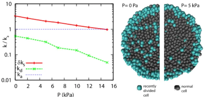
As in the experiments, we observe a steady state that depends on the applied stress. In the numerical simula-tions each division event can be traced and the spatial distribution of divisions can be measured to build virtual cryosections of the simulated spheroids. We observe a strong dependence of the division rate on the distance to the surface of the spheroids and a clear decrease of cell division anywhere in the section but stronger in the core (see Fig.3).
Based on the growth curves and cryosection observa-tions, we present a simple two rate description of the
0.01 0.1 1 10 0 2 4 6 8 10 12 14 16 k / k a P (kPa) δks kd ka P= 0 Pa P= 5 kPa recently divided cell normal cell
FIG. 3: Dissipative Particle Dynamics Simulations. (Left)
Bulk division rate kd, surface rate increment δks and
apop-tosis rates ka as function of the applied pressure P . (Right)
Virtual cryosections of the simulated spheroids for an external pressure P=0 Pa or P=5kPa. The pressure units have been calibrated from the experiments using the saturation pressure.
spheroid growth in the absence or the presence of exter-nal stress: the core of the spheroid is mostly undergoing apoptosis whereas its periphery is proliferating. In this situation, the net growth rate is proportional to the area
(∝ r2) while the net death rate is proportional to the
vol-ume (∝ r3). This surface growth effect leads to a stable
steady state size. The surface localization of the prolif-eration can be obtained using purely mechanical consid-erations. A cell must deform its environment to grow. The deformation is facilitated if the cell is closer to the surface, and this implies that proliferation is favored at the surface. The increased number of cell divisions at the surface drives a flow from the surface of the spheroid toward its center. The flow is a possible explanation for the accumulation of the long lasting apoptotic markers in the center of the spheroid. A mechanical control of cell cycle entrance can also explain the growth of tumor spheroids in free suspension [17]. In this case nutrient depletion causes the formation of a necrotic core which generates death at the inner surface of the viable rim
(∝ r2) i.e. not proportional to the volume thereby not
generating a steady state [17, 18]. Using a fluorescently labelled growth factor (Alexa 555- EGF) we have verified that the transport of these molecules is not affected by stress. This result supports a direct mechanical effect on the division rate.
Our two rate model can be seen as a simplified version of the two rate model of Radszuweit et al. [19]. The net
bulk growth rate is k = kd− ka, where kdand ka are the
division and apoptosis rates respectively. It is a function
of stress. At the surface, the net growth rate kd− ka+
δks is larger and δks has a different stress dependence.
Taking into account surface and bulk growth, the growth equation reads :
∂tN = (kd− ka)N + δksNs (1)
Assuming a constant cell density and a constant thickness λ of the region where the division rate increment is equal
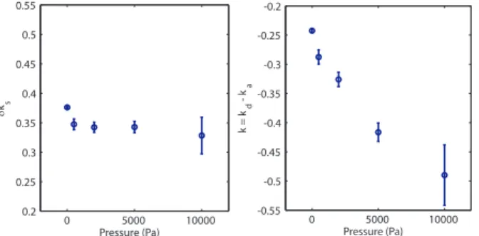
4 0 5000 10000 -0.55 -0.5 -0.45 -0.4 -0.35 -0.3 -0.25 -0.2 Pressure (Pa) k = k d - k a 0 5000 10000 0.2 0.25 0.3 0.35 0.4 0.45 0.5 0.55 Pressure (Pa) δks
FIG. 4: Evolution of growth rates with stress. (Left) Surface
division rate increment δks as a function of stress. (Right)
Bulk growth rate k as a function of stress. For each condi-tion, N ≥ 3 experiments have been recorded. The errors bar are obtained using a jackknifing method and represent the efficiency of the fitting algorithm
to δks, one can express for R > λ the rate of volume
increase as :
∂tV = (kd− ka)V + (36π)1/3δksλV2/3 (2)
For small spheroids (R << λ) the growth rate is
posi-tive and constant (kd+ δkd− ka), this leads to the
previ-ously described exponential growth [17, 18]. In our case the growth curves can readily be fitted by Eq. 2 in the range of large spheroids [20]. The variation with pressure
of the parameters k = kd− ka and δksis given in Fig. 4.
The surface growth rate δksis less affected by stress than
the bulk growth rate k. A similar fit can be performed on the simulations. The bulk and surface growth rates k and
δksare represented on Fig. 3. Although both the surface
and bulk growth rates depend exponentially on pressure, the decay constant of the surface rate is much smaller than that of the bulk rate. While the bulk rate decreases by more than one order of magnitude, the surface rate decreases by a factor 3. This supports the hypotheses of the simulations. In summary, cell division in the core of the spheroid is strongly affected by stress whereas cell division rate increment on the surface of the spheroid depends more weakly on stress.
In conclusion, we have shown by a direct measure-ment of the tissue response to an external stress that the application of an external stress drastically limits the growth of tumoral spheroids. Previous approaches [5, 21] had used the elastic deformation of poro-elastic gels to measure the maximum stress that can be devel-oped by spheroids. The measured stress in these experi-ments is in the same range as in our measureexperi-ments. In a recent study a localized increase of mitochondrial apop-tosis and a reduction of proliferation in presence of stress were also reported [3]. This difference with our results may be due to the fact that in our case we are not con-trolling the rigidity of the surrounding substrate but the applied pressure. This may leads to a different response of the spheroid. Our results favors the idea that direct
mechanical effects can have strong implications in cancer proliferation. This raises the question of the players in the crosstalk between stress and cellular response and in particular of the nature of the stress sensor.
We would like to thank F. Brochard, F. Graner, P. Nassoy, K. Alessandri and J. Kaes for useful discussions. F.M. and G.C. would like to thank Axa Research Fund and CNRS for funding. The group belongs to the CNRS consortium CellTiss.
∗
Electronic address: [email protected]
[1] J. Whitehead, D. Vignjevic, C. F¨utterer, E. Beaurepaire,
S. Robine, and E. Farge, HFSP journal 2, 286 (2008), ISSN 1955-2068.
[2] Z. Demou, Annals of Biomedical Engineering 38, 3509 (2010), ISSN 0090-6964.
[3] G. Cheng, J. Tse, R. K. Jain, and L. L. Munn, PloS one 4, e4632 (2009), ISSN 1932-6203.
[4] T. Roose, P. Netti, L. Munn, Y. Boucher, and R. Jain, Microvascular research 66, 204 (2003), ISSN 0026-2862. [5] G. Helmlinger, P. Netti, H. Lichtenbeld, R. Melder, and
R. Jain, Nature Biotechnology 15, 778 (1997), ISSN 1087-0156.
[6] M. Basan, T. Risler, J.-F. Joanny, X. Sastre-Garau, and J. Prost, HFSP journal 3, 265 (2009), ISSN 1955-205X. [7] J. Ranft, M. Basan, J. Elgeti, J.-F. Joanny, J. Prost,
and F. J¨ulicher, Proceedings of the National Academy
of Sciences of the United States of America 107 (2010), ISSN 1091-6490.
[8] M. Basan, J. Prost, J.-F. Joanny, and J. Elgeti, Physical biology 8, 026014 (2011), ISSN 1478-3975.
[9] W. Mueller-klieser and L. A. Kunz-schughart, Journal of Biotechnology (2010), ISSN 0168-1656.
[10] C. Bonnet-Gonnet, L. Belloni, and B. Cabane, Langmuir 10, 4012 (1994), ISSN 0743-7463.
[11] A. Bouchoux, P.-E. Cayemitte, J. Jardin, G. G´
esan-Guiziou, and B. Cabane, Biophysical journal 96, 693 (2009), ISSN 1542-0086.
[12] K. J. Cowan, Journal of Experimental Biology 206, 1107 (2003), ISSN 00220949.
[13] B. Racz, D. Reglodi, B. Fodor, B. Gasz, A. Lubics, F. Gallyas, E. Roth, and B. Borsiczky, Bone 40, 1536 (2007), ISSN 8756-3282.
[14] M.-B. Nielsen, S. T. Christensen, and E. K. Hoffmann, American journal of physiology. Cell physiology 294, C1046 (2008), ISSN 0363-6143.
[15] Y. Xie, W. Zhong, Y. Wang, A. Trostinskaia, F. Wang, E. E. Puscheck, and D. A. Rappolee, Molecular human reproduction 13, 473 (2007), ISSN 1360-9947.
[16] W. Mueller-Klieser, American Journal of Physiology-Cell Physiology 273, C1109 (1997), ISSN 0363-6143.
[17] D. Drasdo and S. H¨ohme, Physical biology 2, 133 (2005),
ISSN 1478-3975, URL http://www.ncbi.nlm.nih.gov/ pubmed/16224119.
[18] G. Schaller and M. Meyer-Hermann, Physical Review E 71, 1 (2005), ISSN 1539-3755, URL http://link.aps. org/doi/10.1103/PhysRevE.71.051910.
[19] M. Radszuweit, M. Block, J. Hengstler, E. Sch¨oll, and
[20] L. Von Bertalanffy, The Quarterly Review of Biology 32, 217 (1957), URL http://www.jstor.org/stable/ 2815257.
[21] A. Fritsch, M. Hockel, T. Kiessling, K. D. Nnetu, F. Wet-zel, M. Zink, and J. A. Kas, Nat Phys 6, 730 (2010).