HAL Id: inserm-00700119
https://www.hal.inserm.fr/inserm-00700119
Submitted on 23 May 2012
HAL is a multi-disciplinary open access
archive for the deposit and dissemination of
sci-entific research documents, whether they are
pub-lished or not. The documents may come from
teaching and research institutions in France or
abroad, or from public or private research centers.
L’archive ouverte pluridisciplinaire HAL, est
destinée au dépôt et à la diffusion de documents
scientifiques de niveau recherche, publiés ou non,
émanant des établissements d’enseignement et de
recherche français ou étrangers, des laboratoires
publics ou privés.
SmpB as the handyman of tmRNA during
trans-translation.
Brice Felden, Reynald Gillet
To cite this version:
Brice Felden, Reynald Gillet. SmpB as the handyman of tmRNA during trans-translation.. RNA
Biol, 2011, 8 (3), pp.440-9. �10.4161/rna.8.3.15387�. �inserm-00700119�
review
rNA Biology 8:3, 440-449; May/June 2011; © 2011 Landes Bioscience
The Why and How of Trans-Translation in Bacteria
In bacteria, ribosome stalling at the 3'-end of mRNAs lacking termination codons is a serious issue for cell survival. Arrested ribosomes cannot be recycled back to new rounds of translation since they are unable to terminate protein synthesis using release factors. If not rescued, most ribosomes would become inactive very fast during bacterial growth. In all eubacteria and some organelles, there is a specialized quality-control mechanism for rescuing ribosomes arrested during translation performed by the ‘tmRNA-SmpB’ (transfer-messenger RNA-Small protein B) ribonucleoprotein complex.1 In a reaction known as
trans-translation, bi-functional tmRNA possessing both a tRNA and a mRNA domain, in complex with SmpB, recognizes the stalled ribosomes and adds an internally encoded peptide tag to the C terminus of nascent polypeptides for destruction by cellular proteases. The ribosomes dissociate from tmRNA at its internal stop codon and are recycled back to new rounds of translation.2
Ribosome recycling is its primary function whereas protein tag-ging and elimination is being accessory. Recent studies suggest a broader physiological role for trans-translation in monitoring protein folding and co-translational events.3
tmRNA safeguard, activation and shuttling. In all species
encoding and expressing tmRNA, it contains a partial tRNA domain (TLD for tRNA-like domain) aminoacylated at its 3'-end by alanyl-tRNA synthetase, but restricted to a tRNA acceptor branch (acceptor stem and T stem-loop) containing
*Correspondence to: Reynald Gillet; Email: reynald.gillet@univ-rennes1.fr Submitted: 02/09/11; Accepted: 02/25/11
DOI: 10.4161/rna.8.3.15387
in the recent years, a wealth of genetic, biochemical and structural data focusing on various steps of bacterial trans-translation has been reported. The early events, from stalled ribosome recognition, pre-accommodation to translocation have been recently investigated in great details. in comparison, the later events including ‘elongation-termination’ onto tmrNA reading frame and ribosome recycling are much less known. what follows is a summary of those data together with our personal view of the dynamics of trans-translation, with emphasis to the pivotal and leading roles that the SmpB protein has, being the essential conductor during the whole process.
SmpB as the handyman of tmRNA
during trans-translation
Brice Felden1,3 and reynald Gillet2,3,*
1Université de rennes 1; iNSerM U835-UPreS eA2311; Laboratoire de Biochimie Pharmaceutique; 2UMr CNrS 6026 equipe Structure et Dynamique des Macromolécules; 3iFr
140 Génomique Fonctionnelle Agronomie et Santé; rennes, France
Key words: cryo-electron microscopy; protein synthesis; ribosome; SmpB; tmRNA; trans-translation
modified nucleosides, as for canonical tRNAs,4 as well as
sev-eral pseudoknots and helices (Fig. 1A and reviewed in refs. 5 and 6). The small basic SmpB protein is essential for trans-translation, enhances tmRNA aminaocylation and prevents its degradation7,8 binds tmRNA9 and the stalled ribosomes10 and is
required for loading Ala-tmRNA, in complex with EF-Tu11 onto
the empty (or partially empty) A site of the stalled ribosomes. During the past 10 y, a large body of biochemical, genetic and structural data has accumulated on bacterial trans-translation, converging to a reasonably well experimentally supported sce-nario. Alanyl-tmRNA bound to SmpB and EF-Tu enters the A-site of a stalled 70S ribosome having a peptidyl-tRNA in the P-site. tmRNA tRNA domain is near the peptidyl-trans-fer active site and SmpB is at the decoding center, mimicking functionally a ‘codon-anticodon’ interaction and structurally a tRNA anticodon branch corresponding to the anticodon and D stems of a tRNA.12,13 The nascent polypeptide gets
trans-peptidated to the alanine at tmRNA 3'-end and the tag read-ing frame is inserted within the ribosome decodread-ing center. Translation continues to a canonical termination codon onto tmRNA, releasing the aborted nascent protein for destruction by cellular proteases.
Handy SmpB. The SmpB protein is an essential component
for trans-translation in vivo, responsible of a variety of actions in the course of the mechanism. It binds with high affinity to tmRNA in vivo,9 protects it from degradations in the cells
(3-fold shorter half-life of tmRNA in the Δsmpb cells compared with wild-type cells, Hallier et al.18) and enhances its alanylation
efficiency.14 Moreover, SmpB is required for stable association of
tmRNA with the stalled ribosomes in vivo,9 tightly interacts
with the stalled ribosomes in vivo,10 presumably to facilitate the
recruitment of ala-tmRNA into a vacant A site. SmpB 3-dimen-sional structure forms an oligonucleotide-binding (OB) fold made of 6 antiparallel β-strands that create a closed β-barrel exposing 2 conserved RNA-binding domains on opposite sides (Fig. 1B and reviewed in refs. 15–17). Similar OB-folds were detected on other RNA-binding proteins involved in transla-tion, including the initiation factor IF1.15 Of the 160 amino
acids in E. coli SmpB, the C-terminal 30 residues comprise a tail that, while unstructured in solution and not observable in NMR or crystal structures, performs an essential function in trans-translation. SmpB is mandatory at each step of the pro-cess of trans-translation.
review review
Key Questions about the Mechanism: Some Recently Answered, Some
Yet Unanswered
Such a peculiar quality control system calls on to provocative questions about the mechanisms. How the stalled ribosomes, vs. the active and translat-ing ones, are discriminated? What is the accurate sequence of molecular events leading to trans-translation? How ‘tmRNA-SmpB’, that is six times larger than the tRNAs, can make its way through the stalled ribosomes? How the ‘SmpB-tmRNA’ duplex progress from the A to the P sites and from the P to the E sites? What are the functional roles of the pseudoknots? Preventing degradation, pro-viding recognition motives for the ribosome? Is their dynamics of opening and closing required for reading frame selection and progression within the ribosomes? What happens to the tRNA domain of tmRNA once released from the E site? What is the exact functional role of SmpB C-terminal tail?
Tentative models of the detailed mechanism have been proposed in the past years, trying to answer these questions. Based on the latest bio-chemical, genetic and structural studies, this review aims at presenting those proposals, together with our personal views of the dynamics of bacte-rial trans-translation.
Roles of SmpB for recruiting tmRNA to the stalled ribosomes. Specific binding of SmpB into
an empty decoding site on the small subunit is required for tmRNA to be recognized and correctly positioned into a stalled ribosome. While SmpB is required for ala-tmRNA to bind the stalled ribo-somes, the opposite is not true: SmpB can bind tightly to 70S ribosomes in the absence of tmRNA, either in vitro or in vivo.18,19 This raises the
ques-tion of the chronology of the early events lead-ing to trans-translation initiation. Is a pre-formed
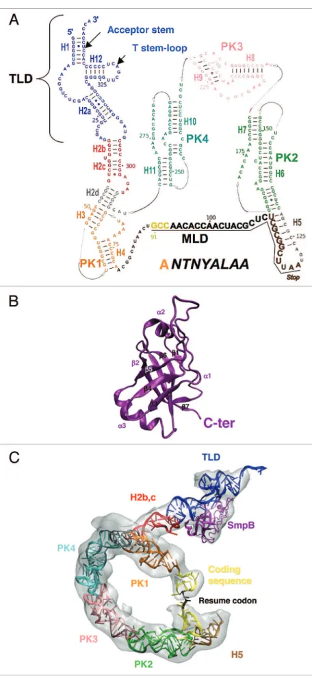
Figure 1. tmrNA and SmpB secondary and tertiary structures. (A) Secondary structure of tmrNA from Ther-mus thermophilus, with emphasis to its structural do-mains (helices H1-H12 and pseudoknots PK1-PK4). The TLD is the trNA domain, the MLD is the mrNA domain and the sequence of the peptide tag is indicated. TLD, blue; H2b-c, red; H2d, gray; PK1, orange; single-strand between PK1 and PK2, black with the resume codon highlighted in yellow; H5, brown; PK2, green; PK3, pink; PK4, light blue. (B) The structure of SmpB contains an oligonucleotide binding fold (OB-fold) made of an antiparallel β-barrel structure with three helices packed outside the core of the barrel. in this respect, SmpB is related to several other proteins associated with the translational apparatus. (C) Three-dimensional view of tmrNA and SmpB derived from the cryo-eM map of the accommodated state (the electronic density is in gray). The color code is as in (A and B).
ribosomes seemed therefore to be the missing link explaining the way trans-translation occurs on full-length mRNAs. However, a few months after this striking discovery, it was shown that the A-site specific cleavage into paused ribosomes could also occur in the absence of RelE or other bacterial toxins, and do not require tmRNA, SmpB, ribonucleases R, E, G and III or (p)ppGpp.1,26
Despite extensive studies, this novel endonucleolytic activity has not yet been attributed, suggesting that the ribosome itself might participate to the process, in a way that is still unknown. Last but not least, trans-translation can, in some cases, be triggered without a previous endonucleolytic activity cleaving the mRNA into the A-site. Indeed, the translation of bacterial mRNA begins while the nascent transcript is being synthesized and the active RNA polymerase partly protects the mRNA downstream the working ribosomes. When translation pauses, the ongoing RNA polymerase synthesizing the mRNA keeps moving and part of the mRNA extending downstream the leading edge of the ribo-some becomes exposed to the cellular endonucleases and to 3'–5' exonucleases. This way, only the nucleotides between the head and the shoulder of the small subunit are maintained protected against rapid degradation. Between the P-site and the 3' edge, this region corresponds to ~15 nucleotides.35 Accordingly,
ribo-somes stalled at mRNAs are targets for trans-translation only if the extension does not exceed 15 nucleotides downstream the A-site. Over this length, the rates of trans-peptidation decrease up to zero.36 Therefore, in this “edge model,” the ‘tmRNA-SmpB’
complex competes with the nucleotides remaining inserted into the mRNA path of the ribosome that may be transiently detached from the A-site because of weak interactions with the mRNA channel.37 Altogether, these situations lead to high frequency of
trans-translation, of about 1 in 250 translation events while ~700 tmRNA are present per cell, corresponding to one tmRNA for 10 to 20 ribosomes.38 This prevalence of trans-translation is not
increased in cells overexpressing tmRNA and SmpB, arguing for a tightly regulated entry of the ‘tmRNA-SmpB’ complex into the A-sites of the stalled ribosomes.
Signatures of ribosome stalling; SmpB interactions with the ribosomal active sites. Finally, despite the wide range of situa-tions described above, ribosome pausing or idling is the common rule governing the process of trans-translation. Because of the strong competition between trans-translation and termination or elongation (more particularly in the “edge model” situation), an accurate identification of these problematic ribosomes by the ‘tmRNA-SmpB’ complex is necessary and needs very specific signatures to discriminate them from the active ones. A vacant A-site is the most obvious signal making a ribosome a good sub-strate for SmpB and therefore ala-tmRNA. So how to detect ribo-somes stalled at the 3' edge and still carrying a codon into the A-site? When the ribosomes stall at or very near the 3'-end of the mRNA it has been suggested that the opening of the mRNA channel may facilitate the subsequent identification and engage-ment of ‘tmRNA-SmpB’, by providing a positive mechanism of identification.1 Although this situation can occur on empty A-site
ribosomes, it is unlikely that ribosomes will distinguish by them-selves long mRNAs from the short ones paused at the 3'edge. Therefore we anticipate that in the “edge model,” opening the quaternary complex made of ‘ala-tmRNA-SmpB-EF-Tu·GTP’
indispensable to trigger trans-translation or does the pre-binding of SmpB pave the way for ala-tmRNA to engage? By monitor-ing the cellular location and expression of endogenous SmpB, we previously reported that it is associated with 70S ribosomes and that this pre-bound SmpB can trigger the recruitment of SmpB-free tmRNA and initiate trans-translation in vitro.18 On the other
hand, the intracellular concentrations of tmRNA and SmpB being roughly similar,20 the formation of a 1:1 complex between
them would be in contradiction with the pre-binding of SmpB to the stalled ribosomes. Interaction of free SmpB with ribosomes is salt sensitive in vivo and therefore could be dependent upon the low stringency conditions used in the purification buffers, while its high binding affinity to tmRNA is unquestionable.21
To our point of view, because SmpB was found in vivo bound to the ribosome and because the intracellular ionic environment can vary, we cannot rule out that this original route for initiating trans-translation might be used, at least under specific cellular conditions.18 In any case, SmpB acts as a cellular sentinel onto the
stalled ribosomes pinpointing those to be rescued.
Stalled ribosomes detection and scanning for an empty A-site. Trans-translation was initially shown to occur on ribosomes stalled at the very 3'-end of incomplete mRNAs lacking a stop codon and therefore carrying an almost or totally empty decod-ing site.2 These non stop mRNAs can result from (1) mutations
causing the lack of in-frame termination codons (2) a prema-ture termination of transcription before the termination codon is reached22 (3) mRNA cleavages by nucleases.23 This situation
occurs also when the stop codon is bypassed by unwanted trans-lational read-through caused by nonsense suppressor tRNAs,24
by the presence of miscoding antibiotics25 or by aberrant
frame-shifts.26 More intriguingly, trans-translation can also be triggered
on intact full-length mRNAs when some internal sites are paused into the ribosomal A-site. These sites can be (1) clusters of rare codons,27 (2) weak termination codons.28-30 They can also be the
result of a flawed co-translational process, such as wrong pro-tein folding or secretion; then, trans-translation is necessary to relieve the subsequent translational arrests, whatever the codon context.3 The mechanism by which trans-translation is activated
on these full-length mRNAs has been elusive until the discovery of the role played by the bacterial toxin RelE in inhibiting protein synthesis under nutrient deprivation conditions. Indeed, during bacterial amino acid starvation, ribosomes are stalled by the binding of deacylated tRNAs to their A sites. The stringent fac-tor RelA then binds to blocked ribosomes and catalyzes synthesis of (p)ppGpp, a secondary messenger that induces the stringent response.31 This situation is cooperating-competing with RelBE
and tmRNA.32 RelE is part of the relBE toxin/antitoxin system
in which it plays the role of the toxin while RelB is the unsta-ble antidote bound to the latter. The concentration in RelB is decreased by the arrest of protein synthesis, leading to the deliv-ery of the stable RelE toxin that in turns cleaves mRNA codons between the second and third nucleotides in the A site of translat-ing ribosomes.33,34 This situation leads to non-stop mRNAs, the
typical substrates of trans-translation (see above). A-site mRNA cleavage by bacterial toxins during the pausing of translating
the tail, which is disordered in solution, forms an α-helix that interacts with the mRNA path downstream from the A-site when entering the ribosome.41,49 This interaction occurs at
pre-accom-modation and it triggers the accompre-accom-modation of tmRNA into the A-site after GTP hydrolysis by EF-Tu, compensating for the lack of a codon during the entering of tmRNA while the upper half of tRNA is mimicked by the tRNA domain (TLD).50 The
position-ing of SmpB into the decodposition-ing site is consistent with the crys-tal structures of the protein bound to the TLD.12,13 Overall, the
‘RNA-protein’ complex mimics the L-shaped conformation of a canonical tRNA. tmRNA terminus corresponds to the acceptor and T arms of the upper part of a tRNA while the positioning of SmpB mimics the anticodon and D stem-loops. The super-imposition of these structures on the A-site of a 70S ribosome orientates the C-terminal domain of SmpB to the decoding site while the β7 strand corresponds to the anticodon loop. These observations confirm the structural mimicry of canonical tRNAs by a ‘SmpB-tmRNA’ complex while the H2 linker helix mimics a tRNA variable arm (Fig. 1C).
Trans-translation dynamics under the eye of a microscope.
Ribosome recognition: the pre-accommodation step. The first views of a complete ‘tmRNA-SmpB’ complex loaded onto a stalled ribosome were determined by cryo-electron microscopy (cryo-EM, Fig. 2). The pre-accommodation step was captured by using kirromycin antibiotic that allows GTP hydrolysis by EF-Tu but stalls the ‘EF-Tu•GDP’ complex with ‘tmRNA-SmpB’ on the ribosome before accommodation into the peptidyl-transferase site. In an initial model, the TLD was placed into the density to interact with the GTPase-associated center (GAC) in the 50S subunit and with protein S12 in the 30S subunit, in a way simi-lar to that of tRNA during the elongation cycle. In this model, SmpB interacts with the elbow and the lower portion of TLD mRNA channel may rather be the task of SmpB competing with
the remaining nucleotides to pave the way for tmRNA to enter. The SmpB protein has a β-barrel core structure and a C-terminal tail that gains structure within the ribosome and its deletion abolishes tagging.39 Based on cryo-EM reconstructions40
and directed hydroxyl radical probing,41 into the A-site, SmpB
mimics a ‘codon-anticodon’ pairing. Accordingly, chemical prob-ing assays and NMR42 have shown that SmpB protects nucleotides
G530, A1492 and A1493 of the 16S rRNA from being modified. These key conserved nucleotides from the decoding site of the small subunit undergo substantial rearrangements in response to the pairing of cognate codons and anticodons,43 binding of IF1,44
binding of antibiotics43 or, to a less extent, recognition of a stop
codon by release factors RF1 or RF2.45-47 However, in spite of the
observed reactivity changes, mutations at these positions do not reduce SmpB binding to the decoding site42 or reduce the rate of
peptidyl-transfer onto tmRNA (A. Buskirk, personal communi-cation). Therefore, other contacts between SmpB and the decod-ing site that are different from those induced durdecod-ing translation elongation, are likely to take place with the surrounding nucleo-tides of the 16S RNA since positively charged SmpB has potential for interacting with negatively charged rRNA. X-ray structures will be necessary to provide detailed insight into the loading of the ‘tmRNA-SmpB’ complex within a stalled ribosome.
The C-terminal tail of SmpB plays a crucial role during trans-translation, which led to an extensive questioning about its func-tion during the early steps of the process. First of all it does not contribute to the binding of the protein to the ribosome, nor to the GTP hydrolysis by EF-Tu as demonstrated by testing various mutated or tail-truncated proteins.10,39,48,49 Therefore, its function
is crucial for the events that are after the initial association with the ribosome but before transpeptidation.39 It is suggested that
Figure 2. Cryo-eM maps of the currently solved functional complexes. Stalled ribosomes (a), pre-accommodation of the ‘tmrNA-SmpB’ complex (b), accommodation (c) and translocation (d). The density attributable to ‘tmrNA-SmpB’ is in red, the 50S subunit is blue, the 30S subunit is yellow, and the P-site and e-site trNAs are depicted in green and orange, respectively, and the problematic mrNA is purple. The semi-transparent ribosomal subunits emphasize the relative positions of the ‘tmrNA-SmpB’ complex, P-site and e-site trNAs in the 3 active sites of the ribosome. For clarity, each of the 4 steps was schematized below the cryo-eM structures.
Swinging toward the accommodation step. Structural infor-mation about the post-accommodated state of the ‘tmRNA-SmpB’ complex comes from cryo-EM studies. In a first study using a truncated version of tmRNA (named PKF for pseu-doknot-free tmRNA) we showed that accommodation leads to the disappearance of SmpB-1 from the large subunit while SmpB-2 remains bound into the decoding site.58 The signal
triggering SmpB-1 departure is explained by the steric clash that the protein would provoke with the P-site tRNA after the release of EF-Tu·GDP and swinging of the TLD into the A-site. At the same time, an independent study by Lindahl’s group has reported the accommodation step in the presence of full-length tmRNA.59 Despite the high heterogeneity of the
samples (as we previously observed in refs. 52 and 58) and the lack of deacylated tRNA into the P-site, the structure of the complex at 15 Å resolution confirms the absence of SmpB-1 and the movements of the ‘TLD-SmpB’ complex as do native aminoacyl-tRNAs during canonical translation. It also reveals that the large ‘arc-shaped’ density made by the pseudoknots and internal ORF of tmRNA remains folded and highly struc-tured around the beak of the 30S subunit (Fig. 2c). We recently revisited ‘tmRNA-SmpB’ accommodation in a novel structure refined at 13 Å resolution.60 From the original cryo-EM
den-sity map we constructed an atomic model that was optimized by ‘molecular dynamic flexible fitting’. As expected, the TLD contacts with the large ribosomal subunit resemble those of the accommodated canonical tRNAs while the lack of a D-stem is compensated for by SmpB. This positioning is instrumental in realigning H2 (the helix connecting the TLD to the arc-like rest of tmRNA) toward the large subunit, in which it makes extended contacts with protein L11. Compared with the pre-accommodated step, SmpB follows the swing of the TLD by a slight rotation of about 30° but stays at the same place into the decoding site, still mimicking an anticodon stem-loop. The ring of pseudoknots does not undergo a large movement and still wraps around the beak of the small subunit.
P-site translocation: tRNA anticodon mimicry and tmRNA frame selection. After transpeptidation, the TLD has to move to the P-site while the internal open reading frame of tmRNA engages into the mRNA path. Until recently, the way such a huge molecule (six times larger than a tRNA when in complex with SmpB), with stable secondary and tertiary structure moves trough the ribosome was still a central question for understand-ing the mechanism of tmRNA function.61 Recently, our group
and that of J. Frank addressed the conformational changes in the ‘tmRNA-SmpB’ complex when it moves through the ribo-some (Fig. 2d), especially those required for the transition from the A- to the P-site.60,62 The translocation step implies dynamical
motions of tmRNA-SmpB but also of the ribosome to (a) allow the complex to cross the hurdles between the A and P-sites (b) swap the mRNA templates (c) select the correct resume codon and maintain the reading frame on tmRNA.
Crossing the hurdles. During ribosomal translocation, ribo-somal subunits need to move relative to each other, underscor-ing the dynamic nature of the ribosome.63 This movement leads
to a ratchet-like rotation of the 30S subunit relative to the 50S and bridges the latter to the 50S subunit. tmRNA helix 2 is
redi-rected along the 30S subunit beak, where pseudoknots PK1 to PK4 form an arc that direct the internal open reading frame close to the entrance of the mRNA channel in the 30S subunit.51 In a
second map with improved resolution (Fig. 2b) and thanks to the docking of the crystal structure of the tRNA domain of tmRNA in complex to SmpB12 we established the occurrence of 2 SmpB
molecules in the complex, one interacting with the 50S subunit at the GTPase-associated center (GAC) near the site where it was previously found (SmpB-1), the other (SmpB-2) to the 30S subunit close to the decoding site.52 This result agrees with the
distribution of the protections induced by the SmpB protein onto tmRNA as well as with biochemical data suggesting that the pro-tein binds to the opposite sites of helix H2 from tmRNA.19,53
Why needing a second SmpB? During standard translation elongation, codon recognition leads to a series of conformational changes that position EF-Tu for GTP hydrolysis.54 Among these
changes, a domain closure of the 30S subunit occurs together with distortions of the tRNA backbone within the anticodon and D stems that are required to simultaneously bind the mRNA codon and EF-Tu.55 The interactions of EF-Tu with the distorted
tRNA and the ribosome activate GTP hydrolysis and the subse-quent dissociation of the protein triggers the accommodation of the tRNA into the A-site. At this step, “proofreading” allows the disengagement of tRNAs interacting weakly (i.e., near-cognate tRNAs) with the decoding center.56 SmpB compensates for the
lack of ‘codon-anticodon’ recognition, therefore being essential during the initial selection of the stalled ribosomes to rescue. “Non-cognate” proofreading is probably unnecessary in this step. An hypothesis would be that SmpB-1 interacting with the 50S forms close contacts with the GAC including the nucleo-tides from the 23S rRNA which interact with the D loop of tRNAs during canonical translation. Thus, this interaction may be required to transfer the “decoding” signal to EF-Tu and to facilitate subsequent GTPase (Guanosine TriPhosphatase) acti-vation.52 The simultaneous binding of 2 SmpB proteins during
pre-accommodation is controversial, mainly because of the ‘1:1’ ratio of SmpB and tmRNA measured within the cells.20 However,
if we consider that SmpB-1 leaves the stalled ribosome as soon as the TLD accommodates into the A-site (see below), the presence of 2 molecules of SmpB during pre-accommodation is compat-ible with the estimated ‘1:1’ ratio of the ‘tmRNA-SmpB’ complex in vivo. Indeed, trans-translation is a multi-step process that can be roughly divided into three main phases: (1) pre-accommoda-tion and accommodapre-accommoda-tion of the TLD into a vacant A-site, (2) template swapping and selection of the reading frame on tmRNA (3) elongation on the 9–35 (depending on the species) internally encoded codons of tmRNA open reading frame and termination. The presence of a ‘2:1’ SmpB/tmRNA ratio during the acceler-ated pre-accommodacceler-ated state is insignificant compared with the remaining steps of the process for which only one SmpB accom-panies tmRNA during its transit through the stalled ribosomes,57
including accommodation, elongation onto tmRNA ORF and termination. Further structural studies will be required to pro-vide a clearer answer to that controversy regarding the number and dynamics of SmpB molecules during pre-accommodation.
the ratchet, the axis of rotation has been located in the vicinity of the central bridge 3 and conse-quently, only the bridges located at the extremities of the 2 subunits (B1a-b; B7b and B8) are disrupted or rearranged during rotation.67 Among them, we
have shown that following ratcheting, disruption of the A-site finger or ASF, part of bridge B1a, allows the large ‘tmRNA-SmpB’ complex to make its way through the narrow pathway of the canonical tRNAs.60 Indeed, this disruption favors the
pas-sage of H2 to the other side of the bridge (Fig. 3). As a consequence, pseudoknot PK1 moves at the entrance of the ASF gate, where H2 was previously positioned, while the 3'-end of PK4 also follows the dynamics of H2. On the other hand, the 5'-end of PK4, together with H5/PK2/PK3 stay immobile, triggering the stretch of the arc-shaped ring of PKs and the unfolding of the internal ORF.
mRNA swapping. According to the general process of trans-translation, the stalled ribosome has to rapidly switch RNA templates to set tmRNA in the mRNA mode. During stalling, the truncated mRNA is stabi-lized into its path mainly through ‘codon:anticodon’ interaction with the peptidyl-tRNA. ‘tmRNA-SmpB’ recruitment is followed by rapid trans-peptidation that destabilizes the P site-bound tRNA, which in turn dissociates the mRNA from the ribosome.68 This
early release of the stalled mRNA has been recently confirmed by cryo-EM. Indeed, the translocation of tmRNA-SmpB to the P-site is accompanied by the disappearance of an extra-density recovered between the head and the back of the small subunit platform, where the Shine-Dalgarno interaction between the stalled mRNA and the 16S RNA occurs.60 Once it is
released, the flawed mRNA has to be rapidly degraded to avoid a new round of flawed translation to start. Noteworthy, trans-translation promotes the degrada-tion of non-stop mRNAs that cause ribosome stall-ing.23 Among all the RNases potentially involved,
the 3' to 5' exoRiboNuclease R is the key enzyme in tmRNA-dependent non-stop mRNA decay, in a pro-cess that requires active trans-translation of the defec-tive mRNA.69
Resume codon selection within tmRNA. As the tmRNA-SmpB complex transits to the P-site, the ribosome has to select the correct reading frame on tmRNA. A wealth of biochemical and genetic data have sug-gested that interactions between SmpB and key nucleotides lying upstream of the resume codon are instrumental in set-ting the new frame.21,70-74 At this step, the position of SmpB
has been mapped by the sites of cleavages induced by hydroxyl radical probing of Fe(II) tethered SmpB mutants. Contrary to the A-site position that protruded into the mRNA path toward the downstream tunnel, they localize almost exclusively around the region of the P-site canonical codon-anticodon interac-tion.41 Strikingly, this localization allows SmpB to have direct
subunit in the direction of the mRNA movement.64 Ratcheting
involves a reversible ~8° inter-subunit rotation and a nearly orthogonal rotation of the head domain of the small subunit.65
The interface between the two subunits rearranges along the ratcheting pathway and imposes stringent limitations on the accessible pathways for tRNAs. This rotation triggers inde-pendent movement of the two extremities of tRNAs, leading to the A/P and P/E hybrid states, necessary for the subsequent translocation of tRNAs. The two subunits of the ribosome are linked together by at least 12 inter-subunit bridges.66 During
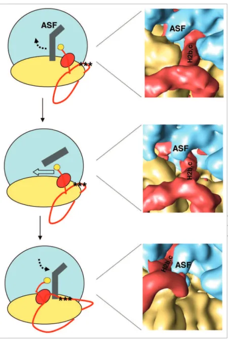
Figure 3. A-site inger (ASF, in gray) motion (see ref. 81 for more details) as the ‘tmrNA-SmpB’ complex (red) translocates to the P site. (Left) Schematic views of how the ‘tmrNA-SmpB’ complex crosses the ASF before, during and after translocation. (Close-up) Cryo-eM maps of stalled 70S ribosomes with ‘tmrNA-SmpB’ accommo-dated (top), the 70S ribosome in the ratcheted state prior to ‘tmrNA-SmpB’ transloca-tion (middle), and the 70S ribosome with ‘tmrNA-SmpB’ translocated into the P site (bottom). During translation, the movement of the ASF is illustrated by a broken arrow and the position of the resume codon corresponds to the 3 black stars.
substantiated between trans-translation and virulence. In spe-cific infections such as gonorrhoea, bacteria cannot survive without trans-translation. Also, trans-translation can contribute to cell viability in the presence of protein synthesis inhibitors,79
because these molecules induce miscoding events or extended stalling during translation. The ‘tmRNA-SmpB’ duplex allows bacteria to recycle the ribosomes stalled by the antibiotics, allowing reusing them onto intact messages, therefore increas-ing cell viability. Trans-translation also impacts the activity of antibacterial drugs that inhibit cell wall synthesis, probably because these drugs induce an overall stress to the bacteria that will be suppressed more efficiently when trans-translation is active.80 Altogether, these data suggest that either tmRNA,
SmpB or the ‘tmRNA-SmpB’ complex, all structures known at the atomic levels, are promising targets for developing novel antibiotics. Molecules that could bind tmRNA, SmpB or both might interfere with their loading onto the ribosomes to res-cue, potentiating the action of the existing drugs and allowing decreasing their active concentrations to limit their side effects. Since trans-translation is missing in the eukaryotes, these drugs should have reduced side effects on the cells and metabolisms of the patient.
Acknowledgments
We thank E. Giudice and F. Weis for generously helping pre-paring the figures used here, K.H. Nierhaus for suggestions on the stringent response and the members of R.G. and B.F. labo-ratories for many stimulating discussions and criticisms. These contacts with the area comprising the 5 nucleotides upstream of
the resume codon (Fig. 4), which precisely places the latter at the ribosomal A-site.60,62
E-site translocation and ribosome exit. The model presented
here lends support to a mechanism in which the next transition of tmRNA-SmpB from the P- to the E-site would follow the same dynamics (Fig. 5). In line with the first translocation, the movement of the TLD to the E-site is likely to drag H2 across bridge B1b, made by the flexible interactions between L5 and S13 64,75 and the crossing of the following module made of PK1
over bridge B1a. Accordingly, an isolated tmRNA-ribosomal complex blocked with tmRNA in the E-site was recently ana-lyzed using chemical probing. The model resulting from this study suggests a positioning of PK1 and H2 nearby the E-site.76
The dynamics of the other modules are more elusive and mainly depend on when helix H5 will come out of the ribosome and unwound to be correctly translated into the mRNA path.
Concluding Remarks
‘tmRNA-SmpB’ movements in the context of the polysomes.
During translation, a cluster of ribosomes bind to a single mRNA, giving birth to an active polyribosome or polysome. When the first ribosome stalls at the 3'-end of a no-go mRNA, it leads to a traffic jam of all the following polysome. Therefore, after salvage of this first ribosome by tmRNA-SmpB, the poly-some moves up on the flawed mRNA that must not be sub-jected to re-initiation at the risk of leading to a translation/ trans-translation vicious circle. Therefore, a competition occurs between the polysome moving forward on the mRNA and deg-radation by RNase R. Another possibility is that the 5'-end of the flawed mRNA is degraded as trans-translation proceeds, preventing re-initiation. Further analysis of stalled polysomes will be needed to answer this question.
Alternative ways of resolving stalled ribosomes.
Trans-translation is not essential in most bacteria, suggesting that there are alternative roads for rescuing the ribosomes that stall on problematic mRNAs. One possibility might be peptidyl-tRNA drop-off promoted by translation factors EF-G, RF3 and RRF, with subsequent hydrolysis of the tRNA by the peptidyl-tRNA hydrolase (Pth). Overexpressing RF3, RRF and the Pth, however, does not stimulate peptidyl-tRNA production,77
argu-ing against the peptidyl-tRNA drop-off hypothesis. Recently, YhdL (or ArfA for alternative ribosome-rescue factor) was iden-tified as an essential protein for E. coli viability in the absence of tmRNA, ArfA taking over the role of the ‘tmRNA-SmpB’ complex.78 Interestingly, ArfA lacks the catalytic residues of the
peptide release factors, therefore requiring an additional ligand able to hydrolyze the bond between the tRNA and the nascent peptide, possibly the Pth. In addition to, the ArfA system may be required during different physiological conditions than trans-translation or, alternatively, when the ‘tmRNA-SmpB’ quality-control mechanism is outreached.
Trans-translation as an antimicrobial drug target. In some
bacteria (e.g., S. typhimurium, Y. pseudotuberculosis) respon-sible of human and/or animal diseases, functional links were
Figure 4. Cryo-eM structure of a translocated ‘tmrNA-SmpB’ complex at 13.5 Å resolution (weis et al. 2010), highlighting the contacts be-tween SmpB and the conserved nucleotides upstream the resume co-don. The electron density corresponding to the ‘tmrNA-SmpB’ complex is in gray and the model reconstructed and itted within the electronic density follows the ribose-phosphate backbone of the rNA and the rib-bon backrib-bone of the protein. The color code is as in Figure 1.
Figure 5. working model of the mechanism of trans-translation based on a wealth of genetic, biochemical and structural data collected over the past 15 y. (a) ribosomes stalled on a problematic message, e.g., a truncated mrNA (blue, with a black scissor), contain an e-site trNA (orange) and a peptidyl-tRNA within the P site (green). Alanylated (yellow marble) tmRNA in complex with EF-Tu•GTP (orange circle) and with SmpB (pink) recognize an empty or partially empty A site, with SmpB mimicking a trNA anticodon branch contacting the small ribosomal subunit. (b) During pre-accommo-dation, a second, transient, SmpB molecule contacts the GTPase center of the large ribosomal subunit, presumably facilitating GTP hydrolysis after binding of the irst SmpB into the decoding site. (c) Accommodation occurs as for canonical trNAs during translation, together with the departure of the second SmpB molecule. (d) The incomplete peptide is transferred from the P site trNA to the alanine located at tmrNA 3'-end. (e) The problematic mrNA is ejected and rapidly degraded by rNases (rNA quality-control system) and ‘tmrNA-SmpB’ translocation from the A to the P site takes place. (f) tmrNA internal resume codon is placed at the ribosomal decoding site with the help of SmpB that provides speciic contacts with nucleotides up-stream of the tag-encoding sequence. (g) Translation of tmrNA internal reading frame continues and ends at a canonical termination codon, releasing the aborted nascent protein for destruction by cellular proteases. (h) The stalled ribosomes are recycled back to new rounds of translation onto intact mrNAs. The remaining color code is as in Figure 2.
‘ANR-09-MIE’ to R.G. and B.F. and from additional funds from the Brittany region and from the Inserm to B.F.
studies were supported by grants from the ‘Agence Nationale pour la Recherche’: the ‘ANR-08JCJC-0027-01’ to R.G., and the
References
1. Moore SD, Sauer RT. The tmRNA system for trans-lational surveillance and ribosome rescue. Annu Rev Biochem 2007; 76:101-24; DOI: 10.1146/annurev. biochem.75.103004.142733.
2. Keiler KC, Waller PR, Sauer RT. Role of a peptide tag-ging system in degradation of proteins synthesized from damaged messenger RNA. Science 1996; 271:990-3; DOI: 10.1126/science.271.5251.990.
3. Hayes CS, Keiler KC. Beyond ribosome rescue: tmRNA and co-translational processes. FEBS Lett 2010; 584:413-9; DOI: 10.1016/j.febslet.2009.11.023. 4. Felden B, Hanawa K, Atkins JF, Himeno H, Muto A,
Gesteland RF, et al. Presence and location of modi-fied nucleotides in Escherichia coli tmRNA: structural mimicry with tRNA acceptor branches. EMBO J 1998; 17:3188-96; DOI: 10.1093/emboj/17.11.3188. 5. Felden B, Himeno H, Muto A, Atkins JF, Gesteland
RF. Structural organization of Escherichia coli tmRNA. Biochimie 1996; 78:979-83; DOI: 10.1016/S0300-9084(97)86720-X.
6. Williams KP, Bartel DP. Phylogenetic analysis of tmRNA secondary structure. RNA 1996; 2:1306-10; PubMed.
7. Shimizu Y, Ueda T. The role of SmpB protein in trans-translation. FEBS Lett 2002; 514:74-7; DOI: 10.1016/ S0014-5793(02)02333-5.
8. Hanawa-Suetsugu K, Takagi M, Inokuchi H, Himeno H, Muto A. SmpB functions in various steps of trans-translation. Nucleic Acids Res 2002; 30:1620-9; DOI: 10.1093/nar/30.7.1620.
9. Karzai AW, Susskind MM, Sauer RT. SmpB, a unique RNA-binding protein essential for the peptide-tagging activity of SsrA (tmRNA). EMBO J 1999; 18:3793-9; DOI: 10.1093/emboj/18.13.3793.
10. Hallier M, Desreac J, Felden B. Small protein B inter-acts with the large and the small subunits of a stalled ribosome during trans-translation. Nucleic Acids Res 2006; 34:1935-43; DOI: 10.1093/nar/gkl097. 11. Rudinger-Thirion J, Giegé R, Felden B. Aminoacylated
tmRNA from Escherichia coli interacts with prokaryotic elongation factor Tu. RNA 1999; 5:989-92; DOI: 10.1017/S135583829999101X.
12. Gutmann S, Haebel PW, Metzinger L, Sutter M, Felden B, Ban N. Crystal structure of the transfer-RNA domain of transfer-messenger RNA in complex with SmpB. Nature 2003; 424:699-703; DOI: 10.1038/ nature01831.
13. Bessho Y, Shibata R, Sekine S, Murayama K, Higashijima K, Hori-Takemoto C, et al. Structural basis for functional mimicry of long-variable-arm tRNA by transfer-messenger RNA. Proc Natl Acad Sci USA 2007; 104:8293-8; DOI: 10.1073/pnas.0700402104. 14. Barends S, Karzai AW, Sauer RT, Wower J, Kraal B.
Simultaneous and functional binding of SmpB and EF-Tu-TP to the alanyl acceptor arm of tmRNA. J Mol Biol 2001; 314:9-21; DOI: 10.1006/jmbi.2001.5114. 15. Dong G, Nowakowski J, Hoffman DW. Structure of
small protein B: the protein component of the tmRNA-SmpB system for ribosome rescue. EMBO J 2002; 21:1845-54; DOI: 10.1093/emboj/21.7.1845. 16. Someya T, Nameki N, Hosoi H, Suzuki S, Hatanaka
H, Fujii M, et al. Solution structure of a tmRNA-binding protein, SmpB, from Thermus thermophilus. FEBS Lett 2003; 535:94-100; DOI: 10.1016/S0014-5793(02)03880-2.
17. Nameki N, Someya T, Okano S, Suemasa R, Kimoto M, Hanawa-Suetsugu K, et al. Interaction analysis between tmRNA and SmpB from Thermus
thermophi-lus. J Biochem 2005; 138:729-39; DOI: 10.1093/jb/
mvi180.
18. Hallier M, Ivanova N, Rametti A, Pavlov M, Ehrenberg M, Felden B. Pre-binding of small protein B to a stalled ribosome triggers trans-translation. J Biol Chem 2004; 279:25978-85; DOI: 10.1074/jbc.M314086200. 19. Ivanova N, Pavlov MY, Bouakaz E, Ehrenberg M,
Schiavone LH. Mapping the interaction of SmpB with ribosomes by footprinting of ribosomal RNA. Nucleic Acids Res 2005; 33:3529-39; DOI: 10.1093/nar/ gki666.
20. Sundermeier TR, Karzai AW. Functional SmpB-ribosome interactions require tmRNA. J Biol Chem 2007; 282:34779-86; DOI: 10.1074/jbc. M707256200.
21. Metzinger L, Hallier M, Felden B. The highest affinity binding site of small protein B on transfer messen-ger RNA is outside the tRNA domain. RNA 2008; 14:1761-72; DOI: 10.1261/rna.1185808.
22. Abo T, Inada T, Ogawa K, Aiba H. SsrA-mediated tag-ging and proteolysis of LacI and its role in the regula-tion of lac operon. EMBO J 2000; 19:3762-9; DOI: 10.1093/emboj/19.14.3762.
23. Yamamoto Y, Sunohara T, Jojima K, Inada T, Aiba H. SsrA-mediated trans-translation plays a role in mRNA quality control by facilitating degradation of trun-cated mRNAs. RNA 2003; 9:408-18; DOI: 10.1261/ rna.2174803.
24. Ueda K, Yamamoto Y, Ogawa K, Abo T, Inokuchi H, Aiba H. Bacterial SsrA system plays a role in coping with unwanted translational readthrough caused by suppressor tRNAs. Genes Cells 2002; 7:509-19; DOI: 10.1046/j.1365-2443.2002.00537.x.
25. Abo T, Ueda K, Sunohara T, Ogawa K, Aiba H. SsrA-mediated protein tagging in the presence of miscoding drugs and its physiological role in Escherichia coli. Genes Cells 2002; 7:629-38; DOI: 10.1046/j.1365-2443.2002.00549.x.
26. Hayes CS, Sauer RT. Cleavage of the A site mRNA codon during ribosome pausing provides a mecha-nism for translational quality control. Mol Cell 2003; 12:903-11; DOI: 10.1016/S1097-2765(03)00385-X. 27. Roche ED, Sauer RT. SsrA-mediated peptide tagging
caused by rare codons and tRNA scarcity. EMBO J 1999; 18:4579-89; DOI: 10.1093/emboj/18.16.4579. 28. Collier J, Binet E, Bouloc P. Competition between
SsrA tagging and translational termination at weak stop codons in Escherichia coli. Mol Microbiol 2002; 45:745-54; DOI: 10.1046/j.1365-2958.2002.03045.x. 29. Hayes CS, Bose B, Sauer RT. Stop codons preceded by
rare arginine codons are efficient determinants of SsrA tagging in Escherichia coli. Proc Natl Acad Sci USA 2002; 99:3440-5; DOI: 10.1073/pnas.052707199. 30. Sunohara T, Jojima K, Yamamoto Y, Inada T, Aiba H.
Nascent-peptide-mediated ribosome stalling at a stop codon induces mRNA cleavage resulting in nonstop mRNA that is recognized by tmRNA RNA 2004; 10:378-86.
31. Wendrich TM, Blaha G, Wilson DN, Marahiel MA, Nierhaus KH. Dissection of the mechanism for the stringent factor RelA. Mol Cell 2002; 10:779-88; DOI: 10.1016/S1097-2765(02)00656-1.
32. Wilson DN, Nierhaus KH. RelBE or not to be. Nat Struct Mol Biol 2005; 12:282-4; DOI: 10.1038/ nsmb0405-282.
33. Pedersen K, Zavialov AV, Pavlov MY, Elf J, Gerdes K, Ehrenberg M. The bacterial toxin RelE displays codon-specific cleavage of mRNAs in the ribosomal A site. Cell 2003; 112:131-40; DOI: 10.1016/S0092-8674(02)01248-5.
34. Neubauer C, Gao YG, Andersen KR, Dunham CM, Kelley AC, Hentschel J, et al. The structural basis for mRNA recognition and cleavage by the ribosome-dependent endonuclease RelE. Cell 2009; 139:1084-95; DOI: 10.1016/j.cell.2009.11.015.
35. Beyer D, Skripkin E, Wadzack J, Nierhaus KH. How the ribosome moves along the mRNA during protein synthesis. J Biol Chem 1994; 269:30713-7; PubMed. 36. Ivanova N, Pavlov MY, Felden B, Ehrenberg M.
Ribosome rescue by tmRNA requires truncated mRNAs. J Mol Biol 2004; 338:33-41; DOI: 10.1016/j. jmb.2004.02.043.
37. Asano K, Kurita D, Takada K, Konno T, Muto A, Himeno H. Competition between trans-translation and termination or elongation of translation. Nucleic Acids Res 2005; 33:5544-52; DOI: 10.1093/nar/ gki871.
38. Moore SD, Sauer RT. Ribosome rescue: tmRNA tagging activity and capacity in Escherichia coli. Mol Microbiol 2005; 58:456-66; DOI: 10.1111/j.1365-2958.2005.04832.x.
39. Sundermeier TR, Dulebohn DP, Cho HJ, Karzai AW. A previously uncharacterized role for small protein B (SmpB) in transfer messenger RNA-mediated trans-translation. Proc Natl Acad Sci USA 2005; 102:2316-21; DOI: 10.1073/pnas.0409694102.
40. Gillet R, Kaur S, Li W, Hallier M, Felden B, Frank J. Scaffolding as an organizing principle in trans-translation. The roles of small protein B and ribosomal protein S1. J Biol Chem 2007; 282:6356-63; DOI: 10.1074/jbc.M609658200.
41. Kurita D, Sasaki R, Muto A, Himeno H. Interaction of SmpB with ribosome from directed hydroxyl radical probing. Nucleic Acids Res 2007; 35:7248-55; DOI: 10.1093/nar/gkm677.
42. Nonin-Lecomte S, Germain-Amiot N, Gillet R, Hallier M, Ponchon L, Dardel F, et al. Ribosome hijack-ing: a role for small protein B during trans-transla-tion. EMBO Rep 2009; 10:160-5; DOI: 10.1038/ embor.2008.243.
43. Ogle JM, Brodersen DE, Clemons WM Jr, Tarry MJ, Carter AP, Ramakrishnan V. Recognition of cognate transfer RNA by the 30S ribosomal subunit. Science 2001; 292:897-902; DOI: 10.1126/science.1060612. 44. Carter AP, Clemons WM Jr, Brodersen DE,
Morgan-Warren RJ, Hartsch T, Wimberly BT, et al. Crystal structure of an initiation factor bound to the 30S ribosomal subunit. Science 2001; 291:498-501; DOI: 10.1126/science.1057766.
45. Weixlbaumer A, Jin H, Neubauer C, Voorhees RM, Petry S, Kelley AC, et al. Insights into translational termination from the structure of RF2 bound to the ribosome. Science 2008; 322:953-6; DOI: 10.1126/ science.1164840.
46. Laurberg M, Asahara H, Korostelev A, Zhu J, Trakhanov S, Noller HF. Structural basis for transla-tion terminatransla-tion on the 70S ribosome. Nature 2008; 454:852-7; DOI: 10.1038/nature07115.
47. Korostelev A, Asahara H, Lancaster L, Laurberg M, Hirschi A, Zhu J, et al. Crystal structure of a translation termination complex formed with release factor RF2. Proc Natl Acad Sci USA 2008; 105:19684-9; DOI: 10.1073/pnas.0810953105.
48. Jacob Y, Sharkady SM, Bhardwaj K, Sanda A, Williams KP. Function of the SmpB tail in transfer-messenger RNA translation revealed by a nucleus-encoded form. J Biol Chem 2005; 280:5503-9; DOI: 10.1074/jbc. M409277200.
49. Kurita D, Muto A, Himeno H. Role of the C-terminal tail of SmpB in the early stage of trans-translation. RNA 2010; 16:980-90; DOI: 10.1261/rna.1916610. 50. Kurita D, Muto A, Himeno H. tRNA/mRNA Mimicry
by tmRNA and SmpB in Trans-Translation. J Nucleic Acids 2011; 2011:130581.
51. Valle M, Gillet R, Kaur S, Henne A, Ramakrishnan V, Frank J. Visualizing tmRNA entry into a stalled ribosome. Science 2003; 300:127-30; DOI: 10.1126/ science.1081798.
73. Miller MR, Healey DW, Robison SG, Dewey JD, Buskirk AR. The role of upstream sequences in select-ing the readselect-ing frame on tmRNA. BMC Biol 2008; 6:29; DOI: 10.1186/1741-7007-6-29.
74. Watts T, Cazier D, Healey D, Buskirk A. SmpB con-tributes to reading frame selection in the translation of transfer-messenger RNA. J Mol Biol 2009; 391:275-81; DOI: 10.1016/j.jmb.2009.06.037.
75. Schuwirth BS, Borovinskaya MA, Hau CW, Zhang W, Vila-Sanjurjo A, Holton JM, et al. Structures of the bacterial ribosome at 3.5 A resolution. Science 2005; 310:827-34; DOI: 10.1126/science.1117230. 76. Bugaeva EY, Surkov S, Golovin AV, Ofverstedt LG,
Skoglund U, Isaksson LA, et al. Structural features of the tmRNA-ribosome interaction. RNA 2009; 15:2312-20; DOI: 10.1261/rna.1584209.
77. Janssen BD, Hayes CS. Kinetics of paused ribosome recycling in Escherichia coli. J Mol Biol 2009; 394:251-67; DOI: 10.1016/j.jmb.2009.09.020.
78. Chadani Y, Ono K, Ozawa S, Takahashi Y, Takai K, Nanamiya H, et al. Ribosome rescue by Escherichia
coli ArfA (YhdL) in the absence of trans-translation
system. Mol Microbiol 2010; 78:796-808; DOI: 10.1111/j.1365-2958.2010.07375.x.
79. Vioque A, de la Cruz J. Trans-translation and pro-tein synthesis inhibitors. FEMS Microbiol Lett 2003; 218:9-14; DOI: 10.1111/j.1574-6968.2003. tb11491.x.
80. Luidalepp H, Hallier M, Felden B, Tenson T. tmRNA decreases the bactericidal activity of aminoglycosides and the susceptibility to inhibitors of cell wall synthesis. RNA Biol 2005; 2:70-4; DOI: 10.4161/rna.2.2.2020. 81. Réblová K, Rázga F, Li W, Gao H, Frank J, Sponer
J. Dynamics of the base of ribosomal A-site finger revealed by molecular dynamics simulations and Cryo-EM. Nucleic Acids Res 2010; 38:1325-40; DOI: 10.1093/nar/gkp1057.
63. Horan LH, Noller HF. Intersubunit movement is required for ribosomal translocation. Proc Natl Acad Sci USA 2007; 104:4881-5; DOI: 10.1073/ pnas.0700762104.
64. Valle M, Zavialov A, Sengupta J, Rawat U, Ehrenberg M, Frank J. Locking and unlocking of ribosomal motions. Cell 2003; 114:123-34; DOI: 10.1016/ S0092-8674(03)00476-8.
65. Zhang W, Dunkle JA, Cate JH. Structures of the ribo-some in intermediate states of ratcheting. Science 2009; 325:1014-7; DOI: 10.1126/science.1175275. 66. Yusupov MM, Yusupova GZ, Baucom A, Lieberman
K, Earnest TN, Cate JH, et al. Crystal structure of the ribosome at 5.5 A resolution. Science 2001; 292:883-96; DOI: 10.1126/science.1060089.
67. Korostelev A, Ermolenko DN, Noller HF. Structural dynamics of the ribosome. Curr Opin Chem Biol 2008; 12:674-83; DOI: 10.1016/j.cbpa.2008.08.037. 68. Ivanova N, Pavlov MY, Ehrenberg M. tmRNA-induced
release of messenger RNA from stalled ribosomes. J Mol Biol 2005; 350:897-905; DOI: 10.1016/j. jmb.2005.05.033.
69. Richards J, Mehta P, Karzai AW. RNase R degrades non-stop mRNAs selectively in an SmpB-tmRNA-dependent manner. Mol Microbiol 2006; 62:1700-12; DOI: 10.1111/j.1365-2958.2006.05472.x.
70. Williams KP, Martindale KA, Bartel DP. Resuming translation on tmRNA: a unique mode of determining a reading frame. EMBO J 1999; 18:5423-33; DOI: 10.1093/emboj/18.19.5423.
71. Lee S, Ishii M, Tadaki T, Muto A, Himeno H. Determinants on tmRNA for initiating efficient and precise trans-translation: some mutations upstream of the tag-encoding sequence of Escherichia coli tmRNA shift the initiation point of trans-translation in vitro. RNA 2001; 7:999-1012; DOI: 10.1017/ S1355838201010342.
72. Konno T, Kurita D, Takada K, Muto A, Himeno H. A functional interaction of SmpB with tmRNA for deter-mination of the resuming point of trans-translation. RNA 2007; 13:1723-31; DOI: 10.1261/rna.604907. 52. Kaur S, Gillet R, Li W, Gursky R, Frank J. Cryo-EM
visualization of transfer messenger RNA with two SmpBs in a stalled ribosome. Proc Natl Acad Sci USA 2006; 103:16484-9; DOI: 10.1073/pnas.0607438103. 53. Wower J, Zwieb CW, Hoffman DW, Wower IK. SmpB: a protein that binds to double-stranded segments in tmRNA and tRNA. Biochemistry 2002; 41:8826-36; DOI: 10.1021/bi0201365.
54. Voorhees RM, Schmeing TM, Kelley AC, Ramakrishnan V. The mechanism for activation of GTP hydrolysis on the ribosome. Science 2010; 330:835-8; DOI: 10.1126/science.1194460. 55. Schmeing TM, Voorhees RM, Kelley AC, Gao YG,
Murphy FV, 4th, Weir JR, et al. The crystal structure of the ribosome bound to EF-Tu and aminoacyl-tRNA. Science 2009; 326:688-94; DOI: 10.1126/ science.1179700.
56. Gromadski KB, Rodnina MV. Kinetic determinants of high-fidelity tRNA discrimination on the ribosome. Mol Cell 2004; 13:191-200; DOI: 10.1016/S1097-2765(04)00005-X.
57. Bugaeva EY, Shpanchenko OV, Felden B, Isaksson LA, Dontsova OA. One SmpB molecule accompanies tmRNA during its passage through the ribosomes. FEBS Lett 2008; 582:1532-6; DOI: 10.1016/j.febs-let.2008.03.049.
58. Weis F, Bron P, Rolland JP, Thomas D, Felden B, Gillet R. Accommodation of tmRNA-SmpB into stalled ribosomes: a cryo-EM study. RNA 2010; 16:299-306; DOI: 10.1261/rna.1757410.
59. Cheng K, Ivanova N, Scheres SH, Pavlov MY, Carazo JM, Hebert H, et al. tmRNA.SmpB complex mimics native aminoacyl-tRNAs in the A site of stalled ribo-somes. J Struct Biol 2010; 169:342-8; DOI: 10.1016/j. jsb.2009.10.015.
60. Weis F, Bron P, Giudice E, Rolland JP, Thomas D, Felden B, et al. tmRNA-SmpB: a journey to the centre of the bacterial ribosome. EMBO J 2010; 29:3810-8; DOI: 10.1038/emboj.2010.252.
61. Ivanov PV, Zvereva MI, Shpanchenko OV, Dontsova OA, Bogdanov AA, Aglyamova GV, et al. How does tmRNA move through the ribosome? FEBS Lett 2002; 514:55-9; DOI: 10.1016/S0014-5793(02)02310-4. 62. Fu J, Hashem Y, Wower I, Lei J, Liao HY, Zwieb C, et
al. Visualizing the transfer-messenger RNA as the ribo-some resumes translation. EMBO J 2010; 29:3819-25; DOI: 10.1038/emboj.2010.255.