HAL Id: hal-00400776
https://hal.archives-ouvertes.fr/hal-00400776
Submitted on 23 Feb 2017
HAL is a multi-disciplinary open access
archive for the deposit and dissemination of
sci-entific research documents, whether they are
pub-lished or not. The documents may come from
teaching and research institutions in France or
abroad, or from public or private research centers.
L’archive ouverte pluridisciplinaire HAL, est
destinée au dépôt et à la diffusion de documents
scientifiques de niveau recherche, publiés ou non,
émanant des établissements d’enseignement et de
recherche français ou étrangers, des laboratoires
publics ou privés.
Squalamine: an appropriate strategy against the
emergence of multidrug resistant gram-negative
bacteria?
Chanaz Salmi, Celine Loncle, Nicolas Vidal, Yves Letourneux, Jacques
Fantini, Marc Maresca, Nadira Taïeb, Jean-Marie Pagès, Jean Michel Brunel
To cite this version:
Chanaz Salmi, Celine Loncle, Nicolas Vidal, Yves Letourneux, Jacques Fantini, et al.. Squalamine:
an appropriate strategy against the emergence of multidrug resistant gram-negative bacteria?. PLoS
ONE, Public Library of Science, 2008, 3 (7), pp.e2765. �10.1371/journal.pone.0002765�. �hal-00400776�
as polymyxin B. In this context and because of its structure, action and its relative insensitivity to efflux resistance mechanisms, we have demonstrated that squalamine appears as an alternate way to combat MDR pathogens and by pass the gap regarding the failure of new active antibacterial molecules.
Citation: Salmi C, Loncle C, Vidal N, Letourneux Y, Fantini J, et al. (2008) Squalamine: An Appropriate Strategy against the Emergence of Multidrug Resistant Gram-Negative Bacteria? PLoS ONE 3(7): e2765. doi:10.1371/journal.pone.0002765
Editor: Floyd Romesberg, The Scripps Research Institute, United States of America Received March 7, 2008; Accepted May 30, 2008; Published July 23, 2008
Copyright: ß 2008 Salmi et al. This is an open-access article distributed under the terms of the Creative Commons Attribution License, which permits unrestricted use, distribution, and reproduction in any medium, provided the original author and source are credited.
Funding: The authors have no support or funding to report.
Competing Interests: The authors have declared that no competing interests exist. * E-mail: [email protected]
Introduction
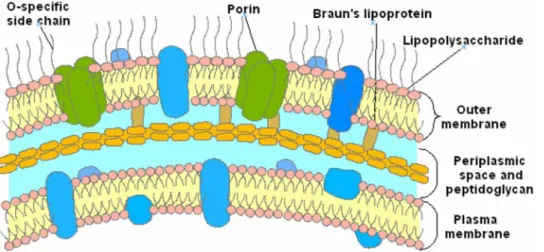
The emergence of multidrug resistant bacteria/pathogens has highlighted the need for the development of new antibiotics.[1–3] In this area, drug resistance of Gram-positive organisms has received significant attention with respect to Gram-negative bacteria which are innately resistant to many common antibiotics due to their envelope structure. In positive and Gram-negative bacteria, resistance to membrane active antibiotics requires major changes in membrane organization, which in turn influence the permeability barrier increasing susceptibility to hydrophobic antibiotics. The outer membrane of Gram-negative bacteria forms an effective barrier to such molecules.[4] Consequently, numerous antibiotics that are active against positive organisms are much less active against Gram-negative bacteria. In the latter case, the outer membrane contains lipopolysaccharide (LPS) which creates the asymmetry of the membrane architecture (Figure 1).[5–7] It is widely held[8] that the permeability barrier of the outer membrane is increased via cross-bridging between LPS and divalent cations.[9,10] Thus, metal ion chelators such as EDTA, certain cationic antimicrobial peptides[11–13] and polyamines[14–16], which can alter the binding of divalent cations, are able to disrupt the organization of the outer membrane, increasing its permeability, and therefore sensitise bacteria to hydrophobic antibiotics. In this context, an attractive approach for the development of antibacterial agents is the use of compounds targeting outer membranes of Gram-negative bacteria since they are not expected to readily induce resistance formation. In recent years, a wide variety of low molecular weight antibiotics including peptides, lipids and
alkaloids have been isolated from diverse animal spe-cies.[11,12,17–19] Among these substances, a water soluble cationic amino sterol namely squalamine1 (7,24-dihydroxylated-24 sulfated cholestane conjugated to spermidine group at C-3) has been isolated from the dogfish shark Squalus acanthias (Figure 2). This compound exhibits potent antimicrobial activity and high minimum haemolytic concentration (.200 mg/mL) suggesting its potential application in human medicine.[20–24] We will report on the the broad spectrum of antibacterial activity of squalamine against sensitive and resistant bacterial strains. We also demon-strate its mechanism of action towards Gram-negative bacteria suggesting that this molecule constitutes one of the most appropriate responses against the questionable emergence of multidrug resistant Gram-negative bacteria and associated noso-comial diseases.
Results and Discussion
Our first study concerning the antimicrobial activities of squalamine 1 demonstrated its efficiency towards fungal and bacterial strains with Minimum Inhibitory Concentrations (MIC) varying from 2.5 to 25 mg/mL (Table 1–2). It is also noteworthy that similar activities have been demonstrated against sensitive and resistant Gram-negative bacteria (Escherichia coli and Pseudomonas aeruginosa). The re-use of ‘‘old’’ drugs such as polymyxins has been proposed as an alternative or rescue therapy for patient infected by MDR strains.[25,26] We have recently reported that two clinical Enterobacter aerogenes isolates have developed resistance to polymyx-ins involving an alteration of LPS after colistin was used during the therapy. This modification did not alter the protein profile of outer
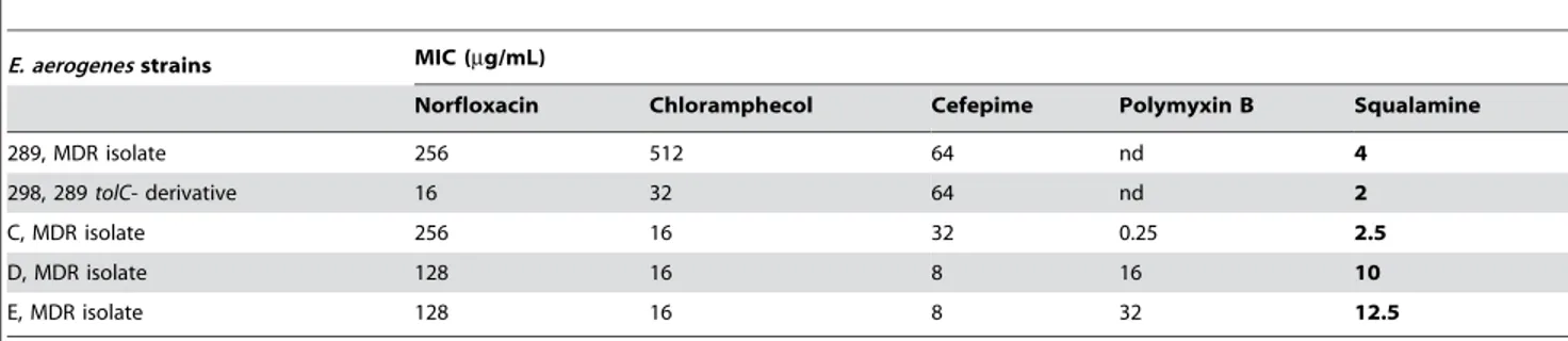
membrane.[27] The first isolate, strain C (Table 3) presenting a polymyxin B susceptibility was sensitive to low concentrations of squalamine 1. Interestingly, clinical isolates D and E that presented a high level of polymyxin resistance (32-fold increase of MIC) exhibited a decrease of squalamine susceptibility with a five-fold increase of the corresponding MIC. This result suggested that the alterations of LPS previously reported in these isolates and causing the resistance towards polymyxin B[27], are able to modulate the squalamine activity. In this context, regarding the other antibiotic families, squalamine offers advantages associated with its activity properties. The squalamine action is preserved even in MDR clinical isolates that overexpress various mecha-nisms of resistance including drug efflux pumps, alteration of membrane permeability caused by absence of porins, enzymatic barrier, all well-known mechanisms which induce high level of resistance towards quinolones, ß-lactams, phenicols, etc (Table 1– 3). For instance: (i) strain 289 was completely devoid of porins, expressed high level of AcrAB-Tol C efflux and a simultaneous overproduction of b-lactamase activity, (ii) strain 298 (289
derivative) exhibited the same phenotype but was deleted of Tol C efflux component, (iii) strain C was porin defficient, overex-pressed AcrAB-Tol C efflux and exhibited a lipopolysaccharide (LPS) wild type profile, (iv) strains D and E had same phenotype plus LPS modifications.[27,28] Thus, the activity of squalamine1 suggests in a first approach that its biological activity results from the synergistic combination of an anionic bile salt with spermidine, each of which independently exhibit considerably less antibiotic activity than squalamine1.[29,30]
Even if strong antibacterial activities have been noticed, the mechanism of action of squalamine towards Gram-negative bacteria remains questionable. Thus, two possible modes of action for such an antibacterial molecule can be underlined (i) competitive binding to a cell-surface exposed receptor (e.g. such as porin)[6,10] involved in key cellular processes and (ii) channel or pore formation in the cytoplasmic membrane. Recently, Katsu et al. examined the structure-activity relationship between original polyamines (naphthylacetylspermine and methoctramine) and the outer membrane of Gram-negative bacteria demonstrating that lipophilic moieties and a number of amino groups in polyamines were important to permeabilisation.[31]
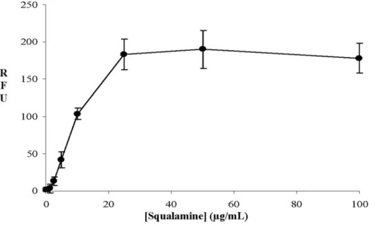
A bioluminescence method was used to determine the effect of squalamine addition on the intracellular pool of bacterial ATP. The detection of external concentration of ATP was then used as a reporter reflecting the permeabilizing effect of squalamine along with the dose-effect relationships. Thus, it clearly appears that for a squalamine concentration of about 20 mg/mL, 80% of the intracellular ATP has been released in the medium suggesting the disruption of the membrane barrier (Figure 3). In addition to ATP release measurements, effect of squalamine on bacterial mem-brane integrity was also assessed using the cell-impermeable DNA/RNA dye propidium iodide (PI) (Figure 4). Results showed
Figure 1. Gram negative bacteria envelope. doi:10.1371/journal.pone.0002765.g001
Figure 2. Structure of squalamine 1. doi:10.1371/journal.pone.0002765.g002
Table 1. Antimicrobial activities of squalamine 1.
Sensitive Strain MIC,mg/mL S. cerevisiae (CIP 28383) C. albicans (CIP 1180-79) S. aureus (CIP 4.83) E. faecalis (CIP 103015) E. hirae (ATCC 10541) E. coli (ATCC 54127) P. aeruginosa (ATCC 15442) E. aerogenes (ATCC 15038) Squalamine 25 .20 3.12 12.5 10 2.5 8 20 doi:10.1371/journal.pone.0002765.t001
that squalamine caused a dose-dependent increase in PI-associated fluorescence. At a 1.25 mg/mL concentration, squalamine did not significantly affect PI-associated fluorescence (263.5-fold increase, p = 0.42). Increase only started to be significant at 2.5 mg/mL (8.063.4 fold increase (p,0.05)) and was maximal at 25 mg/mL squalamine concentration (110.0612.5-fold (p,0.001)). Similarly, CTAB known to cause bacterial permeabilisation, induced equivalent increases in PI-associated fluorescence 100.0611.5-fold increase compared to vehicle-treated bacteria, p,0.001).
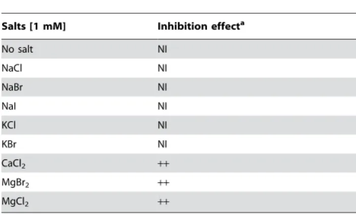
or Ca ).[5] In this context, the effect of various monovalent or divalent ions on bactericidal activity of squalamine 1 has been investigated using 1 mM concentration salts. As shown in Table 4 the addition of monovalent salts did not block the activity of squalamine towards E. coli whereas this later was completely abolished by the same concentration of divalent ions such as MgCl2 or CaCl2. Regarding these divalent salts, it was
demonstrated that the full activity of squalamine is obtained at low concentrations i.e. approximately 0.09 mM.
Table 3. Antibacterial susceptibility of various Multidrug resistant (MDR) E. aerogenes clinical isolates expressing various antibiotic resistance mechanisms.
E. aerogenes strains MIC (mg/mL)
Norfloxacin Chloramphecol Cefepime Polymyxin B Squalamine
289, MDR isolate 256 512 64 nd 4 298, 289 tolC- derivative 16 32 64 nd 2 C, MDR isolate 256 16 32 0.25 2.5 D, MDR isolate 128 16 8 16 10 E, MDR isolate 128 16 8 32 12.5 doi:10.1371/journal.pone.0002765.t003
Figure 3. Measurement of squalamine concentration effect onE. coliATP efflux. doi:10.1371/journal.pone.0002765.g003
Moreover, using bioluminescence method, a noticeable inhibi-tion of the E. coli ATP efflux squalamine-dependent was observed in the presence of divalent ions at various concentrations after 10 minutes of incubation (Figure 6). Thus, NaCl and NaH2PO4
did not lead to any inhibition on E. coli ATP efflux in the presence of squalamine whereas a dramatic inhibition of this efflux was observed in the presence of CaCl2or MgCl2. Moreover, a total
inhibition of the ATP efflux was reached by a concentration of about 5 mM or 2.5 mM for MgCl2or CaCl2, respectively.
Many groups have observed the antibacterial effect of cationic surfaces on Gram-positive as well as Gram-negative bacteria. This suggests that the mechanism is not system-specific, contrary to that which is generally the case with antibiotics. We surmise, as already hinted by others[32], that the death process involves electrostatic interactions and is related to the high density of charges exposed at the surface of bacterial membranes. The architecture of LPS, the main component of outer leaflet of the outer membrane, favors the presence of a large number of negative charges that may stimulate the interactions with cationic substrates.[6,33] The role of LPS is partially suggested with the modulation of squalamine activity in polymyxin resistant isolates and (Table 3).
To propose the existence of a charge-density threshold in the squalamine mode of action, we are helped by recent advances in the understanding of the electrostatic interactions between polyelectrolyte chains and oppositely charged surfaces.[32] It has been gradually realized that adsorption in such cases is driven by the release in solution of the counterions initially confined within the respective electrical double layers. The same process applies to bacteria, which can be crudely considered as large two dimensional polyelectrolytes. Upon adsorption on a cationic solid substrate, the electrostatic compensation of the negative charges of the bacterial envelope is provided by the cationic charges of the substrate, and the bacteria lose their natural counterions.
As previously outlined, squalamine is an amphipathic com-pound which interacts with various membrane glycerophospholi-pids with distinct affinities.[34] As phosphatidylglycerol is the main glycerophospholipid in bacterial membranes whereas
phosphati-dylcholine is more abundant in eukaryotic membranes, this may explain why squalamine could kill bacteria more easily than mammalian cells. Nevertheless, although Gram-positive bacteria have a single membrane that is enriched in phosphatidylglycerol, Gram-negative bacteria also have an external membrane in which the predominant lipid is lipopolysaccharide (LPS). LPS is the major glycolipid recovered from a Folch extract of Gram-negative E. coli bacteria. To study the potential interaction with squalamine and a reconstituted bacterial membrane containing LPS, a lipid extract of E. coli enriched in LPS was spread at the air-water interface where it formed a stable lipid monolayer. Squalamine was then added in the aqueous subphase and its insertion within the LPS film was assessed by surface pressure measurements[35] As shown in Table 5, squalamine penetrated the LPS monolayer at concentrations as low as 0.5 mg/mL. In contrast, higher doses of squalamine were necessary to allow its insertion in monolayers consisting of either neutral glycosphingolipids or gangliosides extracted from lymphocytes. Thus, as far as glycolipids are concerned in early squalamine-membrane interactions, it is clear that bacterial LPS is significantly more active than eucaryotic glycolipids. Squalamine also interacted with matured lipid A (the membrane-anchored backbone of LPS), in a divalent cation-dependent way. Indeed, this squalamine-membrane interaction is highly cationic divalent ion dependent which is consistent with the previously demonstrated lack of activity of squalamine in the presence of such ions in the medium. Moreover, squalamine interacted very poorly with GalCer, but very actively with ceramide (Cer), the membrane-anchored backbone of sphingolip-ids. This may suggest that the insertion of squalamine into eukaryotic membranes could be impaired by the sugar part of glycolipids. Overall these data provide a biochemical basis for the potent activity of squalamine on bacterial Gram-negative and Gram-positive membranes and its relative lack of activity on eukaryotic membranes. Further physicochemical studies will be conducted in the near future in order to decipher the molecular mechanisms (including divalent cation dependence) controlling this striking lipid selectivity.
Figure 4. Effect of squalamine on bacterial membrane integrity assessed by fluorescence measurement of propidium iodide – DNA/ RNA interactions. Results are expressed in relative fluorescence unit (RFU) as Mean6S.D. (n = 3, three independent experiments).
doi:10.1371/journal.pone.0002765.g004
Conclusion
Squalamine is a membrane-active molecule that targets the membrane integrity as demonstrated by the ATP release and dye entry. Consequently, its activity may depend on the membrane lipid composition. It is worthwhile mentioning that the alteration of LPS involved in the polymyxin-resistant clinical isolates moderately changes the squalamine MIC preserving the activity spectrum of the molecule compared to polymyxin B. Thus, if we consider that squalamine acts as a ‘‘membranotropic’’ molecule, it remains possible to observe less susceptible strains like those isolated after polymyxin treatment. However, the resistant variants must preserve a sensitivity level since the adaptation stress requires strong changes in membrane structure which drastically deal with intrinsic membrane stability and the bacterial fitness. In addition, this molecule shows a preserved activity against bacterial pathogens presenting a noticeable MDR phenotype concerning usual antibiotics. Squalamine has membranotropic properties
regarding its bacterial membrane activity and due to its structure containing a cholestanol core it exhibits a moderate level of side effect on eukaryotic cells at doses that kill MDR bacterial pathogens. In this context and because of its structure, action and its relative insensitivity to efflux resistance mechanisms, squalamine may be an alternate way to combat MDR pathogens and by pass the gap regarding the failure of new active antibacterial molecules. This aspect is especially important since some recently described molecules having an active antibacterial spectrum are also substrates for efflux pump systems resulting in a decrease of activity in MDR strains, e.g. peptide deformylase inhibitor, plectasin, platensimycin.[36–38]
Materials and Methods
Determination of minimal inhibitory concentrations
Antimicrobial activity of the compounds was studied by determination of minimal inhibitory concentrations (MIC)
ac-Figure 5. Fluorescence-based microscopic evaluation of the effect of squalamine on bacterial integrity and survival. E. Coli cultures (109/mL) were either left untreated (A) or treated with increasing squalamine concentrations: 1.25mg/mL (B), 2.5 mg/mL (C), 5 mg/mL (D), 10 mg/mL (E), 100mg/ml (F). Suspensions of cultured E. Coli were then stained with the Live/Dead BacLight bacterial Viability kit, as described in material and methods. Scale bar (10mm).
cording to the NCCLS guidelines M7-A2 using the microbroth dilution methods. All the reference strains were issued from the Institut Pasteur collection (Paris). The other reference strains and clinical isolates were from the UMR-MD1 collection and have been previously described.[27,28,39] The bacteria strains were grown on trypticase soy agar (Becton Dickinson) at 37uC in LB or MH broth for E. coli, E. aerogenes and S. aureus or BHI broth for E. faecalis. Inocula were prepared in the respective medium by ajusting the cell density.
Antimicrobial activities of the compounds were determined by using a broth microdilution method performed in sterile 96-well microplates. The molecules were diluted in water and were transferred to each microplate well in order to obtain a two-fold serial dilution and inocullum containing 2–6 105 CFU of each bacteria was added to each well. A number of wells were used for positive controls, inoculum viability and solvent effect. Results were read after 18 hours at 37uC and the MIC was the lowest concentration of the antibacterial agent at which no growth was
detected. MIC values are the mean of three independent experiments.
Measurement of ATP efflux
Squalamine solutions were prepared in doubly distilled water at different concentrations. A suspension of growing E.coli to be studied in LB broth was prepared and incubated at 37uC. 90 mL of this suspension was added to 10 mL of squalamine solution and vortexed for 1 second. 50 mL of luciferin-luciferase reagent (Yelen, France) was immediately added to the precedent mix and luminescent signal quantified with a Lucy luminometer (Yelen, France) for five seconds. ATP concentration was quantified by internal sample addition.
Measurement of ATP efflux inhibition in E. coli in the presence of squalamine (25mg/mL) and various mono and divalent salt solutions
1 M salts (CaCl2, MgCl2, NaH2PO4, NaCl) solutions were
prepared in doubly distilled water and diluted in a 250 mg/mL squalamine solution for 10 minutes at room temperature. Then, ATP efflux was measured with the protocol described above. Results were expressed as ATP efflux percent inhibition relative to the salt free squalamine solution.
Membrane permeability assessment
Over night bacterial suspensions of E. coli in LB were centrifuged 10 min at 10 000 g. Bacterial pellets were resuspended in PBS at 2.56109bacteria per mL, as bacterial permeabilisation assays giving identical results in PBS or LB. Bacteria were added into 96-well black NUNC plate with 0.56109bacteria added per well. The cell-impermeable DNA/RNA dye propidium iodide (PI, Sigma) was then added to bacteria at a final concentration of 30 mM. After 10 min of equilibration at 37uC, bacteria were treated with dye alone or with increasing concentrations of squalamine; CTAB at 58 mg/mL (i.e. 160 mM)[40] being used as positive control of membrane permeabilisation. Finally, fluores-cence was measured after 30 min of incubation at 37uC using a microplate Fluoroscan Ascent spectrofluorometer (excitation at Table 4. Effects of various monovalent or divalent salt
solutions (1 mM) on bactericidal activity of squalamine (2.5mg/mL).
Salts [1 mM] Inhibition effecta
No salt NI NaCl NI NaBr NI NaI NI KCl NI KBr NI CaCl2 ++ MgBr2 ++ MgCl2 ++ a
NI: No inhibition;++: Total inhibition (100% Bacterial survival). doi:10.1371/journal.pone.0002765.t004
Figure 6. ATP efflux inhibition inE. coliin the presence of squalamine (5mg/mL) and various mono and divalent salt solutions. doi:10.1371/journal.pone.0002765.g006
540 nm and emission at 590 nm), as preliminary experiments have shown that maximal fluorescence was obtained at that time independently of the dose of squalamine.
Fluorescence microscopy
In order to investigate membrane damage, we have used the fluorescence based Live/Dead BacLight assay (Molecular Probes). This assay contains a mixture of two nucleic acid stains: a green-fluorescent Stylo 9 stain and a red-green-fluorescent propidium iodide stain. These stains differ to their ability to penetrate healthy bacterial cells. Intact cell membranes stains green, whereas bacteria with damaged membranes stains red. Live and dead bacteria were viewed simultaneously by fluorescence microscopy with suitable optical filter sets. Escherichia coli cells (109CFU/mL) were incubated in the presence of different concentrations of squalamine (1.25 mg/mL, 2.5 mg/mL, 5.0 mg/mL, 10 mg/mL, 100 mg/mL) for 30 minutes at 37uC. Suspensions of treated and untreated cells were stained according to BacLight assay instruction and 5 mL of each bacterial suspensions were subse-quently deposited on slides and analyzed using a fluorescence microscope.
Measurements of squalamine interactions with various bacterial and eucaryotic lipids
The lipids were spread at the air-water interface at an initial surface pressure of 15 mN.m21. After evaporation of the solvent (hexane/chloroform/ethanol; 11:5:4, vol:vol:vol), squalamine was injected in the pure aqueous subphase (volume 800 mL). The variations of the surface pressure were continuously recorded with a fully automated microtensiometer (mTROUGH SX, Kibron Inc.
Helsinki, Finland). All experiments were carried out in a controlled atmosphere at 20uC61uC. The data were analyzed with the Filmware 2.5 program (Kibron Inc. Helsinki, Finland). The accuracy of the system under our experimental conditions was 60.25 mN.m21 for surface pressure. A plus means that the surface pressure increase was above 5 mN.m21after 60 minutes of interaction, a minus means that during the same period of time, the surface pressure increase did not exceed 2 mN.m21. Bacterial and eukaryotic lipids were extracted and submitted to a Folch partition as described previously.[41] Lipopolysaccharide (LPS) was the only glycolipid recovered from the Folch lower phase of the bacterial extract, as demonstrated by high performance thin layer chromatography. The Folch lower phase of lymphocyte lipids contained GlcCer, LacCer, Gb3 and Gb4. The upper phase contained GM1, GM3 and GD3. Pure Lipid A, Cer, GalCer, GM1, GD3 and GT1b were purchased from Sigma. Each experiment was performed three times with similar results.
Acknowledgments
We acknowledge Pr M. Zasloff from Georgetown University, Washington (USA) providing us a sample of squalamine. Thanks to L. Amaral and S. Fanning for their helpful advices. We thank Emilie Donatin and Jacqueline Chevalier for help during determination of bacteriological activities.
Author Contributions
Conceived and designed the experiments: JF MM JMP JMB. Performed the experiments: CS CL NV JF MM NT JMP JMB. Analyzed the data: JMB. Contributed reagents/materials/analysis tools: CS CL. Wrote the paper: NV YL JF MM JMP JMB.
References
1. Armstrong D, Neu H, Peterson LR, Tomasz A (1995) The prospects of treatment failure in the chemotherapy of infectious diseases in the 1990s. Microb Drug Resist 1: 1–4.
2. Bax R, Bywater R, Cornaglia G, Goossens H, Hunter P, et al. (2001) Surveillance of antimicrobial resistance-xhat, how and whither? Clin Microbiol Infect 7: 316–325.
3. Tomasz A (1994) Multiple-antibiotic-resistant pathogenic bacteria. A report on the Rockefeller University workshop. N Engl J Med 330: 1247– 1251.
4. Labischinski H, Barnickel G, Bradaczek H, Naumann D, T. RE, et al. (1985) High state of order of isolated bacterial lipopolysaccharide and its possible contribution to the permeation barrier property of the outer membrane. J Bacteriol 162: 9–20.
5. Vaara M (1992) Agents that increase the permeability of the outer membrane. Microbiol Rev 56: 395–411.
6. Nikaido H (1996) Outer membrane. In: Neidhardt FC, Curtis III R, Ingraham JL, eds. Escherichia coli and Salmonella: cellular and molecular biology. washington: ASM press. pp 29–47.
7. Hancock R (1997) The bacterial outer membrane as a drug barrier. Trends Microbiol 5: 37–42.
8. Murata T, Tseng W, Guina T, Miller SI, Nikaido H (2007) PhoPQ-mediated regulation produces a more robust permeability barrier in the outer membrane of Salmonella enterica serovar typhimurium. J Bacteriol 189: 7213–7222. 9. Vaara M (1993) Outer membrane permeability barrier to azithromycin,
clarithromycin, and roxithromycin in gram-negative enteric bacteria. Anti-microb Agents and Chemother 37: 354–356.
10. Nikaido H (2003) Molecular basis of bacterial outer membrane permeability revisited. Microbiology and Molecular Biology Reviews 67: 593–656. 11. Zasloff M (1992) Antibiotic peptides as mediators of innate immunity. Curr
Opin Immunol 4: 3–7.
12. Zasloff M (1994) Antibacterial molecules from frogs, sharks and man. Phylogenet Perspect Immun Insect Host Def. pp 31–41.
13. Zasloff M (2002) Antimicrobial peptides of multicellular organisms. Nature (London, United Kingdom) 415: 389–395.
14. Burns MR, Wood SJ, Miller KA, Nguyen T, Cromer JR, et al. (2005) Lysine-spermine conjugates: hydrophobic polyamine amides as potent lipopolysaccha-ride sequestrants. Bioorg Med Chem 13: 2523–2536.
15. Hayrinen J, Haseley S, Talaga P, Muhlenhoff M, Finne J, et al. (2002) High affinity binding of long-chain polysialic acid to antibody, and modulation by divalent cations and polyamines. Molecul Immunol 39: 399–411.
16. Sol V, Branland P, Chaleix V, Granet R, Guilloton M, et al. (2004) Amino porphyrins as photoinhibitors of Gram-positive and -negative bacteria. Bioorg Med Chem Lett 14: 4207–4211.
17. Stone R. Deja vu guides the way to new antimicrobial steroid. Science 259: 1125.
18. Ahima Rexford S, Patel Hiralben R, Takahashi N, Qi Y, Hileman Stanley M, et al. (2002) Appetite suppression and weight reduction by a centrally active aminosterol. Diabetes 51: 2099–2104.
19. Boman HG (1991) Antibacterial peptides: key components needed in immunity. Cell 65: 205–207.
20. Brunel JM, Letourneux Y (2003) Recent advances in the synthesis of spermine and spermidine analogs of the shark aminosterol squalamine. Eur J Org Chem. pp 3897–3907.
21. Brunel JM, Salmi C, Loncle C, Vidal N, Letourneux Y (2005) Squalamine: a polyvalent drug of the future? Curr Cancer Drug Targets 5: 267–272. 22. Ding B, Guan Q, Walsh JP, Boswell JS, Winter TW, et al. (2002) Correlation of
the antibacterial activities of cationic peptide antibiotics and cationic steroid antibiotics. J Med Chem 45: 663–669.
23. Moore KS, Wehrli S, Roder H, Rogers M, Forrest JN Jr, et al. (1993) Squalamine: An aminosterol antibiotic from the shark. Proc Natl Acad Sci USA 90: 1354–1358.
24. Rao MN, Shinnar AE, Noecker LA, Chao TL, Feibush B, et al. (2000) Aminosterols from the dogfish shark Squalus acanthias. J Nat Prod 63: 631–635. 25. Zavascki AP, Goldani LZ, Li J, Nation RL (2007) Polymyxin B for the treatment of multidrug-resistant pathogens: a critical review. J Antimicrob and Chemother 60: 1206–1215.
26. Li J, Nation RL, Turnidge JD, Milne RW, Coulthard K, et al. (2006) Colistin: the re-emerging antibiotic for multidrug-resistant gram-negative bacterial infections. Lancet Infect Diseases 6: 589–601.
27. Thiolas A, Bollet C, La Scola B, Raoult D, Pages J-M (2005) Successive emergence of Enterobacter aerogenes strains resistant to imipenem and colistin in a patient. Antimicrob Agents and Chemother 49: 1354–1358.
28. Pradel E, Pages J-M (2002) The AcrAB-TolC efflux pump contributes to multidrug resistance in the nosocomial pathogen Enterobacter aerogenes. Antimicrob Agents and Chemother 46: 2640–2643.
29. Kwon DH, Lu CD (2006) Polyamines increase antibiotic susceptibility in Pseudomonas aeruginosa. Antimicrob Agents and Chemother 50: 1623–1627.
30. Kwon DH, Lu CD (2007) Polyamine effects on antibiotic susceptibility in bacteria. Antimicrob Agents and Chemother 51: 2070–2077.
31. Yasuda K, Ohmizo C, Katsu T (2004) Mode of action of novel polyamines increasing the permeability of bacterial outer membrane. Int J Antimicrob Agents 24: 67–71.
32. Ku¨gler R, Bouloussa O, Rondelez F (2005) Evidence of a charge-density threshold for optimum efficiency of biocidal cationic surfaces. Microbiol 151: 1341–1348.
33. Peschel A (2002) How do bacteria resist human antimicrobial peptides? Trends Microbiol 10: 179–186.
34. Selinsky BS, Zhou Z, Fojtik KG, Jones SR, Dollahon NR, et al. (1998) The aminosterol antibiotic squalamine permeabilizes large unilamellar phospholipid vesicles. Biochimica et Biophysica Acta 1370: 218–234.
35. Garmy N, Taieb N, Yahi N, Fantini J, Journal of Lipid Research (2005), 36–45. (2005) Interaction of cholesterol with sphingosine: Physicochemical character-ization and impact on intestinal absorption. J Lipid Res 46: 36–45.
36. Dean CR, Narayan S, Daigle DM, Dzink-Fox JL, Puyang X, et al. (2005) Role of the AcrAB-TolC efflux pump in determining susceptibility of Haemophilus influenzae to the novel peptide deformylase inhibitor LBM415. Antimicrob Agents and Chemother 49: 3129–3135.
37. Mygind PH, Fischer RL, Schnorr KM, Hansen MT, Soenksen CP, et al. (2005) Plectasin is a peptide antibiotic with therapeutic potential from a saprophytic fungus. Nature (London, United Kingdom) 437: 975–980.
38. Wang J, Soisson SM, Young K, Shoop W, Kodali S, et al. (2006) Platensimycin is a selective FabF inhibitor with potent antibiotic properties. Nature (London, United Kingdom) 441: 358–361.
39. Viveiros M, Dupont M, Rodrigues L, Couto I, Davin-Regli A, et al. (2007) Antibiotic stress, genetic response and altered permeability of E. coli. PLoS One 2: No pp. given.
40. Niven GW, Mulholland F (1998) Cell membrane integrity and lysis in Lactococcus lactis: the detection of a population of permeable cells in post-logarithmic phase cultures. J Appl Microbiol 84: 90–96.
41. Fantini J, Cook DG, Nathanson N, Spitalnik SL, Gonzalez-Scarano F (1993) Infection of colonic epithelial cell lines by type 1 human immunodeficiency virus is associated with cell surface expression of galactosylceramide, a potential alternative gp120 receptor. Proc Natl Acad Sci USA 90: 2700–2704.