HAL Id: inserm-00318936
https://www.hal.inserm.fr/inserm-00318936
Submitted on 5 Sep 2008HAL is a multi-disciplinary open access
archive for the deposit and dissemination of sci-entific research documents, whether they are pub-lished or not. The documents may come from teaching and research institutions in France or abroad, or from public or private research centers.
L’archive ouverte pluridisciplinaire HAL, est destinée au dépôt et à la diffusion de documents scientifiques de niveau recherche, publiés ou non, émanant des établissements d’enseignement et de recherche français ou étrangers, des laboratoires publics ou privés.
A single subunit (GB2) is required for G-protein
activation by the heterodimeric GABA(B) receptor.
Béatrice Duthey, Sara Caudron, Julie Perroy, Bernhard Bettler, Laurent
Fagni, Jean-Philippe Pin, Laurent Prézeau
To cite this version:
Béatrice Duthey, Sara Caudron, Julie Perroy, Bernhard Bettler, Laurent Fagni, et al.. A single subunit (GB2) is required for G-protein activation by the heterodimeric GABA(B) receptor.. Journal of Biological Chemistry, American Society for Biochemistry and Molecular Biology, 2002, 277 (5), pp.3236-41. �10.1074/jbc.M108900200�. �inserm-00318936�
A single subunit (GB2) is required for G protein activation by the
heterodimeric GABA
Breceptor
Béatrice Duthey, Sara Caudron, Julie Perroy, Laurent Fagni, Jean-Philippe Pin and Laurent Prézeau*
CNRS, UPR 9023, Centre CNRS-INSERM de Pharmacologie-Endocrinologie, 34094 Montpellier, France
Running title: GB2 coupling to G-protein in the GABAB receptor
*To whom correspondence should be addressed. :
CNRS, UPR 9023, CCIPE, 141 Rue de la Cardonille, 34000 Montpellier, France Tel: 33 467 14 2933 Fax: 33 467 542 432
Abstract
Although G-protein coupled receptors (GPCRs) have been shown to assemble into functional homo or heteromers, the role of each protomer in G-protein activation is not known. Among the GPCRs, the γ-aminobutyric acid (GABA) type B receptor (GABABR) is the only one
known so far that needs two subunits, GB1 and GB2, to function. The GB1 subunit contains the GABA binding site but is unable to activate G-proteins alone. In contrast the GB2 subunit which does not bind GABA, has an heptahelical domain able to activate G-proteins when assembled into dimers (Galvez et al., EMBO J. 20, 2001, 2152-2159). In the present study, we examined the role of each subunit within the GB1-GB2 heteromer, in G-protein coupling. To that aim, point mutations in the highly conserved third intracellular loop known to prevent G-protein activation of the related Ca-sensing or metabotropic glutamate receptors were introduced into GB1 and GB2. One mutation, L686P introduced in GB2 prevents the formation of a functional receptor, even though the heteromer reaches the cell surface, and even though the mutated subunit still associates with GB1 and increases GABA affinity on GB1. This was observed either in HEK293 cells where the activation of the G-protein was assessed by measurement of IP accumulation, or in cultured neurons where the inhibition of the Ca-channel current was measured. In contrast, the same mutation when introduced into GB1 does not modify the G-protein coupling properties of the heteromeric GABAB receptor
either in HEK293 cells or in neurons. These data show that a single subunit in a dimeric GPCR is critical for coupling to G-proteins.
Introduction
G protein-coupled receptors (GPCR) are coded by the largest gene family of the animal genomes. These receptors detect and transduce accross the plasma membrane, the information carried by a large variety of message molecules, from photons to glycoproteins, from amino-acids to ions, with a very high specificity (1). Accordingly, GPCRs are involved in a large variety of physiological and pathological processes, and as such constitute the targets of a large number of therapeutic drugs. All these receptors have an heptahelical domain (HD) formed by 7 transmembrane helices interconnected by 3 extracellular and 3 intracellular loops. The intracellular face of the receptor contacts the heterotrimeric G-proteins and stimulates it upon agonist activation. Indeed, the second and third intracellular loops have been shown to play a critical role in G-protein activation by forming a pocket in which the C-terminal tail of the α subunit of the G-protein binds (2-4).
Within the last decade, a number of studies reported that several GPCRs can form oligomers, either homo or hetero-oligomers (5). Such protein complexes, when constituted of different subunits, can have specific properties, not shared by the corresponding receptors alone. Indeed, the heteromers can have a specific pharmacology (6, 7), desensitization properties (7, 8), or even can activate specific intracellular pathways (9). However, the respective role of each subunits of the oligomer in G-protein activation is not known.
Among the various GPCRs, the GABAB receptor is the only one identified so far that requires
two subunits, GB1 and GB2, to be functional. This is true both in heterologous expression systems (10-14) and in neurons (15). Such a characteristic of the GABAB recceptor makes it
an excellent model to examine the respective role of each subunit in G-protein coupling. Each GABAB receptor subunit share sequence similarity with the metabotropic glutamate
(mGlu), the calcium sensing (CaS), some putative pheromone and taste receptors. All these receptors have been shown to form dimers (16-18), and all are composed of two main domains, a HD involved in G-protein coupling (19-21), and a large extracellular domain (ECD) where the agonists bind (22-27). But why are two distinct subunits required to get a functional GABAB receptor ? Indeed, GABAB agonists likely bind on GB1 only (26, 27), but
are unable to activate it. The GB2 subunit is required for the correct insertion of the GB1 subunit in the plasma membrane but also for the correct functioning of the receptor (28-30). We recently reported that the GB2-HD can couple to G-protein when associated in dimers, but not the GB1-HD (31). Here, the specific role of the GB1-HD and GB2-HD in G-protein
activation by the heteromeric GABAB receptor was examined. Our data show that the
Material and Methods
Construction of point mutated receptors:
cDNAs encoding wild type GB1 and GB2 were described previously (31). Site-directed mutagenesis of sequences encoding amino acids of i3 loop of GB1 and GB2 was performed on a BamH I-Sal I fragment from pRK-BR1a cloned into pBluescript SK(-) vector (Stratagene, La Jolla, CA) and a Bsp120 I-Xba I fragment from pCi-Neo-BR2 cloned into pBluescript SK(-), using the QuickChange Site-Directed Mutagenesis kit (Stratagene, La Jolla, CA). For each mutagenesis, two complementary 30-mers oligonucleotides (sense and anti-sense; Genaxis Biotechnologie, Nimes, France) were designed to contain the desired mutation in their center. The authenticity of each point mutation and the absence of undesired ones were confirmed by DNA sequencing. Subsequently, a short fragment surrounding the mutation was subcloned in place of the corresponding wild-type fragment of pRK-BR1a or pRK-BR2.
Cell culture and Transfection:
Human embryonic kidney (HEK) 293 cells were cultured and transfected by electroporation as described previously (32). After electroporation, the cells were plated on poly-ornithine-coated dishes. Serum, culture media, and other solutions used for cell culture were from Life Technologies, SARL. (Cergy Pontoise, France).
Cultures of cerebellar granule neurons were prepared from one week old mice and transfected as previously described using Transfast (Promega, Lyon, France) (33), with minor modifications (transfection was performed immediately before platting instead of 24 hours after platting). A plasmid expressing the green fluorescent protein (GFP, pGFP-N1 from Clontech, Heidelberg, Germany) in order to identify transfected neurons from electrophysiological studies. We previously reported that more than 95% of the GFP positive neurons also express the co-transfected plasmids (33). About ten percent of granule cells were transfected under these conditions.
Western Blot
The western blotting protocole was already described (31). 10 µg of membrane were loaded on a tricine-SDS gel for electrophoresis and transferred on nitrocellulose membranes (Amersham Pharmacia Biotech, Orsay, France). After incubation in PBS-milk 5%, the
membranes were incubated with the anti-GB1 antibody (Sigma, L'Isle d'Abeau, France) or the monoclonal anti-HA antibody (1/3000, Roche diagnostics, Meylan, France) at room temperature for 2 hrs. After washing, the membranes were incubated overnight at 4 °C with the anti mouse HRP antibody (Amersham Life Science, Orsay, France). Signal was revealed using an ECL chemioluminescent assay (Amersham Pharmacia Biotech,Orsay, France).
Ligand binding on intact HEK293 cells:
Ligand binding experiments were performed on intact HEK 293 cells as previously described. Briefly, cells were plated after electroporation the day before experiment. Thus, the cells on ice were washed with ice cold binding buffer (20 mM Tris-Cl pH 7.4, 118 mM NaCl, 1.2 mM KH2PO4, 1.2 mM MgSO4, 4.7 mM KCl, 1.8 mM CaCl2) and incubated in the presence of 0.1 nM [125I]-CGP64213 with or without unlabelled ligands at the indicated concentration. The incubation was terminated by washing with ice cold buffer. The cells were disrupted with 0.1M NaOH (400µl) and the bound radioactivity was counted and normalized to the amount of protein in each wells. Non-specific binding was determined in the presence of 1 mM GABA. The concentration of [125
I]-CGP64213 used in displacement experiments (0.1 nM) was approximately 10 times lower than the affinity of this radioligand on GB1. Ki values were calculated according to the equation IC50 = Ki (1+[L]/Kd), Kd being assumed to be
equal to Ki in the case of CGP64213. GABA was obtained from Sigma (L'Isle d'Abeau, France). [125
I]-CGP64213 was labeled to a specific radioactivity of >2,000 Ci mmol-1 (ANAWA AG, Wangen, Switzerland). L-Baclofen was synthesized in the research laboratories of Novartis Pharma in Basel (34). Displacement curves were fitted as previously described (27).
Determination of inositol phosphate (IP) accumulation
HEK 293 cells were transfected as described above with the receptor constructs in pRK5 (2 µg), Gqi9 expression vector (2 µg) and carrier DNA (pRK6, 4µg) (32). Determination of IP accumulation was performed 15 h after transfection as described elsewhere (32).
Electrophysiological recordings:
We used the whole-cell patch-clamp configuration to record Ba2+ currents (IBa) from GFP expressing cerebellar granule cells (co-transfected or not with GABAB receptor
contained (in mM): BaCl2 (20), HEPES (10), tetraethylammonium acetate (10), TTX (3x10 -4
), glucose (10), Na-acetate (120), and MK-801 (1x10⁻³), adjusted to pH 7.4 with Na-OH and 330 mosm with Na-acetate. Drug solutions were prepared in this medium and pH readjusted to 7.4. Patch pipettes were made from borosilicate glass, coated with Sylgard, and their tip fire polished. Pipettes had resistances of 3-5 MOhms when filled with the following internal solution (in mM): Cs-acetate (100), MgCl2 (2), HEPES (10), glucose (15), CsCl (20), EGTA
(20), Na2ATP (2 mM) and cAMP (1mM), adjusted to pH 7.2 with CsOH and 300 mosm with
Cs-acetate.
IBa were evoked by 500 msec voltage-clamp pulses, from a holding potential of –80 mV, to a test potential of 0 mV, applied at a rate of 0.1 Hz. Current signals were recorded using an Axopatch 200 amplifier, filtered at 1 kHz with an 8-pole Bessel filter and sampled at 3 kHz on a Pentium II PC computer. Analyses were performed using the pClamp6 program of Axon Instruments. Barium currents were measured at their peak amplitude and values expressed as mean ± s.e.m. of the indicated number (n) of experiments for comparison using the Student's t test (p ≤ 0.05).
Results
Generation and characterization of GB1 and GB2 mutants
Like any other G-protein coupled receptors, the GABAB-like receptors, such as the
mGlu and CaS receptors contact the G-protein α subunit via their second and third intracellular loops (i2 and i3 loops respectively) (19-21, 36). As shown in Fig.1, the i3 loop is very well conserved among the receptors of this family, and several residues in this loop have already been shown to play a critical role in G-protein coupling in both mGlu1 and CaS receptors. Among these, R796 and F802 of CaS (R775 and F781 of mGlu1) when mutated into W and A/P respectively, generate receptors unable to activate G-protein upon agonist activation (21, 36, 37). As shown in Fig.1 the general characteristic of these residues (basic and hydrophobic respectively) is conserved in both GB1 and GB2 subunits suggesting that they play a similar role as in the CaS and mGlu1 receptors. In order to identify the respective role of GB1 and GB2 in G-protein coupling of the GABAB heteromer, these residues (K791
and I798 of GB1; R679 and L686 of GB2) were mutated into different residues. The following mutants were generated: GB1-K791W; GB1-K791D; GB1-I798S, GB1-I798P; GB2-R679W; GB2-R679D; GB2-L686S and GB2-L686P. All these constructs were tagged at their N-terminal end with either a HA or c-myc epitope, as previously described (29, 31).
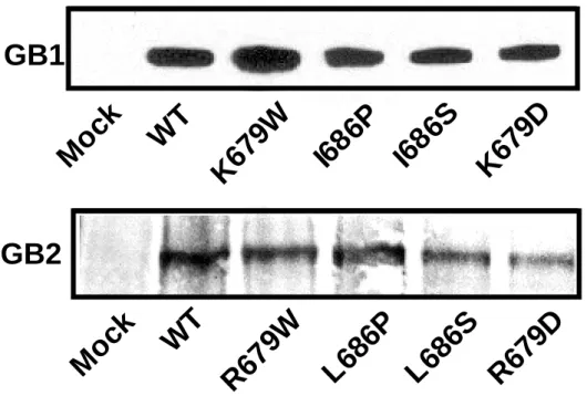
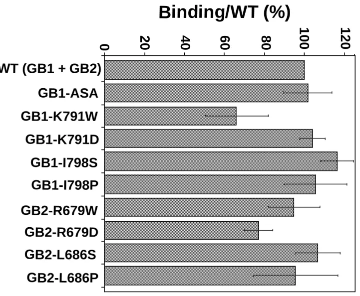
Western blot analysis showed that all mutated receptors were expressed in HEK293 cells (Fig.2a). Binding experiments performed on intact cells with the membrane not permeable high affinity GB1 radioligand [125
I]-CGP64213 indicated that any GB1 mutants were correctly targeted to the plasma membrane when co-expressed with the wild-type GB2 (Fig.2b), but not when expressed alone (data not shown). Because GB1 only reaches the cell surface when associated with GB2 (28-30), these data indicate that the mutations in GB1 do not affect the correct folding of this subunit nor its association with GB2. Similarly, [125
I]-CGP64213 binding can be detected on intact cells expressing any of the mutated GB2 subunits co-expressed with the wild-type GB1 (Fig.2b). This indicates that these mutated GB2 subunits are still able to bring GB1 to the cell surface, like the wild-type GB2.
Mutation L686P in GB2 suppresses the functional coupling of the heterodimer to Gqi9
We then analyzed the effect of the mutations in the i3 of GB1 and GB2 on the G-protein coupling of the heteromeric receptor. To that aim, various combinations of wild-type and mutated GB1 and GB2 subunits where co-expressed with the chimeric G α-protein Gqi9
in HEK293 cells. This chimeric G protein α-subunit corresponds to Gαq in which the last 9 C-terminal residues have been replaced by those of Gαi2. This allows this G-protein construct to couple many Gi/o coupled-receptors to PLC (2), including the heteromeric GABAB
receptor (32). As shown in Fig.3, GABA-induced inositol phosphate (IP) formation was not affected when K791 of GB1 or R679 of GB2 were mutated into Trp or Asp. Only a ten fold decrease in GABA potency (10 fold increase in the EC50 value, Table 1) was noticed with GB2-R679W and GB2-R679D mutants expressed with GB1 (Fig.3b). However, the GABA response was dramatically reduced in cells expressing the GB2 mutants, GB2-L686P and GB2-L686S (by 94% and 83%, respectively), together with the wild-type GB1. Indeed, the maximal GABA response was largely decreased as well as the potency (increased EC50 values, see Table 1) for the GB2-L686S + GB1 expressing cells, and no significant response could be measured in cells expressing GB2-L686P + GB1 (Fig.3b, Table 1). Of interest, the equivalent mutations in GB1 (GB1-I798S and GB1-I798P) did not affect much the GABA response (Fig. 3a). Only a small decrease in the maximal effect of GABA was noticed with the GB1-I798S mutant.
As mentioned above the GB2-L686S and GB2-L686P mutants were able to bring the wild-type GB1 subunits to the cell surface, but it remains possible that the loss of GABA response in cells co-expressing these GB2 mutants and GB1 is due to an instability of the heteromers at the cell surface, or to the absence of allosteric interaction between the two subunits required for function. It has been reported that the association of GB2 with GB1 increases agonist affinity on GB1 by a factor 10 (10, 12, 31). As shown in Fig.4 and in Table 1, both GB2 mutants were able to increase GABA affinity on GB1 like the wild-type GB2, indicating that they not only bring GB1 at the cell surface, but also still allosterically interact with GB1 at the cell surface. Indeed, the Ki value of GABA in displacing [125
I]-CGP64213 bound on intact cells expressing the GB1ASA (a mutant that reaches the cell surface alone (29))
is 10 times higher than that measured on cells expressing the wild-type GB1 with GB2, GB2-L686S or GB2-L686P (Table 1).
Taken together, these data indicate that the mutation of a single residue, L686 into S or P in the i3 loop of GB2 decreases and suppresses, respectively, the ability of the heteromeric GABAB receptor to activate Gqi9. In contrast, the equivalent mutations introduced in the i3
loop of GB1 do not prevent coupling of the heteromer to this G-protein.
GABAB receptors do not naturally couple to the artificial Gqi9 protein. Indeed, in
neurons, GABAB receptors couple to Gi/o proteins and as a consequence inhibit adenylyl
cyclase or Ca-channel activity, or activate G-protein inward rectifying K-channels (38). It is possible that GB2 plays a critical role in the coupling to Gqi9 in HEK293 cells, but not to Gi/o in neurons. We therefore examined the ability of our wild-type and mutated GB1 and GB2 subunits to form a functional GABAB receptor able to inhibit Ca-channels in cerebellar
granule cells. In these neurons, depolarization to 0 mV induced activation of a Ba current (IBa) which is mediated by various types of Ca-channels (35, 39). Under our culture conditions, the GABAB selective agonist baclofen was unable to inhibit the voltage-activated
IBa in control or GFP-expressing neurons (Fig.5a). In contrast, baclofen inhibited the IBa in neurons expressing GB1 and GB2 subunits (Fig.5b, 20 ± 2 % inhibition (n=8)), or neurons expressing GB1-I798P and GB2 (Fig.5c, 19 ± 3 % inhibition (n=9)). However, baclofen did not affect IBa in neurons expressing GB1 and GB2-L686P subunits (Fig.5d), even though both subunits where correctly expressed at the cell surface as revealed by immunostaining of intact cells with the HA antibody (data not shown).
Discussion
In the present study we identified a single residue within the i3 loop of GB2 that plays a critical role for the coupling of the heteromeric GABAB receptor to G-proteins, either in a
cell line, or in primary neurons. In contrast, the equivalent mutation in GB1 does not prevent coupling.
GABAB receptor subunits are part of the so-called family 3 GPCRs. These receptors
do not share the sequence similarity with the other rhodopsin-like or secretin receptor-like GPCRs. One of the characteristic of these receptors is their short and highly conserved i3 loop, in which a signature has been identified ((F/Y/L/I)-N-(E/D)-X-(K/R)) at the bottom of TM6 (Fig.1). Among these residues, the first hydrophobic residue has been shown to play a critical role in G-protein coupling in both mGlu1 and CaS receptors since its mutation into either Ser, Pro in mGlu1 (21), or Ala, His, Glu, Leu or Val (but not Tyr or Trp) in CaS (36) receptor abolished coupling of these receptors to PLC. In the GB1 and GB2 subunits, this Phe residue is replaced by an Ile and a Leu respectively. Our data revealed that this residue in GB2 also plays a critical role in G-protein coupling since its mutation into either Ser and Pro largely decreases and abolishes, respectively, coupling of the heteromeric receptor to either Gqi9 in HEK293 cells or inhibition of Ca-channels in neurons. It may appear surprising that the replacement of Phe in the CaS receptor by Leu, as found in the GB2 subunit prevent CaS receptor functioning. This may be due to a different environment of the Leu residue in the CaS and GB2 proteins. However, the mutation of the Ile residue found at that position in the i3 loop of GB1 into either Ser or Pro (but also into Ala, data not shown) did not affect coupling of the GABAB receptor either in HEK293 cells or in neurons.
Another residue within the i3 loop is highly conserved among the family 3 GPCR, this is the Arg of the conserved sequence K-(T/S)-R at the bottom of TM5 (Fig.1). This Arg of the CaS receptor was found to be mutated into a Trp in families suffering from hypocalciuric hypercalcemia (37). This mutation of either the CaS or mGlu1 receptor was found to prevent coupling to G-proteins (21, 37). Here we show that the mutation of this residue in either GB1 (Lys791) or GB2 (Arg679) into Trp or Asp did not modify the ability of the heteromeric receptor to activate Gqi9. Indeed, only a decrease in the potency of GABA was noticed with the GB2-R679W and R679D mutants, consistent with a decrease in G-protein coupling efficacy (no change in the GABA affinity nor receptor expression level being observed with these mutants). When a careful analysis of the same mutation introduced in mGlu1 was
performed, we found that it resulted in an increase in the basal activity of the receptor (Restituito and Pin, unpublished observation), indicating that this mutation does not prevent mGlu1 receptor coupling to G-protein, but rather stabilize an active state of the receptor. It is possible that the loss of function observed for the R-W mutants of the CaS and mGlu1 receptors by others results from the desensitization of the receptor due to a high basal activity, rather than from a total uncoupling to G-proteins. Taken together, these data show that the conserved basic residue at the bottom of TM5 does not play a critical role in G-protein coupling, but rather modify the coupling efficacy of the receptor to the G-protein.
We recently reported that the HD of GB2 when associated in dimers could activate G-proteins. Indeed, the co-expression of a chimeric construct containing the GB1-ECD and the GB2-HD together with the wild-type GB2 forms a functional GABAB receptor that can
activate a same set of G-proteins as the wild-type heteromer (31). In contrast, no coupling to any tested G-protein could be detected with a receptor containing only GB1-HDs. In the present study we show that GB2-HD plays a crucial role in G-protein coupling if the heteromer. So what is the role of the GB1-HD in GABAB receptor function ?
Our data do not exclude the possibility that GB1 or GB1-I798P can activate a G-protein different from Gqi9 or different from that involved in the inhibition of Ca-channels in neurons. Alternatively, it is also possible that GB1 coupling to G-proteins also requires GB2 coupling. This would be the case if the simultaneous interaction of G-proteins with GB1 and GB2 would be required for G-protein activation. However, the simplest hypothesis would be that GB1 does not couple to the G-protein α-subunit. GB1 could however still be able to interact with the βγ dimer. This could explain our recent data showing that GB1-HD improves the G-protein coupling efficiency of the GB2-HD (31). Of interest, it has been reported recently that GPCRs could act as a lever to tilt the βγ subunit on the α-subunit to stimulate the GDP-GTP exchange (40). This model, as well as the respective size of the heterotrimeric G-protein and a dimeric GPCR (41), will very well fit with the possibility that one subunit in a dimeric GPCR interacts with the G-protein α subunit whereas the other interacts with the βγ subunit. However, although a direct specific cross-linking approach revealed a close contact of rhodopsin with the α-subunit of transducin, no cross-linking was obtained between rhodopsin and the βγ dimer (42, 43). Finally, it remains possible that GB1 does not contact the heterotrimeric G-protein at all, and that it plays a different role in the transduction mechanism of the GABAB receptor, possibly by interacting with other intracellular proteins
Although our data do not identify the precise role of the HD of GB1 in G-protein coupling of the heteromeric GABAB receptor, our data demonstrate that GB2-HD plays a
critical role in this function of the GABAB receptor. This illustrates therefore the importance
References
1 Bockaert, J., and Pin, J.-P. (1999) EMBO J. 18, 1723-1729
2 Conklin, B. R., Farfel, Z., Lustig, K. D., Julius, D., and Bourne, H. R. (1993) Nature
363, 274-276
3 Wess, J. (1997) FASEB J. 11, 346-354
4 Bourne, H. R. (1997) Curr. Opin. Cell Biol. 9, 134-142 5 Bouvier, M. (2001) Nature Rev. 2, 274-286
6 Jordan, B. A., and Devi, L. A. (1999) Nature 399, 697-700.
7 Pfeiffer, M., Koch, T., Schroder, H., Klutzny, M., Kirscht, S., Kreienkamp, H. J., Hollt, V., and Schulz, S. (2001) J Biol Chem 276, 14027-36.
8 Jordan, B. A., Trapaidze, N., Gomes, I., Nivarthi, R., and Devi, L. A. (2001) Proc Natl
Acad Sci U S A 98, 343-8.
9 Mellado, M., Rodriguez-Frade, J. M., Vila-Coro, A. J., Fernandez, S., Martin de Ana, A., Jones, D. R., Toran, J. L., and Martinez, A. C. (2001) Embo J 20, 2497-507.
10 White, J. H., Wise, A., Main, M. J., Green, A., Fraser, N. J., Disney, G. H., Barnes, A. A., Emson, P., Foord, S. M., and Marshall, F. H. (1998) Nature 396, 679-682
11 Jones, K. A., Borowsky, B., Tamm, J. A., Craig, D. A., Durkin, M. M., Dai, M., Yao, W.-J., Johnson, M., Gunwaldsen, C., Huang, L.-Y., Tang, C., Shen, Q., Salon, J. A., Morse, K., Laz, T., Smith, K. E., Nagarathnam, D., Noble, S. A., Branchek, T. A., and Gerald, C. (1998) Nature 396, 674-679
12 Kaupmann, K., Malitschek, B., Schuler, V., Heid, J., Froestl, W., Beck, P., Mosbacher, J., Bischoff, S., Kulik, A., Shigemoto, R., Karschin, A., and Bettler, B. (1998) Nature 396, 683-687
13 Kuner, R., Kohr, G., Grunewald, S., Eisenhardt, G., Bach, A., and Kornau, H. C. (1999)
Science 283, 74-77
14 Ng, G. Y., Clark, J., Coulombe, N., Ethier, N., Hebert, T. E., Sullivan, R., Kargman, S., Chateauneuf, A., Tsukamoto, N., McDonald, T., Whiting, P., Mezey, E., Johnson, M. P., Liu, Q., Kolakowski, L. F., Jr., Evans, J. F., Bonner, T. I., and O'Neill, G. P. (1999) J.
Biol. Chem. 274, 7607-10
15 Filippov, A. K., Couve, A., Pangalos, M. N., Walsh, F. S., Brown, D. A., and Moss, S. J. (2000) J Neurosci 20, 2867-74
16 Romano, C., Yang, W.-L., and O'Malley, K. L. (1996) J. Biol. Chem. 271, 28612-28616 17 Bai, M., Trivedi, S., and Brown, E. M. (1998) J. Biol. Cell. 273, 23605-23610
18 Bai, M., Trivedi, S., Kifor, O., Quinn, S. J., and Brown, E. M. (1999) Proc Natl Acad Sci
U S A 96, 2834-9
19 Pin, J.-P., Joly, C., Heinemann, S. F., and Bockaert, J. (1994) EMBO J. 13, 342-348 20 Gomeza, J., Joly, C., Kuhn, R., Knöpfel, T., Bockaert, J., and Pin, J.-P. (1996) J. Biol.
Chem. 271, 2199-2205
21 Francesconi, A., and Duvoisin, R. M. (1998) J. Biol. Chem. 273, 5615-5624
22 O'Hara, P. J., Sheppard, P. O., Thøgersen, H., Venezia, D., Haldeman, B. A., McGrane, V., Houamed, K. M., Thomsen, C., Gilbert, T. L., and Mulvihill, E. R. (1993) Neuron
11, 41-52
23 Bessis, A.-S., Bertrand, H.-O., Galvez, T., De Colle, C., Pin, J.-P., and Acher, F. (2000)
Prot. Sci. 9, 2200-2209
24 Kunishima, N., Shimada, Y., Tsuji, Y., Sato, T., Yamamoto, M., Kumasaka, T., Nakanishi, S., Jingami, H., and Morikawa, K. (2000) Nature 407, 971-977
25 Malitschek, B., Schweizer, C., Keir, M., Heid, J., Froestl, W., Mosbacher, J., Kuhn, R., Henley, J., Joly, C., Pin, J.-P., Kaupmann, K., and Bettler, B. (1999) Mol. Pharmacol.
56, 448-454
26 Galvez, T., Parmentier, M.-L., Joly, C., Malitschek, B., Kaupmann, K., Kuhn, R., Bittiger, H., Froestl, W., Bettler, B., and Pin, J.-P. (1999) J. Biol. Chem. 274, 13362-13369
27 Galvez, T., Prézeau, L., Milioti, G., Franek, M., Joly, C., Froestl, W., Bettler, B., Bertrand, H.-O., Blahos, J., and Pin, J.-P. (2000) J. Biol. Chem. 275, 41166-41174
28 Calver, A. R., Robbins, M. J., Cosio, C., Rice, S. Q., Babbs, A. J., Hirst, W. D., Boyfield, I., Wood, M. D., Russell, R. B., Price, G. W., Couve, A., Moss, S. J., and Pangalos, M. N. (2001) J Neurosci 21, 1203-1210.
29 Pagano, A., Rovelli, G., Mosbacher, J., Lohmann, T., Duthey, B., Stauffer, D., Ristig, D., Schuler, V., Heid, J., Meigel, I., Lampert, C., Stein, T., Prézeau, L., Pin, J.-P., Froestl, W., Kuhn, R., Kaupmann, K., and Bettler, B. (2001) J. Neurosci. 21, 1189–1202
30 Margeta-Mitrovic, M., Jan, Y. N., and Jan, L. Y. (2000) Neuron 27, 97-106
31 Galvez, T., Duthey, B., Kniazeff, J., Blahos, J., Rovelli, G., Bettler, B., Prézeau, L., and Pin, J.-P. (2001) EMBO J. 20, 2152-2159
32 Franek, M., Pagano, A., Kaupmann, K., Bettler, B., Pin, J.-P., and Blahos II, J. (1999)
Neuropharmacology 38, 1657-1666
33 Ango, F., Albani-Torregrossa, S., Joly, C., Robbe, D., Michel, J.-M., Pin, J.-P., Bockaert, J., and Fagni, L. (1999) Neuropharmacology 38, 793-803
34 Froestl, W., Mickel, S. J., Hall, R. G., von Sprecher, G., Strub, D., Baumann, P. A., Brugger, F., Gentsch, C., Jaekel, J., Olpe, H.-R., Rihs, G., Vassout, A., Waldmeier, P. C., and Bittiger, H. (1995) J. Med. Chem. 38, 3297-3312
35 Perroy, J., Prezeau, L., De Waard, M., Shigemoto, R., Bockaert, J., and Fagni, L. (2000) J
Neurosci 20, 7896-904
36 Chang, W., Chen, T. H., Pratt, S., and Shoback, D. (2000) J Biol Chem 275, 19955-63 37 Pollak, M. R., Brown, E. M., Chou, Y.-H. W., Hebert, S. C., Marx, S. J., Steinmann, B.,
Levi, T., Seidman, C. E., and Seidman, J. G. (1993) Cell 75, 1297-1303
38 Couve, A., Moss, S. J., and Pangalos, M. N. (2000) Mol Cell Neurosci 16, 296-312. 39 Chavis, P., Fagni, L., Bockaert, J., and Lansman, J. B. (1995) Neuropharmacology 34,
929-937
40 Rondard, P., Iiri, T., Srinivasan, S., Meng, E., Fujita, T., and Bourne, H. R. (2001) Proc
Natl Acad Sci U S A 98, 6150-5.
41 Hamm, H. E. (2001) Proc Natl Acad Sci U S A 98, 4819-21.
42 Itoh, Y., Cai, K., and Khorana, H. G. (2001) Proc Natl Acad Sci U S A 98, 4883-7. 43 Cai, K., Itoh, Y., and Khorana, H. G. (2001) Proc Natl Acad Sci U S A 98, 4877-82. 44 White, J. H., McIllhinney, R. A., Wise, A., Ciruela, F., Chan, W. Y., Emson, P. C.,
Billinton, A., and Marshall, F. H. (2000) Proc Natl Acad Sci U S A 97, 13967-72
45 Vernon, E., Meyer, G., Pickard, L., Dev, K., Molnar, E., Collingridge, G. L., and Henley, J. M. (2001) Mol Cell Neurosci 17, 637-45.
46 Nehring, R. B., Horikawa, H. P. M., El Far, O., Kneussel, M., Brandstätter, J. H., Stamm, S., Wischmeyer, E., Betz, H., and Karschin, A. (2000) J. Biol. Chem. 275, 35185–35191
Acknowledgements: We thank Florence Gaven for technical assistance. This work was supported by grants from CNRS, Novartis Pharma (Basel; Switzerland), The program Physique-Chimie du Vivant (PCV) and the Action "Molécules et Cibles Thérapeutique" (AMCT) (all to JPP).
Legend to figures
Figure 1: Alignment of the i3 loop of GABA-B receptor subunits and related receptors. Highlighted in black are residues conserved within this family of receptors. The consensus sequence of the conserved residues in this loop is indicated at the bottom, with b=basic; a=acidic; h=hydrophobic; and X=any residue. These include the sequences of GB1 from human (swall accession number: NP_001461), rat (CAA71398), mouse (AAD22194), D. melanogaster (Q9V3Q9) and C. elegans (from the cosmids Y41G9), of GB2 from human (O75899), rat (O88871), D.melanogaster (Q9Y133) and C. elegans (from cosmid ZK180), the GB3 subunit from D. melanogaster (Q9VPS7), the metabotropic glutamate receptors subtypes 1 (P23385), 2 (P31421), 6 (P35349) and 8 (NP_071538) from rat, from Catfish (CaF, Q9PWE1), subtype A from D. melanogaster (P91685), one subtype from C. elegans (from the cosmid Y4C6A, Q9N4T8), the bovine Ca2+-sensing receptor (P35384), the rat taste receptor T1R1 (Q9Z0R8), the Goldfish amino acid olfactory receptor OR5.24 (AAD46570), as well as the pheromone-like receptors from Goldfish (GFB2 (Q9PSY1) and 14 (O93558)) and Fugu (CA04_Fug, O73647), and the putative rat pheromone receptors GoVN2 (O35266) and V2R1 (O70411).
Figure 2: Expression of the wild-type and mutant GABAB subunits. a. The expression of the
different wild type and mutated GB1 and GB2 subunits was analyzed by Western blot analysis. The proteins were detected with the monoclonal anti GB1 antibody for GB1 subunits and with monoclonal anti HA antibody for the GB2 subunits. b. The expression at the cell surface of the mutated or wild type GB1 subunits was determined by measuring the total [125 I-CGP64213] binding on intact cells expressing the different subunit combinations. Because GB1 reaches the cell surface only when it is co-expressed with GB2, the binding on GB1 indicates that the heteromer GB1 + GB2 is formed and gets to the cell surface. The total binding of the wild type is 3243 ± 750 cpm, and the non-specific binding 233 ± 39 cpm (n=12). Values correspond to the amount of bound radioactivity per well expressed as a percentage of wild type maximal binding. Data are the mean ± s.e.m. of 6 to 12 independent experiments done in triplicates.
Figure 3: G protein coupling of the wild type and mutated GB1 and GB2 subunits. a/ Dose-response curves of GABA on cells expressing the GB1 mutants with the GB2 wild type subunit. b/ Dose response curves of GABA on cells expressing the GB2 mutants with the
GB1 wild-type subunit. Values correspond to the percentage of the maximal agonist-induced IP formation measured in cells expressing the wild type receptor (GB1 + GB2). In those cells, GABA induced a 6.4 fold increase in the basal IP formation. Values are means ± s.e.m. of at least three experiments performed in triplicates.
Figure 4: Displacement curves of [125
I]-CGP64213 with GABA on cells expressing the different mutant receptors. The values correspond to the amount of bound radioactive [125 I]-CGP64213 displaced by increasing concentrations of GABA on the different receptor mutants. Specific [125
I]-CGP64213 binding is plotted as the percentage of the binding obtained in cells expressing the wild-type GABAB receptor. Values are means ± s.e.m. of at least three
experiments performed in triplicates.
Figure 5: Inhibition of IBa in cerebellar granule neurons by different combination of GABAB
subunits. IBa was measured in granule neurons expressing GFP alone (a) or with GB1 and GB2 (b), GB1-I798P and GB2 (c) or GB1 and GB2-L686P (d). The current was measured during a 500 msec depolarizing step to 0 mV in the absence (c) or in the presence (b) of baclofen (100 µM). Scale bars: 200 pA and 200 msec.
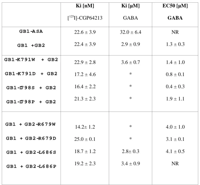
Ki [nM] [125 I]-CGP64213 Ki [µM] GABA EC50 [µM] GABA G B1 -A SA G B1 + G B2 22.6 ± 3.9 22.4 ± 3.9 32.0 ± 6.4 2.9 ± 0.9 NR 1.3 ± 0.3 G B1 -K 7 9 1 W + G B2 G B1 -K 7 9 1 D + G B2 G B1 -I7 9 8 S + G B2 G B1 -I7 9 8 P + G B2 22.9 ± 2.8 17.2 ± 4.6 16.4 ± 2.2 21.3 ± 2.3 3.6 ± 0.7 * * * 1.4 ± 1.0 0.8 ± 0.1 0.4 ± 0.3 1.9 ± 1.1 G B1 + G B2 -R6 7 9 W G B1 + G B2 -R6 7 9 D G B1 + G B2 -L6 8 6 S G B1 + G B2 -L6 8 6 P 14.2± 1.2 25.0 ± 0.1 18.7 ± 1.2 19.2 ± 2.3 * * 2.8± 0.3 3.4 ± 0.9 4.0 ± 1.0 3.1 ± 0.1 4.1 ± 0.5 NR
Table I: Affinity potency values of CGP 64213 and GABA on wild type and mutated GB1 and GB2 subunits. The Ki of CGP64213 and GABA were determined according to displacement of [125
I]-CGP 64213 binding experiments performed on intact cells expressing the indicated subunits combinations (as described in Material and Methods). EC50 values of
GABA as determined from displacement curve of IP3 production Values are means ± SEM of at least three independent determinations. * Not determined. NR: no response.
![Figure 3a020406080100 0 0.01 0.1 1 10 100 1000WTGB1-K791W + GB2GB1-K791D + GB2GB1-I798S + GB2GB1-I798P + GB2IP/Total (%)[GABA] (µM)](https://thumb-eu.123doks.com/thumbv2/123doknet/14649613.551110/25.892.103.776.126.836/figure-wtgb-gb-gb-gb-gb-total-gaba.webp)
![Figure 3b0204060801000 0.01 0.1 1 10 100 1000WTGB1 + GB2-R679WGB1 + GB2-R679DGB1 + GB2-L686SGB1 + GB2-L686PIP/Total[GABA] (µM)](https://thumb-eu.123doks.com/thumbv2/123doknet/14649613.551110/26.892.101.778.163.912/figure-wtgb-wgb-dgb-sgb-pip-total-gaba.webp)
![Figure 4 020406080100 0,01 0,1 1 10 100 1000WTGB1 ASAGB1 +GB2 L686PGB1 +GB2L686S[125I]-CGP64213 Binding(%](https://thumb-eu.123doks.com/thumbv2/123doknet/14649613.551110/27.892.108.780.93.726/figure-wtgb-asagb-gb-pgb-gb-cgp-binding.webp)
