HAL Id: hal-03195956
https://hal.sorbonne-universite.fr/hal-03195956
Submitted on 12 Apr 2021
HAL is a multi-disciplinary open access
archive for the deposit and dissemination of
sci-entific research documents, whether they are
pub-lished or not. The documents may come from
teaching and research institutions in France or
abroad, or from public or private research centers.
L’archive ouverte pluridisciplinaire HAL, est
destinée au dépôt et à la diffusion de documents
scientifiques de niveau recherche, publiés ou non,
émanant des établissements d’enseignement et de
recherche français ou étrangers, des laboratoires
publics ou privés.
Doxycycline inhibits α-synuclein-associated pathologies
in vitro and in vivo
Antonio Dominguez-Meijide, Valeria Parrales, Eftychia Vasili, Florencia
González-Lizárraga, Annekatrin König, Diana Lázaro, Annie Lannuzel,
Stéphane Haik, Elaine del Bel, Rosana Chehín, et al.
To cite this version:
Antonio Dominguez-Meijide, Valeria Parrales, Eftychia Vasili, Florencia González-Lizárraga,
Anneka-trin König, et al.. Doxycycline inhibits α-synuclein-associated pathologies in vitro and in vivo.
Neu-robiology of Disease, Elsevier, 2021, 151, pp.105256. �10.1016/j.nbd.2021.105256�. �hal-03195956�
Neurobiology of Disease 151 (2021) 105256
Available online 8 January 2021
0969-9961/© 2021 The Authors. Published by Elsevier Inc. This is an open access article under the CC BY-NC-ND license (http://creativecommons.org/licenses/by-nc-nd/4.0/).
Doxycycline inhibits
α
-synuclein-associated pathologies in vitro and in vivo
Antonio Dominguez-Meijide
a,b,c,1, Valeria Parrales
d,1, Eftychia Vasili
a, Florencia Gonz´alez-
Liz´arraga
e, Annekatrin K¨onig
a, Diana F. L´azaro
a, Annie Lannuzel
d,f, St´ephane Haik
d,k,
Elaine Del Bel
g, Rosana Chehín
e, Rita Raisman-Vozari
d, Patrick P. Michel
d, Nicolas Bizat
d,h,**,
Tiago Fleming Outeiro
a,i,j,*aDepartment of Experimental Neurodegeneration, Center for Biostructural Imaging of Neurodegeneration, University Medical Center Goettingen, Goettingen, Germany bLaboratory of Neuroanatomy and Experimental Neurology, Dept. of Morphological Sciences, CIMUS, IDIS, University of Santiago de Compostela, Santiago de Compostela, Spain
cNetworking Research Center on Neurodegenerative Diseases (CIBERNED), Madrid, Spain dParis Brain Institute, Inserm U 1127, CNRS UMR 7225, Sorbonne Universit´e, F-75013 Paris, France
eInstituto de Investigaci´on en Medicina Molecular y Celular Aplicada (IMMCA) (CONICET-UNT-SIPROSA), Argentina fUniversity Hospital of Pointe-`a-Pitre, Neurology Department, route de Chauvel, 97139 Abymes, Guadeloupe
gDepartment of Basic and Oral Biology, Faculty of Odontology of Ribeir˜ao Preto, University of S˜ao Paulo (USP), Av do Caf´e s/n, S˜ao Paulo, Brazil hFacult´e de Pharmacie de Paris, Paris University, 4 avenue de l’Observatoire, Paris F-75006, France
iMax Planck Institute for Experimental Medicine, Goettingen, Germany
jTranslational and Clinical Research Institute, Faculty of Medical Sciences, Newcastle University, Framlington Place, Newcastle Upon Tyne NE2 4HH, UK kAP-HP, Cellule Nationale de R´ef´erence des Maladies de Creutzfeldt-Jakob, University Hospital Piti´e-Salpˆetri`ere, Paris F-75013, France
A R T I C L E I N F O Keywords: Alpha-synuclein Parkinson’s disease Doxycycline Aggregation C. elegans A B S T R A C T
Parkinson’s disease (PD) and dementia with Lewy bodies (DLB) are neurodegenerative disorders characterized by the misfolding and aggregation of alpha-synuclein (aSyn). Doxycycline, a tetracyclic antibiotic shows neu-roprotective effects, initially proposed to be due to its anti-inflammatory properties. More recently, an additional mechanism by which doxycycline may exert its neuroprotective effects has been proposed as it has been shown that it inhibits amyloid aggregation. Here, we studied the effects of doxycycline on aSyn aggregation in vivo, in vitro and in a cell free system using real-time quaking induced conversion (RT-QuiC). Using H4, SH-SY5Y and HEK293 cells, we found that doxycycline decreases the number and size of aSyn aggregates in cells. In addition, doxycycline inhibits the aggregation and seeding of recombinant aSyn, and attenuates the production of mitochondrial-derived reactive oxygen species. Finally, we found that doxycycline induces a cellular redistri-bution of aggregates in a C.elegans animal model of PD, an effect that is associated with a recovery of dopa-minergic function. In summary, we provide strong evidence that doxycycline treatment may be an effective strategy against synucleinopathies.
1. Introduction
Parkinson’s disease (PD) is the second most common neurodegen-erative disease, affecting more than ten million people worldwide (Tysnes and Storstein, 2017). PD and some other synucleinopathies like dementia with Lewy bodies (DLB) are characterized by the presence of intraneuronal aggregates composed of the protein alpha-synuclein
(aSyn; (Spillantini et al., 1998)) and the overexpression of this protein
in neurons contributes to these pathologies (Mehra et al., 2019; Outeiro
et al., 2019).
Monomeric aSyn is an intrinsically disordered neuronal protein highly enriched in presynaptic nerve terminals, consistent with the hy-pothesis that aSyn is a regulator of dopaminergic (DA)
neurotransmis-sion (Abeliovich et al., 2000). Upon membrane binding, aSyn promotes
SNARE complex formation and may protect against neurodegeneration (Burr´e et al., 2014). aSyn deficient mice exhibit a reduction in striatal
* Correspondence to: T. F. Outeiro, Department of Experimental Neurodegeneration, University Medical Center Goettingen, 37073 Goettingen, Germany. ** Correspondence to: N. Bizat, Institut du Cerveau et de la Moelle, Hˆopital de la Piti´e-Saplˆetri`ere, 47, Bd de l’Hˆopital, Paris F-75013, France.
E-mail addresses: [email protected] (N. Bizat), [email protected] (T.F. Outeiro).
1 These authors contributed equally to this work.
Contents lists available at ScienceDirect
Neurobiology of Disease
journal homepage: www.elsevier.com/locate/ynbdi
https://doi.org/10.1016/j.nbd.2021.105256
dopamine and an attenuation of dopamine-dependent locomotor response. The molecular mechanisms involved in synucleinopathies, from aSyn misfolding and aggregation to the various cellular effects and
pathologies associated remains unclear (Br´as et al., 2020). This may
happen most likely due to aggregated aSyn disrupting vesicular trans-port, impairing autophagy, causing mitochondrial deficits and affecting
vesicle recycling, neuronal plasticity and synaptic integrity (Bonini and
Giasson, 2005; Liang and Tamm, 2018; Lindstr¨om et al., 2017; Murphy et al., 2000; Winslow et al., 2010). Furthermore, aggregate forms of aSyn can self-propagate within neurons and to interconnected neural networks throughout the nervous system in a prion-like manner, resulting in the spread of pathology throughout the brain and the
pro-gression of PD (Angot et al., 2012). This spread may lead to the seeding
of aggregation in receiving cells, and has been observed even when
exogenous aSyn is used (Desplats et al., 2009; Luk et al., 2009).
Doxycycline is a wide-spectrum antibiotic that belongs to the group
of the tetracyclines reported to be neuroprotective (Reglodi et al., 2017;
Santa-Cecília et al., 2019). The neuroprotective effect was proposed to be due to its anti-inflammatory properties similar to minocycline,
another tetracycline antibiotic (Bortolanza et al., 2018; Cronin and
Grealy, 2017). In fact, in animal models of PD, it has been seen that tetracycline derivatives can protect DA neurons in the substantia nigra (Cho et al., 2009; Du et al., 2001), possibly by restraining the glial
response in the 6-OHDA animal model of PD (Lazzarini et al., 2013). In
addition, doxycycline inhibits fibril formation of amyloid beta (Aβ),
prion protein (PrP), β-microglobulin and aSyn (Costa et al., 2011;
Giorgetti et al., 2011; Gonz´alez-Liz´arraga et al., 2017; Hannaoui et al., 2014; Schmitz et al., 2016). More recently, it has been proposed that doxycycline may also act against amyloid aggregation in
neurodegen-erative diseases (Socias et al., 2018; Stoilova et al., 2013). Even though
the neuroprotective effects of doxycycline are clear in various model systems, the mechanism of its action remains to be elucidated.
The nematode Caenorhabditis elegans (C. elegans) possess a well- known neuronal system and has emerged as a valuable experimental in vivo model for understanding the neurobiological processes involved in neurodegenerative diseases like Parkinson, Alzheimer’s,
Hunting-ton’s and prion diseases (Alexander et al., 2014; Bizat et al., 2010;
Dexter et al., 2012; Gaeta et al., 2019; Hannan et al., 2016; Rand and Nonet, 1997; White et al., 1986). To study in vivo the potential in-teractions that may exist between doxycycline and aSyn, we developed transgenic models expressing the human wild-type aSyn in the GFP- tagged DA neurons.
In the present study, we show that doxycycline reduces the formation of aSyn inclusions by inhibiting the seeding process and by disrupting the seeds, as observed by RT-QuiC and Thioflavin T (ThT) seeding as-says. In addition, doxycycline inhibits the production of reactive oxygen species by mitochondria. Importantly, we found that, in vivo, doxycy-cline modifies the cellular distribution of aSyn inclusions in DA neurons and restores altered motor behaviours.
2. Materials and methods 2.1. Cell-free studies
2.1.1. Generation of pre-formed fibrils
Monomeric aSyn WT was produced by transforming E. coli BL21-DE3 competent cells with plasmids encoding the corresponding cDNA sequence (pET21-aSyn). Purification was performed as previously
re-ported (Masaracchia et al., 2018; Volpicelli-Daley et al., 2014) and the
protein stocks were frozen in single aliquots at − 80 ◦C. The stocks were
subsequently diluted in a buffer containing 100 mM Tris, 500 mM NaCl and 10 mM EDTA to a final concentration of 5 mg/mL. Samples were incubated for seven days in an Eppendorf Thermomixer Comfort
(Eppendorf, USA) at 600 rpm and 37 ◦C. The transition from the
monomeric form to aggregated aSyn was determined by thioflavin T fluorescence absorbance. In brief, thioflavin T was added to a final
concentration of 20 μM, samples were mixed by slowly inverting the
tubes and the mix was added to a black 96-well plate (COSTAR, Corning
Incorporated) in which 100 μL of the reaction mixture was loaded.
Fluorescence intensity measurements were performed in an Infinite M200 fluorescence plate reader (TECAN) at 480 nm. Three replicates of each sample were measured.
2.1.2. Seeding assay
Monomeric aSyn solutions (70 μM) in 20 mM HEPES, 150 mM NaCl,
pH 7.4, were incubated in a Thermomixer comfort (Eppendorf) at 37 ◦C
under orbital agitation at 600 rev./min to form aggregates. These spe-cies were diluted 1/10 when harvested and further incubated with 70
μM of fresh monomers in the absence or presence of 100 μM
doxycy-cline. Aliquots were taken from the seeding aggregation at different
times and mixed with ThT probe, according to LeVine (LeVine 3rd,
1993). Changes in the emission fluorescence spectra with the excitation
wavelength set at 450 nm were monitored using FluoroMax-4 Spectro-fluorometer. We employed similar proportions between oligomers, monomers and ThT as the ones used for the RT-QuiC.
2.2. Real-time quaking induced conversion (RT-QuiC)
RT-QuiC reactions were performed in black 96-well plates (COSTAR,
Corning Incorporated) in which 100 μl of the reaction mixtures were
loaded. The final concentrations in the reaction mix were 150 mM NaCl,
1 mM EDTA, 10 μM ThT, 70 μM SDS, 7 μM monomeric aSyn and 0.7 μM
of PFF in PBS buffer (pH = 7.1). Doxycycline was added at the indicated final concentrations. Plates were covered with sealing tape and
incu-bated at 41 ◦C in a plate reader (Infinite M200 fluorescence plate reader,
TECAN) with intermittent shaking cycles, consisting of one minute orbital shaking at 432 rpm followed by two minutes incubation and one minute pause to measure the ThT fluorescence intensity at 480 nm. Three replicates of each sample were measured for 250 amplification cycles. Pre-formed fibrils, generated by the method proposed by Volpicelli-Daley have been shown to have similar aggregation and
spreading behaviour to in vivo generated seeds (Patterson et al., 2019;
Volpicelli-Daley et al., 2014), furthermore, as in vivo generated seeds need to be purified from patients there are several external factors affecting the homogeneity of the results including the baseline
pathol-ogy (Candelise et al., 2019; Candelise et al., 2020). Thus, the use of in
vitro generated seeds will lead to more comparable results. A sample lacking PFF and SDS in the reaction mix was used as a negative control as the lack of these components will lead to no aggregation after the process.
2.3. Dynamic light scattering
Dynamic light scattering measurements were performed using a DynaPro Nanostar (Whyatt) operating at λ = 633 nm. The measurement
temperature was 25 ◦C. Protein samples contained 7.7 μM of aSyn in RT-
QuiC aggregation buffer. Measurements were always performed in triplicate.
2.4. In vitro studies 2.4.1. Cell culture
Human neuroglioma (H4) cells were maintained in OptiMEM 1× with Glutamax medium (Gibco, Carlsbad, Germany) supplemented with
10% FBS and 1% penicillin/streptomycin (PS) at 37 ◦C in a 5% CO
2 atmosphere.
Human embryonic kidney (HEK293) cells were maintained in DMEM
supplemented with 10% FBS and 1% PS at 37 ◦C in a 5% CO
2 atmosphere.
Human neuroblastoma (SH-SY5Y) cells were used to measure reac-tive oxygen species produced by mitochondria. Cells were grown in
5% CO2. After, the cells were seeded in 48 plates at 60,000 cell/well and
differentiated 24 h after plating by treatment with 10 μM retinoic acid
(Sigma) in DMEM-GlutaMAX supplemented with 1% FBS and 1% PS, at
37 ◦C and 5% CO2 for 5 days. Media were changed every 2 days.
2.4.2. Cell transfection
Twenty-four hours prior to transfection, approximately 100.000 H4 cells were plated per well in a 12-well plate (Costar, Corning, New York, USA). Six hours prior to transfection medium was replaced with fresh
one. Five μL of a 2.5 M CaCl2 solution was added to 45 μL of a solution
containing equal amounts of the plasmids encoding SynT and Synphilin-
1-V5 (L´azaro et al., 2014). 2xHBS calcium phosphate buffer (50 mM N,
N-bis (2-hydroxyethyl)-2-aminoethanesulfonic acid (BES) pH 7.05, 280
mM NaCl, 1.5 mM Na2PO4 x 2 H2O), pH 7.05 was added drop wise to the
solution and vigorously mixed. The mix was incubated for 20 min and added dropwise to the cells while the plate was gently rocked.
Additionally, HEK293 cells were transfected according to the following protocol: twenty-four hours prior to transfection, approxi-mately 50.000 cells were plated per well in a 24-well live imaging plate (Zell-Kontakt, N¨ortern-Hardenberg, Germany). Transfection was per-formed with metafectene according to the manufacturer’s instructions. In brief, a solution containing equal amounts of the plasmids encoding
aSyn-VC and VN-Synphilin-1 (L´azaro et al., 2014) was added to a
so-lution containing metafectene leading to a final DNA:metafectene ratio of 1:3.
2.5. Cell treatment
Seventeen hours after transfection, H4 cells were fed with fresh medium and treated with doxycycline hyclate (Sigma-Aldrich, St. Louis, MO, USA) at the indicated final concentrations. Additionally, some cells were treated with water as it was used as solvent for doxycycline.
Seventeen hours after transfection, HEK293 cells were fed with fresh medium and treated with doxycycline hyclate to a final concentration of
10 μM.
Additionally, exogenous pre-formed fibrils (PFFs) (Volpicelli-Daley
et al., 2014) were added to HEK293 cells stable for the expression of
aSyn-EGFP at 0.1 μM 48 h after culture. Concomitantly, 1 and 10 μM
doxycycline were added to the cells. At the end of the treatment, cells were extensively washed with PBS to remove residual proteins still outside of the cells and immunocytochemistry was performed. 2.6. Mitochondrial superoxide levels
Differentiated SH-SY5Y cells were treated with 25 μL of 70 μM aSyn
aggregates formed in the absence or presence of 100 μM doxycycline.
ROS levels produced by cells were examined by determining mito-chondrial superoxide using MitoSOX Red. This probe selectively targets mitochondria and undergoes oxidation by superoxide to form a stable fluorescent compound. Briefly, SH-SY5Y cells after the different
treat-ments were incubated with 5 μM MitoSOX Red reagent (Invitrogen) for
10 min, then washed and fixed with 4% formaldehyde in PBS before further analysis. Fluorescent images of randomly chosen fields were acquired with identical acquisition parameters using inverted cence microscopy. The experiments were run in triplicate and fluores-cence intensity values calculated by adding the intensities of each pixel in the image and then the average background was subtracted. 2.7. Immunoblotting
For SDS-PAGE, after 250 amplification cycles, a volume of the RT-
QuiC products containing 0.2 μg of aSyn was boiled with Laemmli
buffer at 95 ◦C for 5 min and loaded into 12% Bis-Tris-polyacrylamide
gels and transferred to nitrocellulose membranes. The membranes were incubated overnight with different anti-aSyn primary antibodies with different binding properties: either 1:1000 mouse anti-aSyn (BD
Biosciences), 1:3000 mouse anti-aggregated-aSyn (clone 5G4, Milli-pore), 1:2000 rabbit anti C20-Synuclein (Santa Cruz) or 1:2000 rabbit
anti-αβγSyn (Santa Cruz). For the SDD-PAGE immunoblot, the method
used was a modification of a previously proposed one (Olsson et al.,
2018). In brief: a volume of the RT-QuiC products containing 0.2 μg of
aSyn was loaded into 10% tricine gels. The electrophoresis was per-formed with a running buffer containing 0.1 M Tris base, 0.1 M tricine and 0.1% SDS. Gels were then transferred to nitrocellulose membranes. The membranes were incubated overnight with 1:3000 mouse anti-
aggregated-aSyn (clone 5G4, Millipore) at 4 ◦C. Following three
washes with TBS-Tween, membranes were incubated for two hours with the corresponding HRP conjugated secondary antibody (GE Healthcare). After incubation with the secondary antibody, membranes were washed three times with TBS-Tween and developed in a chemiluminescence system (Fusion FX Vilber Loumat).
2.8. Immunocytochemistry
Twenty-four hours after treatment with doxycycline, transfected H4 cells were washed with PBS and fixed with 4% PFA for 20 min at room temperature (RT), followed by a permeabilization step with 0.1% Triton X-100 (Sigma-Aldrich, St. Louis, MO, USA) for 20 min at RT. Following permeabilization, a blocking step with 1.5% BSA (NZYTech, Lisbon, Portugal) was performed for 1 h. Primary antibodies used were: rabbit anti-aSyn (1:2000, Santa Cruz Biotechnology, Dallas, TX, USA) and anti V5 antibody (1:2000, Life Technologies-Invitrogen, Carlsbad, CA, USA).
After overnight incubation with the primary antibody at 4 ◦C, cells were
washed with PBS and incubated for 2 h at RT with Alexa Fluor 488 donkey anti-rabbit (1:2000, Life Technologies-Invitrogen, Carlsbad, CA, USA) and Alexa Fluor 555 donkey anti-mouse (1:2000, Life Technologies-Invitrogen, Carlsbad, CA, USA). Finally, cells were stained with DAPI (1:5000 in PBS) for 5 min and maintained in PBS for epi-fluorescence microscopy.
Additionally, 48 h after treatment HEK293 cells stably expressing aSyn-EGFP were fixed with 4% PFA for 20 min at RT, followed by a permeabilization step with 0.1% Triton X-100 for 20 min at RT. Following permeabilization, a blocking step with 1.5% BSA was per-formed for 1 h. Primary antibody used was mouse anti-Syn1 (1:1000, BD Biosciences, Heidelberg, Germany). After overnight incubation with the
primary antibody at 4 ◦C, cells were washed with PBS and incubated for
2 h at RT with Alexa Fluor 555 donkey anti-mouse (1:1000, Life Technologies-Invitrogen). Cells were finally stained with DAPI (1:5000 in PBS) for 5 min and mounted in coverslips for epifluorescence microscopy.
2.9. Microscopy
Pictures for quantification and determination of the size of aSyn inclusions were taken on a Leica DMI 6000B microscope, with an objective of 40×. The camera used was a Leica DFC 350 FX R2. The filter modes used were Y3 (666 nm), GFP (516 nm) and A4 (451 nm). The software used was Leica Application Suite AF.
2.10. Determination of the size of aSyn inclusions
Average inclusion size was determined using ImageJ. For each image, individual inclusions were identified, the image was then duplicated and a Gaussian blur filter was applied to one of the images with a sigma radius value of 10. The blurred image was subtracted from the original and the image threshold of the image obtained was adjusted. The resulting image was used to analyse the particle size directly with the analyse particles plug-in.
2.11. Quantification of aSyn inclusions
inclusion pattern and classified into four different groups: cells without inclusions, cells with less than six inclusions, cells between six and ten inclusions and cells with more than ten inclusions. Results were expressed as percentage of the total number of transfected cells obtained from three independent experiments. For the PFF-treated cells, the presence or absence of inclusions was assessed and the data were expressed as percentage of cells with inclusions.
2.12. Electron microscopy
Samples (100 μL) were adsorbed onto glow-discharged 200 mesh
formvar carbon coated copper grids (Electron Microscopy Sciences). Counterstaining was done with 1% uranylacetate in methanol. The excess of liquid was removed and grids were allowed dry. Images were taken in a Philips CM120 electron microscope (Philips Inc.) using a TemCam F416 CMOS camera (TVIPS).
2.13. In vivo studies
2.13.1. Growth and maintenance of C. elegans strains
Animals were generally maintained in Petri dishes on solid nematode growth medium (NGM) seeded with OP50 strain of E. coli as previously
described (Brenner, 1974) and synchronized animals were prepared by
the standard bleach method (Stiernagle, 2006). For liquid culture media,
after hatching, larval stage L1 animals were incubated at 20 ◦C in liquid
S-medium enriched with heat-killed of E. coli OP50 strain as a food
source as previously described (Couillault and Ewbank, 2002;
Stierna-gle, 2006).
2.13.2. Plasmids constructs and generation of transgenic lines
pNB30 (dat-1p::gfp) and pNB50 (dat-1p::SNCAWt) expression vectors
were produced by a gene synthesis manufacturer (GenScript). For amplification, the vectors were transferred in competent bacteria (One
Shot TOP 10, Thermo Fischer Scientific) in LB media overnight at 37 ◦C
under agitation at 200 rpm. For microinjection, the total genomic DNA from plasmids was extracted from fresh transformed bacteria using a plasmid DNA Maxi Prep kit (Nucleobond XtraMaxi EF, Macherey- Nagel). Transgenic control GFP fluorescent marker expressing line
(NB30; nbIs50[dat-1p::gfp]; named controlGFP in this study) and double
transgenic line co-expressing the full length wild-type human gene of aSyn (417 bp) and the GFP proteins (NB50; nbIs70[dat-1p::gfp;dat-1p:: SNCAWt]; named aSyn in this study) were generated by microinjection
following standard transformation techniques (Mello and Fire, 1995).
For this, we injected in the gonads of the animals (i) for the controlGFP
line, GFP fluorescent marker plasmid (NB30) at the final concentration
40 ng/μL and (ii) for the aSyn line, the SNCA gene (NB50) in association
with the GFP plasmid (NB30) at the final concentration of 60 ng/μL and
40 ng/μL respectively. To obtain stable transgenic lines, integration of
extrachromosomal arrays were induced by gamma irradiation exposures (42 Gy; CellRad Faxitron, Edimax) of GFP-expressing preselected L4 animals. Lines showing stable and full transmission of the gfp transgene expression were cloned and backcrossed three times with the N2 wild- type referenced background. The strains N2 and cat-2 (e1112)
(defi-cient mutant of ortholog of human tyrosine hydroxylase) (Lints and
Emmons, 1999) were provided from the Caenorhabditis Genetics Center (University of Minnesota, St. Paul, MN).
2.14. Drug treatment
Larval stage L1 after hatching were grown in liquid media in
pres-ence of doxycycline at the final concentration of 15 μM during 3 days at
20 ◦C under agitation (70 rpm). To avoid small progeny larvae, young
adult animals before egging (approximatively 72 h of incubation) were put on solid NGM media and were individually picked to put back in culture liquid media with doxycycline treatment until day 6. L-Dopa (Sigma-Aldrich, St. Louis, MO, USA) was dissolved in water at 10 mM and
young adult stage animals were incubated at a final concentration of 1
mM during 2 h hours at 20 ◦C under agitation (70 rpm).
2.15. Behavioural analysis
To evaluate dopaminergic behaviours induced by aSyn we used a
method described by Vidal-Gadea et al. (2011). Behavioural test of
young adult animals was preceded by an incubation of 2 h in M9
me-dium without food source at 20 ◦C without doxycycline in presence or
absence of L-Dopa (1 mM). To motivate the movement of the worms, assay plates containing bacteria were prepared by spreading a drop of
OP50 E. coli in the center of the plates followed by incubation at 37 ◦C
overnight. A drop of 5 μL of about 10 worms was placed on the assay
plate and the assays began 5 min after the drop was absorbed by the agar, i.e., at a time where the worms were able to escape and start crawling (recordings were done for 5 min on a dry zone). We performed movies during 1 min (7 fps, AZ100M microscope from Nikon) on a total of 30 worms per condition for each experiment. Crawling velocity was analysed by tracking the centroid of animals by video using the wrMTrck
plugin of ImageJ software (Nussbaum-Krammer et al., 2015). Assays
were performed with three independent experiments, n = 30 animals per experimental condition and per experiment.
2.16. Immunostaining of C. elegans
Hermaphrodite worms were synchronized and young adults were
prepared using a variation of the freeze-crack method from Albertson
(1984). Worms were washed by centrifugation in M9 and placed on a
poly-L-Lysine-coated slide (superfrost) previously incubated with a drop
of poly-L-Lysine (Sigma-Adrich) for 15 min at 60 ◦C. The samples were
incubated with 50 μL of primary antibodies diluted in the blocking
so-lution overnight at 4 ◦C. Monoclonal anti-aSyn antibodies used for this
study were Ab6176 (1:200, Abcam; recognizes the N-terminal part of the
human aSyn protein, red signal in Fig. 4B). The tyrosine hydroxylase
enzyme (TH) was detected with the antibody #22941 (1:200,
Immu-nostar, blue signal in Fig. 4B). After washing, the worms were incubated
in a solution containing the secondary anti-mouse (for TH; Alexa Fluor, Red-548, Life Technologies) and anti-rabbit antibody (for aSyn; Alexa Fluor, Red-647, Life Technologies) for 2 h at RT. Slides were mounted
with 8 μL of anti-fade gold (Life Technologies) and stored at 4 ◦C.
2.17. Quantification of dopaminergic neurons
Quantification of GFP tagged DA neurons was performed in young
adult worms raised at 20 ◦C. Animals fixed in PBS with 4% PFA were
then mounted on slides. The potential neuroprotective effect of doxy-cycline was evaluated with an epifluorescence microscope (Apotome.2 imaging system from Zeiss) using an x100 magnification lens. Quanti-fications were performed from three independent experiments, n = 25 animals per experimental condition and per experiment. In a second analysis, images sections of PDE DA neurons were acquired, the surface area and the intensity of the GFP of each neuron was quantified using the 3D object counter plugin of ImageJ software (90). Date were obtained from three independent experiments, n = 20 animals per experimental condition and per experiment.
2.18. aSyn distribution in DA neurons in C. elegans
To determine the localisation of the aSyn immunosignal, images of DA GFP tagged neurons were captured with an inverted confocal mi-croscope (Leica TCS SP8) using an x63 oil immersion objective. For quantification of aSyn aggregates, stacks of 20 images sections of 0.228
μm per neuron were acquired per animal. Analysis of the aSyn
immu-nostaining distribution was assessed using the 3D object counter plugin of ImageJ software. Assays were performed with three independent exper-iments, n = 20 neurons per experimental condition and per experiment.
2.19. Statistical analyses
All data were obtained from at least three independent experiments and expressed as mean ± SEM or mean ± SD. Comparisons were made using ANOVA followed by Tukey’s or Dunnett’s post-hoc test and Stu-dent’s t-test whenever required. Differences were considered as statis-tically significant at p < 0.05. Statistical analyses were carried out with GraphPad Prism 5 (San Diego, California, USA) and EXCEL (Microsoft, Seattle, WA, USA).
3. Results
3.1. Doxycycline reduces aSyn aggregation in a cell free system
In order to evaluate the effect of different doxycycline concentrations
on aSyn aggregation we used the RT-QuiC aggregation assay (Wilham
et al., 2010). The results obtained showed a dose dependent decrease in ThT fluorescence intensity. Basal values decreased with higher con-centrations of doxycycline pointing not only to inhibition of aggregation but also to a possible change in the conformation of the aggregated aSyn (Fig. 1A). aSyn can also aggregate into polymormphs that are “invisible”
to ThT (De Giorgi et al., 2020).
Samples collected before and after the RT-QuiC reaction were run on SDS-PAGE gels and, upon immunoblotting with the Syn-1 antibody, we observed a clear signal corresponding to monomeric aSyn in the samples
prior to RT-QuiC amplification (Fig. 1B). On the other hand, after RT-
QuiC amplification we generally observed no monomeric, neither mul-timeric aSyn signal. We then incubated the membranes with other anti-
aSyn antibodies that bind to larger epitopes (Perrin et al., 2003) (pan
αβγ-Synuclein or C20-Synuclein), and detected a smear in samples
incubated with 10 μM doxycycline (Fig. 1C). As this suggested the
presence of aggregated species, we incubated the membranes with the 5G4 anti-aggregated aSyn antibody, observing again a smear in the
samples treated with 10 μM doxycycline (Fig. 1 D).
Interestingly, when the samples were run in a semi-denaturing polyacrylamide gel electrophoresis (SDD-PAGE) tricine gel, samples
amplified in the presence of 0.1 μM doxycycline showed smears ranging
from approximately 35 kDa all the way up to the top of the gel while
samples amplified in the presence of 10 μM doxycycline showed only
higher molecular weight species (Fig. 1E). The absence of the smear
when we probed with the Syn-1 antibody might be due to the differences
in the epitopes recognized, as pan αβγ-Synuclein and C20-Synuclein
bind to the C-terminus, and the 5G4 to a different region of the
pro-tein (Fig. 1F,G). Interestingly, this characteristic of the Syn-1 antibody
has been used to selectively detect monomeric aSyn in disassembly
as-says (De Giorgi et al., 2020).
As it has been previously reported that, above equimolar concen-trations, doxycycline, may change the conformation of the protein, leading to the formation of non-fibrillary species we then analysed the
samples using electron microscopy (EM) (Gonz´alez-Liz´arraga et al.,
2017). Samples amplified without doxycycline or with 1 μM doxycycline
showed the presence of fibrils, while samples amplified in the presence
of 10 μM doxycycline showed some fibrils and large non-fibrillary
structures. In the RT-QuiC assay, 1 μM doxycycline showed an effect
after more than 25 cycles (Fig. 1 H).
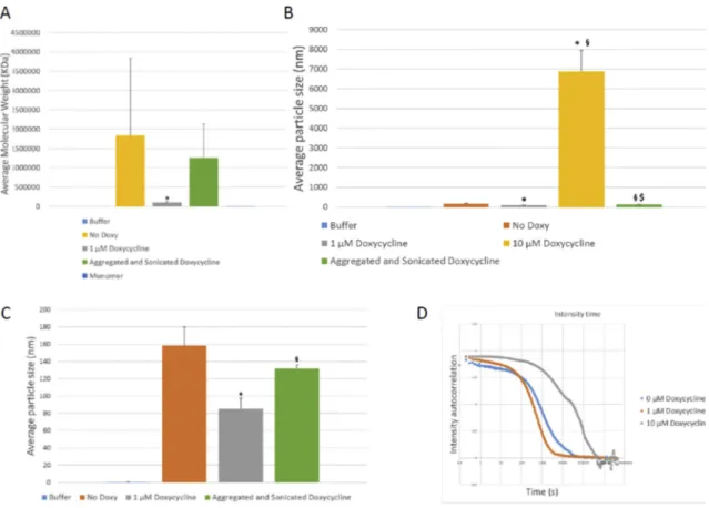
Next, in order to further assess the size of the aggregated species formed, we used dynamic light scattering (DLS). We detected very high molecular weight (HMW) species in samples amplified in the presence of
10 μM doxycycline (Fig. 1I, Fig. A.1). We also assessed the average
molecular weight of the monomers used for amplification, obtaining a result of 16.05 ± 1.8 kDa, consistent with the reported molecular weight of 14.5 kDa.
3.2. Doxycycline reduces seeded aSyn aggregation and the levels of reactive oxygen species generated by mitochondria
Next, in order to further investigate the effect of doxycycline on aSyn aggregation, we tested whether it affected the seeded aggregation of monomeric aSyn, as this might relate to the spreading of aSyn pathology as disease progresses. In vitro, in cell free models, the process of aSyn aggregation follows a typical sigmoidal curve, with a latency time of 16 h followed by an exponential phase that reached a stationary state
around 44 h (Fig. 2A), as previously described (Avila et al., 2014´ ). In
contrast, the presence of aSyn aggregates triggered a rapid aggregation of monomeric aSyn, abolishing the lag phase. However, when doxycy-cline was added to the reaction, the increase in ThT fluorescence in-tensity was completely blocked. This suggests that doxycycline “poisons” the seeding of aSyn aggregates species.
Mitochondrial dysfunction leads to the production of reactive oxygen species and can directly interact with a variety of specific proteins to be
involved in neurodegenerative disorders (Lin and Beal, 2006). For
example, in PD aSyn aggregates may induce neurodegeneration by causing a mitochondrial dysfunction and, thereby, the increased
pro-duction of reactive oxygen species (ROS) (Park et al., 2020; Santos and
Outeiro, 2020; Szeg˝o et al., 2019; Wang et al., 2019). For this reason, we measured intracellular ROS production in differentiated SH-SY5Y cells
treated with aggregates formed in the absence or presence of 100 μM
doxycycline. SH-SY5Y cells, as most neuronal cell lines, are more
sus-ceptible to oxidative stress than glial cells (Wang and Michaelis, 2010).
Using MitoSOX Red, we observed that SH-SY5Y cells exhibited a sig-nificant increase in intracellular ROS when in the presence of aSyn
ag-gregates (Fig. 2B). However, when the cells were treated with
aggregated aSyn, formed in the presence of doxycycline, this effect was almost completely blocked.
3.3. Doxycycline decreases the number of aSyn inclusions in cells In order to assess the effect of doxycycline treatment on aSyn ag-gregation in cell cultures, we used the SynT/Synphil1 model of
in-clusion formation (L´azaro et al., 2014; Masaracchia et al., 2020).
Synphilin-1 is an interacting partner of aSyn and SynT is a C-terminally modified variant of aSyn that is more prone to aggregation. H4 cells
Fig. 1. Effects of the presence of different doxycycline concentrations on in vitro amplification of aSyn by RT-QuiC. A. Effect of doxycycline on aSyn aggregation by RT-QuiC. Increasing concentrations of doxycycline lead to lower ThT fluorescence intensity. B. SDS-PAGE membrane of the RT-QuiC products developed with the BD anti-aSyn antibody. (b: Sample before amplification. a: Sample after amplification). C. SDS-PAGE membrane of the RT-QuiC products developed with the anti-alpha beta gamma aSyn antibody. (b: Sample before amplification. a: Sample after amplification). D. SDS-Page membrane of the RT-QuiC products developed with the 5 g4 anti- aggregated aSyn antibody. (b: Sample before amplification. a: Sample after 250 cycles of amplification). E. Semi-Denaturing gel membrane of the RT-QuiC products. (b: Sample before amplification. a: Sample after 250 cycles of amplification. n: Sample without PFF and SDS after 250 cycles of amplification used as negative control for RT- QuiC). Presence of doxycycline during RT-QuiC amplification lead to the formation of higher molecular weight species above equimolar concentrations. F. Epitopes for the anti-human aSyn (Syn-1 and C20) and anti-αβγSyn antibodies. G. Epitope for the 5G4 anti-aggregated human aSyn antibody. H. Electron microscopy pictures of the samples obtained in the RT-QuiC. (a) In samples without docycycline, abundant fibrillar structures are observed. Scale bar 360 nm. (b) Below equimolar concentrations (1 μM) abundant fibrillar structures are still observed. Scale bar 360 nm. (c) Above equimolar concentrations (10 μM) few fibrils are seen. Scale bar
360 nm. (d) Above equimolar concentrations large non-fibrillar structures are observed. Scale bar 360 nm. I. Average molecular weight obtained by DLS. The presence of 10 μM doxycycline leads to the formation of higher molecular weight species. Data presented as mean ± SD. *p < 0.05 in comparison with 0 μM
Doxycycline. §p < 0.05 in comparison with 1 μM Doxycycline. $p < 0.05 in comparison with 10 μM Doxycycline. Monomers displayed an average value of 16 kDa
were transiently transfected with constructs encoding for Synphilin-1
and SynT, as previously described (L´azaro et al., 2014), in order to
induce the formation of LB-like inclusions (Fig. A.2A), as characterized
by our and other groups (Engelender et al., 1999; Masaracchia et al.,
2020; McLean et al., 2001). Previous studies showed that some
in-clusions are positive to pS219aSyn (Smith et al., 2005; Büttner et al.,
2010; L´azaro et al., 2014; L´azaro et al., 2016). Seventeen hours after transfection, cells were treated with doxycycline at the following
con-centrations: 0.01 μM, 0.1 μM, 1 μM and 10 μM. The presence and
number of inclusions were assessed by immunocytochemistry (Fig. 3A)
24 h after treatment. An average of 50 positive cells for aSyn expression per treatment and experiment were then arbitrary divided into four different groups, depending on the number of inclusions present, i.e. cells without inclusions, cells with 1 to 5 inclusions, cells with 6 to 10
inclusions, and cells with more than 10 inclusions (Fig. 3 A-E). We
observed no statistically significant differences in the percentages of transfected cells showing less than six inclusions with any of the
doxy-cycline concentrations tested (Fig. 3 C). On the other hand, the
Fig. 2. Doxycycline interferes with aSyn seeding and mitochondrial ROS production. A. Doxycycline inhibits αSyn seeding in vitro. Fresh solutions of monomeric αSyn were seeded with aggregates and incubated in the absence (empty squares) or presence of doxycycline (empty diamonds) at 37 ◦C under orbital agitation.
Aggregation was assayed by ThT fluorescence emission. Unseeded aggregation kinetics are also shown (empty circles).
B. Intracellular ROS production visualized with MitoSOX Red probe in differentiated SH-SY5Y cells treated with aSyn aggregates produced in the presence or the absence of doxycycline. Scale bar: 100 μm. MitoSOX Red fluorescence intensities in the same culture conditions as in A *p < 0.05 vs NT.
percentage of cells containing more than 6 inclusions significantly
decreased when cells were treated with 10 μM doxycycline (Fig. 3 D, E).
Interestingly, the percentage of cells showing more than 10 inclusions
increased when cells were treated with 0.01, 0.1 and 1 μM. The total
number of inclusions per cell showed a tendency to increase in relation
with the control cells at 0.1 and 1 μM, with values of 4.05 ± 1.57, 3.97 ±
1.41, 5.86 ± 0.88 and 6.08 ± 1.56 inclusions per cell, respectively (Fig. A.2B). Treatment with 10 μM resulted in a drastic reduction in the
total number of inclusions per cell (1.28 ± 0.39 inclusions per cell). 3.4. Doxycyline reduces the size of aSyn inclusions at concentrations below 10 μM
After observing a slight increase in the percentage of cells showing
more than 10 inclusions (Fig. 3E), we wondered whether this was due to
the formation of smaller inclusions. To assess this, we checked the average inclusions size. Untreated cells showed average values of 7.85
± 0.82 μm2 and cells treated with 0.01 μM of doxycycline showed
average values of 6.80 ± 1.42 μm2. Interestingly, we observed a
sig-nificant decrease in average inclusion area when cells were treated with
0.1 and 1 μM, values been 5.13 ± 0.56 and 3.91 ± 0.79 μm2, respectively
(p = 0.03 for 0.1 μM, n = 3 and p = 0.004 for 1 μM, n = 3). This was
reverted when cells were treated with 10 μM doxycycline, as the mean
area was 10.50 ± 3.72 μm2 (Fig. 3F). The greater average size of
in-clusions in cells treated with 10 μM combined with the lower average
number of inclusions may suggest that, at 10 μM doxycycline, smaller
inclusions are disrupted while a few larger ones still remain, suggesting a breakage phenomenon consistent with the in vitro and EM data. Interestingly, larger inclusions remain at higher concentrations, sug-gesting that doxycycline may reach a point where aSyn is present mainly as oligomers that organize into large cellular inclusions that are not toxic to cells. Furthermore, the total inclusion area in the cells was also smaller, which points to not only a disruption of smaller inclusions but
to an overall lower presence of aggregated aSyn (Fig. A.2C).
To test whether doxycycline was disrupting the aggregates into smaller ones, we performed live cell imaging of cells transfected with aSyn-VC and VN-Synphilin-1 bimolecular fluorescence
complementa-tion constructs (L´azaro et al., 2014) and manuscript in preparation). As
the inclusions were formed through the interaction between aSyn and Synphilin-1, the VN- and VC- fragments of Venus fluorescent protein came into contact, thereby reconstituting the functional fluorophore and
emitting fluorescence (Outeiro et al., 2008). Using this aggregation
model we observed a decrease in particle size over time after treatment
with 10 μM doxycycline (Fig. A.2D), consistent with the disruption of
aSyn inclusions.
3.5. Doxycycline prevents the seeding of aSyn inclusions in PFF-treated cells
It has been reported that the addition of exogenous aSyn PFFs seeds
the aggregation of endogenous aSyn (Volpicelli-Daley et al., 2014).
Thus, based on our previous results, we hypothesized that doxycycline could inhibit this process. To assess this we treated, 48 h after plating, cells stably expressing aSyn-EGFP with PFFs at a final concentration of
100 nM of PFFs with 1 or 10 μM doxycycline. We observed a significant
decrease in the number of inclusions in cells treated with 10 μM
doxy-cycline (Fig. 4A, B) when compared to vehicle-treated cells.
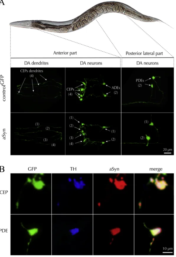
3.6. Expression of human aSyn in the dopaminergic neurons of C. elegans We first characterized the presence of the aSyn in the tagged dopa-minergic neurons. For this, we evaluated the specific targeting pattern of the dat-1p promoter using GFP as a reporter. We observed signals in all
the eight DA neurons previously described (Sulston et al., 1975),
including (i) the four CEP cell bodies, their associated neuritic exten-sions and the two ADEs neurons located in the anterior part of the ani-mals, and (ii) the two PDEs neurons in a more posterior lateral position (Fig. 5A). Using an anti-pan aSyn antibody, we confirmed that the aSyn
transgenic line expressed aSyn in all GFP+DA neurons (Fig. 5B). In
addition, GFP+neurons were also immuno-positive for TH. (Fig. 5B).
3.7. Doxycycline alters the subcellular distribution of aSyn in dopaminergic neurons in C. elegans
The total number of DA neurons tagged with GFP was similar in an-imals expressing aSyn and controls suggesting that the expression of aSyn does not induce DA neuronal loss. However, the expression of aSyn induced both a significant increase in the surface area of the DA cell
bodies of 60.58% (54.54 ± 3.44 μm2 for controlGFP versus 87.58 ± 7.73
μm2 for aSyn line; p = 0.0004) and a significant decrease of GFP density of
23.87% (118.5 ± 12.80 A.U. for controlGFP versus 90.21 ± 5.04 A.U. for
aSyn line; p = 0.03) suggesting a deleterious effect of aSyn expression on
DA neurons (Fig. 6A). Chronic treatment with 15 μM doxycycline did not
induce any change in the number of DA cell bodies or their neuritic processes in comparison to untreated controls. In contrast, this tetracy-cline concentration significantly decreased the swollen morphology of the soma induced by the aSyn transgene. In addition, doxycycline significantly improved the altered density of GFP fluorescent signal
induced by aSyn when comparing to untreated animals (Fig. 6A). In
addition, we performed immunohistochemistry of aSyn on whole animal observing a regular immunolabeling of the aSyn in the soma and along
the axons of the PDEs GFP tagged neurons (Fig. 6B, left panel). The
treatment with doxycycline altered the distribution of the protein in the soma compartment, presenting a punctuated expression pattern (white
arrows) (Fig. 6B, left panel). To evaluate these modifications, we
quan-tified the ratio of the surface area signal between the aSyn associated immunofluorescence and the GFP signal in the soma of PDE neurons. In the presence of doxycycline, we found a significant reduction (55.20%) of the surface area of the aSyn distribution signal relative to the soma of the
PDE neurons (Fig. 6B, right panel), suggesting, in combination with our
previous results, that larger non-toxic aggregates are formed. 3.8. Doxycycline restores motor alteration in aSyn C. elegans
The functionality of DA neurons was evaluated using a specific dopaminergic behavioural test which measure the locomotor velocity of the animal in a plate preceded with a short swimming activity in a liquid
media (Vidal-Gadea et al., 2011). As expected, we found that this
parameter was significant decreased in the cat-2 (e1112) TH null mutant
used as a reference line (Fig. 6C). Most interesting, this parameter was
Fig. 3. Doxycycline reduces aSyn inclusion size and number in H4 cells. A. Representative pictures of immunostained H4 cells without inclusions, with less than six inclusions, between six and ten inclusions and with more than 10 inclusions. Scale bar 25 μm. B. Percentage of cells that showed no inclusions in the absence or in the
presence of doxycycline (0.01–10 μM). C. Percentage of cells that showed less than six inclusions in the presence or not of doxycycline (0.01–10 μM). D. Percentage of
cells that showed between six and ten inclusions in the absence or presence of doxycycline (0.01–10 μM). Treatment with 10 μM doxycycline leads to a significant
decrease in the percentage of transfected cells containing between 6 and 10 inclusions. E. Percentage of cells that showed more than ten inclusions over the total number of transfected cells in the absence or presence of doxycycline (0.01–10 μM). There was a significant decrease in the percentage of cells treated with 10 μM
doxycycline. F. Average area of the inclusions in cells positive for aSyn expression at different doxycycline concentrations. Treatment with 0,1 and 1 μM doxycycline
leads to a significant decrease in the percentage of transfected cells that showed more than ten inclusions. Data presented as mean ± SD. N = 3 in all experiments * p <0.05 relative to control. ** p < 0.005 relative to control. ANOVA and subsequent t-test against control.
strongly reduced in the transgenic line expressing aSyn in DA neurons in
comparison to the control line (Fig. 6C). L-Dopa treatment significantly
restored dopaminergic function in both the cat-2 and aSyn lines. These results suggest that aSyn overexpression in the DA neurons resulted in a DA deficit inducing behavioural dysfunction despite the absence of neuronal loss. Of interest, doxycycline treatment restored locomotor
behaviour specifically on aSyn line (Fig. 6C) compared to the control
strain expressing only GFP (not shown), suggesting that the effect of doxycycline on aSyn distribution in DA neurons improved motor behaviour.
4. Discussion
The aggregation of aSyn is one of the hallmarks of synucleinopathies.
Fig. 4. Doxycycline prevents the formation of aSyn inclusions after treatment with exogenous PFFs. Treatment with PFFs at a concentration of 0.1 μM for 48 h in absence or presence of doxycycline at concentrations of 1 μM and 10 μM. A Presence of aSyn aggregates in HEK293-aSyn:EGFP stable cells treated with PFF and
visualized with anti-aSyn antibody (Scale bar 20 μm). DAPI was used for nuclear staining. B. Quantification of the inclusions formed in n > 200 cells per condition
(mean ± SD) reveals a significant decrease in the number of inclusions after treatment with 10 μM doxycycline. In contrast, treatment with 1 μM of doxycycline had no effect compared to non-treated cells. N = 3 independent experiments ** p < 0.005.
Fig. 5. Targeted expression of aSyn in C. elegans DA neurons. A. Illustration describing a C. elegans control line expressing the GFP reporter gene (controlGFP)
specifically in DA neurons and another line co-expressing the wild-type human SNCA gene and the GFP reporter (αS), in the same neurons. The observation was made
on fixed samples using an epifluorescence microscope fitted with a high magnification (x100) objective. The micrographs depict the expression pattern of the GFP reporter in the two lines and confirm the presence of six DA neurons in the head (CEPs and ADEs) and two DA neurons (PDEs) in the middle of the body. B. Panel showing the concomitant expression of TH (blue), aSyn (red) and GFP (green) in the CEP and PDE neurons. These observations confirm that aSyn is expressed in the soma and the dendrites of DA neurons.
In recent years, several aggregation inhibitors have been developed and
tested, and the search for new ones continues (Dominguez-Meijide et al.,
2020; Hengartner and Fernandez, 2019). Different drugs have been developed and tested, such as pyrazole derivatives and hydrazones (Cukierman et al., 2020; Cukierman et al., 2017; Wagner et al., 2013). Nonetheless, most of the drugs developed are either toxic or show low bioavailability in the brain due to low water solubility or other factors. To circumvent this, new therapeutic strategies like pharmacokinetic modulation (like complexation with cyclodextrins) or repurposing of
existing drugs have been developed (Barros et al., 2018; Heemskerk
et al., 2002). Examples of recent repurposing of existing drugs include inhibitors of the renin-angiotensin system, anti-asthmatic drugs, COX-2
inhibitors and other NSAIDs (Hengartner and Fernandez, 2019; Jang
et al., 2017; Moghaddam et al., 2007; Rodriguez-Perez et al., 2018). Following this trend, the anti-neuroinflammatory properties of
tetracy-clines such as minocycline and doxycycline have been studied (
Borto-lanza et al., 2018; Lazzarini et al., 2013). More recently, it has been proposed that tetracyclines and, specifically, doxycycline, have not only anti-neuroinflammatory properties, but can also inhibit the aggregation
of misfolded aSyn (Gonz´alez-Liz´arraga et al., 2017).
Our results in cell free systems showed that increasing concentra-tions of doxycycline lead to a decrease in ThT fluorescence intensity in an RT-QuiC assay. This is probably due not only to a decrease in the aggregation rate of aSyn, but also to a possible change in the confor-mation of the aSyn aggregates formed. As ThT becomes fluorescent upon binding to beta-sheet structures, a possible change in aSyn aggregation may lead to lower ThT binding and, thereby, to lower ThT fluorescence (Wu et al., 2009). Indeed, it has been proposed that treatment with doxycycline induces a conformational change, as infrared studies demonstrated the presence of parallel beta-sheet instead of the typical antiparallel beta-sheet found in on-pathway toxic oligomeric species (Celej et al., 2012). Furthermore, our WB, EM and DLS results confirm previous observations where it was reported that the presence of doxycycline induces a remodelling of aSyn oligomers into off-pathway
non-toxic, non-seeding, high molecular weight species (
Gonz´alez-Liz-´
arraga et al., 2017). In fact, it has been shown that different aSyn fibril polymorphs can exhibit differential ThT binding, consistent with some
of the high molecular weight species we detected (De Giorgi et al.,
2020). Our results and previous ones also prove that this effect is dose-
dependent (Gonz´alez-Liz´arraga et al., 2017). In addition, our RT-QuiC
results show a decrease in ThT fluorescence intensity at high doxycy-cline concentrations. As these reactions were seeded, this suggests additional effects on the fibrils. Furthermore, we also demonstrate that while aSyn aggregates are very efficient at seeding native aSyn, as
revealed by ThT assays, they lose all seeding ability when incubated with doxycycline.
Our results in cells also show the ability of doxycycline to break intracellular aSyn-positive inclusions, as we observed an increase in the number inclusions per cell that was accompanied by a decrease in their size. This was further confirmed by live cell imaging. The lower per-centage of cells with more than ten inclusions when cells were treated
with 10 μM, and the higher average inclusion size may be either due to
more compact inclusions that cannot be decomposed by doxycycline or to a possible reorganization of aSyn inside individual cells that leads to fewer but larger inclusions. In addition, it has been proposed that exog-enous aSyn fibrils can be internalized and act as templates for the
for-mation of aggregates by addition of endogenous monomeric aSyn (Grassi
et al., 2018; Masaracchia et al., 2018; Volpicelli-Daley et al., 2014). These results, in combination with those observed in cell free systems, further support the idea of doxycycline being able to exert inhibition of seeding. Thus, doxycycline not only degrades already formed aggregates, but it can also inhibit the formation of new ones. Interestingly, our results also suggest that the formation of certain aggregated species may be higher in the aSyn-VC and VN-Synphilin-1 BiFC models those in the SynT/Synph-1 model, as higher doxycycline concentrations in this model still render smaller inclusions. It has been reported that an equimolar concentration of doxycycline to aSyn would be suitable to reach a protective effect (Gonz´alez-Liz´arraga et al., 2017). The concentration of 10 μM may be
already above equimolar levels as the concentration of aSyn in cell
syn-apse is ~50 μM (Perni et al., 2017; Wilhelm et al., 2014). Furthermore,
the neuroprotective and anti-inflammatory doses of doxycycline are
below the ones needed to exert its antibiotic activity (Gonz´alez-Liz´arraga
et al., 2017; Santa-Cecília et al., 2019).
An additional pathological effect of aSyn does not come from its aggregation and propagation. It may also cause mitochondrial dysfunction and enhanced production of ROS, which may, in turn, further modulate aSyn oligomerization and cytotoxicity, leading to a vicious circle. The generation of ROS by aSyn aggregates can affect macromolecules or activate signaling pathways that culminate in central
nervous system neurodegeneration (Park et al., 2020; Wang et al.,
2019). For this reason, the inhibition of oxidative stress could offer an
effective therapeutic approach to mitigate the progression of PD. In concordance with this concept, we show that aSyn aggregates increase the production of ROS in differentiated SH-SY5Y cells. However, aSyn aggregates formed in presence of doxycycline are less prone to induce oxidative stress in cell cultures. In this regard, minocycline and doxy-cycline have been proposed to protect against mitochondrial dysfunc-tion. Furthermore, the proposed mechanism of inhibition of aSyn
Fig. 6. Cellular and behavioural consequences of doxycycline treatment in C. elegans. A. After synchronization, a homogeneous population of young adult animals treated or not with doxycycline (15 μM), were PFA-fixed (4%), mounted on slide and observed with an epifluorescence microscope equipped with a x63 objective.
(Upper panel) Images illustrate the most characteristic features of DA GFP-tagged neurons (PDE, arrows head). (Lower panel) Quantification of the number of GFP+
cell bodies and dendrites, the surface area of GFP+ cell bodies and the somal intensity of the GFP fluorescent signal in control and aSyn-expressing animals. Bar graphs show that aSyn expression did not affect the number of DA neurons but induced significant morphological alterations, such an increase in the somal surface area and a decrease of the GFP fluorescence signal. Such changes were prevented by a chronic exposure to doxycycline. Data are expressed as mean values ± s.e.m. Three independent experiment; n = 25/condition/experiment. Statistical significance was calculated with various tests: (i) a one-way ANOVA followed by a Dunnett’s multiple comparisons tests with all condition versus the untreated control line (controlGFP), *p < 0,05 and ***p < 0,005; (ii) a two-tailed unpaired t-test for aSyn
animals treated or not with doxycycline, #p < 0,05, and ##p < 0,01; n.s: not significant. B. Left panel: Illustrations showing the impact of doxycycline (15 μM) on the
distribution of human aSyn in the soma of DA neurons. Representative images of the wild-type human aSyn immunosignal (red signal) in the PDE-DA GFP tagged neurons (green). In untreated animals the immunosignal for aSyn is distributed over a large part of the cell body in contrast with worms treated with doxycycline where the signal is punctuated and restricted to a small intracellular compartment (white arrows). Right panel: Measurement of the impact of the doxycycline treatment on the surface area occupied by the aSyn immunosignal in the soma of PDE DA neurons. The surface area of the aSyn immunosignal expressed as a ratio to the total somal surface area of GFP positive neurons, appears significantly decreased. Data are mean values ± s.e.m. Three independent experiment, n = 20/con-dition/experiment. Statistical significance was calculated with two-tailed unpaired t-test for the two experimental conditions; ***p < 0,005. C. Impact of the doxycycline treatment on behavioural crawling velocity after swim-to-crawl transition. Note that the loss of velocity in animals expressing wild-type aSyn is significantly prevented by a chronical doxycycline treatment in comparison with the untreated line. The null mutant cat-2 (e1112) /TH was used as positive control for the assays. A treatment of the animals with 1 mM of L-DOPA 2 h before the test prevented the behavioural defect observed in the line expressing aSyn. These results suggest that DA neurons are affected by the presence of the aSyn. Data are mean values ± s.e.m. Three independent experiment, n = 30/condition/exper-iment. Statistical significance was calculated with various tests: (i) a one-way ANOVA followed by a Dunnett’s multiple comparisons test versus the GFP control line (ControlGFP); **p < 0,01, n.s is not significant; (ii) a two-tailed unpaired t-test for cat-2 (e1112) animals treated or not with L-Dopa, ####p < 0,001; (iii) a two-tailed
aggregation may also reduce its detrimental effects on mitochondrial
function (Garcia-Martinez et al., 2010; Santa-Cecília et al., 2016).
In recent years, several aSyn-inducible cell models have been developed, including tet-on and tet-off inducible systems, which are based on the induction or blocking of aSyn expression in the presence of
doxycycline, respectively (Vekrellis et al., 2009; Zhu et al., 2002). These
induction systems have been developed in several cell lines and animal
models (Harding et al., 1997; Takahashi et al., 2007; Vasquez et al.,
2018; Vekrellis et al., 2009). In tet-on systems, addition of doxycycline leads to the expression of aSyn at levels that enable the formation of
inclusions (Vasquez et al., 2018). The concentrations of doxycycline
used in these publications range between 2.25 and 6.7 μM (Harding
et al., 1997; Vasquez et al., 2018). Our results show that a concentration
of 10 μM efficiently modulates aSyn aggregation in cellular models.
Therefore, the concentration of doxycycline required to modulate pro-tein aggregation in our culture setting appears higher than that required for inducing aSyn expression in aSyn-inducible cell models.
To further test the biochemical properties of doxycycline in this context, we generated a new in vivo transgenic model of PD in C. elegans expressing wild-type aSyn in the DA neurons. Unlike previously estab-lished C. elegans models designed to model different features of PD (Cooper and Van Raamsdonk, 2018), and target tissues like muscle (Hamamichi et al., 2008; van Ham et al., 2008), the entire neuronal
system (Kuwahara et al., 2006; Kuwahara et al., 2008; Lakso et al.,
2003), or the DA system (Cao et al., 2005; Lakso et al., 2003), our model
uses the dat-1 promoter. Dat-1 is the ortholog of the human dopamine transporter in the nematode, and is selectively expressed in the DA system. Thus, our model enables the in vivo study of the specific vulnerability of DA neurons expressing aSyn. We verified the correct targeting of the transgene in the cell soma using GFP as a reporter. Human wild-type aSyn, co-expressed with the GFP reporter under the same genetic regulatory system, is present in the soma of the DA neu-rons. In young-adult animals, aSyn did not induce significant DA neuronal loss until the worms reached 6 days of age. In another C. elegans model expressing aSyn in the DA system, the authors described the presence of aSyn-dependent neuronal injuries, probably resulting
from differences in the expression levels of the transgene (Cao et al.,
2005). However, in our model aSyn induced abnormal neuronal
mor-phologies consistent with a dysfunctional DA system, as indicated by a noticeable alteration of DA-associated behaviour, suggesting that over-expression of aSyn impacts DA homeostasis. These irregular neuronal morphologies and pathological behaviour are restored after chronic treatment with doxycycline, consistent with the effects observed in vitro model, and reinforcing the hypothesis that doxycycline may act as a neuroprotective agent.
It has been proposed that C. elegans movement depends not only on DA, but also on cholinergic neuron-dependent muscle contraction and GABAergic neuron-dependent relaxation. However, it has been shown that blockade of DA D2 receptors does not fully limit paralysis triggered
by changes in cholinergic and GABAergic signaling (Refai and Blakely,
2019). The opposite results have also been shown, i.e. changes in
cholinergic and GABAergic signaling due to D2 antagonism (Krum et al.,
2020). Furthermore, it was also shown that changes in DA may affect
GABAergic egg release in the worms (Cermak et al., 2020). These results
demonstrate that further research into these pathways is necessary, but this is beyond the scope of our study.
In our study, we employed a classic transgenesis technique (Mello
and Fire, 1995) to produce our line, resulting in the production of transgenic animals carrying multi-copy arrays of the SNCA transgene, which may induce non-specific neurobiological process. Additionally, some studies showed that expression of aSyn in DA motor neurons of C. elegans recapitulates features of aSyn neuronal propagation. Using the
BiFC assay (Outeiro et al., 2008), we showed the protein could
disseminate to the neighbouring serotoninergic neurons and interact
with another host targeted aSyn (Tyson et al., 2017). These processes
have been extensively described in mammals for synucleinopathies and
other proteinopathies (Peng et al., 2020). Here, we did not observe a
significant neuronal propagation of aSyn, which might be possibly explained by the fact that we used a different promoter gene to induce aSyn expression. In addition, we have not investigated the tissue expression pattern of aSyn in old animals, as this was beyond the scope of the present study.
In C. elegans the treatment with doxycycline induces a significant
effect on development by reducing the growth rate (Letizia et al., 2018),
and it appears to promote longevity via inhibition of mrps-5 ribosomal
protein and atf-4 transcription factor mitochondrial pathways (
Mole-naars et al., 2020). Doxycycline has been described to play role in the inhibition of the translation process effect by suppressing the binding of tRNAs to the ribosomal-machinery system and inducing a global
decrease in protein production (Nelson and Levy, 2011). Thus, we
cannot rule out that the neuroprotective effect of doxycycline in our experimental conditions may also be explained via an indirect activation of the aging-metabolic cellular pathways and the observed effect on mitochondria and ROS levels, as decreased ROS may lead to a lower
level of neuroinflammation and neuroprotective effects (Picca et al.,
2020). Since aging is the major risk factor for PD, it will be interesting to
assess the potential interaction between aging-associated pathways and aSyn in the novel transgenic lines we generated.
Strikingly, we observed modifications in aSyn distribution in the DA neurons in aSyn-transgenic animals chronically incubated with doxy-cycline. In particular, we observed a reduction of the sub-cellular dis-tribution of the protein in the soma. This occurred together with the effects on aSyn aggregation, further strengthening the anti-aggregation properties of doxycycline in vivo, and suggesting additional comple-mentary effects on aSyn to those observed in vitro and in cell-free models. The punctate pattern observed in DA neurons in vivo are consistent with a combined effect of changes in cellular distribution and the formation of fewer and larger inclusions inside the cells, as observed in vitro. These changes lead to an overall decrease in aSyn area in rela-tion to the total surface of the cell soma of the PDE neurons. This is not an independent effect, but the combination of different effects, i.e. in-hibition of aggregation, disruption of pre-formed inclusions, blocking of seeding, and changes in inclusion area leading, ultimately, to the distinct dotted pattern observed in vivo. Additionally, aggregated species of aSyn produced in the presence of doxycycline lost their capacity to stimulate mitochondrial ROS production, which may also account for the ration of DA neuron function. In fact, our results indicate that a resto-ration of motor behaviour by doxycycline can be explained by the combination of the aforementioned decrease in ROS, combined with changes in aSyn processing, seeding and distribution.
As mentioned above, doxycycline has neuroprotective effects in several models of neurodegenerative disease and, in particular, has anti-
aggregation properties (Costa et al., 2011; Paldino et al., 2020; Santa-
Cecília et al., 2019). In a C. elegans amyloidogenesis model induced by the expression of a human pathogenic variant of β2-microglobulin
(D76N of β2-m) in the muscle (Faravelli et al., 2019), doxycycline
de-creases the proportion of oligomeric fraction produced by the β2-m
ag-gregation, resulting in an improvement of the motor symptoms (Raimondi et al., 2017). These findings are consistent with those we report herein.
5. Conclusions
In summary, our data provides novel insight into the mechanisms by which doxycycline may inhibit the aggregation of aSyn by remodelling aSyn oligomers into off-pathway non-toxic, non-seeding, high molecular weight species. We showed this effect not only in vitro but also in cell free models. The extreme need for alternative strategies against PD calls for attempts for repurposing existing drugs. In this context, our data sug-gests that doxycycline may act at different levels, modulating the ag-gregation process and the effects of aSyn agag-gregation, promoting somatic repair, and leading to motor recovery in C. elegans.