HAL Id: hal-03101594
https://hal.sorbonne-universite.fr/hal-03101594
Submitted on 7 Jan 2021
HAL is a multi-disciplinary open access
archive for the deposit and dissemination of
sci-entific research documents, whether they are
pub-lished or not. The documents may come from
teaching and research institutions in France or
abroad, or from public or private research centers.
L’archive ouverte pluridisciplinaire HAL, est
destinée au dépôt et à la diffusion de documents
scientifiques de niveau recherche, publiés ou non,
émanant des établissements d’enseignement et de
recherche français ou étrangers, des laboratoires
publics ou privés.
SARS-CoV-2 Does Not Spread Through Extracorporeal
Membrane Oxygenation or Dialysis Membranes
Martin Dres, Sonia Burrel, David Boutolleau, Guillaume Voiriot, Alexandre
Demoule, Alain Combes, Guillaume Lebreton, Matthieu Schmidt
To cite this version:
Martin Dres, Sonia Burrel, David Boutolleau, Guillaume Voiriot, Alexandre Demoule, et al..
SARS-CoV-2 Does Not Spread Through Extracorporeal Membrane Oxygenation or Dialysis Membranes.
American Journal of Respiratory and Critical Care Medicine, American Thoracic Society, 2020, 202
(3), pp.458-460. �10.1164/rccm.202004-1339LE�. �hal-03101594�
SARS-CoV-2 Does Not Spread Through Extracorporeal Membrane Oxygenation or Dialysis Membranes To the Editor:
Severe acute respiratory syndrome coronavirus 2 (SARS-CoV-2) has become a major worldwide health threat in just a
few weeks (1). ICU admission and the recourse to extracorporeal organ support, such as continuous renal replacement therapy (CRRT) or venovenous extracorporeal membrane oxygenation (VV-ECMO) may be needed in the most severe forms of the disease (2). Because SARS-CoV-2 viremia has been reported in some cases (3), it has been hypothesized that this small virus (average size of 125 nm) (4) could pass through polymethylpentene ECMO
membranes or acrylonitrile/sodium methallylsulfonate CRRT membranes. In this study, we investigated whether SARS-CoV-2 RNA was detected in the dialysis effluent fluid or in the condensate collected from the ECMO membrane exhalation port (gas outlet) when the virus was present in the lower respiratory tract and the plasma.
Methods
We evaluated consecutive patients admitted to three university ICUs in Paris who had severe SARS-CoV-2 infection and required CRRT (hemodiafiltration or hemofiltration), VV-ECMO, or both. Samples were obtained from respiratory tract, plasma, the dialysis effluent fluid, and from 5 to 10 ml of condensate collected
from the ECMO membrane gas outlet within 48 hours after ECMO initiation. Real-time RT-PCR targeting the E (envelope) gene of SARS-CoV-2 was performed as previously described (5). The cycle threshold (CT) values of RT-PCR were used as indicators of the RNA viral load in samples: the lower the CT, the higher the RNA viral load. The estimated probability (95% confidence interval [CI]) and the binomial probability of SARS-CoV-2 in the gas outlet and the dialysisfluid were reported, respectively. Ethical approval was applied to our local ethics committee (CER Sorbonne University, N82020–CER-2020–32).
Results
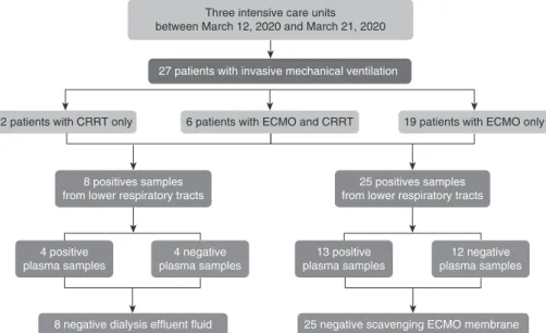
All 27 patients were on mechanical ventilation, and 25/27 were supported by VV-ECMO (20 patients with Quadrox oxygenator [Getinge] and 5 patients with Oxymedos oxygenator [Xenios]). In addition, 8/27 patients received CRRT (Prismaflex, Baxter). CRRT was administered using
hemofiltration in four patients and hemodiafiltration in four patients. Mainfindings are presented in Figure 1. SARS-CoV-2 RNA was detected in all samples from patients’ lower respiratory tract (median CT, 28; 25–75% interquartile range, 22–31) and in the plasma of 13/27 of them (median CT, 29; interquartile range, 29–30). However, SARS-CoV-2 RNA was not detected in the membrane oxygenator gas outlet condensate, whether plasma RNA was positive (n = 13/25) or negative (n = 12/25). Similarly, SARS-CoV-2 RNA was not present in the dialysis effluent of the eight patients on CRRT whether plasma PCR was positive (n = 4/8) or negative (n = 4/8). Therefore, the estimated probability of a positive SARS-CoV-2 RNA in the membrane oxygenator gas outlet condensate and in the dialysisfluid were 0.0 (95% CI, 0.00–0.14) and 0.0 (95% CI, 0.00–0.37), respectively. On the basis of binomial probabilities of our results, the prevalence of a positive SARS-CoV-2 RNA in the ECMO gas outlet and in the dialysisfluid will likely be lower than 11% and 31%, respectively. Individual data for SARS-CoV-2 RNA detection in lower respiratory tracts, plasma, dialysisfluid, and ECMO membrane are given in Table 1 with the CT values resulting from PCR for lower respiratory tracts (column 2) and plasma (column 3).
Discussion
To the best of our knowledge, this is thefirst study that investigated the risks for SARS-CoV-2 dissemination through membranes used for extra corporeal organ support in critically ill patients. Though a recent report revealed that SARS-CoV-2 is almost always present in the lower respiratory tract, sometimes in the feces but never in urine samples (3), ourfindings are reassuring regarding the risk of contamination for ICU professionals when treating patients on VV-ECMO or CRRT. Specifically, ourfindings do not support the routine use of a viral filter on the exhaust of the commonly used polymethylpentene-based ECMO membrane lungs. Prevention and education of
healthcare workers should therefore remain focused on limiting the risks of virus spreading during invasive respiratory procedures, such as high-flow oxygenation, mouth care, intubation, or microbiological sampling of nasopharyngeal, tracheal, or bronchioalveolar secretions. The number of patients with CRRT (n = 8) is limited, but the fact that SARS-CoV-2 PCR was negative in all dialysis effluent is somehow reassuring. Lastly, we cannot rule out that longer ECMO runs could progressively lead to membrane alteration, plasma leakage, and ultimately SARS-CoV-2 aerosolization. However, we purposely chose to investigate the risk of virus spreading within 48 hours after ECMO and CRRT initiation as the viral load—if present in the plasma—is expected to progressively decline afterward. Though ourfindings may not alter practices, they may contribute to address legitimate interrogations raised by caregivers and reinforce adhesion and trust into infection control measure policies, which is likely to play a major role against the outbreak spreading.n
This article is open access and distributed under the terms of the Creative Commons Attribution Non-Commercial No Derivatives License 4.0 (http:// creativecommons.org/licenses/by-nc-nd/4.0/). For commercial usage and reprints, please contact Diane Gern ([email protected]).
Originally Published in Press as DOI: 10.1164/rccm.202004-1339LE on January 11, 2020
CORRESPONDENCE
Table 1. Detection of SARS-CoV-2 RNA in the Condensate Collected from the ECMO Membrane Gas Outlet and in the Dialysis Effluent Fluid
Lower Respiratory Tracts
RT-PCR (CT) Plasma RT-PCR (CT) Dialysis RT-PCR (CT)
ECMO Membrane Gas Outlet RT-PCR (CT)
Patient 1 32 Undetectable No CRRT Undetectable
Patient 2 13 29 Undetectable Undetectable
Patient 3 Positive* 33 Undetectable Undetectable
Patient 4 28 Undetectable Undetectable No ECMO
Patient 5 Positive* Undetectable Undetectable No ECMO
Patient 6 29 30 No CRRT Undetectable
Patient 7 35 Undetectable No CRRT Undetectable
Patient 8 21 Undetectable No CRRT Undetectable
Patient 9 24 28 No CRRT Undetectable
Patient 10 24 Undetectable Undetectable Undetectable
Patient 11 19 30 Undetectable Undetectable
Patient 12 26 30 Undetectable Undetectable
Patient 13 29 Undetectable No CRRT Undetectable
Patient 14 36 Undetectable No CRRT Undetectable
Patient 15 33 Undetectable No CRRT Undetectable
Patient 16 Positive* 50 No CRRT Undetectable
Patient 17 Positive* Undetectable No CRRT Undetectable
Patient 18 18 29 No CRRT Undetectable
Patient 19 18 30 No CRRT Undetectable
Patient 20 27 Undetectable No CRRT Undetectable
Patient 21 15 28 No CRRT Undetectable
Patient 22 30 Undetectable No CRRT Undetectable
Patient 23 30 29 Undetectable Undetectable
Patient 24 29 Undetectable No CRRT Undetectable
Patient 25 23 29 No CRRT Undetectable
Patient 26 33 Undetectable No CRRT Undetectable
Patient 27 32 28 No CRRT Undetectable
Definition of abbreviations: CRRT = continuous renal replacement therapy; CT = cycle threshold; ECMO = extracorporeal membrane oxygenation; SARS-CoV-2 = severe acute respiratory syndrome coronavirus 2.
*Patient tested positive before being transferred in our center; no CT was provided.
25 negative scavenging ECMO membrane 8 negative dialysis effluent fluid
4 positive plasma samples
13 positive plasma samples 8 positives samples
from lower respiratory tracts
25 positives samples from lower respiratory tracts
4 negative plasma samples
12 negative plasma samples
2 patients with CRRT only 6 patients with ECMO and CRRT 19 patients with ECMO only
27 patients with invasive mechanical ventilation
Three intensive care units between March 12, 2020 and March 21, 2020
Figure 1. Detection of severe acute respiratory syndrome coronavirus 2 (SARS-CoV-2) RNA in dialysis effluent fluid or in the exhalation port of the extracorporeal membrane oxygenation membrane according to plasma detection of the viral RNA. CRRT = continuous renal replacement therapy; ECMO = extracorporeal membrane oxygenation.
CORRESPONDENCE
Author disclosures are available with the text of this letter at www.atsjournals.org.
Martin Dres, M.D., Ph.D.*
UMR_S 1158 Neurophysiologie respiratoire exp ´erimentale et clinique Paris, France
and
AP-HP Sorbonne Universit ´e Paris, France Sonia Burrel, M.D., Ph.D. David Boutolleau, M.D., Ph.D. INSERM UMR_S 1136 Paris, France and
AP-HP Sorbonne Universit ´e Paris, France
Guillaume Voiriot, M.D., Ph.D. AP-HP Sorbonne Universit ´e Paris, France
Alexandre Demoule, M.D., Ph.D.
UMR_S 1158 Neurophysiologie respiratoire exp ´erimentale et clinique Paris, France
and
AP-HP Sorbonne Universit ´e Paris, France
Alain Combes, M.D., Ph.D. Guillaume Lebreton, M.D., Ph.D. Matthieu Schmidt, M.D., Ph.D.
UMRS_1166-ICAN Institute of Cardiometabolism and Nutrition Paris, France
and
AP-HP Sorbonne Universit ´e Paris, France
For the Groupe de Recherche Clinique RESPIRE *Corresponding author (e-mail: [email protected]).
References
1. MacLaren G, Fisher D, Brodie D. Preparing for the most critically ill patients with COVID-19: the potential role of extracorporeal membrane oxygenation. JAMA [online ahead of print] 19 Feb 2020; DOI: 10.1001/jama.2020. 2342.
2. Ramanathan K, Antognini D, Combes A, Paden M, Zakhary B, Ogino M, et al. Planning and provision of ECMO services for severe
ARDS during the COVID-19 pandemic and other outbreaks of emerging infectious diseases. Lancet Respir Med 2020;8: 518–526.
3. Wang W, Xu Y, Gao R, Lu R, Han K, Wu G, et al. Detection of SARS-CoV-2 in different types of clinical specimens. JAMA 2020;323: 1843–1844.
4. Fisher D, Heymann D. Q&A: the novel coronavirus outbreak causing COVID-19. BMC Med 2020;18:57.
5. Corman VM, Landt O, Kaiser M, Molenkamp R, Meijer A, Chu DK, et al. Detection of 2019 novel coronavirus (2019-nCoV) by real-time RT-PCR. Euro Surveill 2020;25: 2000045.
Copyright © 2020 by the American Thoracic Society
Extracorporeal Membrane Oxygenation for Critically Ill Patients with COVID-19–related Acute Respiratory Distress Syndrome: Worth the Effort?
To the Editor:
According to Chinese and Italian reports, 15–42% of patients with coronavirus disease (COVID-19) develop acute
respiratory distress syndrome (ARDS), with a 60% mortality rate (1–3). Venovenous extracorporeal membrane oxygenation (VV-ECMO) is therefore considered a rescue therapy to be used in the most severe ARDS, as recommended by the World Health Organization’s interim guidelines for the management of patients with COVID-19 (4). However, without a significant impact on mortality, the benefit of ECMO in ARDS remains controversial (5). Generally, only few data on the use of ECMO in the present pandemic are available (1, 2) with a short follow-up (6, 7). However, accurately selecting patients with COVID-19–related ARDS, who may be good candidates for ECMO support, is important during a pandemic characterized by limited medical resources.
Methods
We prospectively included all patients referred to thefive ICUs of the Strasbourg University Hospital, between March 3 and April 1, 2020, for severe ARDS due to COVID-19 (confirmed by RT-PCR test), and that had been supported by ECMO after failure of optimal medical treatment, including neuromuscular blocking agents, protective ventilation, and high positive end-expiratory pressure (PEEP). According to EOLIA (ECMO to Rescue Lung Injury in Severe ARDS) criteria (5), patients were eligible for ECMO if they developed a refractory ARDS defined by a PaO2/FIO2, 80 mm Hg
or a pH, 7.25 with a PaCO2. 60 mm Hg for more than 6 hours with a FIO2. 80%, despite low-pressure ventilation strategies
and no participation offluid overload. The contraindications for ECMO implantation were an age older than 70 years and severe comorbidities, including severe chronic respiratory failure, severe cardiac failure, and Child Pugh C cirrhosis. Invasive
This article is open access and distributed under the terms of the Creative Commons Attribution Non-Commercial No Derivatives License 4.0 (http:// creativecommons.org/licenses/by-nc-nd/4.0/). For commercial usage and reprints, please contact Diane Gern ([email protected]).
Supported by grants from the Strasbourg University Hospital (Les H ˆopitaux Universitaires de Strasbourg, Direction de la Recherche Clinique et des Innovations).
Author Contributions: P.-E.F. and F.M. had full access to all of the data in the study and take responsibility for the integrity of the data and the accuracy of the data analysis. Concept and design: P.-E.F., A.M., and F.M. Acquisition, analysis, or interpretation of data: P.-E.F., A.M., M.P., S.P., P.-O.L., A.O., P.-M.M., F.S., J.H., and F.M. Drafting of the manuscript: P.-E.F., A.M., M.P., J.H., and F.M. Critical revision of the manuscript for important intellectual content: all authors. Statistical analysis: M.P. Administrative, technical, or material support: all authors. Supervision: P.-E.F., M.P., J.H., and F.M. Originally Published in Press as DOI: 10.1164/rccm.202004-1370LE on June 16, 2020