HAL Id: tel-00455270
https://tel.archives-ouvertes.fr/tel-00455270
Submitted on 9 Feb 2010
HAL is a multi-disciplinary open access
archive for the deposit and dissemination of sci-entific research documents, whether they are pub-lished or not. The documents may come from teaching and research institutions in France or abroad, or from public or private research centers.
L’archive ouverte pluridisciplinaire HAL, est destinée au dépôt et à la diffusion de documents scientifiques de niveau recherche, publiés ou non, émanant des établissements d’enseignement et de recherche français ou étrangers, des laboratoires publics ou privés.
Structural and functional studies of AMSH implicated in
the endosomal sorting pathway and enveloped virus
budding.
Julianna Solomons
To cite this version:
Julianna Solomons. Structural and functional studies of AMSH implicated in the endosomal sorting pathway and enveloped virus budding.. Biomolecules [q-bio.BM]. Université Joseph-Fourier - Grenoble I, 2009. English. �tel-00455270�
Université Joseph Fourier - Grenoble I
Ecole Doctorale Chimie et Sciences du Vivant
Pour obtenir le titre de:
DOCTEUR DE L’UNIVERSITÉ JOSEPH FOURIER - GRENOBLE I
en BIOLOGIE STRUCTURALE ET NANOBIOLOGIE
Soutenance le 26 Novembre 2009
devant le jury:
Président:
Rapporteurs:
Examinateurs:
Thèse préparée au sein de:
EMBL Grenoble
UVHCI (Unit of Virus Host Cell Interactions) UMI3265 UJF-EMBL-CNRS
Par Julianna Solomons
Études Structurale et Fonctionelle d’AMSH
impliquée dans la voie de tri endosomale
et le bourgeonnement des virus enveloppés
Rémy Sadoul
Robin Buckland
John Briggs
Wim Burmeister
Winfried Weissenhorn
Laurence Aubry
Prof.
Dr.
Dr.
Prof.
Prof.
Dr.
Université Joseph Fourier - Grenoble I
Ecole Doctorale Chimie et Sciences du Vivant
To obtain the title of:
DOCTOR OF UNIVERSITÉ JOSEPH FOURIER - GRENOBLE I
in STRUCTURAL BIOLOGY AND NANOBIOLOGY
To be defended 26th Novembre 2009
before a jury of:
President:
Reporters:
Examinators:
Thesis work conducted at:
EMBL Grenoble
UVHCI (Unit of Virus Host Cell Interactions) UMI3265 UJF-EMBL-CNRS
By Julianna Solomons
Structural and Functional Studies of AMSH
implicated in the Endosomal Sorting Pathway
and Enveloped Virus Budding
Rémy Sadoul
Robin Buckland
John Briggs
Wim Burmeister
Laurence Aubry
Winfried Weissenhorn
Prof.
Dr.
Dr.
Prof.
Dr.
Prof.
Acknowledgements Acknowledgements I would like to recognise the scientific direction of my PhD supervisor Winfried Weissenhorn, and thank him for the opportunity to join his research group on such an interesting project. I thank the members of my Thesis Advisory Committee, Stephen Cusack and Klaus Scheffzek for their helpful advice, and my jury members Rémy Sadoul, Laurence Aubry, John Briggs, Wim Burmeister and Robin Buckland for taking time out of their busy schedules to participate in my thesis defense. I am grateful to the EMBL to have accepted me into their PhD Programme, to the UVHCI who welcomed me two years later, and to the DFG for funding my work at the UVHCI. I would like to thank the people of the EMBL Grenoble and UVHCI, past and present, for creating such a pleasant work environment, and the staff of both establishments who ensure the smooth running of the institutes.
I would like to thank Charles Sabin for the crystallography‐related work, and for teaching me about data collection and structure determination, Jennifer McCarthy for her contribution to the proteolysis project, Guy Schoen for electron microscopy, Marc Jamin for MALLS, and Heinrich Göttlinger for the viral budding assays. My profound gratitude to Estela Pineda‐ Molina for her advice, patience and good humour, and Ricardo Pires for helpful discussions, his support and most of all for his understanding. Je remercie Alexandre Dias, pour l’élimination de l’accent anglais dans les parties traduits de mon manuscrit, et d’avoir partagé cette expérience de la thèse avec moi. Most importantly, I would like to thank my parents Stephen and Christine Solomons, whose unfailing love, support, and wisdom provide the foundations for all my endeavours in life. Julianna
Abstract... i Resumé...iii List of Abbreviations ……….………. v Introduction... 1 0.1 The Endosomal Sorting Pathway ... 1 0.2 The ESCRT machinery ... 2 0.3 ESCRT‐0... 3 0.4 ESCRT‐I and ESCRT‐II ... 5 0.5 ESCRT‐III... 6 0.6 Interaction of ESCRT‐III with MIT domain‐containing proteins... 10 0.7 Recruitment of Deubiquitinating enzymes (DUBs)... 11 0.8 AMSH domain architecture and binding partners... 12 0.9 Suggested mechanisms for AMSH in Endosomal Sorting... 14 0.10 The ESCRT machinery in HIV budding ... 15 0.11 A New Role for ESCRT in Cytokinesis ... 17 Aims and Objectives ... 19 Materials and Methods... 21 Results 1 – Determining soluble AMSH constructs... 31 Results 2 – Interaction of the AMSH N‐terminal domain with CHMP3 ... 37 Results 3 – Structure of the AMSH N‐terminal domain CHMP3 Complex ... 51 Results 4 – Pursuit of Full‐Length AMSH... 65 Results 5 – The JAMM domain structure and the AMSH Inter‐domain Interaction ... 73 Discussion ……….. 83 Conclusions... 97 Conclusions (français) ... 99 References...101
Abstract Abstract
Receptors to be down‐regulated via the lysosomal degradation pathway are targeted through a ubiquitin signal to the endosomal membrane, where incorporation into multi‐ vesicular bodies (MVBs) through invagination of the endosomal membrane commits them for degradation. The ESCRT (endosomal sorting complex required for transport) protein subcomplexes ESCRT‐0, ‐I, ‐II and ‐III are responsible for identifying receptor cargo and MVB formation. AMSH (Associated Molecule of the SH3 domain of STAM), an auxiliary protein of the ESCRT machinery, contains a C‐terminal JAMM metalloprotease domain that hydrolyses K‐63 linked ubiquitin chains in vitro, leading to the hypothesis AMSH functions to remove ubiquitin from receptors before their incorporation into MVBs. AMSH also interacts with CHMP proteins of ESCRT‐III through a predicted MIT domain‐containing N‐terminal domain, binding to a C‐terminal auto‐inhibitory domain implicating AMSH in the activation of CHMP proteins for polymerisation to effectuate membrane remodelling. This work shows AMSH can bind to two distinct forms of CHMP3, one corresponding to an open, activated form, the second a closed, auto‐inhibited form. The interaction of the AMSH N‐terminal with both CHMP3 forms has been measured by isothermal titration calorimetry to be of nanomolar affinity, a value orders of magnitude higher than other MIT‐CHMP interactions, with the last 40 amino acids of the CHMP3 C‐terminal required for maximum affinity. The x‐ray crystallographic structure of an AMSH N‐terminal‐CHMP3 complex has been determined to 1.7Å resolution, presenting a mode of CHMP3 binding distinct from previously characterised MIT‐CHMP structures and a deviation from the classical MIT domain architecture, with an additional N‐terminal helix implicated to function in dimerisation. We provide evidence that the N‐terminal domain of AMSH interacts intramolecularly with the enzymatic JAMM domain, and that this interaction stimulates the deubiquitinating activity of the JAMM domain. These results highlight a particularly high affinity binding between AMSH and CHMP3, where the dynamics of CHMP binding and the efficient deubiquitinating activity of AMSH can create a point of regulation in the endosomal sorting pathway.
Key words: AMSH, STAMBP, ESCRT, CHMP, endosomal sorting, multi‐vesicular bodies, deubiquitination, JAMM.
Résumé
Résumé
Les récepteurs marqués pour la voie de dégradation lysosomale sont dirigés vers l’endosome par addition d’ubiquitine. L’invagination de la membrane endosomale incorpore ces récepteurs dans les corps multivésiculaires (Multivesicular bodies, MVBs), et mène à leur dégradation. Les protéines ESCRT (Endosomal Sorting Complex Required for Transport), en subcomplexes ESCRT ‐0, ‐I, ‐II, –III, sont responsables de l’identification de ces récepteurs et de la formation des MVBs. AMSH (Associated Molecule of the SH3 domain of STAM), une protéine auxiliaire de la machine ESCRT, contient un domaine métalloprotéase JAMM au niveau de sa partie C‐terminale. Celui‐ci a montré, in vitro, la capacité d’hydrolyser les chaînes d’ubiquitine de liaison K‐63, laissant supposer une fonction d’élimination des ubiquitines pour AMSH avant l’incorporation des récepteurs dans les MVBs. AMSH interagit aussi avec les protéines CHMP (Charged Multivesicular body Protein) d’ESCRT‐III via un domaine N‐terminal contenant un domaine MIT. De plus, la liaison d’AMSH avec un domaine C‐terminal autoinhibitoire des protéines CHMP a impliqué AMSH dans l’activation de la polymérisation des protéines CHMP, occasionnant un remodelage membranaire suivi de la formation des vésicules.
Ce travail montre que AMSH se lie à deux formes de CHMP3, une correspondant à la forme ouverte et active, l’autre correspondant à la forme fermée et autoinhibée. L’interaction du domaine N‐terminal d’AMSH avec ces deux formes de CHMP3 fut évaluée à l’échelle nanomolaire par isothermal titration calorimetry. La structure cristallographique du complexe AMSH domaine N‐terminal CHMP3 fut résolue à 1.7Å. Celle‐ci présente un mode de liaison CHMP différent des autres structures MIT‐CHMP déjà déterminées et dévie de l’architecture classique du domaine MIT, avec une hélice de plus à l’extrémité N‐terminale impliquée dans la dimérisation. On montre que les domaines N‐terminal et JAMM d’AMSH interagissent entre eux, et que cette interaction stimule l’activité déubiquitinase du domaine JAMM
Mots Clés : AMSH, STAMBP, ESCRT, CHMP, tri endosomale, corps multivesiculaire, deubiquitination, JAMM.
Abbreviations Abbreviations AAA ATPase Associated with various cellular Activities ALIX ALG‐2 Interacting protein X AMSH Associated Molecule of the SH3 domain of STAM AMSH‐LP AMSH‐Like Protein CHC Clathrin Heavy Chain CHMP CHarged Multivesicular body Protein/CHromatin Modifying Protein DUB Deubiquitinating Enzyme EGF Epidermal Growth Factor Eps15 Epsin‐15 ESCRT Endosomal Sorting Complex Required for transport ESP Endosomal Sorting Pathway FYVE Fab1, YGLO23, Vps27, EEA1 GLUE GRAM‐Like Ubiquitin binding in EAP45 HD‐PTP His‐Domain containing Protein Tyrosine Phosphatase HIV Human Immunodeficiency Virus hIST1 Human IST1 homologue Hrs Hepatocyte growth factor‐Regulated tyrosine kinase Substrate ILV IntraLumenal Vesicle JAMM JAB_MPN Motif MIM MIT Interacting Motif MIT Microtubule Interacting MLV Murine Leukemia Virus MVBs MultiVesicular Bodies NLS Nuclear Localisation Signal NZF Npl4 Zinc Finger PI3P PhosphatidylInositol 3‐Phosphate SBM STAM Binding Motif SIV Simian Immunodeficiency Virus SH3 Src‐Homology domain‐3 STAM Signal Transducing Adaptor Molecule TPR Tetratricopeptide TSG101 Tumor Suppressor Gene 101 Ub Ubiquitin UBPY Ubiquitin‐specific Processing Protease Y UEV Ubiquitin E2 Variant UIM Ubiquitin Interacting Motif VHS Vps27/Hrs/STAM Vps Vacuolar Protein Sorting
Introduction
0.1 The Endosomal Sorting Pathway in Receptor Downregulation
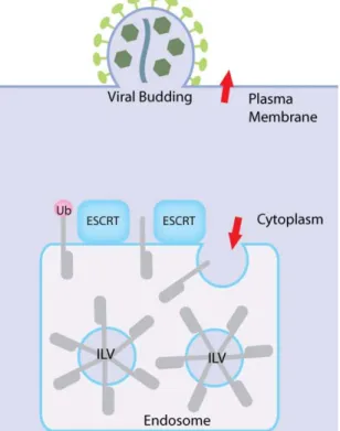
The Endosomal Sorting Pathway (ESP) is an important component of plasma membrane receptor regulation. Recognition of a sequence signal in the cytoplasmic tail of receptors to be downregulated leads to receptor endocytosis and incorporation into the early endosome. From here receptors can suffer one of two fates: recycling back to the cell membrane or trans‐golgi network, or if ubiquitinated, retention at the endosomal membrane. Receptors that are not recycled from the endosome are included into intralumenal vesicles (ILVs) formed by vesiculation of the endosomal membrane (David J Katzmann et al. 2002), creating what is termed a multivesicular body (MVB). Once all receptors have been incorporated into ILVs the mature late endosome delivers receptors for degradation through fusion with the lysosomal membrane (Figure 1). Some integral membrane proteins such as iron and nutrient receptors undergo several cycles of internalization and recycling without ever passing by the lysosome, thus inclusion or exclusion of certain receptors from the endosomal membrane into MVBs must be a tightly regulated event. Recognition of the ubiquitinated cargo and membrane remodeling for MVB biogenesis is carried out by the Endosomal Sorting Complex Required for Transport (ESCRT) machinery, an intricate assembly of more than 60 proteins.
Receptor monoubiquitination, or multiple monoubiquitination, is the classic signature for endosomal sorting, as opposed to the Lys48‐linked polyubiquitin chain signal that targets cytosolic and nuclear proteins to the proteosome (Haglund et al. 2003). It has since been shown that polyubiquitination, through Lys63 specific linkage of ubiquitin chains, can target the membrane receptor epidermal growth factor (EGF) receptor for internalization (F. Huang et al. 2006). Ubiquitination‐deficient mutant EGF receptors are internalized at the same rate as wild‐type receptors but their turnover rates are defective. Localisation experiments show the mutant receptors remain mostly at the early endosome, whilst only wt‐receptors make it to the late endosome, suggesting ubiquitination mutation prevents MVB targeting of receptors, leaving receptors instead to recycle back to the plasma membrane (F. Huang et al. 2006). These studies show how ubiquitination is implicated in lysosomal targeting but not for initial internalization of receptors from the plasma membrane into endosomes.
Introduction Figure 1. Receptor downregulation via the endosomal sorting pathway. Receptors to be down‐regulated at the plasma membrane are endocytosed and delivered to the early endosome. Receptors are then either recycled back to the plasma membrane or trans‐golgi network, or retained at the endosomal membrane through an ubiquitination signal. Invagination of the endosomal membrane incorporates receptors into intralumenal vesicles, forming a compartment termed a multivesicular body. Once all receptors are been incorporated into ILVs, the now mature late endosome delivers receptors for degradation by fusing with the lysosome. 0.2 The ESCRT machinery The components of the ESCRT machinery were first identified from vacuolar sorting mutants in yeast (the vacuole being the yeast functional equivalent to the mammalian lysosome), a method that has continued to be exploited to probe the individual function of ESCRT components (David J Katzmann et al. 2002).
There are four multi‐subunit ESCRT complexes involved in MVB formation, termed ESCRT‐0, ‐I, ‐II, ‐III. These are cytosolic proteins that are recruited to the endosomal membrane for receptor sorting. There are three main requirements of the ESCRT machinery; targeting to the endosomal membrane, recognition of ubiquitinated receptors to be sorted, and MVB biogenesis. ESCRT‐ 0,‐ I and ‐II serve to recognize and anchor the target receptor at the endosomal membrane, whilst ESCRT III is responsible for vesicle biogenesis, and for
Introduction
interaction with the AAA ATPase Vps4 responsible for ESCRT disassembly. Together with an intricate network of auxiliary proteins, such as deubiquitinating enzymes, these ESCRT complexes bring about receptor incorporation into MVBs at the endosomal membrane (Figure 2). The constituent proteins of the ESCRT complexes are listed with equivalent yeast and human nomenclature in Table 1, along with some key domain features. Figure 2. The cytosolic ESCRT complexes, ESCRT‐0, ‐I, ‐II, ‐III are recruited to the endosomal membrane for receptor sorting. The ESCRT machinery must be targeted to the endosomal membrane, recognise ubiquitinated receptors to be sorted, and instigate MVB biogenesis. ESCRT‐ 0, ‐ I and ‐II recognize and anchor the target receptor at the membrane, ESCRT III packages the receptor into an inward budding vesicle for final ILV formation and interacts with accessory proteins like deubiquitinating enzymes and the AAA ATPase Vps4 responsible for ESCRT disassembly. Together with an intricate network of auxiliary proteins ESCRT complexes sort receptors for degradation. 0.3 ESCRT‐0 ESCRT‐ 0 is composed of Hrs (hepatocyte growth factor‐regulated tyrosine kinase substrate), STAM (signal‐transducing adaptor molecule), and Eps15 (epsin‐15). The Hrs/STAM/Eps15 complex is the initiator of MVB biogenesis, providing the first anchor point to the endosomal membrane through the endosome‐specific PI(3)P (phosphatidylinositol3‐phosphate) lipid binding motif FYVE (Fab1, YGL023, Vps27, and EEA1) of Hrs (Gaullier et al. 1998) (Bache et al. 2003).
Introduction
Human Yeast Domains /Features
ESCRT‐0 Hrs STAM Vps27 Hse1 FYVE, UIM UIM, VHS,SH2 ESCRT‐I Tsg101 Vps28 Vps37 Mvb12 Vps23 Vps28 Vps37 UEV Ubiquitin‐binding motif ESCRT‐II EAP30 EAP20 EAP45 Vps22 Vps25 Vps36 GLUE ESCRT‐III CHMP1A, 1B CHMP 2A, 2B, 2C CHMP3 CHMP 4A, 4B CHMP5 CHMP6 CHMP7 hIST1 Did2 Vps2 Vps24 Snf7 Vps60 Vps20 MIM MIM MIM MIM MIM MIM1, MIM2 Associated proteins Vps4A, Vps4B ALIX, HD‐PTP UBPY/USP8, AMSH/STAMBP, AMSH‐ LP Vps4 Bro1 Doa4 AAA ATPase Deubiquitinating enzyme Table 1. Constituent proteins of each ESCRT complex are listed with the equivalent human and yeast nomenclatures, along with key domain features. Monoubiquitinated receptors are recognized by the Ubiquitin Interacting Motif (UIM) of Hrs (Shih et al. 2002), and the UIM and VHS (Vps27/Hrs/Stam) domain of STAM (Bache et al. 2003)(Mizuno et al. 2003)(Hong et al. 2009). Hrs is a 540 kDa hexamer (Pullan et al. 2006), binding two ubiquitin molecules per monomer via a unique, double‐sided UIM (Hirano et al. 2006), giving a total of 12 ubiquitin binding sites. The yeast homologue of Hrs, Vps27p, contains two UIMs, and the crystal structure of the second of these revealed that whilst Vps27p UIM2 is a monomeric helix in solution, the ubiquitin complex structure had formed a four‐helix bundle (Fisher et al. 2003). Oligomerisation of ubiquitin binding proteins could explain how high affinity can be obtained through collective low ubiquitin affinity motifs (Kd
Introduction 0.1 – 1mM) (Fisher et al. 2003). Similarly, each ubiquitin subunit of K63‐linked diubiquitin can bind a UIM, so polyubiquitin chains binding several UIMs can increase affinity (Varadan et al. 2004). Hrs also binds the clathrin coat of the endosomal membrane through a clathrin binding box at its C‐terminal, thus stabilising the ubiquitinated receptor at the endosomal membrane and preventing its recycling back to the plasma membrane (Pullan et al. 2006). Finally, through the interaction of the UEV (ubiquitin E2 variant) domain of Vps27 (Hrs) with Vps23 (Tsg101), Hrs recruits the ESCRT‐I complex (David J Katzmann et al. 2003). 0.4 ESCRT‐I and ESCRT‐II The ESCRT‐I complex was first established as a cytosolic heterotrimeric complex composed of Vps37, Vps28 and Tsg101 (Vps23 in yeast). Recognition of ubiquitinated cargo is through the ubiquitin E2 variant domain (UEV) of Vps23 (D J Katzmann et al. 2001). The highly basic N‐terminal of Vps37 orients ESCRT‐I on the membrane through endosomally enriched lipid phosphatidylinositol 3‐phosphate (PI3P) interaction. Vps23 and Vps37 interact through a hydrophobic packing interaction, as do Vps23 and Vps28, with no direct contact made between Vps37 and Vps28 (Kostelansky et al. 2006)(Kostelansky et al. 2007). Later a fourth subunit, Mvb12, was identified (Oestreich et al. 2007)(Morita, Sandrin, Alam et al. 2007), interacting with Vps23 and Vps37, but not Vps28, and also containing a novel ubiquitin‐ binding motif (Shields et al. 2009). Both the ternary and quaternary complex has a 1:1:1:1 stoichiometry (Kostelansky et al. 2006)(Kostelansky et al. 2007).
ESCRT‐II consists of one copy of EAP30 (Vps22) and EAP45 (Vps36), and two copies of EAP20 (Vps25), with two PPXY motifs of Vps25 contacting Vps22 and Vps36 (Teo et al. 2004)(Im & Hurley 2008). The NZF (npl4 zinc finger) ubiquitin binding domain of the Vps36 GLUE (GRAM‐like ubiquitin binding in EAP45) domain binds to the C‐terminal of Vps28 (Kostelansky et al. 2006), whilst ESCRT‐II targeting to the endosomal membrane is carried out by the GLUE domain in concert with an N‐terminal helix of Vps22 interacting with PI3P of the membrane (Im & Hurley 2008).
Introduction
0.5 ESCRT‐III
ESCRT‐III in humans is composed of 11 Charged Multivesicular Body Proteins (CHMPs), CHMP1A and 1B, CHMP2A and 2B, CHMP3, CHMP4A, 4B and 4C, CHMP5, and CHMP6, all around 220 amino acids in length. CHMP7 was discovered later, being twice as many amino acids in length than the other CHMPs, and described to have greatest similarity with CHMP6, binding CHMP4B as CHMP6 does (Horii et al. 2006). Recently an eighth member, hIST, was identified as an ESCRT‐III member isoform, binding to both ESCRT‐I and CHMP1A and CHMP1B of ESCRT‐III (Agromayor et al. 2009)(Bajorek, Schubert et al. 2009). Where appropriate, use of the yeast ESCRT‐III homologues in described experiments is indicated in brackets. CHMP6 is N‐myristoylated, providing a membrane anchor for CHMP6 (Markus Babst et al. 2002). The N‐terminal portion of CHMP6 binds to EAP20 (Vps25) of ESCRT‐II and CHMP4B of ESCRT‐III independent of N‐myristoylation (Teo et al. 2004)(Yorikawa et al. 2005)(Im et al. 2009), providing a second independent point of recruitment to the endosomal membrane.
The CHMP proteins are characterized by a distinct charge polarization; a basic N‐terminal portion of pI ~10 and an acidic C‐terminal portion of pI ~4, the N‐terminal region spanning a greater proportion of the overall molecule. In 2006 the first structure of an ESCRT‐III protein was released, that of the basic N‐terminal portion of CHMP3, revealing a four helix bundle composed of a long helical hairpin and two shorter helices, with a further C‐terminal helix extending out perpendicular to the core (Figure 3A)(Muzioł et al. 2006). The recent structure of hIST1 residues 1‐189 asks a reinterpretation of the data of Muziol et al (Bajorek, Schubert et al. 2009). Here, the fifth helix is assigned in a cis position, rather than in trans in Muziol et al.’s structure (Figure 3E and Figure 3F). The presence of two extra helices, termed αA and αB show the link between helix 4 and helix 5, thus leading to the repositioning of helix 5. However, although the structure of hIST1 allows us to position a small fragment of a C‐terminal helix in relation to the rest of the basic N‐terminal domain, helping to speculate about the location of the rest of the C‐terminal domain in relation to the N‐terminal domain, structural data of an intact CHMP protein is lacking.
Electrostatic potential maps identified a very basic charged exposed surface as a possible membrane interface within the CHMP3 structure (Figure 3B)(Muzioł et al. 2006). Mutation of key basic residues within this interface redistributed CHMP3 from predominantly plasma membrane and vesicular localization to the cytosol, implicating these residues in targeting of CHMP3 to endosomal membranes. The differential conservation of key basic residues
Introduction
within this interface in the CHMP proteins suggests indirectly individual preferences for membrane composition between the human CHMP members. This orientation to the membrane would leave the other very acidic charged surface exposed on the opposite side. This is the surface where the final C‐terminal of CHMP3 is speculated to be located, and would show how CHMP3 is orientated on the membrane to allow access of binding partners to its final C‐terminal helix.
Two dimer interfaces were identified in the CHMP3 crystal structure, one from dimerisation of CHMP3 during crystallisation (Figure 3C), and the other formed by the crystal packing (Figure 3D). Mutation of the dimer interfaces relocated CHMP3 to a predominantly cytosolic location in cells, showing the dimer interfaces are important in CHMP3 targeting to endosomes. Conservation of dimerisation residues between CHMP3 and CHMP2 suggests heterodimers could form in the same manner as the homodimers seen in the crystal structure.
CHMP2 (Vps2), CHMP3 (Vps24), CHMP4 (Snf) and CHMP6 (Vps20) form large heterooligomeric complexes when membrane bound, including distinct subcomplexes of CHMP2/CHMP3 (Vps2/Vps24), and CHMP4/CHMP6 (Snf7/Vps20). The presence of the CHMP2/CHMP3 (Vps2/Vps24) subcomplex is required for the interaction with the CHMP4/CHMP6 (Snf7/Vps20 subcomplex. In solution CHMP2 (Vps2) and CHMP3 (Vps24) do not interact, but CHMP2 (Vps2) and CHMP3 (Vps24) co‐dependantly localise to the endosomal membrane (Markus Babst et al. 2002). CHMP6 (Vps20) co‐localisation to endosomal membrane bound CHMP4 (Snf7) is dependent on CHNP3 (Vps24), and CHMP2 (Vps2) and CHMP3 (Vps24) membrane localization is also dependant on the presence of CHMP4 (Snf7) and CHMP6 (Vps20) (Markus Babst et al. 2002). CHMP4 (Snf7) endosomal localisation is dependant on CHMP6 (Vps20), but the inverse is not true. Thus although there are individual interactions of the subunits with the endosomal membrane, this is much enhanced through cooperative binding of ESCRT‐III proteins with the membrane. Overexpression of individual subunits does not increase their incorporation into the complex as compared with other subunits, suggesting an equimolar stoichiometry at the endosomal membrane. The C‐terminal domain of CHMP proteins interacts with their own N‐terminal domain and this has been proposed as a mechanism of autoinhibition (Zamborlini et al. 2006). Removal of the C‐terminal portion to mimic activated CHMP proteins produces polymers upon incubation with the respective CHMP binding partner; CHMP2 with CHMP3, and CHMP1 with hIST1. The CHMP membrane‐binding surface is presented on the external face of the
Introduction
tubule, indicating how CHMP polymerisation on the inside of a membrane deformation could induce vesicle formation (Lata, Schoehn et al. 2008)(Bajorek, Schubert et al. 2009). Out of CHMP3 and CHMP2 only C‐terminally truncated CHMP2A polymerised alone, forming ring structures instead of tubes (Lata, Schoehn et al. 2008). Purifying Vps24, the yeast homologue of CHMP3, above concentrations of 10 mg/mL in vitro can induce polymer formation, but again the same phenomenon was not observed for the human CHMP3 protein (Ghazi‐Tabatabai et al. 2008). CHMP4 (Snf7) spontaneously polymerises at the endosomal and plasma membranes when over‐expressed, with membrane curvature away from the cytoplasm observed when CHMP4 is expressed without the C‐terminal autoinhibitory domain (Hanson et al. 2008). A new proposal from the Emr group, based on their work with the yeast ESCRT‐III homologues, pushes CHMP4 (Snf7) as the key building block for ESCRT‐III polymers, where CHMP6 (Vps20) serves to anchor the CHMP4 (Snf7) polymer at the endosomal membrane, and Vps25 of ESCRT‐II binding to CHMP6 (Vps20) on the membrane invokes a conformational change that could be interpreted as an activation event (Saksena et al. 2009). Both CHMP4 (Snf7) and CHMP6 (Vps20) demonstrated a conformational change in response to membrane binding, supporting evidence for membrane‐stimulated activation of CHMP proteins.
CHMP4 (Snf7) and CHMP6 (Vps20) accumulate on the endosomal membrane in the absence of CHMP2 (Vps2) and CHMP3 (Vps24), implicating the CHMP2/CHMP3 (Vps2/Vps24) subcomplex in recruitment of Vps4 for ESCRT‐III disassembly (M Babst et al. 1998)(Markus Babst et al. 2002). Vps4B was shown to disassemble in vitro polymer tubes of C‐terminally truncated CHMP2A and full‐length CHMP3 in the presence of ATP and Mg2+, binding within the central cavity of the tube via the intact C‐terminal domain of CHMP3 (Lata, Schoehn et al. 2008). In the yeast Vps24 (yeast homologue to CHMP3) filaments, Vps4 had no effect on polymer tube diassembly except when the C‐terminal domain of Vps24 was replaced with the C‐terminal domain of Vps2 (yeast homologue to CHMP2), further indicating a specific role for CHMP2 in the recruitment of Vps4 to the CHMP2/CHMP3 heterodimer, at least in the yeast system (Ghazi‐Tabatabai et al. 2008). According to the Emr group’s model, the CHMP2/CHMP3 (Vps2/Vps24) subcomplex is postulated to cap CHMP4 (Snf7) polymerisation before recruitment of Vps4 by CHMP2 (Vps2) disassembles CHMP polymers (Teis et al. 2008)(Saksena et al. 2009). The capacity of three ESCRT subunits, CHMP6 (Vps20), CHMP4 (Snf7) and CHMP3 (Vps24) to achieve membrane scission was finally demonstrated by the Hurley lab early this year, defining the role of CHMP2 (Vps2) to be after polymerisation to recruit Vps4 for CHMP polymer disassembly (Wollert et al. 2009).
Introduction
There is a strong accumulation of evidence that CHMP proteins CHMP3, CHMP4 and CHMP6 are responsible for vesicle formation and scission, with CHMP tubule formation elucidating how CHMP proteins may form and close the pinching neck of a budding vesicle; polymer formation deforms the membrane into a vesicle, and subsequent recruitment by CHMP2 of Vps4 releases CHMP subunits and closes the neck. In vivo it is likely other adaptor proteins are involved to carefully regulate the morphology and timing of this process. ALIX (ALG‐2 interacting protein X) forms crescent shaped dimers that bind to CHMP4 polymers and is speculated to serve as a scaffold for membrane remodelling (Pires et al. 2009). Figure 3. Crystal structure of dimeric CHMP39‐183 and hIST11‐189. A. Crystal structure of CHMP9‐183. Ribbon representation of the four helices bundle constituting the CHMP9‐ 183 monomer. A fifth helix is portrayed packing perpendicularly to the helical bundle, the linking disordered loop represented by a dashed line. B. Electrostatic potential map of CHMP9‐183 dimer 1. The basic surface exposed on one side of the dimer mediates CHMP3 targeting to the membrane and is required for efficient retroviral budding.
C. Ribbon representation of the CHMP39‐183 dimer. The antiparallel dimerisation interface is mainly mediated by the long helical hairpin. D. A second dimerisation interface via the tips of the helical hairpins arises from the crystal lattice. E. Side view of the first dimer interface. The perpendicular packing of the fifth helix to the helical hairpin tips is highlighted. F. Ribbon diagram and helix nomenclature for hIST11‐189 structure. Adapted from (Muziol et al. 2006)(Bajorek, Schubert et al. 2009).
Introduction
0.6 Interaction of ESCRT‐III with MIT domain‐containing proteins – the MIT‐MIM interaction
The AAA (ATPase Associated with various cellular Activities) ATPase Vps4, catalyses dissociation of ESCRT‐III complexes. Human Vps4, composed of subunits Vps4A and Vps4B, associates into a bowl‐shaped dodecamer of two stacking hexameric rings (Yu et al. 2008).
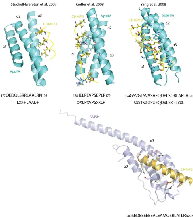
It was the structure of the MIT (Microtubule Interacting) domain of Vps4A that gave the first clues to how MIT domain ‐ CHMP interactions are mediated. The Vps4A MIT domain consists of a three‐helix bundle reminiscent of a classical TPR (tetratricopeptide) motif. Through alanine mutation studies Scott et al discovered a highly conserved leucine residue responsible for CHMP1B binding (Scott et al. 2005). In a classical TPR motif this leucine residue would be responsible for binding of the fourth helix, which led the authors to postulate that CHMP binding through the C‐terminal helix may act to complete the TPR motif. In 2007 the structures of yeast Vps4 in complex with Vps2 (yeast homologue to CHMP2), Vps4A in complex with CHMP1B, and Vps4B in complex with CHMP2B, revealed that a CHMP C‐terminal peptide, comprising the last 20 residues of the C‐terminal, binds to the Vps4 MIT domain to complete the TPR motif, with the orientation of the CHMP C‐ terminal helix being in the opposite direction (Stuchell‐Brereton et al. 2007)(Obita et al. 2007), with the consensus LXX+LAAL+ sequence required for Vps4 binding termed the MIT Interacting Motif (MIM).
The structure of CHMP1B in complex with spastin MIT identified another binding interface in the MIT domain (Yang et al. 2008), using helices 1 and 3 of the three‐helix bundle, in contrast to helices 2 and 3 of the Vps4 MIT. Interestingly, spastin has unique specificity for CHMP1B, showing no binding even to its closest homologue CHMP1A (Yang et al. 2008), with several key residues non‐conserved between the two proteins. The interaction is one of higher affinity than previous measurements binding CHMP1B to the MIT domain of Vps4, with Kd 12 µM compared to 33 µM for Vps4 (Stuchell‐Brereton et al. 2007). Although both the Vps4 and spastin interaction with CHMP1B are predominantly hydrophobic in nature, an increase in affinity of spastin for CHMP1B can be explained by a larger buried surface area. The affinity of binding of full‐length CHMP6 is comparable with that of the CHMP1B Vps4 interaction, but optimal binding is achieved by deleting CHMP6 down to a 165−181 fragment (removing the MIM consensus sequence) resulting in a Kd 5.8 μM (Kieffer et al. 2008). The
identification of key interaction residues further towards the N‐terminal of the C‐terminal domain of CHMP6 prompted the authors to term this motif the MIM2 sequence. The NMR
Introduction
solution structure of a Vps4A MIT CHMP6166−181 MIM2 complex shows binding between
helices 1 and 3 of the MIT domain, the same binding arrangement seen for the CHMP1B spastin complex, but clearly demonstrating different sequence specificity determination. Intriguingly, a recent paper showed that hIST1 contains both MIM1 and MIM2 domains (Bajorek, Morita et al. 2009), thus the presence of two MIM domains in some CHMP proteins may create varying affinities for MIT domains. 0.7 Recruitment of Deubiquitinating enzymes (DUBs) Ubiquitin recovery from ubiquitinated receptors before inclusion into MVBs is an important prerequisite for maintaining the cellular ubiquitin pool. In yeast the ubiquitin thioesterase Doa4 was identified responsible for removing the ubiquitin moieties through studies of doa4 mutants. Cellular ubiquitin levels were found depleted in Doa4 mutants, an effect alleviated by mutations in endosomal sorting components (Amerik et al. 2000). It is not clear how Doa4 is recruited to the endosomal membrane, either directly through CHMP protein interaction, or via binding to Bro1 (Bowers et al. 2004).
ALIX, a Bro1 domain containing protein, was initially proposed as the human homologue of yeast Bro1, yet it was whilst investigating ALIX depletion effects on receptor sorting that another Bro1 domain‐containing protein, His‐domain containing protein tyrosine phosphatase (HD‐PTP), was identified as functionally required for sorting (Doyotte et al. 2008). Like ALIX, HD‐PTP binds CHMP4B of ESCRT‐III and TSG101 of ESCRT‐I (Ichioka et al. 2007). Later there was even found a third Bro1 domain‐containing protein called Brox, also capable of interacting with CHMP4 (Ichioka et al. 2008). The structure of ALIX in complex with C‐terminal fragments from CHMP4A, CHMP4B and CHMP4C shows how the arrangement of hydrophobic residues in the C‐terminal domain of CHMP4 proteins, in particular the conserved Trp220, confers Bro‐1 domain specificity in CHMP4s, in contrast to the MIT domain preference of CHMPs 1‐3 (McCullough et al. 2008). This interaction showed an affinity comparable with that of Vps4 CHMP interactions of 44 ±6 μM for ALIX (Bro1 V domain) construct with CHMP4A205‐222. This adds another dimension to the ESCRT machinery; different CHMP proteins recruit different accessory effecter molecules.
So far three candidate DUBs have been identified as interacting with the ESP; AMSH (Associated Molecule of the SH3 domain of STAM), AMSH‐LP (AMSH‐like protein) and UBPY/USP8 (ubiquitin‐specific processing protease Y). UBPY is a 1118 amino acid ubiquitin thioesterase, binding to the same SH3 domain motif of STAM as AMSH (M Kato et al. 2000),
Introduction
and CHMP 1A, CHMP1B and CHMP7 of ESCRT‐III (Row et al. 2007). In contrast to AMSH, UBPY shows no discrimination between K‐48 linked and K‐63 linked ubiquitin chains, and does not bind to CHMP3 (Row et al. 2006)(Row et al. 2007).
AMSH‐LP, as the name suggests, contains similar features to that of AMSH; it is a 436 amino acid protein containing a C‐terminal JAMM domain. One distinct difference is the replacement of an essential Lysine residue in the SBM of AMSH by a threonine in AMSH‐LP (residue 250), meaning AMSH‐LP no longer binds STAM (Kikuchi et al. 2003). AMSH‐LP was confirmed to interact with clathrin, as does AMSH (Nakamura et al. 2006), but contrastingly does not bind CHMP1A, CHMP1B, CHMP2A or CHMP3 (Agromayor & Juan Martin‐Serrano 2006). In 2008 the structure of the JAMM domain of AMSH‐LP was published, both alone and in complex with Lys‐63 linked di‐ubiquitin, revealing the structural basis for selective hydrolysis of Lys‐63 linked ubiquitin chains (Y. Sato et al. 2008).
0.8 AMSH domain architecture and binding partners
AMSH, also known as STAM Binding Protein (STAMBP), is a 424 amino acid protein expressed in all mammalian tissues, which as the name suggests was first identified as a binding partner of STAM of ESCRT‐0 (N Tanaka et al. 1999). Since then it has been shown to bind the N‐terminal domain of clathrin heavy chain (McCullough et al. 2006)(Nakamura et al. 2006) and a range of CHMPs of the ESCRT‐III complex: CHMP1A, CHMP1B, CHMP2A, and CHMP3 (Agromayor & Juan Martin‐Serrano 2006), CHMP4B (and weakly CHMP4A) (Tsang et al. 2006), and hIST (Agromayor et al. 2009). It is interesting to observe AMSH binding to both CHMPs 1‐3 and CHMPs 4 in light of McCullough et al.’s suggestion of segregation between CHMP 1‐3 binding to MIT domains and CHMPs 4 to Bro1 domains, and there are still conflicts in the literature over the solidity of AMSH’s interaction with CHMP4A and CHMP4B, with pull‐down assays showing the C‐terminal of CHMP4B does not interact with AMSH (Zamborlini et al. 2006).
STAM binding is through a centrally located proline rich STAM‐binding motif (SBM) PXXXRXXKP (Figure 4)(M Kato et al. 2000). A bipartite nuclear localization signal of KR[YTKEYTEYNEE]KKK is located in residues 112‐127 of the N‐terminal, and is required for nuclear localization of AMSH (Figure 4) (N Tanaka et al. 1999)(Kikuchi et al. 2003).
Introduction Figure 4. AMSH domain architecture. A central proline rich motif, including a critical lysine residue, is required for STAM binding (SBM). At the N‐ terminal a MIT domain mediates CHMP interactions. Deubiquitinating activity is provided by a zinc‐binding JAB‐MPN motif at the C‐terminal. A bipartite nuclear localisation signal targets AMSH to the nucleus, and a clathrin interaction domain has also been identified between residues 139 and 171. Immunoprecipitation and subsequent pull‐down assay identified clathrin heavy chain (CHC) interacting with AMSH (Nakamura et al. 2006)(McCullough et al. 2006). Deletion mutants highlight residues 139‐171 of AMSH responsible for CHC binding (Figure 4) (Nakamura et al. 2006). Deletion of the clathrin binding site, as well as RNA interference depletion of CHC, prevents AMSH targeting to endosomes, but deletion of the SBM from AMSH failed to affect its localization to endosomes, highlighting a STAM‐independent recruitment of AMSH to the endosomal membrane by clathrin anchoring (Nakamura et al. 2006). STAM also binds CHC, and along with Hrs’ capacity to bind clathrin, a tripartite binding of clathrin by AMSH and ESCRT‐0 is proposed as an initial anchor point for endosomal sorting (McCullough et al. 2006).
Database alignments identify in the AMSH C‐terminal a JAB_MPN zinc‐binding metalloprotease domain displaying the capacity to hydrolyse tetraubiquitin chains in vitro (Figure 4). A clear preference for K‐63 linked chains over K‐48 chains is observed, signalling an association of AMSH with non proteosome‐linked ubiquitin regulated pathways (McCullough et al. 2006). A D348A mutation abolishes catalytic activity, probably by removing zinc binding as this aspartic acid lies within an EXNHS/THX7SXXD zinc binding motif
(Tran et al. 2003)(Ambroggio et al. 2004). The in vitro deubiquitinating activity of AMSH is enhanced in the presence of STAM, but unaffected upon incubation with CHMP3 (McCullough et al. 2006). AMSH can simultaneously interact with STAM and CHMP3, providing a temporal connection between an efficient deubiquitinating activity of AMSH and CHMP binding (McCullough et al. 2006).
Introduction
AMSH binds CHMP3 independantly of the JAB_MPN domain and SBM, requiring residues 1‐ 191 of the N‐terminal domain for this interaction (Agromayor & Juan Martin‐Serrano 2006) (Tsang et al. 2006). Sequence homology analysis of the Vps4 MIT domain with AMSH identified an AMSH MIT domain spanning residues 34‐113 (Tsang et al. 2006). Important sequence discrepancies between Vps4 and AMSH MIT domains, such as the lack of conservation of a key leucine residue (Leu64) required for optimal CHMP1B binding by Vps4 suggest a sequence based mechanism for diffferential CHMP binding by MIT containing proteins (Scott et al. 2005)(Tsang et al. 2006).
0.9 Suggested mechanisms for AMSH in Endosomal Sorting
The role of AMSH within the endosomal sorting pathway has yet to be determined, with conflicting evidence supporting several roles presented in the literature.
AMSH could act as a negative regulator, with the potential to ‘rescue’ ubiquitinated receptors (McCullough et al. 2004). This is supported by short‐interfering RNA experiments, where knockout of AMSH results in increased EGF receptor degradation, and by the in vitro deubiquitination of ubiquitinated EGF receptors by AMSH (McCullough et al. 2004). In this model, AMSH would serve to oppose the action of the E3 ligases responsible for ubiquitination, where an increase in AMSH activity would shift the balance towards receptor recycling and a decrease in the degradation fate.
Initially UBPY was proposed as the negative regulator of the ESP. RNA interference depletion of UBPY accelerated degradation of EGF receptors, whilst over expression of UBPY reduced EGF receptor levels of ubiquitination and delayed EGF receptor degradation in EGF stimulated cells (Mizuno et al. 2005). Row et al. also found that ubiquitinated proteins accumulate on endosomes when catalytically inactive UBPY was over expressed, and siRNA treatment of UBPY increases total levels of ubiquitinated protein, but the observation that free cellular levels of ubiquitin remained unchanged led them to investigate whether UBPY may be acting as a regulator of the ubiquitination of endosomal components rather than endosomal cargo, for ubiquitination is also the mechanism for degradation of the ESP proteins. A subsequent depletion of STAM in UBPY knockdown cells, coupled with the recovery of ubiquitinated STAM in proteosome inhibited cells led them to conclude that UBPY may be regulating STAM polyubiquitination and targeting for proteosomal degradation, a theory concurring with UBPY processing of K‐48 linked ubiquitin chains and a
Introduction
later report by Mizuno et al. that Eps15, an ESCRT‐0 associated protein, is a substrate for UBPY (Row et al. 2006)(Mizuno et al. 2006).
AMSH could be responsible for removing the ubiquitin signal from the cargo just before incorporation into vesicles in a role analogous to that of Doa4 in the yeast ESP. This theory is supported by the accumulation of ubiquitinated cargo and the block of EGF receptor degradation when catalytically inactive AMSHD348A is expressed (Kyuuma et al. 2007)(Agromayor & Juan Martin‐Serrano 2006), and also correlates with in vitro deubiquitination of ubiquitinated EGF receptors by AMSH (McCullough et al. 2004). Further evidence supporting this proposed role of AMSH is presented in the following section, which analyses data from studies of endosomal sorting components in retroviral budding studies. 0.10 The ESCRT machinery in HIV budding Interest in the ESCRT protein machinery was heightened by the discovery that components of the ESP protein machinery are required for budding of enveloped retroviruses such as HIV. Enveloped viral particle budding can be considered topologically similar to that of MVB formation, both processes involving a deformation of membrane away from the cytosol (Figure 5). Much of the work done to characterize the functions of ESCRT proteins has exploited viral budding as a way of following functionality of the ESP.
Figure 5. Enveloped viral budding is topologically similar to MVB biogenesis, both processes requiring a deformation of membrane away from the cytosol.
Introduction
For retroviral virions to egress from the infected host cell, they require late assembly (L) domains located within gag, the gag gene product (H G Göttlinger et al. 1991). In HIV, the PTAP and LYPXL motifs of the p6 region of gag are responsible for recruiting TSG101 of ESCRT‐I and ALIX respectively (Garrus et al. 2001)(J Martin‐Serrano et al. 2001)(Bettina Strack et al. 2003), TSG101 in turn recruiting Vps28 to the site of viral budding (Juan Martin‐ Serrano et al. 2003). This is the same PTAP motif used by Vps27 (Hrs) of ESCRT‐0 to contact Vps23 (TSG101) of ESCRT‐I, showing clearly how the virus mimics the ESP to hijack components (Kostelansky et al. 2006). Retroviruses also use ubiquitination to mimic the ESP, with ubiquitinated gag found in viral particles of HIV, SIV (Simian Immunodeficiency Virus) and MLV (Murine Leukemia Virus)(Ott et al. 1998). The p6 PTAP and PPXY motifs are critical for ubiquitination of gag, with PPXY recruiting HECT E3 ubiquitin ligases for conjugation of ubiquitin to HIV gag (B Strack et al. 2000). However, somewhat contradictorily, increased viral release correlates with decreased levels of ubiquitinated gag, suggesting deubiquitination by DUBs is required before gag incorporation and virion release, a hypothesis supported by the observation of little ubiquitinated gag found in released viral particles (Juan Martin-Serrano 2007).
CHMP3 membrane targeting is important in efficient retroviral budding (Muzioł et al. 2006). Deletions into the C‐terminal domain, which would remove the autoinhibitory binding to CHMP3 N‐terminal, convert CHMP3 into a dominant‐negative inhibitor of HIV‐1 budding. The same effect is seen with incubation of AMSH with full‐length CHMP3, with stronger inhibition occurring upon incubation with a catalytically inactive AMSHD348A mutant, an
effect abolished by point mutations in the CHMP3 binding site of AMSH (Zamborlini et al. 2006). AMSH was also observed to bind more strongly to CHMP proteins when catalytically inactive (Agromayor & Juan Martin‐Serrano 2006). Expression of AMSHD348A inhibits retroviral budding in a dominant‐negative manner and induces the accumulation of ubiquitinated HIV and MLV gag, yet AMSH is non‐essential for viral budding as was shown by knockdown studies (Agromayor & Juan Martin‐Serrano 2006). Inhibition of retroviral budding by AMSHD348A was unaffected by deletion of the STAM binding site, correlating with data that the function of STAM binding is to enhance AMSH catalytic activity and supporting an ESCRT‐III related effect of the AMSHD348A mutant on retroviral budding (Agromayor & Juan
Martin‐Serrano 2006)(McCullough et al. 2006). Finally, CHMP3 binding is not required for AMSH’s in vitro deubiquitinating activity, but in vivo expression of an AMSH mutant lacking CHMP3‐binding ability (AMSH83‐424) resulted in accumulation of ubiquitinated cargo on aberrant endosomes (Kyuuma et al. 2007). These findings suggest AMSH activates CHMP3 through release of the autoinhibition mechanism, and show how the deubiquitinating
Introduction
activity of AMSH affects endosomal receptor degradation and viral budding in a CHMP3 dependant manner, with both CHMP3 binding and enzymatic activity required for efficient endosomal sorting.
The requirement of CHMP3 binding for the endosomal deubiquitinating activity of AMSH combined with evidence AMSH activates CHMP3 by relieving its autoinhibitory state, suggests AMSH’s deubiquitinating activity is localised towards the latter stages of the endosomal sorting pathway in concert with CHMP3 activation and involvement in vesicle formation, which would correspond to a role in removal of ubiquitin from receptors just prior to their incorporation into MVBs.
0.11 A New Role for ESCRT in Cytokinesis
In 2007 a new role for ESCRT proteins was demonstrated in cytokinesis (Carlton & Juan Martin‐Serrano 2007). CHMP3 of the ESCRT‐III complex was shown to localize at the midbody and be functionally required (Morita, Sandrin, Chung et al. 2007)(Dukes et al. 2008), whilst CHMP1B is required for spastin recruitment to the midbody for microtubule severation (Yang et al. 2008). CHMP1A is also required for cell abscission, as is its interaction with its heterodimeric partner hIST, suggesting CHMP polymerization is required (Agromayor et al. 2009) (Bajorek, Morita et al. 2009)(Bajorek, Schubert et al. 2009).
This is not the first time the CHMP proteins have been implicated in subnuclear processes. In 2001 Stauffer et al. reported localisation of CHMP1A to the nucleus as well as the cytoplasm through a bipartite nuclear localization signal (residues 20‐35), and overexpresson of CHMP1 arrested cells in S‐phase, indicating two parallel roles for CHMP1 in cytoplasmic endosomal sorting, and nuclear gene regulation. Interestingly, CHMP1A was also shown to recruit the polycomb repressor ring finger protein BMI1 (Stauffer et al. 2001).
Cellular distribution studies found AMSH predominantly in the cytoplasm, particularly localising to the proximity of the nuclear membrane (F. Itoh et al. 2001). Both DUBs AMSH and UBPY have since been shown to act during cytokinesis, with UBPY primarily at the midbody and AMSH shown to act at both the midbody and the midbody ring (Mukai et al. 2008). A relocalisation of ubiquitin during cytokinesis from the midbody to the midbody ring has been observed (Pohl & Jentsch 2008) and this could link in with the different location of these DUBs. Relocation of ubiquitin and thus localization of DUBs could sequentially mobilise associated proteins such as CHMPs to effectuate midbody ring constriction.
Aims and Objectives Aims and Objectives The overall aim of the thesis work was to gain structural and functional data on AMSH. In particular, we wanted to elucidate the mechanism for interaction with CHMP3, investigating the proposition that AMSH activates CHMP3 for polymerisation. We also wanted to probe the function of the enzymatic domain and to interpret this activity in the context of the full‐ length protein. Here I present the objectives of the thesis work: • To isolate soluble AMSH for crystallisation and structural studies. • To verify CHMP3 binding to AMSH, to define the binding site on each molecule and assay the affinity of the AMSH CHMP3 interaction. • To crystallise AMSH, or a defined CHMP3 binding domain, in complex with full‐length CHMP3, in order to understand if AMSH could play a part in activation of CHMP3, and depending on the activation state crystallised, to gain information either on the arrangement of the CHMP3 C‐terminal autoinhibitory domain to the N‐terminal, or to determine how binding of AMSH could bring about CHMP3 activation.
• To define a soluble JAMM domain for structural studies in order to determine the structural basis of specificity for K‐63 linked ubiquitin chains.
• To verify the deubiquitinating activity of AMSH, and to compare this with the activity of the JAMM domain alone.