RESEARCH OUTPUTS / RÉSULTATS DE RECHERCHE
Author(s) - Auteur(s) :
Publication date - Date de publication :
Permanent link - Permalien :
Rights / License - Licence de droit d’auteur :
Institutional Repository - Research Portal
Dépôt Institutionnel - Portail de la Recherche
researchportal.unamur.be
University of Namur
Importance of measuring pharmacologically active metabolites of edoxaban
Siriez, Romain; Alpan, Lütfiye; Elasaad, Kossay; Devel, Philippe; Laloy, Julie; Dogne,
Jean-Michel; Douxfils, Jonathan
Published in:
Journal of Thrombosis and Thrombolysis
DOI:
10.1007/s11239-019-02030-5 Publication date:
2020
Document Version
Peer reviewed version
Link to publication
Citation for pulished version (HARVARD):
Siriez, R, Alpan, L, Elasaad, K, Devel, P, Laloy, J, Dogne, J-M & Douxfils, J 2020, 'Importance of measuring pharmacologically active metabolites of edoxaban: development and validation of an ultra-high-performance liquid chromatography coupled with a tandem mass spectrometry method', Journal of Thrombosis and
Thrombolysis, vol. 49, no. 3, pp. 395-403. https://doi.org/10.1007/s11239-019-02030-5
General rights
Copyright and moral rights for the publications made accessible in the public portal are retained by the authors and/or other copyright owners and it is a condition of accessing publications that users recognise and abide by the legal requirements associated with these rights. • Users may download and print one copy of any publication from the public portal for the purpose of private study or research. • You may not further distribute the material or use it for any profit-making activity or commercial gain
• You may freely distribute the URL identifying the publication in the public portal ? Take down policy
If you believe that this document breaches copyright please contact us providing details, and we will remove access to the work immediately and investigate your claim.
Importance of measuring pharmacologically active metabolites of edoxaban: development and validation of an ultra-high-performance liquid chromatography coupled with a tandem mass spectrometry method. Running short title: Simultaneous quantification of edoxaban and edoxaban-M4 metabolite by ultra-high-performance liquid chromatography-tandem mass spectrometry method in human plasma.
Romain Siriez1, Lütfiye Alpan1,Kossay Elasaad1, Philippe Devel1, Julie Laloy2, Jean-Michel Dogné1, Jonathan
Douxfils1, 3
1 University of Namur, Department of Pharmacy, Namur Thrombosis and Hemostasis Center (NTHC), Namur
Research Institute for LIfe Sciences (NARILIS), Namur, Belgium; 2 University of Namur, Department of
Pharmacy, Namur Nanosafety Center (NNC), Namur Research Institute for Life Sciences (NARILIS), Namur, Belgium; 3 Qualiblood s.a., Namur, Belgium.
Corresponding author:
Romain Siriez, PhD Student
University of Namur, Department of Pharmacy, Namur Thrombosis and Hemostasis Center (NTHC), Namur Research Institute for Life Sciences (NARILIS), B-5000, Belgium.
E-Mail: [email protected]
Telephone number: +32 81/72.43.25 Fax number: +32 81/72.42.99
ORCID
Romain Siriez https://orcid.org/0000-0002-5003-6939 Kossay Elasaad https://orcid.org/0000-0002-7713-2605 Julie Laloy https://orcid.org/0000-0001-9570-6070 Jonathan Douxfils https://orcid.org/0000-0002-7644-5298
Keywords: Edoxaban, Edoxaban-M4, Ultra-High-Performance Liquid Chromatography/tandem mass
Abstract
Introduction:Although DOACs do not require regular measurements of their blood concentrations, clinical situations may require an assessment of their concentration. Among the factor Xa inhibitors, edoxaban is the only compound for which some metabolites (e.g. edoxaban-M4) are reported to be pharmacologically active. Therefore, their contribution could interfere with assays used for the estimation of edoxaban concentration. In addition, drug interactions may alter the metabolite/parent compound ratio making the sole estimation of edoxaban concentration, a poor assessment of the overall anticoagulation.
Aims: To develop a validated UHPLC-MS/MS method to quantify simultaneously edoxaban and its more relevant
M4-metabolite in human plasma.
Methods: Electrospray ionization and chromatographic separation were optimized for the simultaneous dosage of
edoxaban and edoxaban-M4. The method was validated according to regulatory guidelines for bioanalytical method validation.
Results: The total run time was 6 minutes. The method was validated for calibration curves, precision, accuracy,
carry-over, selectivity, matrix effect and short-time stability.
Conclusion: This method permits quantification of edoxaban and edoxaban-M4 providing complementary
information about the inhibitory effect of this active metabolite in chronometric or chromogenic assays. Although patients treated with edoxaban exhibits usually low concentrations of active metabolites, the measurement of edoxaban-M4 is interesting; especially in case of drug interactions. Indeed, concomitant prescriptions of edoxaban and carbamazepine or rifampicin is frequent and may lead to disturbance of the estimations of edoxaban concentration by chromogenic anti-Xa assays. Therefore, patients are at risk of having inadequate control of anticoagulation supporting the need of measuring the most representative edoxaban metabolite concomitantly to the parent compound.
Abbreviations
%Dev Percentage of relative standard deviation CV Coefficient of variation
DMSO Dimethyl sulfoxide DOACs Direct oral anticoagulants EMA European Medicines Agency ESI Electro spray ionization
ESI+ Electro spray on positive ionization mode FDA Food and Drug Administration
ICH International Council of Harmonization IS Internal standard
LC Liquid chromatography LLOQ Lower limit of quantification ME Matrix effect
MRM Multiple reaction monitoring MS Mass spectrometry
NPP Normal pooled plasma PBS Phosphate buffer saline PPP Platelet poor plasma QCs Quality controls
UHPLC-MS/MS Ultra-high-performance liquid chromatography coupled with mass spectrometry
ULOQ Upper limit of quantification VKAs Vitamin K antagonists VTE Venous thromboembolisms
1. Introduction
To date, ultra-high-performance liquid chromatography coupled with mass spectrometry (UHPLC-MS/MS) is quite expensive and time consuming, and thus measurements of DOACs have been performed with chromogenic FXa- or FIIa-based assays [1-8]. These tests initially responded to the clinical need by providing good sensitivity, high reproducibility and a short turn-around time [3]. Nevertheless, such tests are not able to discriminate between the inhibitory effect of the parent compounds and its metabolite which can become clinically relevant as already demonstrated with dabigatran and its acylglucuronide metabolite [9]. While apixaban and rivaroxaban do not have active metabolites, this is not the case for edoxaban [10]. Edoxaban received its market authorisation in Europe in June 2015 under the brand name of Lixiana for the prevention of stroke and systemic embolism in patients with
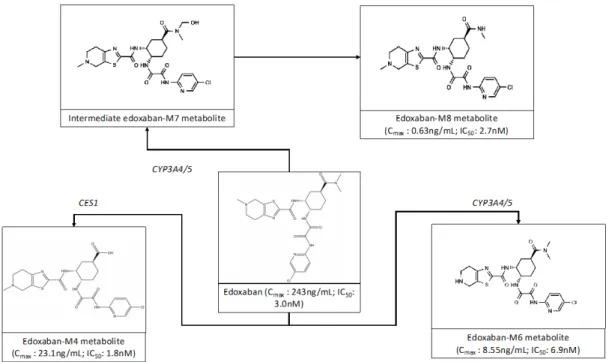
non-valvular atrial fibrillation as well as for the treatment of venous thromboembolisms (VTE) and pulmonary embolism (PE) [5]. Edoxaban has the particularity to release active metabolites which could be present at a sufficient level in the blood to have an impact on haemostasis. These metabolites are named edoxaban-M4, -M6 and -M8 [11]. The metabolism occurs through carboxylesterase-1 (CES-1), cytochrome P450 3A4/5 (CYP3A4/5) and non-enzymatic hydrolysis [12]. Although DOACs do not require regular measurements of their blood concentrations, clinical situations may require an assessment of their concentration (detection of drug accumulation in acute renal or hepatic failure; planning the timing of urgent invasive procedure; recurrence of stroke or bleedings). Currently, prothrombin time (PT) or activated partial thromboplastin time (aPTT) measurement are proposed for the estimation of the intensity of some DOAC. Nevertheless, theses assays are strongly impacted by inter-reagent and inter-individual variability and must not be used in first intention to assess the intensity of anticoagulation with DOACs [5]. Current chromogenic tests for edoxaban are calibrated with standards containing 3 or 4 different levels of edoxaban. However, these standards do not include or consider active metabolites. This approach is questioning since, according to the Food and Drug Administration (FDA) guideline on Bioanalytical Validation [13], a validated method should meet the acceptance criteria regarding selectivity and specificity. Chromogenic anti-Xa assays have been reported to be sensitive to the presence of the M4-metabolite [14] and thus, the specificity criteria is certainly an issue. Normally, the assay should be free of potential interfering substance including metabolites [13]. This lack of specificity could lead to overestimation or underestimation of edoxaban concentrations depending on the clinical situation. Namely, the inhibitory constant (Ki) of these metabolites is not the same as the parent compound and thus, depending on the parent/metabolite ratio, the inhibitory effect on functional assay, like chromogenic anti-Xa assay, for a same initial amount of edoxaban may differ. Therefore, there is a need to develop sensitive and specific assays for edoxaban [3, 5].
In this context, we developed a validated UHPLC-MS/MS method to measure concentration of edoxaban and its active major metabolite-M4 in human plasma [13, 15].
2. Material and Methods
2.1. Preparation of the Normal Pooled Plasma (NPP)
Sixty individuals were included in the study. The exclusion criteria were thrombotic and/or hemorrhagic events, antiplatelet and/or anticoagulant medication, pregnancy and uptake of drugs potentially affecting the platelet and/or coagulation factor functions during the two weeks prior to the blood drawn. The study protocol is in accordance with the Declaration of Helsinki and has been approved by the Ethical Committee of the CHU UCL Namur, Yvoir, Namur, Belgium (B03920096633). Blood was taken by venipuncture in the antecubital vein and collected into 0.109M sodium citrate (9:1 v/v) tubes (Vacuette®, Greiner Bio-One International, Kremsmünster,
Austria) using a 21-gauge needle (Terumo, Leuven). The platelet poor plasma (PPP) was obtained from the supernatant fraction of blood tubes after a double centrifugation for 15 minutes at 2,500g at room temperature. Immediately after centrifugation, PPP from the 60 donors were brought together to obtain the NPP which was frozen at -80°C without any delay. Treatment of plasmas by proteins precipitation was performed within 4 hours after thawing [5].
2.2. Instrumentation and analytical conditions
All optimized parameters and protocol are provided in supplementary material (
Supplementary material and Supplementary figures 1-2).2.3. Method validation
2.3.1. Calibration curve
Calibration curves were constructed from 3 to 500ng/mL for edoxaban and from 3 to 150ng/mL for edoxaban-M4 with six levels of non-zero concentration standards [13, 15]. Calibration ranges were chosen to cover (supra)-therapeutic ranges for edoxaban and edoxaban-M4. Calibration standards were run in triplicate in consecutive runs. The calibration curves were fitted using a weighted least-squares non-linear regression method by measuring the peak-area ratio of the analytes to the internal standard (IS). The acceptance criterion for each back-calculated standard concentration was ± 15% deviation from the nominal value, except for the lower limit of quantification (LLOQ), for which a deviation of ± 20% was permitted.
The intra-day precision and accuracy of the assay were estimated by analysing in a single run of 3 replicates of each QC within 1 day. The inter-day precision and accuracy were assessed by repeating the analysis of three concentration levels of QC samples on 5 consecutive days. Accuracy was expressed as a percentage of relative standard deviation (%Dev), while the precision was expressed as the coefficient of variation (%CV). The accuracy and precision were required to be within a ±20% range for LLOQ and within a ±15% for higher concentrations of edoxaban. For edoxaban-M4 measurement, accuracy and precision were required to be within a ±25% range for LLOQ and within a ±20% for higher concentrations [13, 15].
2.3.3. Carry-over
The carry-over of analytes and IS were evaluated by injecting a blank sample immediately after a sample of upper limit of quantification. Carry-over should not exceed 20% of the signal of the LLOQ for analytes and 5% for IS [13, 15].
2.3.4. Lower limit of quantification
The LLOQ was defined as the lowest concentration of edoxaban or edoxaban-M4 that could be quantitatively determined with acceptable precision and accuracy. Acceptance limits were defined as accuracy of 80-120% and precision ≤20%.
2.3.5. Selectivity
Selectivity was evaluated using six independent blank human plasmas to confirm the absence of potential interfering substances in the matrix [13, 15]. Chromatograms were visually compared with the corresponding spiked plasma.
2.3.6. Specificity
Specificity was assessed to ensure that the method is free of interference due potential residual molecules (e.g. other anticoagulants) [13, 15]. Measurements of spiked samples with 100ng/mL apixaban, edoxaban, edoxaban-M4, rivaroxaban and dabigatran were performed in a single run to assess the potential interference by cross-reacting molecules.
To evaluate ME%, intermediate solutions were post-spiked in water or serum and compare with intermediate solutions post-spiked in NPP. The use of serum as alternative matrix is often encountered in clinical trials and the assessment of this matrix effect may inform on the possibility to perform the measurement in serum if needed.
2.3.8. Extraction recovery
Extraction recovery was determined by spiking known amounts of edoxaban, edoxaban-M4 and IS into NPP and into the extracted NPP and exposing the spiked NPP to the extraction procedure.
2.3.9. Short time stability
Edoxaban and edoxaban-M4 stability was assessed using QCs samples. Intermediate solutions of QCs were stored at -20°C and 4-7°C and plasmatic solutions were stored at -80°C and analysed on day 0 and after one month. Solutions were considered to be stable when average measured concentrations were within the limit of 80-120% of the nominal concentrations [13, 15].
2.4. Statistical analysis
The UHPLC-MS/MS calibration curve was performed using a non-linear regression with a weighting function of 1/x. Statistical analyses and graphics were computed using GraphPad Prism 5.0D® for Mac OS X.
3. Results
3.1. Optimization of the UHPLC-MS/MS method
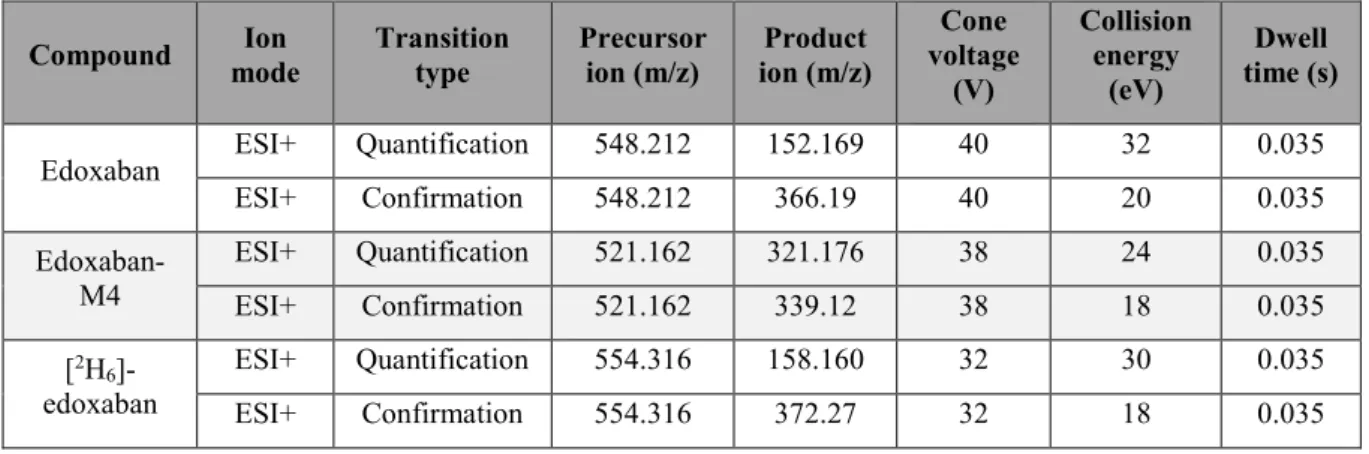
MS detection parameters were optimized by direct infusion of the reference compounds using the electrospray interface in the positive mode for edoxaban and edoxaban-M4 (Table 1). Selection of chromatographic
conditions was optimized to suit an effective chromatographic resolution of analytes with symmetrical peak shapes, short retention time and overall short run time.
3.2. Method validation of the UHPLC-MS/MS method
3.2.1. Calibration curves, linearity and lower limit of quantification
According to results reported in Supplementary figure 3, non-zero calibrators were between ±15% of nominal
concentrations and LLOQ were between ±20% of the nominal concentrations in each validation run. Although one calibration point is out of the range, 75% and a minimum of six non-zero calibrator levels meet the above criteria in each validation run [13, 15]. For routine purpose, the LLOQ was set at 3ng/mL for edoxaban and edoxaban-M4.
3.2.2. Precision and accuracy
Accuracy and precision obtained for inter-day and intra-day assays were within the limits of guidelines with calibration curves performed in NPP (Supplementary table 1 & Supplementary figures 4-7).
3.2.3. Carry-over
There was no detectable carry-over for analytes and IS even at high concentration (500ng/mL).
3.2.4. Selectivity and specificity
No co-eluting components peaks from endogenous or exogenous compounds were observed at the retention time of edoxaban, edoxaban-M4 and [2H6]-edoxaban (Supplementary figure 8 & 9).
3.2.5. Matric effect (ME%)
Non-relevant ME% (<15%) for edoxaban and IS were observed contrary to edoxaban-M4 measurements when these molecules were spiked in water. Serum matrix did not impact edoxaban and edoxaban-M4 measurements with our analytical method.
3.2.6. Stability
Stability of edoxaban and edoxaban-M4 was assessed for 28 days (-20°C) and for 155 days (4-7°C and -80°C). These compounds remained stable for the tested period and accuracy at each level was ±20% (some points are outside the range but represent less than 20% of all tested concentration levels) [13, 15].
4. Discussion
Although the scientific literature contains articles about liquid chromatography measurement of DOACs, few of them address the problem of the active metabolites and propose a simultaneous quantification of edoxaban and edoxaban-M4 in the same analytical run meeting criteria of the International Council of Harmonization (ICH), the European Medicines Agency (EMA) and FDA guidelines for ME, carry-over and stability [14, 16-18]. Our validation provides the pharmaceutical industries, the manufacturers and the physicians with a robust, sensitive and specific method for the measurement of edoxaban and its M4 metabolite.
Despite this must-needed validation, some limitations of have to be recognized. This method allows the measurement of edoxaban and edoxaban-M4 concentration without considering edoxaban-M6. Nevertheless, regarding the IC50 (6.9nM) and the Cmax of edoxaban-M6 (8.55ng/mL), the impact on chromogenic assays should
be negligible contrary to the impact of the edoxaban-M4 which has a lower IC50 (1.8nM) and a higher Cmax
(23.1ng/mL), in the absence of CYP inducers [10, 11, 19] in patients treated with edoxaban at 60mg once daily. Currently, no manufacturer has been able to offer us the edoxaban-M6 in sufficient quantity and with acceptable quality. Another limitation is that we only reported short time stability. Further investigations are underway to confirm the long-term stability of intermediate solutions of edoxaban/edoxaban-M4 and plasmatic solutions of analytes.
The different pharmacodynamic profiles and the interest of measuring the active metabolites
Among the factor Xa inhibitors, edoxaban is the only compound with pharmacologically active metabolites (edoxaban-M4, -M6 and -M8). These metabolites may contribute to the global anticoagulant activity and could interfere with chromogenic assays usually used for the estimation of the plasma level of edoxaban [5, 14]. Taking into consideration their respective IC50 towards human factor Xa, these metabolites would inhibit factor Xa at
different degree. Namely the IC50 of edoxaban, and the metabolites4 (M4; D212393), 6 (M6; D211402) and
-8 (M-8; D21-2135) are 3.0, 1.-8, 6.9 and 2.7nM, respectively (Figures 1 & 2) [10]. While these differences may
only have a small impact in a population where the metabolic pathways are not inhibited or induced (the relative presence of edoxaban-M4 due to CES1 and edoxaban-M6 and -M8 due to CYP3A4/5 are less than 10% each [10]), the metabolite to parent ratio is increased in presence of inducers of CYP3A4/5 and P-gp. A potential interest of synchronously measuring edoxaban and edoxaban-M4 is to obtain complementary information about the impact of the active metabolite in chronometric or chromogenic assays.
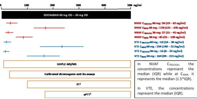
This validated method could be applicable in common clinical situation or situations requiring a close monitoring of edoxaban and its most active metabolite. This is especially important since at low concentration (<30ng/mL) a deviation of more than 50% has been observed (anti-Xa vs LC-MS/MS) [14], suggesting that anti-Xa assays are not able to provide reliable results in these low values. This becomes of clinical interest in light of the low edoxaban concentrations observed at CTrough in the various indications (Figure 3) [3, 20-22]. Also, such low concentrations
(i.e. 30 or 50ng/mL) are threshold for decision making (i.e. before urgent surgery with high bleeding risk or to guide antidote administration) and any inaccuracy may lead to mismanagement or inappropriate clinical judgments [23].
The issue of drug interactions
In addition, this technique could be interesting for adult population with drug-drug interactions which are frequently reported in the daily routine (e.g. co-treatment with quinidine, verapamil, ketoconazole, rifampin, …,)
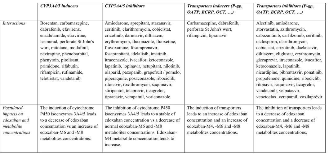
[10, 12]. Indeed, pharmacokinetic interactions with edoxaban have already be highlighted, attesting that exposure to edoxaban and its active metabolites is significantly disturbed when edoxaban is administered with CYP3A4/5 inducers (e.g. rifampin) or inhibitors (e.g. cyclosporine, erythromycin) [10, 12, 14, 19, 24, 25] (Table 2). For
example, co-administration of rifampin and edoxaban leads to an increased concentration of edoxaban-M4 (23.1 ng/mL without rifampin vs 108ng/mL with rifampin) which is not formed through the CYP3A4/5 pathway. According to Mendell et al. the increased concentration is linked to a potential induction of CES1 through upregulation of the pregnane X receptor gene and to the inhibitory effect of rifampin on organic anion transporting polypeptide 1B1 (OATP1B1) [19].
Anti-epileptic agents are also frequently administered concomitantly with anticoagulant in the NVAF setting [26]. Indeed, the cause of symptomatic epilepsy due to strokes and cerebrovascular diseases is estimated to be at 30-40% [27, 28]. With the exception of lamotrigine, pregabalin, zonisamide and lacosamide which have a safer pharmacokinetic profile, phenobarbital, phenytoin and carbamazepine may cause pharmacological interactions due to their impact on CYP3A4 and P-glycoprotein (P-gp) [28-30]. Although such drugs should be avoided, concomitant prescriptions of edoxaban and carbamazepine, first-intention treatment, still remains. Therefore, patients are at risk of exhibiting drug-drug interactions leading to inadequate control of the degree of anticoagulation or disturbed measurement of edoxaban concentration. This supports the need of measuring the most representative active metabolites according to special circumstances to known the real contribution of each compound in order to highlight abnormal edoxaban or metabolite levels [28-30].
To note, an interim analysis of our ongoing study (data not show) evaluating the impact of co-administration of cytochrome 3A4 inducers and edoxaban on edoxaban-M4/edoxaban ratios, demonstrates that the concentration of edoxaban-M4 is increased compared to patients without such interactions. This increase is more pronounced at low concentrations of edoxaban and leads to a significant increase of the ratio edoxaban-M4/edoxaban. Deviations between chromogenic anti-Xa assessments and mass spectrometry measurements appears to be high in those with drug interactions (i.e. > 20%). Investigations are planned to confirm and compare these results to other chronometric and chromogenic assays.
To conclude, the evidence from the literature and the data reported in regulatory documents that have led to the marketing authorization of edoxaban [31, 32] questions about the suitability of calibrators that do not consider the usual proportion of the active metabolites when reporting results as edoxaban equivalent.
In conclusion, this validated UHPLC-MS/MS method allows an accurate quantification of edoxaban and its active-metabolite, edoxaban-M4, in human plasma according to the recommendations of the ICH, EMA and FDA. Calibration ranges permit simultaneous estimations of edoxaban and edoxaban-M4 concentrations in patient’s plasma. Although edoxaban treated patients exhibit usually low levels of active metabolites, some clinical situations may require an accurate estimation of the exposure for decision making. In addition, drug-drug interactions or hepatic insufficiency should stimulate investigations to explore abnormal levels of metabolites with different pharmacodynamic profile than edoxaban. Additional studies are needed to compared results obtained by this validated UHPLC-MS/MS method vs current coagulation/chromogenic assays in plasma from patients treated with edoxaban including those with drug interactions or with levels close to decision making thresholds.
Acknowledgments
The authors would like to acknowledge Christelle Vancraeynest, Vincent Maloteau, Lionel Pochet and Jonathan Evrard for their contribution to this work.
Conflict of Interest Disclosures
Among the authors, J. Douxfils is CEO and founder of QUALIblood s.a. and reports personal fees from Portola Pharmaceuticals, Stago, Roche, Roche Diagnostics and Daiichi-Sankyo, outside the submitted work. The other authors have no conflicts of interest to disclose.
References
1. Siriez, R., et al., Development of new methodologies for the chromogenic estimation of betrixaban
concentrations in plasma. Int J Lab Hematol, 2019.
2. Siriez, R., et al., Betrixaban: Impact on Routine and Specific Coagulation Assays-A Practical
Laboratory Guide. Thromb Haemost, 2018. 118(7): p. 1203-1214.
3. Douxfils, J., et al., Laboratory testing in patients treated with direct oral anticoagulants: a
practical guide for clinicians. J Thromb Haemost, 2018. 16(2): p. 209-219.
4. Douxfils, J. and R.C. Gosselin, Laboratory Assessment of Direct Oral Anticoagulants. Semin Thromb Hemost, 2017. 43(3): p. 277-290.
5. Douxfils, J., et al., Edoxaban: Impact on routine and specific coagulation assays. A practical
laboratory guide. Thromb Haemost, 2016. 115(2): p. 368-81.
6. Douxfils, J., et al., Impact of apixaban on routine and specific coagulation assays: a practical
laboratory guide. Thromb Haemost, 2013. 110(2): p. 283-94.
7. Douxfils, J., et al., Assessment of the impact of rivaroxaban on coagulation assays: laboratory
recommendations for the monitoring of rivaroxaban and review of the literature. Thromb Res,
2012. 130(6): p. 956-66.
8. Douxfils, J., et al., Impact of dabigatran on a large panel of routine or specific coagulation
assays. Laboratory recommendations for monitoring of dabigatran etexilate. Thromb Haemost,
2012. 107(5): p. 985-97.
9. Skeppholm, M., et al., On the monitoring of dabigatran treatment in "real life" patients with atrial
fibrillation. Thromb Res, 2014. 134(4): p. 783-9.
10. Parasrampuria, D.A. and K.E. Truitt, Pharmacokinetics and Pharmacodynamics of Edoxaban, a
Non-Vitamin K Antagonist Oral Anticoagulant that Inhibits Clotting Factor Xa. Clin
Pharmacokinet, 2016. 55(6): p. 641-55.
11. Food and Drug Administration, Clinical Pharmacology and Biopharmaceutics review(s). 2014. 12. Parasrampuria, D.A., et al., Edoxaban drug-drug interactions with ketoconazole, erythromycin,
and cyclosporine. Br J Clin Pharmacol, 2016. 82(6): p. 1591-1600.
13. Food and Drug Administration. Bioanalytical Method Validation - Guidance for Industry. 2018 [cited 2019 6 Feb]; Available from:
https://www.fda.gov/downloads/drugs/guidances/ucm070107.pdf.
14. He, L., et al., Determination of edoxaban equivalent concentrations in human plasma by an
automated anti-factor Xa chromogenic assay. Thromb Res, 2017. 155: p. 121-127.
15. EMA, ICH Topic Q2 (R1) Validation of Analytical Procedures: Text and Methodology - NOTE
FOR GUIDANCE ON VALIDATION OF ANALYTICAL PROCEDURES: TEXT AND METHODOLOGY (CPMP/ICH/381/95). 1995.
16. Rohde, G., Determination of rivaroxaban--a novel, oral, direct Factor Xa inhibitor--in human
plasma by high-performance liquid chromatography-tandem mass spectrometry. J Chromatogr B
Analyt Technol Biomed Life Sci, 2008. 872(1-2): p. 43-50.
17. Lagoutte-Renosi, J., et al., A simple and fast HPLC-MS/MS method for simultaneous
determination of direct oral anticoagulants apixaban, dabigatran, rivaroxaban in human plasma.
18. Foerster, K.I., et al., Simultaneous quantification of direct oral anticoagulants currently used in
anticoagulation therapy. J Pharm Biomed Anal, 2018. 148: p. 238-244.
19. Mendell, J., et al., The Effect of Rifampin on the Pharmacokinetics of Edoxaban in Healthy Adults. Clin Drug Investig, 2015.
20. Ruff, C.T., et al., Association between edoxaban dose, concentration, anti-Factor Xa activity, and
outcomes: an analysis of data from the randomised, double-blind ENGAGE AF-TIMI 48 trial. The
Lancet, 2015. 385(9984): p. 2288-2295.
21. Verhamme, P., et al., Dose reduction of edoxaban preserves efficacy and safety for the treatment
of venous thromboembolism. An analysis of the randomised, double-blind HOKUSAI VTE trial.
Thromb Haemost, 2016. 116(4): p. 747-53.
22. Weitz, J.I., et al., Randomised, parallel-group, multicentre, multinational phase 2 study comparing
edoxaban, an oral factor Xa inhibitor, with warfarin for stroke prevention in patients with atrial fibrillation. Thromb Haemost, 2010. 104(3): p. 633-41.
23. Levy, J.H., et al., When and how to use antidotes for the reversal of direct oral anticoagulants:
guidance from the SSC of the ISTH. J Thromb Haemost, 2016. 14(3): p. 623-7.
24. CBIP, Centre Belge d’Information Pharmacothérapeutique - Bon usage des médicaments -
Interactions des médicaments. 2018.
25. Vazquez, S.R., Drug-drug interactions in an era of multiple anticoagulants: a focus on clinically
relevant drug interactions. Hematology Am Soc Hematol Educ Program, 2018. 2018(1): p.
339-347.
26. Wang, J.Z., et al., Incidence and management of seizures after ischemic stroke: Systematic review
and meta-analysis. Neurology, 2017. 89(12): p. 1220-1228.
27. Jabareen, A., et al., Treatment with antiepileptic drugs in patients with stroke. A change in clinical
practice may be required. J Neurol Sci, 2018. 395: p. 4-7.
28. Galgani, A., et al., Pharmacokinetic Interactions of Clinical Interest Between Direct Oral
Anticoagulants and Antiepileptic Drugs. Front Neurol, 2018. 9: p. 1067.
29. Di Gennaro, L., et al., Carbamazepine interaction with direct oral anticoagulants: help from the
laboratory for the personalized management of oral anticoagulant therapy. J Thromb
Thrombolysis, 2019.
30. Stollberger, C. and J. Finsterer, Interactions between non-vitamin K oral anticoagulants and
antiepileptic drugs. Epilepsy Res, 2016. 126: p. 98-101.
31. European Medicines Agency. Lixiana - EMEA/H/C/002629/0000. 2015 2015 Apr 25 [cited 2019 Oct 23]; Available from: https://www.ema.europa.eu/en/documents/assessment-report/lixiana-epar-public-assessment-report_en.pdf.
32. Food and Drug Administration. Savaysa - Clinical Pharmacology and Biopharmaceutics
Table legends
Table 1: MS/MS parameters for edoxaban, edoxaban-M4 and corresponding internal standard Table 2: Non-exhaustive list of drug-drug interactions with edoxaban
Figure legends
Figure 1: Postulated edoxaban metabolism for active metabolites
Figure 2: Chemical structure of edoxaban (DU-176b), edoxaban-M4 and internal standard ([2H6]-edoxaban)
Tables
Table 1: MS/MS parameters for edoxaban, edoxaban-M4 and corresponding internal standard1
Compound mode Ion Transition type Precursor ion (m/z) ion (m/z) Product voltage Cone (V) Collision energy (eV) Dwell time (s)
Edoxaban ESI+ Quantification 548.212 152.169 40 32 0.035 ESI+ Confirmation 548.212 366.19 40 20 0.035 Edoxaban-M4 ESI+ Quantification 521.162 321.176 38 24 0.035 ESI+ Confirmation 521.162 339.12 38 18 0.035 [2H6 ]-edoxaban ESI+ Quantification 554.316 158.160 32 30 0.035 ESI+ Confirmation 554.316 372.27 32 18 0.035
Table 2: Non-exhaustive list of drug-drug interactions with edoxaban2
CYP3A4/5 inducers CYP3A4/5 inhibitors Transporters inducers (P-gp,
OATP, BCRP, OCT, …) Transporters inhibitors (P-gp, OATP, BCRP, OCT, …)
Interactions Bosentan, carbamazepine, dabrafenib, efavirenz, enzalutamide, etravirine, lesinurad, perforate St John's wort, mitotane, modafinil, nevirapine, phenobarbital, phenytoin, pitolisant, primidone, rifabutin, rifampicin, rufinamide, telotristat, vandetanib
Amiodarone, aprepitant, atazanavir, ceritinib, clarithromycin, cobicistat, crizotinib, darunavir, diltiazem, erythromycin, fluconazole, fluoxetine, fluvoxamine, fosamprenavir,
fosaprepitant, idelalisib, imatinib, itraconazole, ivacaftor, ketoconazole, lapatinib, lopinavir, netupitant, nilotinib, olaparid, pazopanib, grapefruit / pomelo, piperaquine, posaconazole, ribociclib, ritonavir, roxithromycin, saquinavir, stiripentol, telaprevir, ticagrelor, tipranavir, verapamil, voriconazole
Carbamazepine, dabrafenib, perforate St John's wort, rifampicin, tipranavir
Alectinib, amiodarone, atorvastatin, azithromycin,
cabozantinib, carfilzomib, ceritinib, ciclosporin, clarithromycin, cobicistat, crizotinib, daclatasvir, diltiazem, eliglustat, erythromycin, glecaprevir, itraconazole, ivacaftor, ketoconazole, lapatinib,
nicardipine, pibrentasvir, ponatinib, propafenone, quinidine, ribociclib, ritonavir, saquinavir, ticagrelor, vandetanib, velpatasvir,
venetoclax, verapamil, voxilaprévir
Postulated impacts on edoxaban and metabolite concentrations
The induction of cytochrome P450 isoenzymes 3A4/5 leads to a decrease of edoxaban concentration vs an increase of edoxaban-M6 and -M8 metabolites concentrations.
The inhibition of cytochrome P450 isoenzymes 3A4/5 leads to a stable of edoxaban concentration vs a decrease of normal edoxaban-M6 and -M8
metabolites concentrations. Edoxaban-M4 metabolite concentration tends to increase.
The induction of transporters leads to an increase of edoxaban concentration and an increase of edoxaban-M4, -M6 and -M8 metabolites concentrations.
The inhibition of transporters leads to a decrease of edoxaban
concentration and a decrease of edoxaban-M4, -M6 and -M8 metabolites concentrations.
2 10. Parasrampuria, D.A. and K.E. Truitt, Pharmacokinetics and Pharmacodynamics of Edoxaban, a Non-Vitamin K Antagonist Oral Anticoagulant that Inhibits Clotting Factor Xa. Clin Pharmacokinet, 2016. 55(6): p. 641-55. 24. CBIP, Centre Belge d’Information Pharmacothérapeutique - Bon usage des médicaments - Interactions des médicaments. 2018.
Supplementary table 1: Accuracy and precision for edoxaban and edoxaban-M43
Nominal concentration
(ng/mL) Within-Run Accuracy (%) - Day 1 Within-Run Accuracy (%) - Day 2 Within-Run Accuracy (%) - Day 3 Within-Run Accuracy (%) - Day 4 Within-Run Accuracy (%) - Day 5
Edoxaban 3 -9 16 -1 5 -3 10 -14 2 1 -4 4 75 -2 7 5 1 9 250 -3 1 13 -1 -2 500 -7 -2 -1 3 -5 Edoxaban-M4 3 -22 7 4 6 2 10 -13 -7 0 -3 -13 75 -16 -4 -4 8 -3 125 -11 -7 9 12 -6 150 -13 -7 -4 7 -6 Nominal concentration
(ng/mL) Within-Run Precision (%) - Day 1 Within-Run Precision (%) - Day 2 Within-Run Precision (%) - Day 3 Within-Run Precision (%) - Day 4 Within-Run Precision (%) - Day 5
Edoxaban 3 4 0 2 2 3 10 0 3 2 2 2 75 3 3 1 2 1 250 1 2 0 1 2 500 2 2 0 1 2 Edoxaban-M4 3 4 1 6 4 6 10 0 4 3 2 3 75 6 3 2 1 3 125 1 1 0 0 1 150 0 2 0 0 2 Nominal concentration
(ng/mL) Between-Run Accuracy (%) - Edoxaban in NPP Within-Run Precision (%) - Edoxaban in NPP Between-Run Accuracy (%) - Edoxaban-M4 in NPP Within-Run Precision (%) - Edoxaban-M4 in NPP -
Edoxaban 3 2 2 - - - 10 0 2 - - - 75 4 2 - - - 250 2 1 - - - 500 -2 1 - - - Edoxaban-M4 3 - - 0 4 - 10 - - -7 3 - 75 - - -3 3 - 125 - - -1 1 - 150 - - -5 1 -
Figures
Figure 1: Postulated edoxaban metabolism for active metabolites 4
4 CES1: carboxylesterase-1; CYP3A4/5: Cytochrome P450 isoenzyme 3A4/5 ; IC50: half-maximal inhibitory
Figure 2: Chemical structure of (A) edoxaban (DU-176b), (B) edoxaban-M4 and (C) internal standard ([2H6]-edoxaban)
(A)
(B)
(C)
Figure 3: Laboratory testing of edoxaban and expected plasma concentrations after therapeutic doses5
5 Red and blue lines represent plasma concentrations at peak and trough in NVAF and VTE, respectively.
Orange boxes represent ranges of applicability of the corresponding test. ‡This represents the range of quantitation for sensitive reagents. Depending on the reagent, the sensitivity may be lower. aPTT, activated partial thromboplastin time; CMAX, maximum plasma concentration during the dosing interval; CTROUGH,
minimum plasma concentration during the dosing interval; IQR, interquartile range; NVAF, non-valvular atrial fibrillation; PT, prothrombin time; VTE, venous thromboembolism. Notes: (i) Data on plasma concentration were extracted from Ruff et al. 20. Ruff, C.T., et al., Association between edoxaban dose, concentration,
anti-Factor Xa activity, and outcomes: an analysis of data from the randomised, double-blind ENGAGE AF-TIMI 48 trial. The Lancet, 2015. 385(9984): p. 2288-2295. , Weitz et al. 22. Weitz, J.I., et al., Randomised, parallel-group, multicentre, multinational phase 2 study comparing edoxaban, an oral factor Xa inhibitor, with warfarin for stroke prevention in patients with atrial fibrillation. Thromb Haemost, 2010. 104(3): p. 633-41. and
Verhamme et al. 21. Verhamme, P., et al., Dose reduction of edoxaban preserves efficacy and safety for the
treatment of venous thromboembolism. An analysis of the randomised, double-blind HOKUSAI VTE trial.
Supplementary Figures
Supplementary figure 1: Gradient program for the mobile phase, with UPLC-grade water with 0.1% formic acid (A) and UPLC-grade methanol with 0.1% formic acid (B)
Supplementary figure 2: MRM chromatograms of edoxaban, edoxaban-M4 and [2H6]-edoxaban
MRM chromatograms of [2H6]-edoxaban (12ng/mL), edoxaban (500ng/mL) and edoxaban-M4 (150ng/mL) using optimized MS and chromatographic conditions.
Supplementary figure 3: Calibration Curve – Edoxaban and edoxaban-M4
(A)
(B)
%Dev - Quadratic calibration curve of (A) edoxaban and (B) edoxaban-M4 in NPP with non-linear ponderation 1/x. For edoxaban, red and green lines represent ranges of ±20% or ±15%, respectively. For edoxaban-M4, red and green lines represent ranges of ±25% or ±20%, respectively.
Supplementary figure 4: Accuracy – Within and between run for edoxaban
(A)
(B)
(A) Within-Run Accuracy (%) - Calibration curve of edoxaban in NPP. Red and green lines represent ranges of ±20% or ±15%, respectively. (B) Between-Run Accuracy (%) - Calibration curve of edoxaban in NPP. Red and green lines represent ranges of ±20% or ±15%, respectively.
Supplementary figure 5: Accuracy – Within and between run for edoxaban-M4
(A)
(B)
(A) Within-Run Accuracy (%) - Calibration curve of edoxaban-M4 in NPP. Red and green lines represent ranges of ±25% or ±20%, respectively. (B) Between-Run Accuracy (%) - Calibration curve of edoxaban-M4 in NPP. Red and green lines represent ranges of ±25% or ±20%, respectively.
Supplementary figure 6: Precision – Within and between run for edoxaban
(A)
(B)
(A) Within-Run Precision (%) - Calibration curve of edoxaban in NPP. Red and green lines represent ranges of ±20% or ±15%, respectively. (B) Between-Run Precision (%) - Calibration curve of edoxaban in NPP. Red and green lines represent ranges of ±20% or ±15%, respectively.
Supplementary figure 7: Precision – Within and between run for edoxaban-M4
(A)
(B)
(A) Within-Run Precision (%) - Calibration curve of edoxaban-M4 in NPP. Red and green lines represent ranges of ±25% or ±20%, respectively. (B) Between-Run Precision (%) - Calibration curve of edoxaban in NPP. Red and green lines represent ranges of ±25% or ±20%, respectively.
Supplementary figure 8: Selectivity
Selectivity assessment aims to verify that the substance being measured is the intended analyte to minimize or avoid interference. The analyze of blank samples of the appropriate biological matrix from six individual sources did not highlight any interference at retention time of edoxaban, edoxaban-M4 and the internal standard (chromatograms were visually checked to assess interferences). According to the visual interpretation of these chromatograms, no interference at predicted retention times of edoxaban, M4 or IS occurs in individual plasma without these molecules of interest.
Individual plasma 1 – No edoxaban – No edoxaban-M4 – No internal standard.
Individual plasma 3 – No edoxaban – No edoxaban-M4 – No internal standard.
Individual plasma 5 – No edoxaban – No edoxaban-M4 – No internal standard.
Supplementary figure 9: Representative chromatograms with retention times of apixaban, rivaroxaban, edoxaban, edoxaban-M4 and dabigatran
![Figure 2: Chemical structure of (A) edoxaban (DU-176b), (B) edoxaban-M4 and (C) internal standard ([ 2 H 6 ]-edoxaban) (A) (B) (C)](https://thumb-eu.123doks.com/thumbv2/123doknet/14488421.717012/21.892.126.458.243.1099/figure-chemical-structure-edoxaban-edoxaban-internal-standard-edoxaban.webp)