On two new linstowiid cestodes from Australian dasyurid
marsupials
IAN BEVERIDGE lnstitut de Zoologie, University of Neuchatel, Switzerland
ABSTRACT
Two new species of the genus Oochoristica Luhe, 1898 from Australian dasyurid marsupials are described and their affinities discussed. The new species are O. antechini sp. nov. from Antechinus macdonnellensis and O. eremophlia sp. nov. from Antechinus rosamondae.
Very few species of the genus Oochoristica Luhe, 1898 (sensu Delia Santa, 1956) are known from Australia. O. trachysauri (MacCallum, 1921) Baer, 1927 and O. vacuolata Hickman, 1954 have been described from two species of reptile belonging to the family Scincidae,
Trachysaurus rugosus and Egernia whitii respectively (Baer, 1927; Johnston, 1932; Hickman,
1954), whilst a single species, O. nyctophili Hickman, 1954 has been described from the bat Nyctophilus geoffroyi in Tasmania.
The absence of species of Oochoristica amongst Australian mammals, which is surprising in view of their relative abundance in South American marsupials, may be more apparent than real as many of the species of small insectivorous marsupials likely to harbour linstowiid cestodes have apparently not been examined for helminths (Mackerras, 1958).
The present paper records two new species of the genus Oochoristica from members of the dasyurid genus Antechinus, in this instance from two species occurring in rather remote and arid inland regions.
MATERIALS AND METHODS
Animals were trapped alive and transported to the laboratory where they were main-tained until autopsy. As soon as possible after the death of an animal, the gastro-intestinal tract was removed and examined for helminths. Any cestodes found were relaxed briefly in water and fixed in a mixture of formalin, alcohol and acetic acid. Whole cestodes were stained in Mayer's haemalum, acid carmine or celestine blue and mounted in balsam. Serial longitudinal and transverse sections were made of each species at a thickness of
10 fim, and were stained with haematoxylin or haematoxylin and eosin. Drawings were made with the aid of a camera lucida.
Throughout the text, measurements are given in mm, as the range followed by the mean in parentheses.
A scale line of 0-1 mm is shown on each figure except figures 2 and 11 which have a scale line of 0-01 mm.
Present address: Dept. of Veterinary Paraclinical Sciences, University of Melbourne, Parkville 3052, Victoria, Australia.
DESCRIPTIONS Anoplocephalata Khodlodkowski
Linstowiidae Mola, 1929
Oochoristica Luhe, 1898
Oochoristica antechini sp. nov. (Figs. 1-8)
Host: Antechinus macdonnellensis (Spencer, 1896) (Marsupialia: Dasyuridae). Location in host: intestine.
Type locality: 27 Km. west of Refrigerator Well, Tanami Road, Northern Territory,
Australia.
Material examined: 16 complete specimens from two hosts. Collected by P. Woolley,
July, 1975.
Types deposited: South Australian Museum, Adelaide, South Australia.
Gravid specimens are 23-70 (36) long and have a maximum width of 4. The scolex is entirely unarmed and is bluntly conical anteriorly, being 0-52-0-78 (0-62) in diameter and bearing two suckers on each of the dorsal and ventral surfaces. The suckers are oval in shape, with thick muscular margins which are incomplete at the anterior extremities. The suckers measure 0-28-0-36 (0-31) by 0-20-0-25 (0-24). The neck region is demarcated in some specimens by a constriction posterior to the scolex but in other specimens the scolex merges imperceptibly into the unsegmented neck region. The segmentation is distinct and the width of the proglottides increases rapidly along the strobila. Segmentation is of the craspedote type with a narrow, straight-edged velum extending over the adjacent proglottis. There are 100-110 proglottides in gravid strobilae; the first mature proglottis is about the 50th and the genital organs have generally involuted by about the 70th proglottis. Mature proglottides measure 1-8-2-3 (2-1) by 0-28-0-45 (0-37), whilst gravid proglottides measure 2-5-3-7 (3-3) by 0-50-0-85 (0-55).
The longitudinal musculature is well developed. The inner longitudinal muscles are arranged in about 40 discrete bundles around the outer edge of the medulla. The bundles are oval in transverse section and contain 10-20 muscle fibres each. The outer longitudinal muscles form irregularly disposed fibre bundles peripheral to the inner longitudinal muscles. The bundles are smaller than those of the inner ring and contain fewer fibres. Towards the periphery, the bundles are replaced by individual fibres or pairs of fibres. Transverse muscles occur in small numbers as individual fibres on either side of and between the inner longi-tudinal muscle bundles. The dorso-ventral musculature is rather weakly developed and consists of individual fine fibres crossing medulla and cortex at irregular intervals.
The dorsal and ventral longitudinal osmoregulatory canals are situated close to the lateral boundaries of the cortex. The dorsal canal is narrow and sinuous, having a diameter of 0-01-0-02 (0013) in mature proglottides. The ventral canal is wider, with a diameter of 0-02-0-03 (0-025) and is less sinuous. A narrow transverse canal connects left and right ventral canals at the posterior margin of each proglottis. The ventral canals also give rise to a complex network of minute canals which cross the proglottis on the ventral surface, forming irregular anastomoses. The diameter of the accessory canals ranges from 0-008-0-010. The genital ducts pass between the longitudinal osmoregulatory canals.
The genital pores alternate irregularly. They are situated in the anterior half of the lateral proglottis margin, dividing the margin in the approximate ratio of 1: 2. The prominent genital atrium is circular or oval in dorso-ventral view and is lined with tegument. Around the atrium there is a condensation of muscular and parenchymatous tissues, the muscle fibres being arranged in a radial fashion.
FIGS. 1-5. O. antechini sp. nov.
FIG. 1. Scolex. FIG. 2. Egg. FIG. 3. Cirrus sac. FIG. 4. Female genitalia and genital ducts. FIG. 5. Mature proglottis.
The cirrus sac is long and narrow, measuring0-30-0-35 (0-33) by 0-04-0-05 (0047) and passes medially just beyond the longitudinal osmoregulatory canals. The cirrus is unarmed and is only slightly coiled. The distal extremity is of a slightly greater internal diameter than the mid-region, whilst the proximal extremity is slightly dilated to form a diminutive internal seminal vesicle. A retractor muscle extends from the proximal extremity of the cirrus sac towards the proglottis centre. The vas deferens is narrow and greatly coiled in the region of the cirrus sac. It runs medially and posteriorly towards the female genitalia, crossing the ovary and seminal receptacle before dividing into fine vasa efferentia. The testes are rounded, measuring 0-044-0-073 (0-057) in diameter and numbering 27-45 (38) per proglottis. They are distributed in a band between the longitudinal osmoregulatory canals, mainly posterior to the ovary. Some testes overlie the vitellarium, but never the ovary. In some instances the testes are discontinuous posterior to the vitellarium in which case they form two distinct, laterally-situated groups.
8 olm ilm
*
• ' ) ° ft I ^
- - P O 1mm J 01mmFIGS. 6 and 7. O. antechini sp. nov., uterus and eggs. Fig. 6 shows early stage of egg formation with eggs present in lumen of uterus. Fig. 7 shows a later stage in development, after the uterus has disappeared, with eggs, now surrounded by a capsule, scattered through parenchyma. FIG. 8. Transverse section of cortex of O. antechini sp. nov., showing musculature. FIG. 9. Longitudinal section of O. eremophila sp. nov. showing musculature.
The vagina opens independently into the genital atrium posterior to the cirrus sac. There is no vaginal sphincter. The vagina runs as a narrow duct posterior to the cirrus sac then posteriorly and medially towards the apex of the ovary where it dilates into an elongate seminal receptacle. The ovary is fan-shaped, measuring 0-33-0-45 (0-40) by 0-18-0-25 (0-21) and is composed of numerous clavate lobules. It is not divided into two major lobes as in some other members of the genus. The vitellarium is roughly reniform in shape, mildly lobulate and measures 0-14-0-23 (0-18) by 0-08-0-11 (009). It is situated posterior to the apex of the ovary extending aporally from it. The ovarian duct almost immediately joins the duct from the seminal receptacle to form a short fertilisation duct dorsal to the ovary. The coiled vitelline duct joins the fertilisation duct and the common duct passes anteriorly to pierce Mehlis' gland which lies between the vitellarium and the apex of the ovary. From Mehlis' gland, the uterine duct passes anteriorly, dorsal to the ovary, terminating at the latter's anterior margin.
The uterus develops initially as a many-branched sac-like structure, rapidly extending through the medullary parenchyma and beyond the longitudinal osmoregulatory canals. The uterus is bounded by a very fine delimiting membrane and the eggs (or zygotes at this stage) consist of large cells with prominent nuclei and nucleoli and lie free in the lumen. Subsequently the uterus disappears and the developing eggs are found scattered individually through the proglottis. The developing embryo appears as a cluster of cells and chromatin-like granules and is surrounded by a wide, clear space delimited externally by a capsule. In fully gravid proglottides, the oncosphere measures 0-042-0-068 (0-046) by 0-034-0-039 (0036) and the oncospheral hooks are 0-015-0-021 (0-018) long. A thick embryophore surrounds the oncosphere as well as a thick outer capsule measuring 0-057-0-070 (0-062) in diameter.
Oochoristica eremophila sp. nov. (Figs. 9-13)
Host: Antechinus rosamondae Ride, 1964 (Marsupialia: Dasyuridae). Location in host: intestine.
Type locality: Woodstock Station via Marble Bar, Western Australia.
Material examined: specimens from three hosts comprising 4 entire strobilae and
numerous fragmented specimens. Collected by P. Woolley, Novem-ber, 1975.
Types deposited: South Australian Museum, Adelaide, South Australia.
Gravid specimens of this species vary in length from 16-24 (20) and are composed of 37-49 (42) proglottides. The worms are extremely fragile and diaphanous, and readily fragment when handled. The scolex is broadly rounded anteriorly and measures 0-26-0-37 (0-32) in diameter. Both the scolex and the four oval suckers it bears are unarmed. The suckers measure 0065-0-143 (0-104) by 0-062-0-117 (0-094). The scolex merges imper-ceptibly into the unsegmented neck region. Segmentation is distinct and is of the acraspid type. Mature proglottides are as long as or slightly longer than wide, roughly barrel-shaped and measure 0-28-0-60 (0-45) by 0-17-0-49 (0-39), whilst gravid proglottides vary from 0-63-1 -05 (0-89) by 0-55-0-75 (0-65) and are usually much longer than wide. In entire worms,
10-20 proglottides contain fully developed eggs.
The longitudinal musculature is very feebly developed. Outer longitudinal muscles were not found, presumably being extremely fine. The inner longitudinal muscles form a narrow irregular band of loosely-connected, fine fibres adjacent to the medulla. The fibres are not apparently grouped in bundles but are scattered in a ring around the corticomedullary junction. The dorso-ventral muscles are present as fine, individual muscle fibres crossing cortex and medulla at irregular intervals. Transverse muscles were not seen.
The longitudinal osmoregulatory canals are paired on either side of the strobila. The ventral canal is sinuous and measures 0-023-0-027 (0-025) in diameter. The dorsal canal which is narrower and more sinuous has a diameter of 0008-0013. Towards the posterior margin of each proglottis, a narrow transverse canal varying from 0-008-0-010 in diameter connects the ventral canals of each side of the strobila. A similar canal is present towards the anterior end of each proglottis, however, in this case, the canal is of greater diameter, 0-010-O-029, and is more sinuous. It begins about the level of the genital atrium, loops anteriorly, close to the ventral canal, then extends laterally across the proglottis and runs posteriorly for some distance, parallel to the longitudinal canal on the other side of the proglottis before actually joining with it. The two transverse canals make simple connections with the longitudinal canals and do not show the duplications and other variations seen in some other species of the genus.
The genital ducts pass the longitudinal osmoregulatory canals dorsally.
The genital primordium appears within the first two or three proglottides and the first fully mature proglottis is about the 20th. The genital pores alternate irregularly. The genital atrium is distinct but small and is not supplied with extra musculature. It is situated in the anterior third of the lateral proglottis margin, dividing the margin in an approximate ratio of 2: 1. Occasionally a small genital papilla is present surrounding the atrium. The cirrus sac is roughly ellipsoidal in shape, invariably reaching and usually crossing the longitudinal osmoregulatory canals. The cirrus sac measures 0-078-0-143 (0-114) by 0 044-0 047 (0-045). The cirrus is unarmed and is coiled within the cirrus sac. No retractor muscle was found attached to the cirrus sac. There is neither internal nor external seminal vesicle. The narrow vas deferens, leaving the cirrus sac passes anteriorly and medially, describing 7-8 elongate coils. The more anterior coils may extend into the adjacent proglottis. From near the mid-line, after a reduction in diameter, the vas deferens passes posteriorly, crossing the ovary dorsally and passing slightly to the poral side of the vitellarium before dividing into minute vasa efferentia which run to the testes. The testes are rounded and measure 0031-0052 (0-042) in diameter. They number 28-33 (31) and are distributed in one or two transverse layers posterior to the female genitalia. Their distribution is limited laterally by the longi-tudinal osmoregulatory canals. Anteriorly, the band of testes is 4-5 wide, diminishing pos-teriorly to 1-3. The testes extend to the anterior margin of the vitellarium, frequently overlying this organ and Mehlis' gland. No testes have been found overlying the ovary.
10
01mm
11
001mm
13
0.1
mm12
0.1mm
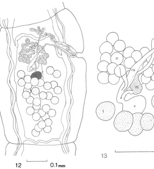
FIG. 12. 0. eremophila sp. nov., mature proglottis.
FIG. 13. O. eremophila sp. nov., female genitalia and genital ducts.
KEY: dvm = dorso-ventral muscle, ilm = inner longitudinal muscle, f = fertilisation duct, m = Mehlis' gland, o = ovary, olm = outer longitudinal muscle, p = parenchyma, sr = seminal receptacle, t = testis, tm = transverse muscle, u = uterus, ud = uterine duct, v = vitellarium.
The vagina opens independently into the genital atrium posterior to the cirrus sac. No vaginal sphincter is present. The vagina runs as a narrow tube posterior and dorsal to the cirrus sac and just beyond the proximal extremity of the cirrus sac, changes course to run posteriorly and medially, crossing the ovary dorsally and enlarging to form an elongate seminal receptacle. The ovary is situated in the centre of the proglottis at about the level of the genital atrium. It is not subdivided into two major lobes, but is fan-shaped and composed of about 25 clavate lobules, most lobules with their longitudinal axes in the transverse plain of the proglottis such that in whole mounts they are seen end-on. The ovarian lobules surround the seminal receptacle on three sides, but on the dorsal side, a short oviduct curves posteriorly, undergoes a considerable reduction in diameter and enters
the posterior extremity of the seminal receptacle. The fertilisation duct is short and as it curves posteriorly, it joins the vitelline duct and then passes anteriorly to Mehlis' gland which is situated between ovary and vitellarium. The ovary measures 0-156-0-169 (0-161) by 0-117-0-135 (0-127). The vitellarium is a small spherical to ovoid body measuring 0-062-0-081 (0-070) by 0-047-0-057 (0-049) and is situated posterior to and slightly to the poral side of the midline of the ovary. From Mehlis' gland, the uterine duct passes anteriorly, dorsal to the ovary, terminating in a slight enlargement close to the anterior margin of the ovary. A uterus was found neither in whole mounts nor in serial sections. Eggs begin to appear in proglottides still containing fully developed male and female genital organs, scattered throughout the parenchyma. It is likely that if a uterus exists, it does so for a short period of time only. Oncospheres measure 0-021-0030 (0-029) by 0-022-0-025 (0023). The oncosphere is surrounded by a thick embryophore and a thin outer capsule measuring 0-046-0-061 (0-052) in diameter. There is great variability within any one proglottis in the degree of maturity of the eggs. Some eggs are almost invariably mature, whilst others in the same proglottis consist of aggregations of relatively undifferentiated cells. Either eggs vary greatly in the time they take to mature or egg production within a proglottis continues over a relatively long period.
The specific epithet is derived from two Greek words meaning "desert-loving".
DISCUSSION
Both species of cestode described above have been referred to the linstowiid genus
Oochoristica Liihe, 1898 sensu Delia Santa (1956), each having an unarmed rostellum, eggs
scattered throughout the parenchyma in gravid proglottides and the genital ducts passing between or dorsal to the longitudinal osmoregulatory canals. Oochoristica has been variously divided into numerous genera by Spasskii (1951) and Yamaguti (1959), depending upon the systematic position of the host (mammal or reptile) and to some extent on the type of segmentation. Delia Santa (1956) however maintained the genera Mathevotaenia Akhumian, 1946, Atriotaenia Sandground, 1926, Cycloskrjabinia Spasskii, 1951, Oschmarenia Spasskii, 1951 and Semenoviella Spasskii, 1951 as synonyms of Oochoristica. Dollfus (1954) proposed the retention of the genus Mathevotaenia for those species parasitising mammals, however Stunkard (1961), whilst accepting the division in principle, merged the genera Mathevotaenia and Atriotaenia giving the latter priority. Stunkard (1965) also erected the genus
Paratrio-taenia for the single species P. oedipomatidis Stunkard, 1965, however, he concluded that
the systematic position of the new genus was uncertain with respect to family as well as to genera. In the present paper, the definition of the genus used is that of Delia Santa (1956) and hence the generic name of Oochoristica is used for the two cestodes described even though their hosts are mammals. However, in the discussion of affinities, generic names are used as first published and species described as belonging to Mathevotaenia or Atriotaenia are discussed under these names.
Delia Santa (1956) recognised 18 species of Oochoristica from mammals. To these must be added the following—Atriotaenia baltazardi Quentin, 1967, Mathevotaenia aegyptica Mikhail and Fahmy, 1968, M. aethechini Dollfus, 1954, M. brasiliensis Kugi and Sawada, 1970, M. hardoiensis Johri, 1961, M. immatura Rego, 1963, M. paraechinis Nama, 1975,
Oochoristica deserti Millemann, 1955, O. kerivoluae Prudhoe and Manger, 1969, O. pedunculata Chandler, 1952, O. wallacei Chandler, 1952, and Parathotaenia oedipomatidis
Initial comparisons of testis number and distribution in species of Oochoristica from mammals indicate that O. antechini most closely resembles O. mephitis Skinker, 1935,
O. pennsylvanica Chandler and Melvin, 1951, and O. procyonis Chandler, 1942, all from
North American mammals, whilst O. eremophila resembles O. rodentium (Joyeux, 1927) and O. incisa Railliet, 1899. Although O. deserti has a very wide range in testis number, covering the range of both new species, it differs from O. antechini in having the genital ducts pass the longitudinal osmoregulatory canals dorsally and from O. eremophila in the morphology of the osmoregulatory system and the ovary.
O. antechini can be distinguished from O. mephitis by its irregularly alternating genital
pores, from O. pennsylvanica by the much larger size of the cirrus sac and from O. procyonis in that the genital ducts pass between the osmoregulatory canals and not dorsal to them. The presence of accessory osmoregulatory canals in O. antechini also serves to separate it from each of these species.
O. eremophila differs from O. rodentium in the presence of a seminal receptacle and in
the shape of the gravid proglottides, and from O. incisa in the size of the eggs, the smaller vitelline gland and the morphology of the osmoregulatory system. It also differs from both these species in that the genital ducts pass the osmoregulatory canals dorsally.
Although the species parasitising reptiles have been omitted from consideration, O.
eremophila does have a superficial similarity to O. vacuoiata described from a Tasmanian
lizard (Hickman, 1954). It differs most markedly from O. vacuoiata in the structure of the ovary, being bilobed in O. vacuoiata and fan-shaped with numerous clavate lobules in
O. eremophila. The two species differ further in the relationships between the genital ducts
and the osmoregulatory canals. The only other species of Oochoristica known from Australia,
0. trachysauri and O. nyctophili are distinguished from both new species by large differences
in the numbers of testes.
The two new species described provide striking contrasts in the degree of development of the musculature (Figs. 8,9) and the form of segmentation. O. antechini has a well developed longitudinal muscle system, a wide cortex and craspedote segmentation, whilst O. eremophila is an extremely fragile worm, has very feeble longitudinal musculature and has acraspedote segmentation. Spasskii (1951) attempted to divide the genus Oochoristica on the basis of host preference and form of segmentation. The two species described above fail to fit this division since, although they are both from mammals, one species has acraspedote segmenta-tion, a feature which is supposedly restricted to the more "primitive" species parasitising reptiles. It is for this reason that the generic definition adopted in the present description is that of Delia Santa (1956) and not of Spasskii (1951).
Although it may be argued that the occurrence of O. eremophila in a mammal may be the result of incidental infection by what is normally a reptile parasite, its occurrence in three different host animals makes this possibility unlikely. However, very little is known of the cestode parasites of Australian lizards and the possibility cannot be entirely dis-counted. O. antrozoi Voge, 1954, originally described from a bat has also been reported from a lizard in South America (Flores-Barroeta et a!., 1958) and would provide an in-stance of one parasite occurring in the two groups of hosts, however the second description does not correspond exactly with the original and two species may be involved.
O. antechini and O. eremophila are the first linstowiid cestodes to be reported from
dasyurid marsupials though the genus of cestode is common in the didelphoid marsupials of South America. The genus is apparently absent in the related forest-dwelling species
been surveyed (Beveridge and Barker, 1975) and in which the only cestodes found belonged to the genus Hymenolepis. The significance of the observations is yet unclear, but at present this is undoubtedly due to the fragmentary knowledge we have of the cestodes of dasyurid marsupials.
ACKNOWLEDGEMENTS
My sincere thanks are due to Dr. P. Woolley, Dept. of Zoology, Latrobe University, Melbourne, who collected the specimens described in this paper. The work was carried out whilst the author was in receipt of a C.S.I.R.O. post-doctoral fellowship.
REFERENCES
BAER, J. G. (1927) Monographie des cestodes de la famille des Anoplocephalidae. Bullitin biologique de la France et Belique, Supplement 10, 1-241.
BEVERIDGE, I. and BARKER, I. K. (1975) Acuariid, capillariid and hymenolepidid parasites of the dasyurid marsupial Antechinus stuartii Macleay, 1841, from south-eastern Australia. Journal of Helminthology, 49, 211-227.
DELLASANTA, E. (1956) Revision du genre Oochoristica Liihe (Cestodes). Revue Suisse de Zoologie, 63, 1-113.
DOLLFUS, R. P. (1954) Miscellanea helminthologica Maroccana XVIII. Quelques cestodes du groupe Oochoristica aucutorum recoltes du Maroc. Archives de llnstitut Pasteur de Maroc, 4, 657-711. FLORES-BARROETA, L., HILDAGO, E. and BRENES, R. R. (1958) Cestodos de Vertebrados IV.
Revista de biologia tropica, 6, 55-78.
HICKMAN, J. L. (1954) Two new cestodes (Genus Oochoristica) one from the lizard, Egernia whitii, the other from the bat, Nyctophilus geoffroyi. Proceedings of the Royal Society of Tasmania, 88, 81-104. JOHNSTON, T. H. (1932) The parasites of the "Stumpy-tail lizard, Trachysaurus rugosus. Proceedings of
the Royal Society of South Australia, 56, 62-70.
MACKERRAS, M. J. (1958) Catalogue of Australian mammals and their recorded internal parasites. Proceedings of the Linnean Society of New South Wales, 83, 101-160.
SPASSKII, A. A. (1951) Essentials of Cestodology. I. Anoplocephalata. Ed. K. I. Skrjabin. Akademia Nauk CCCP.
STUNKARD, H. W. (1961) Cycloskrjabinia taborensis (Loewen, 1934), a cestode from the red bat, Lasiurus borealis (Miiller, 1776), and a review of the family Anoplocephalidae. Journal of Parasitobgy, 47, 847-856.
STUNKARD, H. W. (1965) Paratriotaenia oedipomatidis gen. et sp. n. (Cestoda) from a marmoset. Journal of Parasitology, 51, 545-551.
YAMAGUTI, S. (1959) Systema Helminthum. II. Cestodes. Interscience Publishers. Received 15 May, 1976.