Carcinogenesisvol.il no.7 pp.1237-1239, 1990
SHORT COMMUNICATION
UimcoiuipMinig of B N A excision
po!y(AID)P-ribaise)-dlepIeftedl mm;
a n d inundeosoinnij
cells
ig m
Georg Mathis and Felix R.Althaus1
University of Zurich (Tierspital), Institute of Pharmacology and Biochemistry, Winterthurerstrasse 260, CH-8057 Zurich, Switzerland 'To whom correspondence should be addressed
The repair of DNA damage in eukaryotic cells is closely
coupled with local changes of chromatin structure such that
newly synthesized repair patches transiently appear in 'free'
DNA domains with increased accessibility to enzymatic and
chemical probes. We have isolated these domains from
mammalian cells repairing bulky DNA adducts. During the
first 3 h of repair, excision of adducts occurred exclusively
in free DNA and was closely linked with the appearance of
newly synthesized repair patches. Following depletion of
chromatin-bound poly(ADP-ribose), the repositioning of
repair patches into these domains was completely blocked,
although overall repair patch synthesis was unaltered.
Concomitantly, DNA adducts were no longer excised and
tended to accumulate in free DNA domains. Our results
suggest a tight coupling of the excision step with the formation
of free DNA domains by a mechanism involving poly
ADP-rnbosylation of chromatin proteins.
In chromatin of mammalian cells, newly synthesized DNA repair
patches exhibit a transient micrococcal nuclease hypersensitivity.
This hypersensitivity is thought to reflect local disruptions in the
tightly packed nucleosomal organization of chromatin, causing
exposure of 'free' DNA domains (for review see 1,2). The
function of these domains as well as the mechanisms involved
in their formation are unknown. We have speculated that the
post-translational poly ADP-ribosylation of chromatin proteins
might be involved in the formation of free DNA domains in DNA
excision repair. Poly ADP-ribosylation is catalyzed by the enzyme
poly(ADP-ribose)polymerase (1,3,4; EC 2.4.2.30). Following
activation by DNA nicks, this enzyme operates in a strictly
processive manner (5). Continuous treatment of living cells with
benzamide, a competitive inhibitor of this enzyme (3,4,6), results
in the degradation of chromatin-bound ADP-ribose polymers (7)
by the enzyme poly(ADP-ribose)glycohydrolase (3,4). Using
non-replicating adult rat hepatocytes in primary monolayer
culture, we have established conditions for the complete depletion
of chromatin-associated poly(ADP-ribose) (7,8). Hepatocytes
survive up to 9 days under these conditions and maintain
expression of liver-specific functions (8). Thus,
poly(ADP-ribose)-depleted hepatocytes represent a convenient model system
to study the role of poly ADP-ribosylation in specific steps of
DNA repair.
We have previously shown that unfolded free DNA domains
can be isolated from the chromatin of intact mammalian
cells by taking advantage of their preferential accessibility to
8-methoxypsoralen (9). Upon intercalation into free DNA
domains of living cells, 8-methoxypsoralen can be photoactivated
to form bifunctional DNA adducts crosslinking the two DNA
strands. Crosslinked (free) DNA domains can then be isolated
quantitatively following a denaturation/renaturation treatment and
subsequent nuclease SI digestion of non-crosslinked DNA strands
(for details see 9).
Here we have isolated free DNA domains from non-replicating
hepatocytes (9), which had been induced to repair bulky DNA
adducts by treatment with a low dose of the ultimate carcinogen
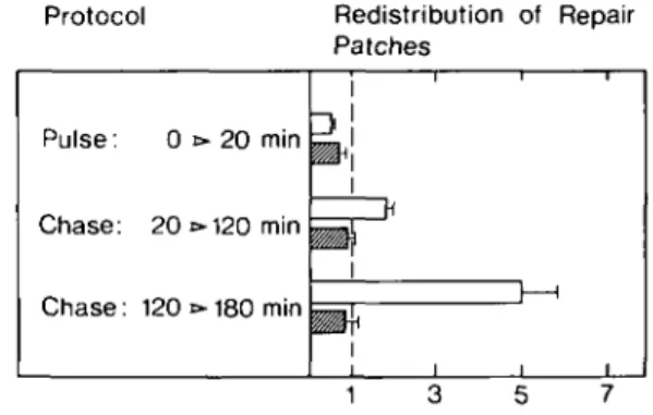
A/-acetoxy-2-acetylaminofluorene. Figure 1 shows that newly
synthesized repair patches, pulse-labeled for an initial 20 min
repair period, gradually accumulate in free DNA domains during
subsequent chase periods. This observation is in accordance with
previous results based on nuclease probing of chromatin (1,2),
or direct isolation of these domains (9).
No repositioning of repair patches into free DNA domains was
observed in poly(ADP-ribose)-depleted hepatocytes (7; Figure
1, legend). This depletion did not affect the overall synthesis of
repair patches (Figure 1, legend). Thus, the synthesis and
repositioning of repair patches in poly(ADP-ribose)-depleted
chromatin of hepatocytes were uncoupled.
Figure 2 provides a comparison of excision activity in total
chromatin (Figure 2A) and free DNA (Figure 2B) of
poly(ADP-ribose)-depleted and undepleted hepatocytes. In the first 3 h of
Protocol Pulse: 0 Chase: 20 Chase: 120 •»20 t»120 t»180 min min min Redistribution of Repair Patches
i '
i i i—i \ \ • i i iFig. 1. Redistribution of repair patches relative to free DNA domains in poly(ADP-ribose)-depleted and undepleted hepatocytes. Parenchymal liver cells were isolated from adult rat hepatocytes and cultured for 24 h in the presence or absence of 8 mM benzamide as previously described (9,31). The level of chromatin-bound poly(ADP-ribose) in benzamide-treated cells was < 10 fmol ADP-ribosyl residues/106 cells, as determined by the method of Jacobson and co-workers (32,33). Hepatocytes were then exposed to 50 /M yV-acetoxy-2-acetylaminofluorene (NCI Chemical Carcinogen Repository, Bethesda, MD, USA) and newly synthesized repair patches were pulse-labeled with [methyl-3H]thymidine (49 Ci/mmol, 20 /tCi/3 ml medium) for 20 min. Incorporation of radioactivity was stopped by two changes of medium containing 1.2 fiM unlabeled thymidine, followed by a chase in the continuous presence or absence of benzamide. At the end of these incubations, free DNA domains were isolated from hepatocellular chromatin as previously described (9) and analyzed for their content of repair patches (9). The numbers plotted on the abscissa represent the ratios of radioactivity (d.p.m.//ig DNA) incorporated into free DNA to the radioactivity incorporated into bulk DNA. The values reflect the relative accumulation of repair patches in free DNA (9) (a value of 1 indicates no accumulation) and represent the mean ± SEM of four independent experiments with separate cell preparations. The overall repair incorporation was 511.2 ± 85.4 mmol (controls) and 408.2 ± 94.8 mmol (depleted cells) [methyl-3H]thymidine/mol adduct formed (mean ± SEM, n = 3). Black bars: control cells; shaded bars: poly(ADP-ribose)-depleted cells.
G.Mathis and F.R.AIthaus 100-~ 75- 100-o < 0 20 120 Repair Time (min)
Fig. 2. Excision of deoxyguanosine adducts in total DNA and free DNA domains of poly(ADP-ribose)-depleted and undepleted hepatocytes exposed to A/-acetoxy-2-acetylaminofluorene (AAAF). Hepatocytes were incubated for 20 min with [G-3H]AAAF (232 mCi/mmol, total concentration 50 ^M) and the amount of deoxyguanosine adducts was quantified following DNA purification (9). (A) Adduct removal in total DNA; (B) adduct removal in free DNA domains. O O, poly(ADP-ribose)-depleted cells; O O , undepleted control cells. The results represent the means ± SEM of three independent experiments involving separate cell preparations.
0 20 120 Repair Time (min)
180
Fig. 3. Relative distribution of the remaining adducts in free DNA domains of poly(ADP-ribose)-depleted and undepleted hepatocytes after various repair intervals. Hepatocytes were incubated with radiolabeled A'-acetoxy-2-acetyl-aminofluorene as described in Figure 2 and the deoxyguanosine adduct concentration was quantified in free DNA and expressed relative to their content in total DNA. O O, poly(ADP-ribose)-depleted cells; O O, undepleted cells. The values plotted on the ordinate represent the ratios of [G-3H]AAAF radioactivity in free DNA to the radioactivity contained in bulk DNA. The results represent the means ± SEM of three independent experiments involving separate cell preparations.
repair, the rate of excision of deoxyguanosine adducts formed
during a 20 min incubation of hepatocytes with
/V-acetoxy-2-acetylaminofluorene was almost identical in total chromatin and
free DNA. However, no excision of these adducts occurred
during the same time period in poly(ADP-ribose)-depleted cells
(Figure 2). Hence the repositioning of repair patches (Figure 1)
seems to be coupled with adduct excision (Figure 2) by a
mech-anism involving de novo poly ADP-ribosylation of chromatin
proteins. This is compatible with the idea that unfolded DNA
domains are a preferential site of excision activity (Figure 2).
The results in Figure 3 suggest that some unfolding of
chromatin occurs also in poly(ADP-ribose)-depleted cells. A
slight accumulation of unexcised DNA adducts in the free DNA
fraction was obtained in poly(ADP-ribose)-depleted cells, while
no significant accumulation was seen in undepleted control cells.
Thus, the unfolded domains formed in poly(ADP-ribose)-depleted
cells are enriched in DNA adducts and largely deficient in repair
patches (cf. Figures 1 and 3).
Our results thus reveal a tight functional linkage between
structural chromatin rearrangements and the excision step, and
dissect the process of repair patch synthesis from the patch
repositioning relative to nucleosomally organized chromatin
regions. Moreover, our results tentatively identify a requirement
for poly ADP-ribosylation in co-ordinating excision repair with
structural rearrangements of chromatin. The role of this
post-translational protein modification is poorly understood, but all
protein acceptors of poly(ADP-ribose) hitherto identified (for
review see 4) share the capacity to bind to nucleic acids. Poly
ADP-ribosylation of these proteins in vitro reversibly alters their
DNA binding affinity (10-15). Enzymatic addition and removal
of ADP-ribose polymers on chromatin preparations in vitro
induces reversible relaxation of polynucleosomes (16), the
relaxation state being directly related to the size of histone-bound
ADP-ribosyl polymers (17-19). Histones are also a physiological
target of ADP-ribose modifications in carcinogen-treated
mammalian cells in vivo (20,21). Thus, the reversible
modification of DNA-protein interactions (14,15,22,23) by
protein ADP-ribosylation may be the underlying molecular
mechanism for the transient formation of free DNA domains in
eukaryotic excision repair.
As far as our findings ascribe poly ADP-ribosylation a role
in DNA excision repair, they contrast the hypothesis of Shall
and associates who envisioned poly ADP-ribosylation as a
direct regulatory mechanism of ligation activity in the repair of
alkylation damage in DNA (24,25). However, other reports could
not confirm such a role for poly(ADP-ribose) in DNA excision
repair (26-30). This contradictory phenomenology may be
reconciled on the premise that poly ADP-ribosylation affects local
disruptions of chromatin structure and this secondarily affects
DNA repair reactions (4,26-30).
Acknowledgements
We thank Phyllis Panzeter for critically reviewing this manuscript. This work was supported by grant 3.161.088 from the Swiss National Foundation for Scientific Research, and the Jubilaumsstiftung of the University of Zurich.
References
l.Friedberg.E.C. (1985) DNA Repair. W.H.Freeman, New York. 2.Smerdon,M.J. (1990) In Lambert.M.W. and Laval.J. (eds), DNA Repair
Mechanisms and their Biological Implications in Mammalian Cells. Plenum, New York, pp. in press.
3. Ueda,K. and Hayaishi,O. (1985) ADP-ribosylation. Annu. Rev. Biochem., 54, 73-100.
4. Althaus.F.R. and Richter,C. (1987) Molecular Biology, Biochemistry, and Biophysics, Vol. 37: ADP-ribosylation of Proteins: Enzymology and Biological Significance. Springer-Verlag, Berlin.
5. Naegeli,H. and Althaus,F.R. (1989) Poly ADP-ribosylation of proteins: processivity of a posttranslational modification. J. Biol. Chem 264 14382-14385.
6. Rankin,P.W., Jacobson.E.L., Benjamin,R.C., Moss,J. and Jacobson,M.K. (1989) Quantitative studies of inhibitors of ADP-ribosylation in vitro and in vivo. J. Biol. Chem., 264, 4312-4317.
7. Alvarez-Gonzalez,R. and Althaus.F.R. (1989) Poly(ADP-ribose)catabolism in mammalian cells exposed to DNA-damaging agents. Mutat. Res., 218 67-74.
8. Althaus.F.R., Lawrence,S.D., He,Y.Z., Sattler.G.L., Tsukada,Y. and Pitot,H.C. (1982) Effects of altered [ADP-ribose]n metabolism on expression of fetal functions by adult rat hepatocytes. Nature, 300, 366-368. 9. Mathis,G. and Althaus.F.R. (1986) Periodic changes of chromatin organization
associated with rearrangement of repair patches accompanying DNA excision repair of mammalian cells. J. Biol. Chem., 261, 5758-5765.
10. Gaal,J.C. and Pearson,C.K. (1985) Eukaryotic nuclear ADP-ribosylation reactions. Biochem. J., 230, 1-18.
Uncoupling of DNA excision repair
11. Burzio,L.O., Koide,S.S., Puigdomenech,P. and Ruiz-CarriUo,A. (1980) ADP-ribosylation of rat liver HI and nucleosomal core histones. In Smulson,M.E. and Sugimura,T. (eds), Novel ADP-ribosylaiions of Regulatory Enzymes and Proteins. Elsevier/North Holland, New York, pp. 345-356.
12. Yoshihara.K., Hashida.T., Tanaka,Y., Matsunami,N., Yamaguchi,A. and Kamiya,T. (1981) Mode of enzyme-bound poly(ADP-ribose) synthesis and histone modification by reconstituted poly(ADP-ribose)polymerase — DNA cellulose complex. J. Biol. Chem., 256, 3471-3478.
13.Ohashi,Y.J. (1986) Effect of ionic strength on chain elongation in ADP-ribosylation of various nucleases. J. Biochem., 99, 971—979.
14. Ferro.A.M. and Olivera,B.M. (1982) Poly(ADP-ribosylation) in vitro. Reaction parameters and enzyme mechanism. J. Biol. Chem., 257, 7808-7813.
15. Zahradka,P. and Ebisuzaki.K. (1982) A shuttle mechanism for DNA-protein interactions. The regulation of poly(ADP-ribose)polymerase. Eur. J. Biochem., 127, 579-585.
16. De Murcia.G., Huletsky.A., Lamarre,D., Gaudreau.A., PouyetJ., Daune,M. and Poirier,G.G. (1986) Modulation of chromatin superstructure induced by poly(ADP-ribose) synthesis and degradation. J. Biol. Chem., 261, 7011-7018. 17. Butt,T.R. and Smulson.M. (1980) Relationship between nicotinamide adenine dinucleotide concentration and in vitro synthesis of poly(adenosine diphosphate ribose) on purified nucleosomes. Biochemistry, 19, 5233-5242. 18. Aubin.R.J., Frechette,A., de Murcia,G., Mandel,P., Lord.A., Grondin,G.
and Poirier,G.G. (1983) Correlation between endogenous nucleosomal hyper(ADP-ribosyl)ation of histone HI and the induction of chromatin relaxation. EMBOJ., 2, 1685-1693.
19. Huletsky,A., De Murcia.G., Muller.S., Hengartner.M., Menard.L., Lamarre,D. and Poirier,G.G. (1989) The effect of poly(ADP-ribosyl)ation on native and Hl-depleted chromatin. J. Biol. Chem., 264, 8878-8886. 20. Kreimeyer.A., Wielckens,K., Adamietz.P. and Hilz.H. (1984) DNA
repair-associated ADP-ribosylation in vivo. J. Biol. Chem., 259, 890-896. 21. Adamietz,P. and Rudolph,A. (1984) ADP-ribosylation of nuclear proteins
in vivo. Identification of histone H2B as a major acceptor for mono- and poly(ADP-ribose) in dimethyl sulfate-treated hepatoma AH 7974 cells. J. Biol. Chem., 259, 6841-6846.
22. Mathis.G. and Althaus.F.R. (1987) Release of core DNA from nucleosomal core particles following (ADP-ribose)n-modification in vitro. Biochem.
Biophys. Res. Commun., 143, 1049-1053.
23. Althaus,F.R., Collinge.M., Loetscher.P., Mathis.G., Naegeli.H., Panzeter.P. and Realini,C. (1990) The poly ADP-ribosylation system of higher eukaryotes: how can it do what? Adv. Mutagen. Res., 1, 22-30.
24. Creissen,D. and Shall,S. (1982) Regulation of DNA ligase activity by poly(ADP-ribose). Nature, 296, 271-272.
25. Shall,S. (1989) ADP-ribosylation of proteins: a ubiquitous cellular control mechanism. Biochem. Soc. Trans., 17, 317-322.
26. Ohashi.Y., Ueda.K., Kawaichi.M. and Hayaishi.O. (1983) Activation of DNA ligase by poly(ADP-ribose) in chromatin. Proc. Natl. Acad. Sci. USA, 80, 3604-3607.
27. Moran.M.F. and Ebisuzaki.K. (1985) Inhibition of poly(ADP-ribose)poly-merase causes increased DNA strand breaks without decreasing strand rejoining in alkylated HeLa cells. FEBS Lett., 190, 279-282.
28. Morgan.W.F., Djordjevic.M.C, Milam.K.M., Schwartz.J.L., Borek,C. and Cleaver,J.E. (1985) Is there a role for ADP-ribosylation in DNA repair? In Althaus.F.R., Hilz.H. and Shall.S. (eds), ADP-ribosylation of Proteins. Springer-Verlag, Berlin, pp. 244-251.
29. Cleaver,J.E. and Park,S.D. (1986) Enhanced ligation of repair sites under conditions of inhibition of poly(ADP-ribose) synthesis by 3-aminobenzamide. Mutat. Res., 173, 287-290.
30. Teraoka.H., Sumikawa.T. and Tsukada,K. (1986) Purification of DNA ligase II from calf thymus and preparation of rabbit antibody against calf thymus DNA ligase II. J. Biol. Chem., 259, 6888-6892.
31. Althaus.F.R., Lawrence.S.D., Sattler,G.L. and Pitot.H.C. (1982) ADP-ribosyltranferase activity in cultured hepatocytes: interactions with DNA repair. J. Biol. Chem., 257, 5528-5535.
32. Jacobson.M.K., Payne,D.M., Alvarez-Gonzalez,R., Juarez-Salinas,H., Sims,J.L. and Jacobson,E.L. (1984) Determination of in vivo levels of polymeric and monomeric ADP-ribose by fluorescence methods. Methods Enzymol, 106, 483-494.
33. Alvarez-Gonzalez.R., Eichenberger.R., Loelscher.P. and Althaus,F.R. (1986) A new highly selective physicochemical assay to measure NAD+ in intact cells. Anal Biochem., 156, 473-480.
Received on February 7, 1990; revised on April 4, 1990; accepted on April 9, 1990