Vol. 16, 1997 Notes 675
Eosinophilia in Patients
Infected with the Human
Immunodeficiency Virus
A. Tietz 1, L. Sponagel 1, E Erb e ,
H. Bucher 1, M. Battegay 1, W. Zimmerli 3.
The prevalence and significance of peripheral blood eosinophilia in patients infected with the hu- man immunodeficiency virus (HIV) were evaluated. Fifteen of 119 consecutive patients had absolute eosinophil counts of > 450/ram 3. During a mean fol- low-up period of 419 days eosinophilia could be identified as secondary to a parasitic infection in only one patient. Correlation with disease stage showed a higher rate of advanced disease in pa- tients with absolute eosinophilia. In a multivariate regression analysis, only low CD4+ cell counts, not the CDC disease stage or the use of antiretroviral therapy or primary prophylaxis, contributed signif- icantly to the prevalence of eosinophilia. It is con- cluded that expen-sive laboratory investigations in asymptomatic patients with advanced-stage HIV disease are neither necessary nor cost effective.
The occurrence of peripheral blood eosinophilia in patients with human immunodeficiency type 1 (HIV-1) infection has been described previously (1-4). Our daily clinical work suggested that eosinophilia is more common in late-stage infec- tion. Previous retrospective studies have shown ei- ther no significant difference in its incidence as re- lated to various disease stages (1, 2) or a strong as- sociation with advanced disease (3, 5).
The major hypotheses for an increased prevalence of peripheral blood eosinophilia in advanced HIV-1 infection include the Thl-Th2 shift of HIV-infected CD4+ lymphocytes, associated with higher levels of interleukin-4 and -5 and a subse- quent increase in immunogtobulin E (IgE) levels (5-8); parasitic disease not detectable by routine
1 Medical Outpatient Clinic 2 Institute of Medical Microbio- logy, and 3 Division of Infectious Diseases, University Hos- pitals Basel, Petersgraben 4, CH-4031 Basel, Switzerland.
laboratory methods (4); the infection of CD4+ receptor-positive eosinophils by HIV (since eosinophils are able to produce their own growth factor) (9, 10); and the numeric decline of other blood cell lines, leading to a reactive proliferation of eosinophils (11). The aim of this study was to evaluate the prevalence, possible etiolog3; and correlation with demographic and biologic vari- ables of eosinophilia in HIV-infected patients.
Materials and Methods. We performed a retro- spective analysis of charts from 119 Caucasian HIV-l-positive patients who were monitored in a prospective cohort study (the Swiss HIV Cohort Study) at our center. Semiannual follow-up visits included a history and physical examination, complete blood cell counts, lymphocyte differen- tiation, chemograms, serologic assays, and, if nec- essary additional diagnostic tests.
Peripheral blood eosinophilia was defined as an absolute eosinophil count of > 450/mm 3 on at least two subsequent measurements. No specific rou- tine program for the investigation of eosinophil- ia is used at our clinic, but all evaluations includ- ed analysis of multiple stool samples and serolog- ic tests for common HIV-associated parasites as well as skin biopsies in a few cases with concur- rent dermatosis. Basic features of the patients with eosinophilia were identified and compared to those of the control subjects. The 15 case subjects were then paired in a 1:1 fashion with an HIV- positive control group matched for age, sex, transmission mode, and stage of disease. Levels of interleukin-4, interleukiu-5, interferon-s, and IgE were measured in serum samples of both groups with modified enzyme-linked assays (12, 14). Serum samples for determination of cy- tokine profiles and IgE levels were collected at the time of detection of eosinophilia.
Results underwent univariate and multivariate regression analysis (p < 0.05) with a statistical soft- ware program (SPSS for Windows, Version 6.0; SPSS, USA).
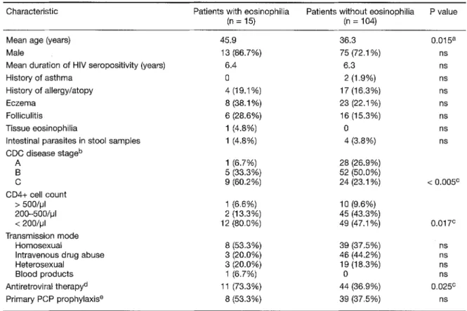
Results and Discussion. O f 119 subjects evaluat- ed 15 (12.6%) had absolute eosinophilia (range, 451-2940/~tl; median, 720/~1). Intergroup compar- ison showed a significantly higher proportion of patients with low CD4+ cells and CDC disease stage C in the group with eosinophilia. Table 1 summarizes the characteristics of both groups. Patients with eosinophilia were significantly old- er, and a higher proportion was using antiretrovi- ral medication, which is consistent with the in- creased prevalence of advanced-stage disease in
676 Notes Eur. J. Clin. Microbiol. Infect. Dis.
Table 1: Characteristics of HIV-positive patients with and without eosinophilia.
Characteristic Patients with eosinophilia Patients without eosinophilia P value
(n = 15) (n = 104)
Mean age (years) 45.9
Male 13 (86.7%)
Mean duration of HIV seropositivity (years) 6.4
History of asthma 0
History of allergy/atopy 4 (19,1%)
Eczema 8 (38.1%)
Folliculitis 6 (28.6%)
Tissue eosinophilia 1 (4.8%)
Intestinal parasites in stool samples 1 (4.8%) CDC disease stage b A 1 (6.7%) B 5 (33.3%) C 9 (60.2%) CD4+ cell count > 500/~JI 1 (6.6%) 200-500/IJI 2 (13.3%) < 200/IJI 12 (80.0%) Transmission mode Homosexual 8 (53.3%)
Intravenous drug abuse 3 (20.0%)
Heterosexual 3 (20.0%) Blood products 1 (6.7%) Antiretroviral therapy d 11 (73.3%) Primary PCP prophylaxis e 8 (53.3%) 36.3 0.015 a 75 (72.1%) ns 6.3 ns 2 (1.9%) ns 17 (16.3%) ns 23 (22.1%) ns 16 (15.3%) ns 0 ns 4 (3.8%) ns 28 (26.9%) 52 (50.0%) 24 (23.1% < 0.005 c 10 (9.6%) 45 (43.3%) 49 (47.1%) 0.017 c 39 (37,5%) ns 46 (44.2%) ns 19 (18.3 %) ns 0 ns 44 (36.9%) 0.025 c 39 (37.5%) ns
a Levene's t-test for equality of variances.
b CDC disease stage definition: A, asymptomatic; B, minor opportunistic diseases; C, acquired immunodeficiency syndrome; c ManteI-Haenszel test for linear association.
d Antiretroviral drugs used were zidovudine, didanosine, and zalcitabine. e Cotrimoxazole or dapsone/pyrimethamine.
ns, not significant; PCR Pneumocystis carinii pneumonia.
this group. The results of the multivariate logistic regression analysis indicate that a CD4+ cell count below 200/jal is significantly associated with an increased risk for peripheral blood eosinophil- ia (Table 2). The use of antiretroviral therapy (reverse transcriptase inhibitors: zidovudine, didanosine, or zalcitabine) did not contribute to the model significantly, in contrast to the findings
from the univariate analysis. The duration of seropositivity was similar in both groups, indicat- ing coincidence between rapid progression and eosinophilia.
On retrospective evaluation, one patient had severe eczema and erythrodermia with tissue eosinophilia. He also had a history of travel in the
Table 2: Multivariate anaJysis of variables associated with peripheral blood eosinophilia in HtV-positive patients (Cox proportional hazards model).
Variable Adjusted risk ratio 95% Cl P value
CD4+ cell count < 200/IJI 2.81 1.09-7.24 0.03
Disease stage C 1.82 0.95-3.51 0.07
Antiretroviral therapy a 0.62 0.29-1.34 ns
Primary PCP prophylaxis b 2.09 0.98-4.89 0.09
a Zidovudine, didanosine, or zalcitabine. b Cotrimoxazole or dapsone/pyrimethamine.
Vol. 16, 1997 Notes 677
tropics and transient Trichuris trichiura infection detected by stool examination. During a median follow-up time of 419 days (range, 89-1769 days), we detected no other cases of secondary eosinophilia. Therefore, we propose that in the ab- sence of epidemiologic, anamnestic, or clinical symptoms indicating parasitic disease, allergic disorders, neoplasms, or skin disease, three stool samples for bacteria, ova, and parasites are suffi- cient for evaluation. In view of our findings, a com- plete series of tests including chest radiographs, ab- dominal ultrasound, serologic evaluation for par- asitic disease, or even bone m a r r o w aspiration is unnecessary.
Interleukin-4, interleukin-5, and interferon-~ lev- els were undetectable in both groups, i.e., subjects with eosinophilia and the m a t c h e d control sub- jects. T h e r e was no statistically significant differ- ence b e t w e e n the groups in immunoglobulin E levels (20.6 + 12.9 ng/ml in eosinophilic patients vs. 13.0 _+ 7.5 ng/ml in patients without eosinophil- ia; p = 0.32 with the two-tailed Student's t-test). As to the possible reasons for the higher rate of eosinophilia in patients with late-stage disease, our data were not able to reproduce any cytokine mod- ulations attributed to the Thl-Th2 shift hy- pothesis. This finding m a y be due to our use of se- rum instead of supernatants of T cells for our ana- lyses. Patients with eosinophilia had IgE levels similar to those found in the control subjects which precludes us from interpreting eosinophil- ia in H I V infection as indicative of an allergic re- action. Nevertheless, since severely i m m u n o c o m - promised patients are m o r e likely to receive multidrug regimens for the t r e a t m e n t of H I V infection and diseases associated with it, eosino- philia could d e n o t e a reaction to multiple-drug therapy.
In conclusion, the prevalence of eosinophilia in pa- tients with HIV-1 infection was 12.6%; m o r e than two-thirds of these patients were severely immu- nocompromised. T h e r e was no significant eleva- tion of serum IgE levels, suggesting a nonallergic cause. Our results indicate that eosinophilia is most probably associated with H I V infection when epidemiological or clinical features indica- tive of other conditions with eosinophilia are absent.
References
1. Sanchez-Borges M, Orozco A, DiBagio E, Tami I, Suarez-Chacon R: Eosinophilia in early-stage HIV-infec- tion. Journal of Allergy and Clinical Immunology 1993, 92: 494-495.
2. Van der Graaf W, Borleffs JCC: Eosinophilia in patients with HIV-1 infection. European Journal of Haematology 1994, 52: 246-247.
3. Caterino DeAranjo C: HIV-1 infection and eosinophilia. Immunology Today 1994, 15: 498-499.
4. Wardlaw A J: Eosinophils in the 1990s: new perspectives in their role in health and disease. Postgraduate Medi- cal Journal 1994, 70: 536-552.
5. Smith K J, Skelton HG, Drabick J J, McCarthy WF, Wagner KF: Hypereosinophilia secondary to immuno- dysregulation in patients with HIV-1 disease. Archives of Dermatology 1994, 130:119-121.
6. Clerici M, Shearer GM: A T h l ~ T h 2 switch is a critical step in the etiology of HIV infection. Immunology Today 1993, 14: 107-111.
7. Clutterbuck E J, Hirst EMA, Sanderson C J: Human interleukin-5 regulates the production of eosinophils in human bone marrow cultures. Blood 11989, 73: 1504-1512.
8. Freedman AR, Gibson FM, Fleming SC, Spry C J, Griffin GE: Human immunodeficiency virus infection of eosino- phils in human bone marrow cultures. Journal of Exper- imental Medicine 1991, 174: 1661-1664.
9. Lucey DR, Dorsky DI, Nicholson-WeIler A, Weller P: Hu- man eosinophils express CD4 protein and bind HIV-1 gp120. Journal of Experimental Medicine 1989, 169: 327-332.
10. Steffen M J, Ebersole JL: Sequential ELISA for cytokine levels in limited volumes of biological fluids. Biotech- niques 1996, 21:504-509.
11. Cohen A J, Steigbigel RT: Eosinophilia in patients infect- ed with the human immunodeficiency virus. Journal of Infectious Diseases 1996, 174: 615-618.
12. Sanderson C J: Interleukin-5, eosinophils and disease. Blood 1992, 79: 3101-3109.
13. Sample S, Chernoff DN, Lenahan GA, Lanahan GA, Serwonska MH, Rangi S, Sherman JW, Sooy CD, Hol- lander H, Goetzi E J: Elevated serum concentrations of IgE antibodies to environmental antigens in HIV-seropos- itive male homosexuals. Journal of Allergy and Clinical Immunology 1990, 86: 876-880.
14. Kemeny DM, Richards D, Durnin S, Johannson A: Ultrasensitive enzyme-linked immunosorbent assay (ELISA) for the detection of picogram quantities of IgE. Journal of Immunological Methods 1989, 120: 251-258.
Acknowledgement
These data were presented in part as a poster at the XIth International Congress on AIDS, Vancouver, Canada, 7-13 July 1996.