Discordant Increases in CD4
+T Cells in Human Immunodeficiency Virus–
Infected Patients Experiencing Virologic Treatment Failure: Role of Changes
in Thymic Output and T Cell Death
Denise Lecossier,1Francine Bouchonnet,1 Pascal Schneider,2Franc¸ois Clavel,1
and Allan J. Hance,1for the Centre de Recherche Integre´ sur le VIH Bichat-Claude Bernard
1INSERM U552, Hoˆpital Bichat-Claude Bernard, Paris, France; 2Institute of Biochemistry, University of Lausanne, Epalinges, Switzerland
Some patients infected with human immunodeficiency virus (HIV) who are experiencing antiretroviral treatment failure have persistent improvement in CD4+
T cell counts despite high plasma viremia. To explore the mechanisms responsible for this phenomenon, 2 param-eters influencing the dynamics of CD4+
T cells were evaluated: death of mature CD4+
T cells and replenishment of the CD4+T cell pool by the thymus. The improvement in CD4+T cells
observed in patients with treatment failure was not correlated with spontaneous, Fas li-gand–induced, or activation-induced T cell death. In contrast, a significant correlation between the improvement in CD4+
T cell counts and thymic output, as assessed by measurement of T cell receptor excision circles, was observed. These observations suggest that increased thymic output contributes to the dissociation between CD4+
T cell counts and viremia in patients failing antiretroviral therapy and support a model in which drug-resistant HIV strains may have reduced replication rates and pathogenicity in the thymus.
Treatment of patients infected with human immunodeficiency virus type 1 (HIV-1), using a combination of antiretroviral drugs that includes a protease inhibitor, leads to a rapid decrease in virus load and an improvement in the absolute number of cir-culating CD41T cells. In a proportion of individuals, however, virus load either fails to decline or rapidly rebounds after treat-ment with combined antiretroviral agents. Despite this apparent treatment failure, some, but not all, of the patients have sub-stantial and persistent improvement in their CD41T cell counts, so-called discordant responses [1–3].
The mechanisms responsible for discordant treatment re-sponses are not well understood. Two recent studies evaluating patients with persistently detectable plasma viral mRNA after treatment have found that the reduction in virus load from pretreatment levels correlated with the improvement in CD41 T cell counts [4, 5]. A decrease in virus load, however, is not the only factor explaining discordant responses, because, for any given reduction in virus load, a wide range of improvement
Received 4 October 2000; revised 11 December 2000; electronically pub-lished 8 March 2001.
Presented in part: Fourth International Workshop on Drug Resistance and Treatment Strategies, Sitges, Spain, June 2000 (abstract 192).
Informed consent was obtained from all patients; the studies were per-formed in accordance with the human experimentation guidelines of our institution.
Reprints or correspondence: Dr. Allan J. Hance, INSERM U552, IMEA INSERM, Hoˆpital Bichat-Claude Bernard, 46, rue Henri Huchard, 75018 Paris, France (hance@bichat.inserm.fr).
The Journal of Infectious Diseases 2001 183:1009–16
q 2001 by the Infectious Diseases Society of America. All rights reserved.
0022-1899/2001/18307-0003$02.00
in CD41 T cell counts can be observed, and some patients develop discordant responses despite no reduction or actual increases in plasma virus load.
A variety of processes have been identified that may con-tribute to T cell depletion in HIV infection, including the direct cytopathic effect of the virus [6], the elimination of infected cells by the immune response [7], virus-induced apoptosis of uninfected cells [8, 9], induction of abnormalities in cell traf-ficking [10], and impairment of the generation of T cells within the thymus [11–13]. Demonstrating the involvement of>1 of these pathways in the paradoxical increase in circulating CD41 T cells in patients with virologic treatment failure would directly support the conclusion that the given mechanism has a real impact on T cell depletion in the course of HIV infection. At present, however, no information is available concerning the contribution of changes in these pathways to the improvement in CD41T cell counts in patients with discordant responses. To explore this question, we identified a cohort of patients who, after being treated for the first time with a protease inhibitor in conjunction with 2 reverse-transcriptase inhibitors, experi-enced confirmed virologic failure. In these patients we evaluated (1) spontaneous and activation-induced death of cultured pe-ripheral blood lymphocytes and (2) thymic output, as assessed by quantification of T cell receptor excision circles.
Patients and Methods
Study participants. The group of patients with virologic treat-ment failure consisted of 18 adult patients followed up at Hoˆpital Bichat-Claude Bernard who met the following criteria: (1) the
pa-tient had been treated with a protease inhibitor for the first time in combination with>2 reverse-transcriptase inhibitors; (2) plasma HIV RNA levels had been measured before initiating therapy with the protease inhibitor; (3) treatment with protease inhibitors had been continued for>1 year after virologic treatment failure had become evident; and (4) on the day of evaluation, plasma HIV RNA had returned to levels of>104
copies/mL and had rebounded to within 1 log10of the pretreatment levels or was higher than pretreatment levels. All patients in our cohort who were identified as meeting the inclusion criteria were evaluated. Initial CD4 T cell counts or changes in CD4 T cell counts were not used as criteria for inclusion. These patients (all men) had amean5 SDage of
years. 415 9
Twelve HIV-1–infected patients who had never received antire-troviral therapy were evaluated. These patients (8 men and 4 women) had an average age of415 13years. On the day of eval-uation, CD41T cell counts were 6cells/L (range,
3055 214 3 10
cells/L), and virus load was log10copies/ 6
17–6193 10 4.55 1.1
mL (range,!2.3–6.4 log
10copies/mL). Twelve healthy volunteers not infected by HIV served as controls. These individuals (4 men and 8 women) had an average age of365 9years. Informed con-sent was obtained from all participants.
Evaluation of cell death. Mononuclear cells were isolated from peripheral blood mononuclear cells (PBMC) by centrifugation on Ficoll-Paque (Pharmacia). Cells were resuspended at 6cells/
13 10 mL in complete medium (RPMI-1640 containing 25 mM HEPES, 10% fetal calf serum, 2 mM glutamine, 200 U/mL penicillin G, and 250 mg/mL streptomycin) and 0.5-mL aliquots cultured in 5-mL polypropylene culture tubes (Falcon 2005, Becton Dickinson). To test the sensitivity of T cells to death induced by cross-linking receptors of the tumor necrosis factor (TNF) receptor family, cells were cultured with 100 ng/mL (final concentration) of fusion pro-teins containing the extracellular domain of the corresponding cog-nate ligand linked to a Flag epitope (FasL-Flag, TRAIL-Flag TNF-a–Flag, or TWEAK-Flag) in the presence of 2 mg/mL anti-Flag monoclonal antibody (M2, Sigma) [14]. To evaluate activa-tion-induced cell death, 4 mg/mL activating anti-CD3 monoclonal antibody (Beckman-Coulter) was added. Cultures were maintained at 377C in 95% air/5% CO2. Samples were evaluated either before or after 48 h of culture. The cells were resuspended, and 100-mL aliquots were removed and were incubated at 47C for 45 min with fluorescein isothiocyanate (FITC)–conjugated anti-CD4, phyco-erythrin-conjugated anti-CD8, and PC5-conjugated anti-CD14 monoclonal antibodies, using the concentrations suggested by the manufacturer (Beckman Coulter). Fifty microliters of Flow-Count Fluorospheres (Coulter) was added, and the samples were analyzed by cytometry (FACScan, Becton-Dickinson). Debris was excluded during acquisition, and 50,000 total events were evaluated. The number of events corresponding to viable CD41 T cells, viable CD81T cells, and fluorospheres was determined by setting appro-priate gates, and, by use of this information, the total number of viable CD41and CD81T cells remaining in the culture at the time of analysis was calculated.
Cells in gates used to identify viable CD41 and CD81T cells contained !1% positive cells after incubation with
FITC-conju-gated Annexin V (Beckman Coulter). Incubation of Jurkat cells with the fusion proteins containing the extracellular domain of Fas-ligand or TRAIL under the conditions used in these experiments
led to massive apoptosis after 24 h of culture (data not shown). The fusion proteins containing the extracellular domains of TNF-a TNF-and TWEAK hTNF-ave been shown elsewhere to induce TNF-apoptosis in WEHI-164 fibrosarcoma cells and interferon-g–treated HT29 colon carcinoma cells, respectively, in the presence or absence of anti-FLAG antibody [14].
Evaluation of thymic output. To evaluate thymic output, T cell receptor excision circles (TRECs) were quantified, using a modi-fication of the approach described by Douek et al. [15]. TRECs are episomal DNA circles formed as by-products of the rearrange-ment of the T cell receptor locus during T cell developrearrange-ment. In this study, the “signal-joint” TREC, a TREC formed in 70% of all T cells expressing abTCR, which results from recombination between the dRec and wJa loci, was evaluated. The percentage of T cells expressing the CD41 CD45RA1 CD142 “naive” phenotype was determined by flow cytometry. To measure TRECs, CD41T cells were purified from PBMC by using magnetic beads coated with anti-CD4 monoclonal antibodies (CD4 Positive Isolation Kit, Dy-nal), following the manufacturer’s instructions (purity195%), and were stored at2807C. DNA was extracted (QIAamp DNA Blood Mini Kit, Qiagen), and the number of TRECs was determined by real-time polymerase chain reaction (PCR). Each reaction con-tained 13 TaqMan Universal PCR Master Mix, 5 mM MgCl2, and 200 nM of each primer and probe (50 mL final volume): TREC-F2, 50-GCAACTCGTGAGAACGGTGA; TREC-R1, 50 -CTTTC-AACCATGCTGACACCTC; TREC probe, 50 -(6-FAM)-CCGTG-CCAGCTGCAGGGTTTAGG(Tamra)(phosphate). As an index of total cells, the number of copies of the albumin gene (ALB; present in only 2 copies per cell and without pseudogenes) [16] was determined in parallel reactions, as described above, by use of 200 nM each of the following primers and probe: ALB-F1, 50
-GTGAA-CAGGCGACCATGCT; ALB-R1, 50
-GCATGGAAGGTGAAT-GTTTCAG; ALB probe, 50 -(VIC)-TCAGCTCTGGAAGTCGA-TGAAACATACGTTC(Tamra)(phosphate). For both systems, cy-cling parameters were as follows: 507C for 2 min and 957C for 10 min, followed by 40 cycles at 957C for 15 s and 587C for 1 min. In each case, the number of cycles required to reach threshold fluorescence (Ct) was determined. For quantification of both TREC and ALB, serial dilutions of DNA extracted from thymic tissue obtained from a 4-month-old infant were used. The difference in Ct(DCt) for the amplification of albumin and TREC in this stan-dard was constant over a 100-fold range of dilution of the template, which indicates that the 2 amplicons had the same efficiency of amplification [17]. Thus, by comparing the Y intercept of the stan-dard curves, the preparation was found to have 37.5 albumin se-quences per 1 TREC sequence. To calculate TREC/103
CD41 CD45RA1 T cells, the following formula was used: TREC/103 CD41CD45RA1Tcells p [(10003 numberof TREC sequences)/ (number of albumin sequences/2)]/[% CD45RA1CD41T cells/100]. Each sample was evaluated in duplicate at 3 dilutions of template, and the values were averaged. To assess the reproducibility of the technique, the same preparation of DNA from normal PBMC was evaluated in 15 consecutive experiments and gave values of
TREC/106
cells ( ). TRECs were undetectable
2345 70 mean5 SD
in T cell lines when this technique was used.
Statistical methods. All results are reported as mean5 SD, unless otherwise indicated. Spontaneous cell death was determined by comparing the number of viable cells present before and after
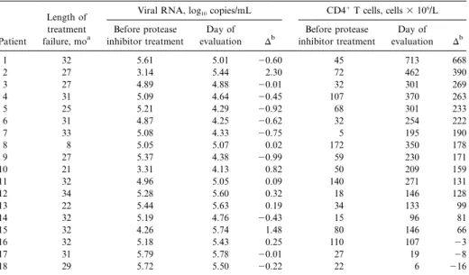
Table 1. Plasma human immunodeficiency virus (type I) RNA levels and circulating CD41T cell counts in patients with virologic treatment failure before treatment with a protease inhibitor and on the day of evaluation.
Patient
Length of treatment failure, moa
Viral RNA, log10copies/mL CD4
1T cells, cells3 106/L Before protease inhibitor treatment Day of evaluation Db Before protease inhibitor treatment Day of evaluation Db 1 32 5.61 5.01 20.60 45 713 668 2 27 3.14 5.44 2.30 72 462 390 3 27 4.89 4.88 20.01 32 301 269 4 31 5.09 4.64 20.45 107 370 263 5 25 5.21 4.29 20.92 68 301 233 6 31 4.87 4.25 20.62 32 254 222 7 33 5.08 4.33 20.75 5 195 190 8 8 5.05 5.07 0.02 172 350 178 9 27 5.37 4.38 20.99 59 230 171 10 21 3.31 4.13 0.82 50 209 159 11 32 4.96 5.05 0.09 140 271 131 12 34 5.28 5.60 0.32 18 146 128 13 22 5.44 5.63 0.19 34 133 99 14 32 5.19 4.76 20.43 15 96 81 15 32 4.26 5.74 1.48 80 146 66 16 32 5.18 5.43 0.25 110 107 23 17 31 5.79 5.78 20.01 27 19 28 18 29 5.72 5.50 20.22 22 6 216 a
Number of months that viral RNA was continuously detectable before evaluation.
b
Change between the intiation of therapy and the day of evaluation.
48 h of culture. Cell loss induced by ligands of the TNF family and activation-induced cell death were determined by comparing the number of T cells remaining at 48 h in cultures performed without or with the corresponding ligands or the activating anti-CD3 monoclonal antibody. Both cell viability and thymic output were evaluated for 13 patients; for the remaining patients only cell viability (n p 3) or thymic output (n p 2) was studied. Compar-isons between groups were made by analysis of variance; posttest comparisons (performed only ifP!.05) were made using the Dun-nett multiple comparisons test. Results for HIV-infected and -un-infected individuals were compared using the Mann-Whitney U test. Correlations were evaluated by linear regression. For all anal-yses,P p .05was considered significant. To confirm the robustness of conclusions drawn from regression analysis, all analyses were repeated using the nonparametric Spearman correlation and after excluding individual outlier points. In no case did these procedures change the interpretation of the findings.
Results
Clinical characteristics of patients with virologic treatment fail-ure. We sought to identify factors influencing the extent of improvement in circulating CD41T cell counts in HIV-infected patients with persistently elevated virus loads after antiretro-viral therapy. To minimize the impact of partial treatment re-sponses on our results, the study was restricted to patients whose plasma HIV RNA was>104copies/mL and had returned to levels that were within 1 log10of pretreatment levels or higher than pretreatment levels.
All patients had been treated with>1 nucleoside analogue reverse-transcriptase inhibitors before receiving protease inhib-itors, and all had detectable plasma HIV-1 RNA when therapy
with a protease inhibitor was initiated (ritonavir [n p 10], in-dinavir [n p 5], saquinavir [n p 2], or nelfinavir [n p 1]). Thir-teen patients had an initial decrease of>1 log10in plasma HIV-1 RNA after treatment with protease inhibitors, and virus became undetectable (!200 copies/mL) in 6 patients. Evidence of virologic treatment failure was apparent within 6 months of treatment in 17 of 18 patients, and, at the time of evaluation, plasma HIV-1 RNA had been continuously detectable for12 years in 16 of 18 patients (table 1; mean,285 6months). Dur-ing the year before evaluation, virus load was fairly stable in these individuals, even in patients for whom therapeutic changes were made (mean change in virus load in the 12 months before evaluation:20.2 5 0.6log10copies/mL).
The evolution of virus load and absolute CD41T cell counts between the initiation of therapy and the time of evaluation are shown in table 1. On the day of evaluation, mean virus load was5.05 0.6 log10 copies/mL; the average change since initiation of therapy was10.03 5 0.8log10copies/mL (range, 0.99 to12.3 log10copies/mL). Before treatment with protease inhibitors, CD41 T cell counts were generally low (605 CD41T cells/L) and were! cells/L in 14 of
6 6
463 10 1003 10
18 patients. After therapy with protease inhibitors, CD41T cell counts increased in most patients despite virologic treatment failure. At the time of evaluation, the average increase in CD41 T cells was11795 162 3 106cells/L, and CD41T cell counts had remained higher than pretreatment values for 15 of 18 patients. Nevertheless, considerable variability in the increase in CD41T cell counts was observed (DCD41Tcells3 106/L: 2100 to 0,n p 3; 1 to 100,n p 3; 101 to 200, n p 6; 201 to 300,n p 4;1300,n p 2.
Figure 1. Loss of CD41and CD81T cells in culture correlates with virus load but not with improvement in CD41T cell counts. Peripheral blood mononuclear cells from patients with virologic treatment failure were cultured for 48 h, and the percentages of CD41(A and C) and CD81 (B and D) T cells lost during culture were determined. These results were correlated with plasma virus load on the day of evaluation (A and B) and with the improvement in CD41T cell counts observed since the initiation of treatment with a protease inhibitor (C and D). No correlation was observed between the loss of CD41T cells in culture and the change in CD4 T cells (r p .06 P2 ; 1.3). No correlation was found between the loss of CD81T cells in culture and the change in CD41T cells by linear regression (r p .08 P2 ; 10.2), when the nonparametric Spearman correlation was used (r p2.32 P; 1.2) or after individual outlier points were excluded.
CD41and CD81T cells during the 48-h culture period was variable for HIV-infected individuals but, when considered for the patients as a group, was not significantly different from that observed for uninfected control subjects (P p 0.5for both com-parisons). For patients with virologic treatment failure, the spontaneous death during culture of CD81T cells was signif-icantly greater than that of CD41 T cells (26%5 27% and , respectively; by paired analysis). In these
9%5 14% P!.002
patients, the spontaneous loss during culture of both CD41and CD81T cells was correlated significantly with virus load (figures 1A, 1B). Most T cells with light scatter properties of dead cells were stained with annexin V, which suggests that T cell loss was occurring, at least in part, through apoptotic mechanisms (data not shown). It is important to note, however, that no correlation was observed between the spontaneous death during culture of either CD41or CD81T cells and the improvement in CD41T cell counts observed in patients with virologic treat-ment failure (figures 1C, 1D;P1.2for both comparisons).
Fas ligand–induced death of both cultured CD41and CD81 T cells from HIV-infected individuals was significantly in-creased, compared with that of the corresponding T cell pop-ulations from uninfected controls (figure 2A). For patients with virologic treatment failure, the increase in death induced by culture with Fas ligand over spontaneous cell death was of similar magnitude for CD41and CD81T cells (cell loss induced
by Fas ligand for CD41 cells, 21%5 19%; for CD81 cells, ; ). As expected, annexin V staining confirmed 17%5 14% P1.2
that many of these T cells were dying by apoptosis. There was no difference, however, in Fas-mediated cell death during cul-ture between untreated HIV-infected patients and patients with virologic treatment failure, and the extent of Fas-mediated death did not correlate with the improvement in CD41T cell counts observed in patients with virologic treatment failure (fig-ure 2B;P1.3for both comparisons). Culture of T cells in the presence of the death-inducing ligands TRAIL, TNF, and TWEAK did not significantly increase the death of either CD41 or CD81T cells from HIV-infected or -uninfected individuals (figure 2A).
For patients with virologic treatment failure, the loss of cul-tured T cells stimulated through CD3 was increased, compared with that of unstimulated cells; the magnitude of this effect was comparable for both CD41and CD81T cells (8%5 16%and decreases in cell number, respectively, compared 16%5 15%
with those in unstimulated cultures). Activation-induced cell death of cultured CD81 T cells from patients with virologic treatment failure was increased, compared with that of CD81 T cells from uninfected controls (P!.05). Again, no correlation was observed between activation-induced cell death during cul-ture of either CD41or CD81 T cells and the extent of im-provement in CD41T cell counts occurring in patients with
Figure 2. Effect of stimulation of receptors of the tumor necrosis factor (TNF) receptor family on survival of CD41T cells in vitro. Peripheral blood mononuclear cells from uninfected controls (open bars), patients with virologic treatment failure (solid bars), and human immunodeficiency virus (HIV)–infected untreated patients (hatched bars) were cultured for 48 h with fusion proteins containing the ex-tracellular domain of the indicated ligands linked to a Flag epitope in the presence of the M2 anti-Flag monoclonal antibody (A). The num-ber of viable CD41T cells was determined, and the percentage of decrease in cell numbers was determined relative to that of parallel cultures not containing ligands or anti-Flag antibody. M2 indicates cultures containing the anti-Flag antibody in the absence of ligand. Results are presented asmean5 SE. For patients with virologic treat-ment failure, no correlation was observed by linear regression between the percentage of decrease in CD41T cells resulting from culture with Fas-ligand fusion protein and the change in CD41T cell counts after beginning treatment with a protease inhibitor (r p .01 P2 ; 1.7), when the nonparametric Spearman correlation (r p2.05 P; 1.8) was used or after individual outlier points were excluded (B).
Figure 3. Relationships between the increase in CD41T cell counts and T cell receptor excision circle (TREC) levels, virus load, and age. CD41 T cells were purified from peripheral blood of patients with virologic treatment failure, DNA was extracted, and TREC/103CD41 CD45RA1T cell counts were determined by real-time polymerase chain reaction. These results were correlated with (A) the improvement in CD41T cell counts after beginning treatment with a protease inhibitor and (B) plasma virus load at the time of evaluation. The relationship between the improvement in CD41T cell counts and patient age is shown in C. NS, not significant.
virologic treatment failure (P1.2for both comparisons; data not shown).
Evaluation of TRECs. In vitro studies have shown that treatment with antiproteases leads to the emergence of viruses with impaired replicative capacity in the thymus [18], suggesting that increased regeneration of T cells may contribute to the increase in CD41T cell counts occurring in some patients with virologic treatment failure. To test this hypothesis, dRec/wJa “signal-joint” TRECs were evaluated in patients with treatment failure, an approach developed by Douek et al. [15]. As reported elsewhere, TREC/103CD41CD45RA1T cell counts were lower in untreated HIV-infected individuals than in uninfected control subjects, although considerable overlap was observed between the 2 groups (U p 23[Mann-Whitney U test];P!.05). Con-siderable variability in TREC levels was seen for patients with virologic treatment failure, but, when the patients were consid-ered as a group, they were not significantly different from
TREC levels of uninfected control subjects. As shown in figure 3A, however, a significant correlation was observed between TREC levels and the improvement in CD41 T cell counts (r p .48 P2 ; !.005). In contrast, TREC levels did not correlate with plasma virus load (figure 3B) or with the change in virus load after antiprotease therapy (data not shown).
Several studies have shown that TREC levels decrease with age, an observation that we confirmed both for patients with
virologic treatment failure and for uninfected control subjects (P!.01andP!.03, respectively). If TREC levels are directly linked to increased T cell counts in patients with virologic treat-ment failure, as suggested by the strong correlation, one would predict a greater increase in CD41T cell counts in the younger individuals. Consistent with this hypothesis, we found that the increase in CD41T cell counts in patients with virologic treat-ment failure was inversely correlated with age (figure 3C).
Discussion
When combined antiretroviral therapy fails to control viral replication in treated patients, peripheral CD41T cell counts can remain paradoxically high, clearly above those recorded before the initiation of therapy. Although higher CD4 counts are more common in patients whose plasma viremia remains below pretherapy levels [4, 5], such “dissociated” response pro-files are observed in a significant proportion of patients expe-riencing virologic therapy failure, including individuals whose virus load returns to pretreatment levels. In this study, we have examined 2 possible mechanisms for this phenomenon: (1) re-generation of CD41T cells by the thymus in spite of high levels of virus replication and (2) a decrease in spontaneous or in-duced peripheral CD41T cell death.
Several authors have suggested that an impairment of thymic function could contribute to the pathogenesis of HIV-1 infec-tion [11, 12, 15, 19–21]. Recently, several authors have used the quantification of TREC in circulating T cells as a surrogate marker for thymic output, to further evaluate this hypothesis [15, 22, 23]. Both Douek et al. and Zhang et al. demonstrated that TREC levels are lower in HIV-infected individuals than in age-matched uninfected control subjects [15, 22], and Hat-zakis et al. showed that a progressive decline in the concentra-tion of TREC is characteristic of patients with progressive dis-ease [23]. After successful antiretroviral therapy, TREC levels have been shown to rise in conjunction with CD41T cells [15], compatible with the possibility that thymic output can con-tribute to the restoration of circulating T cells, at least in some individuals [21, 24]. In our study, we found that TREC levels were lower in untreated HIV-infected patients than in unin-fected controls, although considerable overlap in the 2 groups was observed, as previously described. In patients in whom the improvement in CD41T cells occurred despite the persistence of high levels of circulating virus, we observed a significant correlation between the improvement in CD41T cell counts after treatment and TREC levels.
It is recognized that factors other than thymic output can affect the TREC:“naive” T cell ratio. In particular, increases in the proliferation of naive T cells and the reconversion of “memory” T cells to a naive phenotype [24–27], by increasing the total number of naive cells, can decrease the TREC:naive T cell ratio independent of thymic output, and the flux through these pathways can be increased by stimulation of the immune
system [26, 27]. Thus, in the context of HIV-1 infection, changes in the extent of virus-induced immune stimulation must be taken into consideration. For example, when virus load is dra-matically reduced by effective antiretroviral therapy, the in-creased proliferation of naive T cells returns toward normal levels [26] and can produce an increase in the TREC:naive T cell ratio [27]. Several findings suggest that this was not a con-founding variable in our study. First, all of our patients had a persistently high plasma virus load that, on average, was similar before and after treatment, which suggests that these individuals were subject to intense, persistent, and relatively constant im-mune stimulation. The increased sensitivity of T cells from these patients to Fas ligand–induced cell death is compatible with the persistence of strong virus-induced immune response (see below). Second, we observed a fairly strong correlation between age and both the improvement in CD41 T cell counts and TREC/103 CD41CD45RA1T cells in patients with virologic treatment failure. Thymic output is known to be age dependent [21, 28], whereas no evidence for age-dependent changes in the other factors influencing the total size of the naive T cell pool has been presented. Together, these results suggest that differ-ences in thymic output explain the variation in the TREC:naive T cell ratio seen in our patients.
Although the mechanisms through which thymic output may have been restored in these patients were not directly addressed by our studies, recent experimental evidence suggests that a decrease in HIV replicative capacity may be involved. It is now well established that viruses escaping antiretroviral therapy ac-cumulate mutations in the reverse transcriptase and/or in the viral protease that often reduce their overall replicative capacity [29, 30]. It is interesting to note that the extent of impairment of viral replication is highly dependent on the nature of the target cells. In particular, Stoddart et al. have recently shown that viruses carrying resistant protease sequences from treated patients replicated almost normally in phytohemagglutinin-stimulated human PBMC but failed to replicate and to deplete thymic T cells in thymic implants in SCID-hu Thy/Liv mice [18]. According to this model, reduced HIV replicative capacity in thymocytes would improve thymic function, whereas near-normal viral replication in mature peripheral T cells would explain the high plasma virus load seen in our patients. Other, more indirect mechanisms could also contribute to the resto-ration of thymic function. Cytokines, including interleukin (IL)–7, are known to be required for thymopoiesis but are also required for high-level HIV-1 replication in thymocytes [31, 32]. IL-7 levels have recently been found to be negatively correlated with peripheral CD41T cell counts in HIV-infected individuals [33, 34]. Thus, improved thymic output resulting from reduced HIV-1 fitness in the thymus might further reduce local virus replication by feedback inhibition of IL-7 production.
Further studies will be required to better define the overall contribution of improved thymic output to the restoration of CD41T cell counts in these patients with discordant responses
and the mechanisms responsible for this effect. Nevertheless, we propose that decreased viral replicative capacity due to ac-cumulated drug resistance mutations, together with adequate residual thymic function (e.g., younger patients without irre-versible thymic damage), both contribute to improved CD41 T cell counts through the restoration of thymic output.
In contrast to the findings concerning thymic output, we found no evidence that a reduction in the death of T cells contributes to discordant immunovirologic responses. Several earlier studies quantifying apoptotic cells have shown that the spontaneous apoptosis of cultured T cells from HIV-infected individuals is increased compared with that of T cells from uninfected control subjects, although the effect is generally smalI (∼2-fold increase), and considerable overlap between HIV-infected patients and uninfected controls has been found [9, 35–40]. In this study, the number of viable cells present before and after 48 h of culture was determined, an approach that permits the quantification of total cell loss—regardless of the mechanism—in contrast to earlier studies in which only relatively intact cells in an early stage of apoptosis were eval-uated. Using this approach, we did not observe a significant difference in spontaneous cell death between HIV-infected pa-tients and uninfected controls. The differences in the techniques used, the relatively small number of patients evaluated in our study, and the small number of HIV-infected individuals with very low CD41T cell counts—patients who have the highest levels of spontaneous apoptosis—are possible explanations for this discrepancy. Consistent with earlier studies, we found that both CD41and CD81T cells from HIV-infected individuals were considerably more sensitive to Fas ligand–induced apop-tosis than were T cells from uninfected controls and that ac-tivation-induced cell death of CD81T cells from HIV-infected patients was greater than that of T cells from uninfected control subjects.
Of particular importance for this study were the observations that the extent of spontaneous cell death, Fas ligand–induced cell death, and activation-induced cell death showed no cor-relation with the improvement in CD41T cell counts in patients experiencing virologic treatment failure. Thus, our findings do not support the idea that the selection of drug-resistant viral variants changes their propensity to activate pathways, result-ing in the accelerated elimination of T cells. This is interestresult-ing, because all of our patients were being treated with HIV protease inhibitors at the time of their evaluation, so our observations suggest that these antiviral compounds do not by themselves significantly affect the levels of T cell death, for example, through a nonspecific inhibitory action on cellular proteases involved in apoptosis [41].
The mechanisms responsible for accelerated T cell death in HIV-infected individuals remain controversial. The direct kill-ing of infected cells by HIV-1 may play a role [6]. In this regard, we observed a fairly strong correlation between virus load and spontaneous death of cultured CD41T cells in the HIV-infected
patients and patients with virologic treatment failure. Against this possibility, however, we also observed that virus load cor-related with the spontaneous death of CD81T cells, that spon-taneous death was greater for CD81than for CD41T cells in patients with virologic treatment failure, and that activation-induced apoptosis of CD81, but not CD41, T cells was in-creased in these patients. These findings better fit the idea that virus-induced activation of the immune system increases the number of T cells susceptible to death through apoptotic or other mechanisms [40]. Our observation that Fas-mediated and activation-induced cell death remains elevated in patients with virologic treatment failure offers a possible explanation for the observation that T cell counts do not return to normal levels in patients with discordant responses, despite improved thymic output.
Together, our findings suggest that an improved thymic out-put may contribute to the restoration of circulating CD41T cells seen in some patients with persistently elevated virus loads after antiretroviral therapy. These results further support the potential usefulness of therapeutic strategies designed to restore thymic function in HIV-infected individuals.
Acknowledgments
The authors gratefully acknowledge Sophie Matheron, Ve´ronique Joly, Xavier Duval, Marie-He´le`ne Pre´vot, Christine Mandet, Caroline Silberstein, and Sandrine Masson for their help in obtaining consent from the patients and assembling clinical data; Jerome Estaquier and Jean-Daniel Lelievre for assistance with cytofluorometric studies; and Sonia Berrih-Aknin (Hoˆpital Marie Lannelongue, Le Plessis-Robinson, France), for providing thymic tissue.
References
1. Kaufmann D, Pantaleo G, Sudre P, Telenti A. CD4-cell count in HIV-1-infected individuals remaining viraemic with highly active antiretroviral therapy (HAART). Lancet 1998; 351:723–4.
2. Piketty C, Castiel P, Belec L, et al. Discrepant responses to triple combination antiretroviral therapy in advanced HIV disease. AIDS 1998; 12:745–50. 3. Perrin L, Telenti A. HIV treatment failure testing for HIV resistance in clinical
practice. Science 1998; 280:1871–3.
4. Renaud M, Katlama C, Mallet A. et al. Determinants of paradoxical CD4 reconstitution after protease inhibitor-containing antiretroviral regimen. AIDS 1999; 13:669–76.
5. Deeks SG, Barbour JD, Martin JN, Grant RM. Delayed immunologic de-terioration among patients who virologically fail protease inhibitor-based therapy [abstract 236]. In: Program and abstracts of the 7th Conference on Retroviruses and Opportunistic Infections (San Francisco). Alexan-dria, VA: Foundation for Retrovirology and Human Health, 2000:120. 6. Gandhi RT, Chen BK, Straus SE, Dale JK, Lenardo MJ, Baltimore D.
HIV-1 directly kills CD41T cells by a Fas-independent mechanism. J Exp Med 1998; 187:1113–22.
7. Klein MR, van der Burg SH, Pontesilli O, Miedema F. Cytotoxic T lym-phocytes in HIV-1 infection: a killing paradox? Immunol Today 1998; 19: 317–24.
8. Ameisen JC, Capron A. Cell dysfunction and depletion in AIDS: the pro-grammed cell death hypothesis. Immunol Today 1991; 12:102–5.
9. Kaplan D, Sieg S. Role of the Fas/Fas ligand apoptotic pathway in human immunodeficiency virus type 1 disease. J Virol 1998; 72:6279–82. 10. Pakker NG, Notermans DW, de Boer RJ, et al. Biphasic kinetics of peripheral
blood T cells after triple combination therapy in HIV-1 infection: a com-posite of redistribution and proliferation. Nat Med 1998; 4:208–14. 11. Bonyhadi ML, Rabin L, Salimi S, et al. HIV induces thymus depletion in
vivo. Nature 1993; 363:728–32.
12. Hellerstein MK, McCune JM. T cell turnover in HIV-1 disease. Immunity 1997; 7:583–9.
13. Wolthers KC, Schuitemaker H, Miedema F. Rapid CD41T-cell turnover in HIV-1 infection: a paradigm revisited. Immunol Today 1998; 19:44–8. 14. Schneider P, Holler N, Bodmer JL, et al. Conversion of membrane-bound
Fas(CD95) Ligand to its soluble form is associated with down regulation of its proapoptotic activity and loss of liver toxicity. J Exp Med 1998; 187:1205–13.
15. Douek DC, McFarland RD, Keiser PH et al. Changes in thymic function with age and during the treatment of HIV infection. Nature 1998; 396: 690–5.
16. Hawkins JW, Dugaiczyk A. The human serum albumin gene: structure of a unique locus. Gene 1982; 19:55–8.
17. Relative quantitation of gene expression. In: User bulletin 2, ABI PRISM 7700 sequence detection system. Foster City, CA: Perkin Elmer Corpo-ration, 1997: 13–5.
18. Stoddart C, Mammano F, Moreno M, et al. Lack of fitness of protease inhibitor-resistant HIV-1 in vivo [abstract 4]. In: Program and abstracts of the 6th Conference on Retroviruses and Opportunistic Infections (Chi-cago). Alexandria, VA: Foundation for Retrovirology and Human Health, 1999:67.
19. Smith KY, Valdez H, Spritzler LJ, et al. Thymic size and lymphocyte res-toration in patients with human immunodeficieney virus infection after 48 weeks of zidovudine, lamivudine and ritonavir therapy. J Infect Dis 2000; 181:141–7.
20. Clark DR, De Boer RJ, Wolthers KC, Miedema. F. T cell dynamics in HIV-1 infection. Adv Immunol HIV-1999; 73:30HIV-1–27.
21. Haase AT. Population biology of HIV-1 infection: viral and CD41T cell demographics and dynamics in lymphatic tissues. Annu Rev Immunol 1999; 17:625–56.
22. Zhang L, Lewin SR, Markowitz M, et al. Measuring recent thymic emigrants in blood of normal and HIV-l-infected individuals before and after ef-fective therapy. J Exp Med 1999; 190:725–32.
23. Hatzakis A, Touloumi G, Karanicolas R, et al. Effect of recent thymic em-igrants on progression of HIV-1 disease. Lancet 2000; 355:599–604. 24. Haynes BF, Hale LP, Weinhold KJ, et al. Analysis of the adult thymus in
reconstitution of T lymphocytes in HIV-1 infection. J Clin Invest 1999; 103:453–60.
25. Wills MR, Carmichael AJ, Weekes MP, et al. Human virus specific CD81 CTL clones revert from CD45ROhigh
to CD45RAhigh
in vivo: CD45RAhigh CD81T cells comprise both naive and memory cells. J Immunol 1999; 162:7080–7.
26. Hazenberg MD, Cohen Stuart JWT, Otto SA, et al. T-cell division in human immunodeficiency virus (HIV)-1 infection is mainly due to immune ac-tivation: a 1ongitudinal analysis in patients before and during highly active antiretroviral therapy (HAART). Blood 2000; 95:249–55.
27. Hazenberg MD, Otto SA, Steuart JW, ct al. Increased cell division but not thymic dysfunction rapidly affects the T cell receptor excision circle
con-tent of the naive T cell population in HIV-1 infection. Nat Med 2000; 6: 1036–42.
28. Mackall CL, Fleisher TA, Brown MR, et al. Age, thymopoiesis, and CD41 T-lymphocyte regeneration after intensive chemotherapy. N Engl J Med 1995; 332:143–9.
29. Croteau G, Doyon L, Thibeault D, McKcrcher G, Pilote L, Lamarre D. Impaired fitness of human immunodeficiency virus type 1 variants with high-level resistance to protease inhibitors. J Virol 1997; 71:1089–96. 30. Kaufmann D, Mun˜oz M, Bleiber G. A. Virological and immunological
char-acteristics of HIV treatment failure. AIDS 2000; 14:1767–74.
31. Cheˆne L, Nugeyre MT, Barre´-Sinoussi F, Israe¨l N. High-level replication of human immunodeficiency virus in thymocytes requires NF-kB activation through interaction with thymic epithelial cells. J Virol 1999; 73:2064–73. 32. Cheˆne L, Nugeyre MT, Guillemard E, Moulian N, Barre´-Sinoussi F, Israe¨l N. Thymocyte-thymic epithelial cell interaction leads to high-level repli-cation of human immunodeficiency virus exclusively in mature CD41 CD82CD31thymocytes: a critical role for tumor necrosis factor and interleukin-7.J Virol 1999; 73:7533–42.
33. Napolitano LA, Grant RM, Schmidt D. Circualating interleukin-7 levels are correlated with T-lymphopenia and viral load in HIV-1-infected individ-uals: implications for disease progression [abstract 325]. In: Program and abstracts of the 7th Conference on Retroviruses and Opportunistic In-fections (San Francisco). Alexandria, VA: Foundation for Retrovirology and Human Health, 2000:138.
34. Fry TJ, Connick E, Landay A, et al. A potential role for IL-7 in T-cell homeostasis in HIV infected patients [abstract LB1]. In: Program and abstracts of the 7th Conference on Retroviruses and Opportunistic In-fections (San Francisco). Alexandria, VA: Foundation for Retrovirology and Human Health, 2000:235.
35. Mayaard L, Otto SA, Jonker RR, Mijnster MJ, Keet RP, Miedema F. Pro-grammed death of T cells in HIV-1 infection. Science 1992; 257:217–9. 36. Oyaizu N, McCloskey TW, Coronesi M, Chirmule N, Kalyanaraman VS,
Pahwa S. Accelerated apoptosis in peripheral blood mononuclear cells (PBMCs) from human immunodeficiency virus type-1 infected patients and in CD4 cross-linked PBMCs from normal individuals. Blood 1993; 82:3392–400.
37. Meyaard L, Otto SA, Keet IPM, Roos MTL, Miedema F. Programmed death of T cells in human immunodeficiency virus infection. No correlation with progression to disease. J Clin Invest 1994; 93:982–8.
38. Katsikis PD, Wunderlich ES, Smith CA, Herzenberg LA, Herzenberg LA. Fas antigen stimulation induces marked apoptosis of T lymphocytes in human immunodeficiency virus-infected individuals. J Exp Med 1995; 181: 2029–36.
39. Estaquier J, Idziorek T. Zou W, et al. T helper type 1/T helper type 2 cytokines and T cell death: preventive effect of interleukin 12 on activation-induced and CD95 (FAS/APO-I)-mediated apoptosis of CD41T cells from human immunodeficiency virus-infected persons. J Exp Med 1995; 182:1759–67. 40. Gougeon ML, Lecoeur H, Dulioust A. et al. Programmed cell death in pe-ripheral lymphocytes from HIV-infected persons. J Immunol 1996; 156: 3509–20.
41. Sloand EM, Kumar PN, Kim S, Chaudhuri A, Weichold FF, Young NS. Human immunodeficiency virus type 1 protease inhibitor modulates ac-tivation of peripheral blood CD41T cells and decreases their susceptibility to apoptosis in vitro and in vivo. Blood 1999; 94:1021–7.