2010/189
Bile acid retention and activation of endogenous hepatic
farnesoid-X-receptor in the pathogenesis of fatty liver disease
in ob/ob-mice
Ina V. Martin1,2,a,b
, Johannes Schmitt2,b
, Alexander Minkenberg1, Joachim C. Mertens2, Bruno Stieger3,5,
Beat Mullhaupt2,4and Andreas Geier1,2,4,5,*
1Department of Internal Medicine III, University Hospital
Aachen (UKA), D-52074 Aachen, Germany
2Department of Internal Medicine, Division of
Gastroenterology and Hepatology, University Hospital Zurich (USZ), CH-8091 Zurich, Switzerland
3Department of Internal Medicine, Division of Clinical
Pharmacology and Toxicology, University Hospital Zurich (USZ), CH-8091 Zurich, Switzerland
4Swiss Hepatopancreatobiliary (HPB)-Center, University
Hospital Zurich (USZ), CH-8091 Zurich, Switzerland
5Zurich Center for Integrative Human Physiology (ZIHP),
University of Zurich, CH-8001 Zurich, Switzerland
* Corresponding author e-mail: [email protected]
Abstract
The nuclear bile acid receptor FXR (farnesoid-X-receptor) has recently been implicated in the pathophysiology of non-alcoholic fatty liver disease because selective FXR-agonists improve glucose and lipid metabolism in rodent models of obesity. However, the regulation of FXR and other relevant nuclear receptors as well as their lipogenic target genes in fatty liver is still not revealed in detail. Livers were harvested from 14-week-old maleob/ob mice and wild-type controls.
Serum bile acids were quantified by radioimmunoassay. mRNA and protein expression of transporters and nuclear receptors was analyzed by reverse transcriptase-polymerase chain reaction and Western blotting, whereas DNA binding to the IR-1 element was examined by electrophoretic mobil-ity shift assay. In this study we show: (i) bile acid retention inob/ob mice, (ii) a resulting FXR upregulation and binding
to the IR-1 element inob/ob animals and (iii) concomitant
activation of the fatty acid synthase as a potential lipogenic FXR target genein vivo. The present study suggests a
poten-tial role of hepatic bile acid retention and FXR activation in the induction of lipogenic target genes. Differences between intestinal and hepatic FXR could explain apparent contradic-tory information regarding its effects on fatty liver disease.
Keywords: bile acid transporters; cholestasis; FXR;
non-alcoholic fatty liver disease; nuclear receptors;ob/ob
mouse.
Present address: Department of Internal Medicine II, Division of a
Nephrology and Immunology, RWTH Aachen University, D-52074 Aachen, Germany.
These authors contributed equally to this work. b
Introduction
Non-alcoholic fatty liver disease (NAFLD) is an increasingly recognized condition that affects 10–24% of the general pop-ulation and comprises liver disorders ranging from steatosis and non-alcoholic steatohepatitis (NASH) to advanced fibro-sis and cirrhofibro-sis (Angulo, 2002; Adams et al., 2005; Stefan et al., 2008; Erickson, 2009). The increasing global burden of obesity and related diseases highlights the necessity to define targeted future strategies of intervention based on pathophysiology.
Bile acids are physiological ligands of the nuclear bile acid receptor FXR (farnesoid-X-receptor) that can be referred to as an intracellular bile acid sensor which is the key regulator in bile acid homeostasis (Makishima et al., 1999). Recent studies in genetic mouse models of obesity and diabetes (ob/ob and db/db mice) highlight a link between FXR
acti-vation and development of the metabolic syndrome because treatment with its synthetic ligand GW4064 significantly improves insulin sensitivity (Cariou et al., 2006; Zhang et al., 2006). Hepatocellular bile acid retention is mainly caused by reduced canalicular bile salt secretion as the rate-limiting step in transport. Decreased expression of canalicular trans-porters including the bile-salt export pump (BSEP/Bsep; encoded by ABCB11) and the conjugate export pump (mul-tidrug resistance-associated protein; MRP2/Mrp2, encoded by ABCC2) have been described in various form of chole-stasis (reviewed in Geier et al., 2007) and also in rodent models of fatty liver disease and diabetes (Pizarro et al., 2004; Geier et al., 2005b). As an adaptive response, accu-mulating bile acids mediate indirect negative feedback reg-ulation of hepatocellular bile acid uptake transporters such as the high-affinity Naq
-dependent bile-salt transporter NTCP/Ntcp (encoded by SLC10A1) by an FXR-dependent activation of the transcriptional repressor small heterodimer partner (Shp) (Geier et al., 2007). Furthermore, bile acid-activated FXR represents a negative transcriptional regulator of cholesterol 7-a-hydroxylase (CYP7A1) expression, which represents the rate-limiting enzyme in thede novo synthesis
of bile acids from cholesterol (reviewed in Chiang, 2009). Interestingly, general FXR-/-mice not only exhibit elevated
serum bile acid concentrations but also develop fatty liver which is severely aggravated when fed a 1% cholesterol diet (Sinal et al., 2000; Lambert et al., 2003). Several reports confirm an inverse correlation between bile acid-dependent FXR-pathway activation and plasma triglyceride levels because bile acid-feeding in different models of hypertrigly-ceridemia decreased de novo lipogenesis through
down-regulation of sterol regulatory element binding protein-1c (SREBP-1c) resulting in reduced plasma triglycerides (Wata-nabe et al., 2004; Bilz et al., 2006).
1442 I.V. Martin et al.
Article in press - uncorrected proof
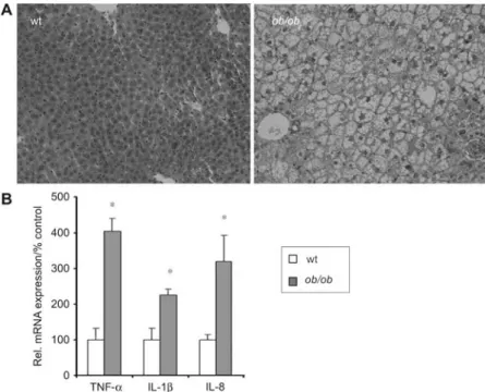
Figure 1 Liver histology and expression of inflammatory cytokines inob/ob mice.
(A) Representative hematoxylin and eosin staining of liver sections from wild-type andob/ob mice (200=magnification). Accumulation of
hepatocellular lipid vacuoles are present inob/ob livers (right) compared with wild-type livers with normal histological appearance (left).
(B) Analysis of inflammatory cytokine gene expression. Quantitative RT-PCR was performed with RNA samples isolated from liver of controls andob/ob-mice (ns6). The asterisk indicates p-0.05 as determined by Student’s t-test.
Despite the central role of FXR and other nuclear recep-tors in bile acid and lipid metabolism, their endogenous expression in fatty livers is still elusive. These nuclear recep-tors undergo marked changes in their expression and activity during cholestasis and inflammation (Trauner et al., 1998; Denson et al., 2000; Geier et al., 2003; Ghose et al., 2004; Geier et al., 2005b,c; Zollner et al., 2005) both of which are characteristic of NAFLD also. We hypothesize that bile acid accumulation in fatty livers leads to the activation of nuclear receptors, particularly FXR, and respective target genes under these specific conditions.
Therefore, the aim of this study was to characterize the expression of FXR and other class II nuclear receptors in fatty livers of ob/ob mice and to elucidate potential effects
of accumulating bile acids on lipogenic FXR-target genes under these conditions.
Results
Ob/ob mice develop liver steatosis without inflammation
Leptin-deficientob/ob mice spontaneously developed hepatic
steatosis at 14 weeks of age. In these animals hematoxylin and eosin staining of paraffin-embedded tissue sections dem-onstrated a severe hepatic steatosis without signs of histo-pathological inflammation compared with unaffected wild-type controls (Figure 1A). The liver/body weight ratio of 14-week-old ob/ob mice (9.0"0.2%) was increased in
comparison with wild-type controls (5.0"0.4%; ns6 each). mRNA expression levels of proinflammatory cytokines are increased in ob/ob-mice compared with control animals
w2.26"0.26-fold for interleukin (IL)-1b and 4.05"0.32-fold for tumor necrosis factor-a (TNF-a), 3.19"0.74-fold for IL-8, respectively; ns6;p-0.05 eachx. No change was observed
for IL-12 (data not shown) (Figure 1B).
Decreased bile acid transporter expression renders fatty livers in ob/ob mice cholestatic
Whetherob/ob mice are cholestatic, as recently reported for fa/fa Zucker rats, has not been investigated so far (Geier et
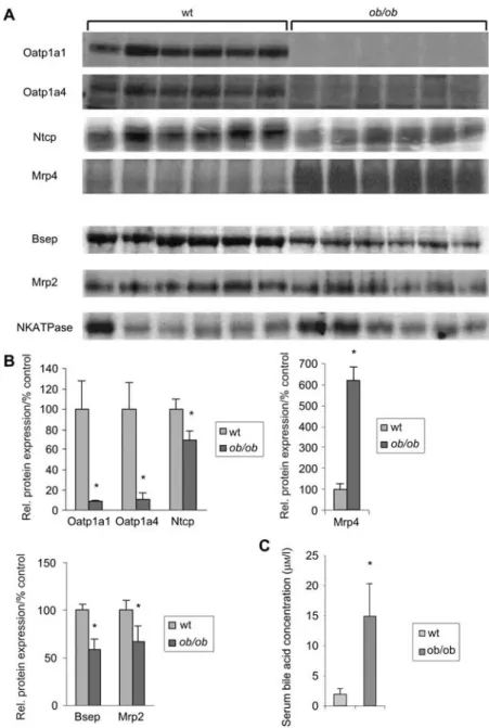
al., 2005a). Serum bile acid concentrations as measured by radioimmunoassay (RIA) are increased sevenfold in ob/ob
mice in comparison with wild-type controls (14.9"5.4 vs. 1.9"1.0mM; ns6; ps0.00080) (Figure 3).
Subsequently, hepatic bile salt and organic anion trans-porter expression inob/ob mice has been determined at both
mRNA and protein expression level using real-time reverse transcriptase-polymerase chain reaction (RT-PCR) and West-ern blot analysis (Figures 2 and 3). RNA expression of baso-lateral uptake transporters was significantly decreased for Oatp1a1 and Oatp1a4 (organic anion transporter) compared with control mice to 2"1% and 55"6%, respectively. Sim-ilarly, protein expression was reduced to 9"1% (Oatp1a1) and 11"6% (Oatp1a4) (bothp-0.05). Whereas Ntcp mRNA
expression was largely unchanged, Ntcp protein levels were significantly decreased inob/ob-mice compared with
wild-Figure 2 Hepatic transporter gene expression is altered inob/ob mice.
Quantitative TaqMan RT-PCR was performed with RNA samples isolated from liver of controls andob/ob-mice (ns6). The asterisk indicates p-0.05 as determined by Student’s t-test.
type controls (70"9%; p-0.05). In contrast to other
baso-lateral transporters, Oatp2b1 mRNA was significantly increased to 188"30% of controls. Expression of basolateral export systems was strongly upregulated at the mRNA (Mrp3 332"45%; Mrp4 1086"170%) and protein level (Mrp4 618"65%;p-0.05) each compared with controls.
On the contrary to other rodent models of cholestasis, but similar to fa/fa rats, expression of canalicular transporters
was mainly decreased at the protein level. Whereas Bsep and Mrp2 mRNA expression levels were even increased in
ob/ob mice to 234"40% and 168"16% of wild-type controls
(p-0.05) microsomal protein levels were decreased to
59"10% for Bsep and 67"16% for Mrp2 (p-0.05).
Consistent with decreased transporter protein abundance and bile acid retention in cholestatic ob/ob mice Cyp7a1
mRNA expression is profoundly suppressed compared with non-cholestatic lean animals to 10"4% (ps0.0001) (Figure
4A).
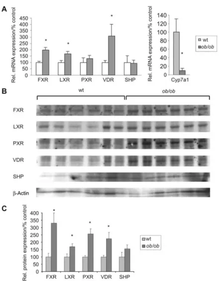
FXR and other nuclear receptors are upregulated in ob/ob mice
In contrast to rodent models of inflammatory liver disease nuclear receptor activity is not suppressed in obesefa/fa rats
(Geier et al., 2005a,b). To examine the influence of bile acid-retention and induction of proinflammatory cytokines on nuclear receptors in ob/ob mice, we analyzed both mRNA
and protein expression of several nuclear receptors implicat-ed in bile acid-signaling including FXR, pregnane X receptor (PXR) and vitamin D receptor (VDR) (Figure 4). Most prom-inently, FXR nuclear protein was increased to 330"70% (mRNA expression 196"21%) inob/ob mice compared with
controls (p-0.05 each). VDR and PXR expression were
sim-ilarly increased at both nuclear protein (225"43% and
258"34%, respectively) and mRNA levels (309"27% and 130"26%, respectively). Likewise, liver X receptor (LXR) protein and mRNA levels increased by 146"20% and 169"19% compared with the wild-type.
Finally, Shp mRNA and protein expression was analyzed to determine whether FXR activation results in the induction of this target gene. In contrast to other rodent models of cholestasis, Shp mRNA and protein expression inob/ob mice
were not significantly increased and comparable with their control littermates (Figure 4A).
FXR DNA-binding activity and lipogenic target genes including fatty acid synthase are increased in ob/ob mice
To investigate whether the observed increase in FXR nuclear protein results in increased DNA binding activity and acti-vation of respective target genes, we analyzed binding to the FXR-responsive element IR-1 which is present in mouse pro-moters of the Bsep (bile-salt export pump) and Fas (fatty
acid synthase) gene (Ananthanarayanan et al., 2001; Mat-sukuma et al., 2006). Using electrophoretic mobility shift assays (EMSAs) DNA binding activity to the IR-1 element is doubled in nuclear extracts from obese mice (196"3% compared with lean controls;p-0.05) (Figure 5). Following
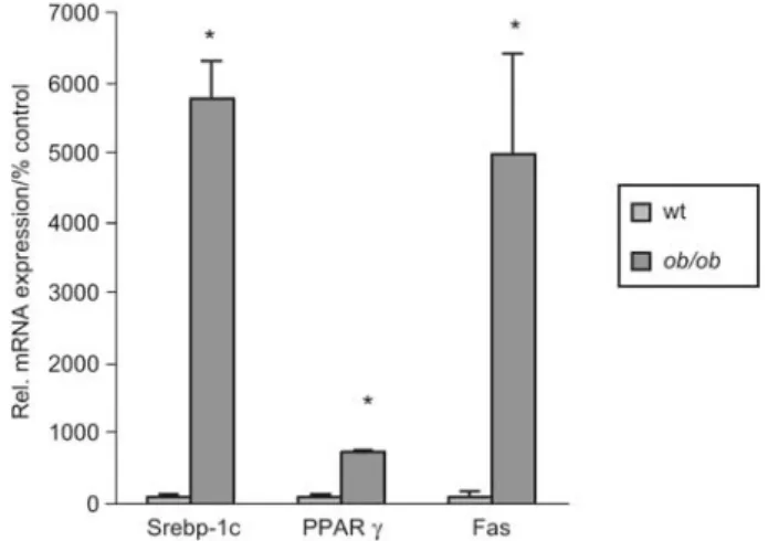
the trend of an increased mRNA expression of Bsep (see Figure 2), FAS mRNA expression, another FXR target gene, was upregulated to 4982"1432% (p-0.05) (Figure 6)
con-sistent with activated IR-1 binding.
As expected, other lipogenic genes including peroxisome proliferator-activated receptor-g (PPAR-g) mRNA and its target transcript Srebp-1c were upregulated in ob/ob mice
58-fold and seven-fold (both p-0.05 each), respectively
1444 I.V. Martin et al.
Article in press - uncorrected proof
Figure 3 Western blot analysis of hepatic transporter protein expression inob/ob mice and serum bile acid concentration in ob/ob mice.
(A) Western blotting: microsomal protein of wild-type andob/ob mice (ns6 each) was separated by SDS-PAGE (protein loading 50mg/ lane), blotted onto a PVDF membrane and detected by specific antibodies. (B) Densitometric analysis: quantification of relative protein expression compared with wild-type animals. The asterisk indicatesp-0.05 as determined by Student’s t-test. (C) Serum from wild-type
andob/ob animals was collected. Serum bile acids were measured by RIA. Asterisk indicates p-0.05 as determined by Student’s t-test.
Discussion
Previous evidence from obesefa/fa Zucker rats as an
estab-lished model of NAFLD, obesity and diabetes highlights the possibility that bile acid retention could play a role in the pathogenesis of the underlying metabolic disease (Pizarro et al., 2004; Geier et al., 2005b). We now used theob/ob mouse
model of fatty liver disease to further investigate potential bile acid-mediated effects on nuclear receptor activity and lipogenic target genes in more detail. This model resembles human fatty liver disease without histological inflammatory infiltrates and is characterized only by a subclinical extent of intrahepatic cytokine activation. The major new findings
of the present study are: (i) the bile acid retention inob/ob
mice rendering these animals by definition cholestatic, (ii) the resulting FXR upregulation and binding to the IR-1 element inob/ob animals and (iii) the concomitant activation
of the fatty acid synthase as lipogenic potential FXR target genein vivo.
In the current study, we describe bile acid retention and FXR activation as a potential trigger in the pathogenesis of fatty liver disease inob/ob mice. This retention of bile acids
is well in accordance to preliminary data from human patients with fatty liver disease which are also characterized by an increase in serum bile acid concentrations (Aranha et al., 2008; Kocabayoglu et al., 2009). Cholestasis in mice
Figure 4 Nuclear receptor mRNA and protein expression inob/ob mice.
(A) mRNA expression: quantitative TaqMan RT-PCR was performed with RNA samples isolated from liver of controls andob/ob-mice
(ns6). (B) Western blot analysis: nuclear protein and whole cell extract (SHP) were separated by SDS-PAGE (protein loading 10mg/lane), blotted onto a PVDF membrane and detected by specific antibodies. (C) Densitometric analysis: quantification of relative protein expression shown in B compared with wild-type mice. The asterisk indicatesp-0.05 as determined by Student’s t-test.
with fatty livers is accompanied by a downregulation of can-alicular bile acid transporter Bsep and Mrp2 proteins as the rate-limiting step in bile acid secretion. Furthermore, there seems to be a lack in the cholestatic feedback inhibition of Ntcp inob/ob mice (Figures 2 and 3). Differences between
maintained RNA expression and slightly decreased protein levels could be explained by post-translational modification or compartmental changes in protein location. A moderate decrease in Ntcp and Bsep protein expression is in accor-dance with previous observations infa/fa rats (Pizarro et al.,
2004; Geier et al., 2005b). Dysregulation of NTCP is even more prominent in humans where higher mRNA expression levels have been linked to disease progression in human NAFLD (Kocabayoglu et al., 2009). Of note, Cheng and co-workers observed in ob/ob mice a decreased Ntcp (mRNA
and protein) and Bsep (mRNA) expression and an even induced Mrp2 protein (mRNA unchanged compared with lean controls) (Cheng et al., 2008) which do not parallel other findings in humans (Martin et al., 2009), rats (Pizarro
et al., 2004; Geier et al., 2005b) and mice with fatty livers. Discrepancies could be explained by different breeding con-ditions which are particularly relevant owing to established changes in the intestinal barrier of ob/ob mice leading to
changes in portal lipopolysaccharide levels that can contrib-ute to hepatic inflammatory damage (Brun et al., 2007). Nev-ertheless, Cheng et al. report similar alterations in the expression of other basolateral bile acid transporters in
ob/ob mice including the downregulation of Oatp1a1 and the
induction of the overflow systems Mrp3 and Mrp4 in accor-dance with the present study (Cheng et al., 2008).
The underlying molecular mechanisms leading to trans-porter dysregulation might be different from (obstructive) cholestasis. Hepatic FXR expression is upregulated inob/ob
mice at both the mRNA and protein levels and, as expected, in the presence of bile acid retention FXR binding to the corresponding IR-1 element is activated (Figures 4 and 5). This is in line with a transcriptional activation of the FXR target geneBsep via IR-1 binding but decreased Bsep protein
1446 I.V. Martin et al.
Article in press - uncorrected proof
Figure 5 DNA-binding activity of FXR to the corresponding IR-1 element.
DNA-binding analysis was performed with nuclear proteins obtained from liver tissue of control and ob/ob mice (ns4 each).
Nuclear protein was incubated with biotin-labeled oligonucleotides representing the IR-1 element. Specificity of binding was confirmed by inclusion of specific competitor (SC) oligonucleotide or an unre-lated oligonucleotide at a 300-fold molar excess (NSCsnon-specific competitor).
Figure 6 Hepatic mRNA expression of lipogenic genes inob/ob
mice.
Quantitative TaqMan RT-PCR was performed with RNA samples isolated from liver of controls andob/ob-mice (ns6). Asterisk
indi-catesp-0.05 as determined by Student’s t-test.
necessitates the postulation of further post-translational pro-cessing under these conditions (Figures 2, 3 and 5). How-ever, we could not detect an upregulation of Shp as another FXR-target, as previously shown for obstructive cholestasis (Zollner et al., 2005) either on the mRNA or protein level. These results regarding an absent Shp mRNA induction have been previously reported in obese ob/ob and db/db mouse
models (Zhang et al., 2006; Miao et al., 2009). Miao and co-workers could further demonstrate that Shp protein abun-dance is highly controlled by ubiquitin-proteasomal degradation in wild-type animals whereas ob/ob mice are
largely protected against Shp ubiquitination.
Irrespective of ameliorating effects of FXR activation on hepatic inflammation, fibrosis during steatohepatitis and lipid
abnormalities in the mouse model (Cipriani et al., 2009; Zhang et al., 2009) the activation of FXR could also play a role for the dysregulation of lipogenesis in the pathogenesis of fatty liver disease. Recently, a FXR-responsive element (IR-1) has been identified within the mouse Fas promoter
which mediates a bile acid-dependent upregulation of the
Fas gene, in addition to the established insulin-induced
acti-vation (Wang and Sul, 1998; Matsukuma et al., 2006). In the present study extremely high levels of Fas transcripts have been observed in ob/ob mice in the presence of increased
IR-1 (FXR) binding (Figures 5 and 6). A moderately increased Fas expression in both wild-type and obese KK-Ay mice upon long-term feeding of cholic acid with a
concurrent decrease of Srebp-1 support this concept in gen-eral (Watanabe et al., 2004).
Consistent with Fas activation the lipogenic transcription factor Srepb-1c and its upstream activators LXR, PPARg and PXR are increased at the mRNA level (Figures 4 and 6). In primary mouse hepatocytes it has been shown that the acti-vation of FXR by bile acids or synthetic agonists represses the expression of Srebp-1c and its lipogenic target genes in a Shp-dependent manner (Watanabe et al., 2004).
Under the special conditions present in the livers ofob/ob
mice, activation of hepatic FXR could therefore affect the fatty acid homeostasis of hepatocytes, by activating FAS expression additionally to established Srebp-1c effects.
It is important to note that the role of FXR in the complex pathophysiological scenario of fatty liver disease is not con-clusively clarified so far. Whereas a variety of data suggest the potentially beneficial role of bile acids in treatment of NAFLD one cannot yet unambiguously evaluate their use-fulness (Orlando et al., 2007). Several interventional and knockout studies demonstrated beneficial effects of FXR activation. The general absence of FXRin vivo has profound
consequences on systemic lipid metabolism because general
Fxr-/- mice have increased serum triglycerides, cholesterol
and free fatty acids and develop fatty livers (Sinal et al., 2000; Lambert et al., 2003; Ma et al., 2006). On one hand, a inverse correlation between the activation of FXR path-ways and plasma triglyceride levels exists because bile acid-feeding in different models of hypertriglyceridemia decreased de novo lipogenesis through downregulation of
Srebp-1c resulting in reduced plasma triglycerides (Watanabe et al., 2004; Bilz et al., 2006). On the other hand, treatment of hyperlipidemic hamsters did not significantly decrease Srebp-1c expression despite a reduction in triglyceride syn-thesis (Bilz et al., 2006) and hepatic Srebp-1c mRNA levels are not increased inFxr-/-mice as to be expected (Zhang et
al., 2004; Duran-Sandoval et al., 2005a,b; Lefebvre et al., 2009).
Studies in db/db and ob/ob mice have shown that
treat-ment with the synthetic FXR-specific ligand GW4064 sig-nificantly improves insulin sensitivity and reduces hepatic lipid accumulation (Cariou et al., 2006; Zhang et al., 2006). Treatment with the FXR agonist GW4064 significantly represses hepatic CYP7A1 in mice with liver-specific dele-tion ofFxr (DeltaL) but not in mice with intestinal deletion
of FXR in intestine but not in the liver could mediate hepatic effects of GW4064 (Kim et al., 2007). Keeping in mind that GW4064 is characterized by a poor pharmacokinetic profile with poor intestinal absorption into the circulation these stud-ies do not necessarily rule out that hepatic FXR and its acti-vation in fatty livers could play a causal role in the pathophysiology of fatty liver disease. To finally answer this question comparable studies using intestinal and liver spe-cific FXR deletion in obese mice might be necessary.
In summary, our study opens the possibility that bile acid retention could contribute to the development of fatty liver disease. Understanding the multifaceted function of FXR in lipid homeostasis might contribute to pathophysiological and therapeutic concepts for a targeted treatment of fatty liver disease in the future.
Materials and methods
Animals
Eight-week-old maleob/ob mice (B6.V-Lepob
/J) and age- and gen-der-matched control animals C57BL/6J were purchased from The Jackson Laboratory (Bar Harbor, ME, USA). All mice were housed in pathogen-free animal facilities under a standard 12-h light, 12-h dark cycle with access to regular rodent chow and autoclaved tap waterad libitum for 6 weeks. The mice were sacrificed, and most
of the liver tissue was immediately frozen in liquid nitrogen and a small portion was immersion fixed in 4% formalin. Subsequently, paraffin-embedded sections were analyzed after hematoxylin and eosin staining for the degree of hepatic steatosis. The animals received humane care and the study protocols were approved by the local Government’s Animal Care Committee.
mRNA isolation and real-time RT-PCR
Total RNA was isolated from liver by standard phenol chloroform extraction procedure using UltraspecTM
(Biotecx Lab, Houston, TX, USA) according to manufacturer’s instructions. mRNA was reverse-transcribed to cDNA using the Transcriptor First Strand cDNA Syn-thesis Kit (Roche, Mannheim, Germany). cDNA was used for RT-PCR with SYBR Green Reagent (Invitrogen, Karlsruhe, Ger-many) and specific primer pairs on a 7300 ABI PRISM Real-Time PCR System and with ABI PRISM 7300 SDS software (Applied Biosystems, Foster City, CA, USA). Cyp7a1 expression was ana-lyzed using ABI TaqMan probes. Expression was normalized against 18S. All primer sequences are available from the authors upon request.
Western blotting
Nuclear and microsomal protein fractions were prepared as described previously (Gartung et al., 1997). Similar amounts of microsomal and nuclear protein (50mg and 10 mg, respectively) were separated by SDS-PAGE, transferred to a polyvinylidene flu-oride (PVDF) membrane and probed with the following antibodies: Oatp1a1 (Eckhardt et al., 1999), Oatp1a4 (Reichel et al., 1999), Ntcp (Stieger et al., 1994), BSEP (Gerloff et al., 1998), Mrp2 (Madon et al., 2000), Mrp4 (Rius et al., 2003), FXR (SantaCruz, CA, USA, clone Q-20, sc-1205), LXR (Abcam, Cambridge, UK, ab-28478), PXR (SantaCruz, USA, clone A-20, sc-7737), VDR (SantaCruz, USA, clone C-20, sc-1008), SHP (SantaCruz, USA,
clone Q-14, sc-15283). Na/K ATPase (Abcam, Cambridge, UK, ab-7671) and b-actin (Sigma, St. Louis, MO, USA, A2066) antibodies were used as loading control for microsomal and total protein. After incubation with species-specific HRP-conjugated secondary anti-body (Dako, Hamburg, Germany) immune complexes were detected using the ECL detection kit (GE Healthcare, Freiburg, Germany). Densitometric quantification of Western blots was performed using Quantity One software (Bio-Rad, Munich, Germany).
Electrophoretic mobility shift assay
Nuclear protein extracts were prepared as described previously (Geier et al., 2002). DNA binding analyses were conducted using the Lightshift Chemiluminescent EMSA kit (Pierce Biotechnology, Rockford, IL, USA) according to the manufacturers’ protocol. The following oligonucleotides were used as probes in the analyses: IR-1, sense 59-CTT TAG GCC ATT GAC CTA TAA-39 and antisense 59-TTA TAG GTC AAT GGC CTA AAG-39 (Geier et al., 2005b). These oligonucleotides were end-labeled using the biotin 39 end DNA labeling kit (Pierce Biotechnology). Binding reactions con-sisted of 1= binding buffer, 50 ng/ml poly dIdC, 20 fmol biotin-labeled DNA and 5 mg nuclear protein in a 20 ml reaction. Competition experiments included 6 pmol unlabeled oligonucleotide (300-fold molar excess). Densitometric quantification was per-formed using Quantity One software (Bio-Rad).
Bile acid quantification
Serum bile acids were measured by RIA using a Bile Acid RIA Kit (MP Biomedicals, Ilkirch, France) according to the manufacturer’s specifications.
Statistical analysis
Statistical significance (p-0.05) between control animals and ob/ob-mice was determined by Student’s t-test. Data represent the
mean"standard deviation.
Acknowledgments
The authors thank Sonja Strauch, Joba Arikkat and Lia Hofstetter for their excellent technical assistance. This work was supported by the Swiss National Science Foundation (SNF) grant 310000-122310/1 (to A.G.) and the Foundation for Research at the Medical Faculty, University of Zurich (to A.G.).
References
Adams, L.A., Angulo, P., and Lindor, K.D. (2005). Nonalcoholic fatty liver disease. Can. Med. Assoc. J.172, 899–905.
Ananthanarayanan, M., Balasubramanian, N., Makishima, M., Man-gelsdorf, D.J., and Suchy, F.J. (2001). Human bile salt export pump promoter is transactivated by the farnesoid X receptor/bile acid receptor. J. Biol. Chem.276, 28857–28865.
Angulo, P. (2002). Nonalcoholic fatty liver disease. N. Engl. J. Med.
346, 1221–1231.
Aranha, M.M., Cortez-Pinto, H., Costa, A., da Silva, I.B., Camilo, M.E., de Moura, M.C., and Rodrigues, C.M. (2008). Bile acid levels are increased in the liver of patients with steatohepatitis. Eur. J. Gastroenterol. Hepatol.20, 519–525.
1448 I.V. Martin et al.
Article in press - uncorrected proof
Bilz, S., Samuel, V., Morino, K., Savage, D., Choi, C.S., and Shul-man, G.I. (2006). Activation of the farnesoid X receptor improves lipid metabolism in combined hyperlipidemic ham-sters. Am. J. Physiol. Endocrinol. Metab.290, E716–E722.
Brun, P., Castagliuolo, I., Di Leo, V., Buda, A., Pinzani, M., Palu, G., and Martines, D. (2007). Increased intestinal permeability in obese mice: new evidence in the pathogenesis of nonalcoholic steatohepatitis. Am. J. Physiol. Gastrointest. Liver Physiol.292,
G518–G525.
Cariou, B., van Harmelen, K., Duran-Sandoval, D., van Dijk, T.H., Grefhorst, A., Abdelkarim, M., Caron, S., Torpier, G., Fruchart, J.C., Gonzalez, F.J., et al. (2006). The farnesoid X receptor mod-ulates adiposity and peripheral insulin sensitivity in mice. J. Biol. Chem.281, 11039–11049.
Cheng, Q., Aleksunes, L.M., Manautou, J.E., Cherrington, N.J., Scheffer, G.L., Yamasaki, H., and Slitt, A.L. (2008). Drug-metabolizing enzyme and transporter expression in a mouse model of diabetes and obesity. Mol. Pharm.5, 77–91.
Chiang, J.Y. (2009). Bile acids: regulation of synthesis. J. Lipid Res.
50, 1955–1966.
Cipriani, S., Mencarelli, A., Palladino, G., and Fiorucci, S. (2010). FXR activation reverses insulin resistance and lipid abnormali-ties and protects against liver steatosis in Zucker (fa/fa) obese rats. J. Lipid Res.51, 771–784.
Denson, L.A., Auld, K.L., Schiek, D.S., McClure, M.H., Mangels-dorf, D.J., and Karpen, S.J. (2000). Interleukin-1b suppresses retinoid transactivation of two hepatic transporter genes involved in bile formation. J. Biol. Chem.275, 8835–8843.
Duran-Sandoval, D., Cariou, B., Fruchart, J.C., and Staels, B. (2005a). Potential regulatory role of the farnesoid X receptor in the metabolic syndrome. Biochimie87, 93–98.
Duran-Sandoval, D., Cariou, B., Percevault, F., Hennuyer, N., Gref-horst, A., van Dijk, T.H., Gonzalez, F.J., Fruchart, J.C., Kuipers, F., and Staels, B. (2005b). The farnesoid X receptor modulates hepatic carbohydrate metabolism during the fasting-refeeding transition. J. Biol. Chem.280, 29971–29979.
Eckhardt, U., Schroeder, A., Stieger, B., Hochli, M., Landmann, L., Tynes, R., Meier, P.J., and Hagenbuch, B. (1999). Polyspecific substrate uptake by the hepatic organic anion transporter Oatp1 in stably transfected CHO cells. Am. J. Physiol. 276,
G1037–G1042.
Erickson, S.K. (2009). Nonalcoholic fatty liver disease. J. Lipid Res.
50 (Suppl.), S412–S416.
Gartung, C., Schuele, S., Schlosser, S.F., and Boyer, J.L. (1997). Expression of the rat liver Naq
/taurocholate cotransporter is reg-ulatedin vivo by retention of biliary constituents but not their
depletion. Hepatology25, 284–290.
Geier, A., Kim, S.K., Gerloff, T., Dietrich, C.G., Lammert, F., Kar-pen, S.J., Stieger, B., Meier, P.J., Matern, S., and Gartung, C. (2002). Hepatobiliary organic anion transporters are differen-tially regulated in acute toxic liver injury induced by carbon tetrachloride. J. Hepatol.37, 198–205.
Geier, A., Dietrich, C.G., Voigt, S., Kim, S.K., Gerloff, T., Kullak-Ublick, G.A., Lorenzen, J., Matern, S., and Gartung, C. (2003). Effects of proinflammatory cytokines on rat organic anion trans-porters during toxic liver injury and cholestasis. Hepatology38,
345–354.
Geier, A., Dietrich, C.G., Grote, T., Beuers, U., Prufer, T., Fraun-berger, P., Matern, S., Gartung, C., Gerbes, A.L., and Bilzer, M. (2005a). Characterization of organic anion transporter regula-tion, glutathione metabolism and bile formation in the obese Zucker rat. J. Hepatol.43, 1021–1030.
Geier, A., Dietrich, C.G., Voigt, S., Ananthanarayanan, M., Lam-mert, F., Schmitz, A., Trauner, M., Wasmuth, H.E., Boraschi, D.,
Balasubramaniyan, N., et al. (2005b). Cytokine-dependent reg-ulation of hepatic organic anion transporter gene transactivators in mouse liver. Am. J. Physiol. Gastrointest. Liver Physiol.289,
G831–G841.
Geier, A., Zollner, G., Dietrich, C.G., Wagner, M., Fickert, P., Denk, H., van Rooijen, N., Matern, S., Gartung, C., and Trauner, M. (2005c). Cytokine-independent repression of rodent Ntcp in obstructive cholestasis. Hepatology41, 470–477.
Geier, A., Wagner, M., Dietrich, C.G., and Trauner, M. (2007). Prin-ciples of hepatic organic anion transporter regulation during cho-lestasis, inflammation and liver regeneration. Biochim. Biophys. Acta1773, 283–308.
Gerloff, T., Stieger, B., Hagenbuch, B., Madon, J., Landmann, L., Roth, J., Hofmann, A.F., and Meier, P.J. (1998). The sister of P-glycoprotein represents the canalicular bile salt export pump of mammalian liver. J. Biol. Chem.273, 10046–10050.
Ghose, R., Zimmerman, T.L., Thevananther, S., and Karpen, S.J. (2004). Endotoxin leads to rapid subcellular re-localization of hepatic RXRalpha: a novel mechanism for reduced hepatic gene expression in inflammation. Nucl. Recept.2, 4.
Kim, I., Ahn, S.H., Inagaki, T., Choi, M., Ito, S., Guo, G.L., Klie-wer, S.A., and Gonzalez, F.J. (2007). Differential regulation of bile acid homeostasis by the farnesoid X receptor in liver and intestine. J. Lipid Res.48, 2664–2672.
Kocabayoglu, P., Bechmann, L.P., Kilicarslan, A., Erhard, J., Kah-raman, A., Schlattjan, M., Odenthal, M., Gerken, G., Geier, A., and Canbay, A. (2009). In NASH patients the bile acid trans-porter NTCP is upregulated and associated with fibrosis. Hepa-tology50, 793A.
Lambert, G., Amar, M.J., Guo, G., Brewer, H.B. Jr., Gonzalez, F.J., and Sinal, C.J. (2003). The farnesoid X-receptor is an essential regulator of cholesterol homeostasis. J. Biol. Chem. 278,
2563–2570.
Lefebvre, P., Cariou, B., Lien, F., Kuipers, F., and Staels, B. (2009). Role of bile acids and bile acid receptors in metabolic regulation. Physiol. Rev.89, 147–191.
Ma, K., Saha, P.K., Chan, L., and Moore, D.D. (2006). Farnesoid X receptor is essential for normal glucose homeostasis. J. Clin. Invest.116, 1102–1109.
Madon, J., Hagenbuch, B., Landmann, L., Meier, P.J., and Stieger, B. (2000). Transport function and hepatocellular localization of mrp6 in rat liver. Mol. Pharmacol.57, 634–641.
Makishima, M., Okamoto, A.Y., Repa, J.J., Tu, H., Learned, R.M., Luk, A., Hull, M.V., Lustig, K.D., Mangelsdorf, D.J., and Shan, B. (1999). Identification of a nuclear receptor for bile acids. Science284, 1362–1365.
Martin, I.V., Minkenberg, A., Canbay, A., Muellhaupt, B., and Geier, A. (2009). Regulation of bile salt transporters in non-alcoholic fatty liver disease in ob/ob-mice and human fatty livers. J. Hepa-tol.50 (Suppl. 1), A719.
Matsukuma, K.E., Bennett, M.K., Huang, J., Wang, L., Gil, G., and Osborne, T.F. (2006). Coordinated control of bile acids and lipo-genesis through FXR-dependent regulation of fatty acid syn-thase. J. Lipid Res.47, 2754–2761.
Miao, J., Xiao, Z., Kanamaluru, D., Min, G., Yau, P.M., Veenstra, T.D., Ellis, E., Strom, S., Suino-Powell, K., Xu, H.E., et al. (2009). Bile acid signaling pathways increase stability of Small Heterodimer Partner (SHP) by inhibiting ubiquitin-proteasomal degradation. Genes Dev.23, 986–996.
Orlando, R., Azzalini, L., Orando, S., and Lirussi, F. (2007). Bile acids for non-alcoholic fatty liver disease and/or steatohepatitis. Cochrane Database Syst. Rev. CD005160.
Pizarro, M., Balasubramaniyan, N., Solis, N., Solar, A., Duarte, I., Miquel, J.F., Suchy, F.J., Trauner, M., Accatino, L.,
Ananthana-rayanan, M., et al. (2004). Bile secretory function in the obese Zucker rat: evidence of cholestasis and altered canalicular trans-port function. Gut53, 1837–1843.
Reichel, C., Gao, B., Van Montfoort, J., Cattori, V., Rahner, C., Hagenbuch, B., Stieger, B., Kamisako, T., and Meier, P.J. (1999). Localization and function of the organic anion-transporting polypeptide Oatp2 in rat liver. Gastroenterology117, 688–695.
Rius, M., Nies, A.T., Hummel-Eisenbeiss, J., Jedlitschky, G., and Keppler, D. (2003). Cotransport of reduced glutathione with bile salts by MRP4 (ABCC4) localized to the basolateral hepatocyte membrane. Hepatology38, 374–384.
Sinal, C.J., Tohkin, M., Miyata, M., Ward, J.M., Lambert, G., and Gonzalez, F.J. (2000). Targeted disruption of the nuclear receptor FXR/BAR impairs bile acid and lipid homeostasis. Cell 102,
731–744.
Stefan, N., Kantartzis, K., and Haring, H.U. (2008). Causes and metabolic consequences of fatty liver. Endocr. Rev.29, 939–960.
Stieger, B., Hagenbuch, B., Landmann, L., Hochli, M., Schroeder, A., and Meier, P.J. (1994).In situ localization of the hepatocytic
Naq
/taurocholate cotransporting polypeptide in rat liver. Gastro-enterology107, 1781–1787.
Trauner, M., Arrese, M., Lee, H., Boyer, J.L., and Karpen, S.J. (1998). Endotoxin downregulates rat hepatic ntcp gene expres-sion via decreased activity of critical transcription factors. J. Clin. Invest.101, 2092–2100.
Wang, D. and Sul, H.S. (1998). Insulin stimulation of the fatty acid synthase promoter is mediated by the phosphatidylinositol
3-kinase pathway. Involvement of protein 3-kinase B/Akt. J. Biol. Chem.273, 25420–25426.
Watanabe, M., Houten, S.M., Wang, L., Moschetta, A., Mangels-dorf, D.J., Heyman, R.A., Moore, D.D., and Auwerx, J. (2004). Bile acids lower triglyceride levels via a pathway involving FXR, SHP, and SREBP-1c. J. Clin. Invest.113, 1408–1418.
Zhang, Y., Castellani, L.W., Sinal, C.J., Gonzalez, F.J., and Edwards, P.A. (2004). Peroxisome proliferator-activated receptor-g coac-tivator 1a (PGC-1a) regulates triglyceride metabolism by acti-vation of the nuclear receptor FXR. Genes Dev.18, 157–169.
Zhang, Y., Lee, F.Y., Barrera, G., Lee, H., Vales, C., Gonzalez, F.J., Willson, T.M., and Edwards, P.A. (2006). Activation of the nuclear receptor FXR improves hyperglycemia and hyperlipi-demia in diabetic mice. Proc. Natl. Acad. Sci. USA 103,
1006–1011.
Zhang, S., Wang, J., Liu, Q., and Harnish, D.C. (2009). Farnesoid X receptor agonist WAY-362450 attenuates liver inflammation and fibrosis in murine model of non-alcoholic steatohepatitis. J. Hepatol.51, 380–388.
Zollner, G., Wagner, M., Fickert, P., Geier, A., Fuchsbichler, A., Silbert, D., Gumhold, J., Zatloukal, K., Kaser, A., Tilg, H., et al. (2005). Role of nuclear receptors and hepatocyte-enriched transcription factors for Ntcp repression in biliary obstruction in mouse liver. Am. J. Physiol. Gastrointest. Liver Physiol.289,
G798–G805.