Effect of Non-Chilling Temperature and Light Intensity during Growth of
Squash Cotyledons on the Composition of Thylakoid Membrane Lipids and
Fatty Acids
Yinong Xu and Paul-Andre Siegenthaler'
Laboratoire de Physiologie ve'ge'tale, University de NeuchStel, Rue Emile Argand 13, CH-2007 NeuchStel, Switzerland
The lipid composition, in particular the content of fat-ty acids in phosphatidylglycerol (PG), is believed to be crucial for the understanding of the chilling injury mechan-ism occurring in the thylakoid membrane (TM) of higher plants. In this investigation, we have studied the effect of growth conditions (e.g. temperature and light) on the com-position of glycerolipid classes and their respective fatty acids during the maturation period of squash cotyledons. We have found that the changes in the lipid fatty acid com-position of TM which are induced by different temperature and light growth conditions occurred only during the devel-opment of cotyledons but not when these latter had reach-ed their maturity. The major changes were an increase of 18:3 and a decrease of 16:0 in galacto- and sulfolipids, and an increase of 16:1(30 and 18:3 with a concomitant decrease of 16:0 and 18:1 in PG, when the temperature was low (20°C compared to 30°C). We conclude that low tem-perature conditions of growth induced an increase of the end acyl products [18:3 and/or 16:1(301 in PG and galacto-lipids. The possible mechanism is discussed in terms of the relative temperature dependence of fatty acid synthesis and desaturation processes. The light intensity of growth affected only the fatty acid composition of PG, i.e. when it was high (350 compare to 100/onol m~2s~1), an increase of 16:1(30 at the expense of 16:0 was observed. Whatever the growth conditions, the level of 16:1(30 increased dur-ing the maturation of cotyledons and was characterized by an increasing linear correlation (r = 0.99) with the fresh weight. In contrast, a decreasing linear relationship (r = — 0.99) was found between the fresh weight and 16:0. Thus, we propose that a constant level of 16:1(30 is a good criterion for defining the chloroplast maturity and that it is highly advisable to study the effect of temperature and light on the lipid composition when the cotyledons have reached their maturity. Under these conditions, these effects can be considered regardless of developmental factors.
Abbreviations: DGDG, digalactosyldiacylglycerol; m:n, fatty acid containing m carbons and n cis double bonds; MGDG, monogalactosyldiacylglycerol; MOPS, 4-morpholinopropane-sulfonic acid; PFD, photon flux density; PG, phosphatidylglycer-ol; SQDG, sulfoquinovosyldiacylglycerphosphatidylglycer-ol; TLC, thin-layer chro-matography; 14:0, myristic acid; 16:0, palmitic acid; 16:1(30. /d3-//wi.s-hexadecenoic acid; 18:0, stearic acid; 18:1, oleic acid;
18:2, linoleic acid; 18:3, linolenic acid.
1 To whom correspondence should be addressed.
Key words: /d'-frarw-hexadecenoic acid — Cucurbita
moschata Durch — Development — Galactolipids —
Maturity — Phosphatidylglycerol.
Low temperature is one of the major environmental factors which limits the growth, the geographical distribu-tion and productivity of plants on the earth. Most plants of temperate climate have the ability to survive at chilling temperature (e.g. from 5 to 15°C) and are called chilling-resistant plants. In contrast, most tropical and subtropical plants are unable to survive under such temperatures and are considered as chilling-sensitive plants (Lyons 1973, Levitt 1980, Murata and Nishida 1990). However, the resist-ance of chilling-sensitive plants can be increased by a pre-treatment at low, but not injurious temperature (Anderson et al. 1994, Kodama et al. 1995). Several mechanisms have been proposed to explain the chilling sensitivity of plants. One of the first hypothesis was that bulk membrane lipid phase transitions at a critical temperature result in the for-mation of gel phase lipids (Lyons 1973). Consequently, the gel phase leads to increased permeability or leakiness of cellular and organelle membranes, loss of compartmenta-tion and physiological funccompartmenta-tions, then to cell death. More recently, Murata (1983) established a correlation between the content of desaturated molecular species of phospha-tidylglycerol [16:0/16:0, 16:0/16:1(30, 18:0/16:0 and 18:0/ 16:1(30] and the chilling sensitivity of plants, i.e. chilling-sensitive plants contain much higher proportions of desatu-rated PG than resistant plants. These "high-melting point" PG molecular species are supposed to form gel phase do-mains in the thylakoid membranes at chilling temperatures. Furthermore, an addition of small amounts (as little as 1 to
2%) of disaturated phospholipids to mixtures of leaf
mem-brane polar lipids significantly increases the temperature at which phase transition occurred in the mixture (Raison and Wright 1983). Experiments with transgenic plants have con-firmed the causal link between the amount of desaturated PG and low-temperature-induced injury (Murata et al. 1992).
However, several results are difficult to interpret in terms of the "high-melting point" PG molecular species hy-pothesis: (1) The correlation between the amount of desatu-rated PG and chilling sensitivity is not unambiguous
cause of the lack of a quantitative measure for chilling sensitivity (Somerville 1995); (2) Not all chilling-sensitive plants contain high proportion of desaturated PG, such as solanaceous and other 16:3-plants and C4 grasses (Roughan 1985); (3) When the level of desaturated PG was determined in several plant species (Murata et al. 1982, Roughan 1985, Kenrick and Bishop 1986) no special precautions were taken to grow plants under similar light and temperature conditions. However, it is known that these two combined growth parameters can influence greatly the content of the fatty acids and lipid molecular species (Dubacq and Tremolieres 1983 and references therein; Xu et al. 1992). Moreover, in earlier studies, the developmental stage of the plant tested is rarely known or denned; (4) According to Murata's hypothesis, low-temperature acclimated plants which are more chilling-resistant should contain less high-melting point PG molecular species than the control plants. This is apparently not the case for the plant species Nerium
oleander L. (Orr and Raison 1987). (5) In a mutant of Arabidopsis (fabl), leaf PG contains 43% of high-melting
point molecular species, a higher percentage that is found in many chilling-sensitive plants. However, the mutant was completely unaffected, when compared with wild-type con-trols, by a range of low-temperature treatments that rapid-ly led to the death of other chilling-sensitive plants (Wu and Browse 1995).
As a further step in understanding the relationship be-tween chilling sensitivity and the level of membrane polar lipids, we have studied the composition of acyl lipids and fatty acids in thylakoids from squash cotyledons. In this in-vestigation, we demonstrate that in the growth tempera-ture/glycerolipid composition relationship, it is very impor-tant to define exactly the growth conditions of plants as well as the development stage of the cotyledons. In addi-tion, the presence of the end acyl products (16:1(3/) and/or 18:3) in galactolipids and PG of squash plants grown at low temperature will be discussed in terms of the tempera-ture dependence of the enzymes involved in fatty acid syn-thesis and desaturation.
Materials and Methods
Plant materials and growth conditions—Plants of squash (Cucurbita moschata Durch. cv Shirakikuza), a chilling-sensitive
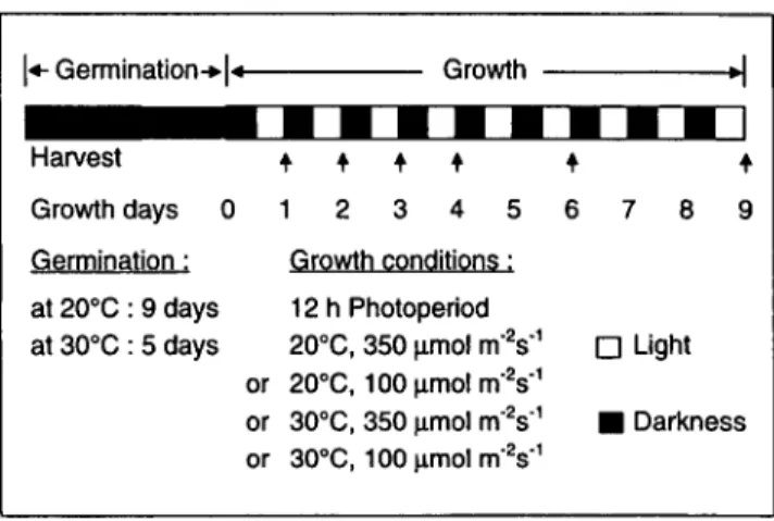
plant (Murata et al. 1982), were grown from seeds in soil (Mio plant Natura, Migros, Switzerland). The seeds were germinated in darkness at 30°C and the seedlings grown in controlled environ-ment growth chambers (Sanyo Gallenkamp, U.K., Cabinet Model PG 660) under a 12 h photoperiod and various temperatures (20 or 30°C) and light (100 or 350^mol m~2 s"1) conditions as in-dicated in Figure 1. Chloroplasts were immediately isolated from cotyledons collected after the desired daily light period.
Isolation of thylakoid membranes—All operations were
per-formed at 4°C. The cotyledons (20 g, 40 to 120 cotyledons) were ground shortly (5 s) using a Waring Blendor in 160 ml of a
grin-• grin-• grin-• grin-• grin-•
Harvest Growth days ( Germination: at 20°C: 9 days at 30°C : 5 daysE
or or or•
1D
21 • • •
3 4 5 Growth conditions: 12 20 20 30 30 h ( C, 'C, C, 'C, 3hotoperiod 350 umol m'2s"1 100 umol m'2s'1 350 umol m"2s'1 100 umol m ' V1• • • I
6 7 8 9 D Light • DarknessFig. 1 Germination and growth conditions of squash plants. Cotyledons were harvested after 1, 2, 3, 4, 6 and 9 days of growth and thylakoids were isolated immediately for lipid analyses as de-scribed in Material and Methods.
ding medium containing 330 mM sorbitol, 30 mM MOPS-KOH (pH7.8), 2mM EDTA-Na2 and 0.15% BSA. The mixture was filtered through six layers of cheesecloth, and the filtrate was subse-quently centrifuged at l,910xg for 1 min. The supernatant was discarded by aspiration whilst the intact chloroplast pellet was re-suspended with a smooth brush in a lysis medium containing 10 mM Tricine-KOH (pH 7.8) first in 4 ml, then diluted with 30 ml of the same solution. After 1 min, isotony was restored by adding 4 ml of 3 M sorbitol, followed by mixing and centrifuging at 4,300 xg for 2 min. After discarding the supernatant by aspira-tion, thylakoids were resuspended in a washing medium contain-ing 300 mM sorbitol and 10 mM Tricine-KOH (pH 7.8) and fur-ther centrifuged at 4,300xg for 2 min. The thylakoid pellet was resuspended in the same washing medium and adjusted to about 2 mg chlorophyll per ml. The chlorophyll concentration was deter-mined according to Bruinsma (1961).
Lipid extraction and purification—Lipids were extracted
from thylakoid membranes according to Siegenthaler et al. (1989). The lipid classes were separated from each other by TLC on silica gel plates (pre-coated silica gel plates, Merck 5626). The develop-ing solvents were: acetone/toluene/H2O (91 : 30 : 8, by volume) for separating PG and chloroform/methanol/25% NH3/H2O (65 : 35 : 3 : 2, by volume) for separating MGDG, DGDG and SQDG. The plates were dried shortly in air and lightly sprayed with 0.01% primuline and viewed under UV light. Zones of interest (MGDG, DGDG, SQDG and PG) were scraped into glass tubes and the quantification of each lipid class was carried out by determining its fatty acids content by gas chromatography (Xu and Siegen-thaler 1996).
Determination of fatty acids—The individual thylakoid
mem-brane lipids separated by TLC were transesterified with 5% H2SO4 in MeOH for 1 h at 85CC. The fatty acid methyl esters were sepa-rated on a Hewlett-Packard 5890 gas chromatography supplied with an hydrogen flame ionization detector and a capillary column FFAP (30 m; i.d. 0.53 mm). The column was isothermally run at 190°C and the detector was held at 230°C. Arachidic acid (Sigma) was used as an internal standard.
Results
Effect of cotyledon development upon the fatty acid composition of glycerolipid classes—Table 1 shows the
changes of fatty acid composition in MGDG, DGDG and
SQDG during the cotyledon maturation when seedlings were grown, after germination, at isothermal temperature (20 or 30°C) and at two photon flux densities (100 or 350 //mol m~2 s~'). During growth and under all temperature
and light conditions, the content of 18:3 increased mainly
Table 1 The effect of the cotyledon development on the fatty acid composition of thylakoid membrane glycerolipids (MGDG, DGDG and SQDG) Lipids MGDG DGDG SQDG Temp. 30 30 20 20 30 30 20 20 30 30 20 20 Growth condition °C PFD,//molm~2s~' 350 100 350 100 350 100 350 100 350 100 350 100 Growth days 1 2 3 1 2 3 1 2 3 1 2 3 1 2 3 1 2 3 1 2 3 1 2 3 1 2 3 1 2 3 1 2 3 1 2 3 16:0 1.5 1.6 1.6 1.5 1.6 1.7 1.1 1.1 1.0 1.1 1.0 1.1 9.6 9.5 9.5 9.9 9.4 9.3 5.0 6.3 6.2 7.2 6.4 6.2 26.7 26.2 27.2 26.6 26.9 27.6 25.3 24.3 24.9 25.2 23.4 23.5 Fatty acid 18:0 0.0 0.0 0.0 0.0 0.0 0.0 0.0 0.0 0.0 0.0 0.0 0.0 1.4 0.7 0.5 1.6 0.9 0.7 1.0 0.4 0.4 1.0 0.5 0.4 5.8 4.8 4.6 6.4 5.7 4.7 6.0 5.8 5.3 6.0 5.9 5.4 composition 18:1 1.3 0.7 0.7 1.1 0.5 0.6 0.8 0.3 0.2 0.9 0.4 0.3 1.0 0.7 0.6 0.9 0.7 0.7 0.6 0.3 0.2 0.6 0.4 0.3 3.6 2.3 2.9 3.2 2.9 2.9 2.8 2.2 1.8 2.9 2.5 2.0 (mol%) 18:2 5.7 2.6 2.7 6.4 2.9 2.8 7.1 2.6 1.8 6.9 2.7 2.1 5.2 3.1 2.8 5.9 3.6 3.5 6.0 3.1 1.9 5.8 3.5 2.6 11.8 7.2 6.5 13.1 8.5 7.5 12.1 7.2 5.0 11.9 7.7 6.1 18:3 91.5 95.1 95.0 90.9 95.0 94.9 90.9 96.0 97.0 91.0 95.9 96.5 82.9 86.0 86.5 81.7 85.5 85.9 87.5 89.9 91.3 85.4 89.2 90.4 52.0 59.5 58.8 50.6 56.0 57.3 53.7 60.5 62.9 54.0 60.5 62.9
80 60 40 o E, £
8
ci d CD att y 20 0 16 12 -16:0 -*-16:1(3t) 0 1 2 3 4 5 6 7 8 9 10Seedling growth time (days)
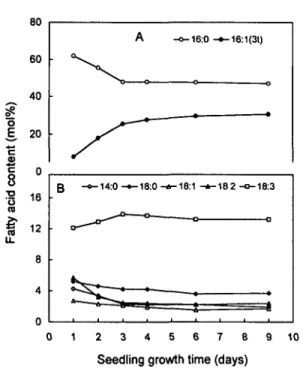
Fig. 2 Changes in fatty acid composition of phosphatidylglycer-ol in thylakoid membranes during cotyledon development. Plants were germinated and grown at 20°C. The photoperiod was 12 h and the PFD 350^mol m~2 s~'. A: changes in the content of
16:0 and 16:1(3/); B: changes in the content of 14:0, 18:0, 18:1, 18:2 and 18:3.
at the expense of 18:2 and to a lesser degree of 18:1 and 18:0 in the three lipid classes. These changes occurred espe-cially during the first two days of growth. In contrast, no significant change was observed at the level of 16:0 during the first three days of growth.
Phosphatidylglycerol displayed the most complex pat-tern in fatty acid composition. Seven fatty acids were in-deed found in thylakoid membranes of squash, i.e. 14:0, 16:0, 16:1(30, 18:0, 18:1, 18:2 and 18:3. Fig. 2 shows, as an example, the changes in the composition of fatty acids oc-curring during the first 9 days of growth at 20°C and 350 photons m~2 s"1. The comparison of the results of
Fig. 2B and Table 1 reveals that the behaviour of 18:3 and 18:2 in PG was very close to that found in the other lipid classes, i.e. an increase in 18:3 content from 12.1 to 13.9± 0.4 mol% at the expense of 18:2 (from 5.7 to 2.9±0.2) dur-ing the first three days of growth. However, when seedldur-ings were grown at a higher temperature (30°C), the content of 18:1 increased from 3 to 6.6±0.4 mol% whilst the level of 18:3 remained constant at about 8.4±0.3mol% (results not shown). The most significant change occurred at the level of the C,6 series as shown in Fig. 2A. During the first
3 days of growth, there was a significant decrease in 16:0 with a concomitant increase in 16:1(30- After 3 days, the molar ratio 16:0/16:1(30 remained constant.
Relative content of glycerolipid classes during seedling growth—Preliminary experiments revealed that whatever
growth conditions were (see Fig. 1), the content of MGDG, DGDG, SQDG and PG remained constant. Cotyledons were collected 6 times during growth (as shown in Fig. 1) and the content of lipid classes was measured in each cotyle-don thylakoid samples. The mean values ( ± standard devia-tion) which are reported in Table 2 show that, within the limits of environmental conditions used, the relative molar content of each lipid class did not depend on the growth temperature and light energy as well as on the maturity degree of the cotyledons.
Effect of growth temperature and light on the fatty acid composition—We have observed previously (Table 2
and Fig. 2) that beyond 4 days of seedling growth, what-ever the growth conditions, the content of each lipid class and of each fatty acid (within one lipid class and one growth conditions) remained constant. This means that the development of cotyledons did not affect the composition of thylakoid glycerolipids and fatty acids beyond 3 days of growth. This situation appears to be ideal for studying the effect of growth temperature and/or light on these biochem-ical parameters, regardless of developmental factors.
Cotyledons were harvested after 4, 6 and 9 days of growth, i.e., when they had reached their full maturity. The content of thylakoid glycerolipids and their respective fatty acids was measured. Data are presented as mean
Table 2 The glycerolipid composition of thylakoid membranes isolated from squash cotyledons which were grown under different temperature and light conditions
Temp. 30 30 20 20 Growth condition °C PFD, / / m o l m "2s " ' 350 100 350 100 MGDG 53.9±0.9 54.4±0.7 51.8±1.1 53.1±0.7
Lipid composition (mol.%) DGDG 29.5 ±1.4 29.1 ±1.1 31.7±0.7 30.2±0.6 SQDG 5.7±0.2 5.7±0.4 5.8±0.2 6.0±0.2 PG 10.9±0.6 10.8±0.3 10.8±0.7 10.8±0.6 Cotyledons were collected after 1, 2, 3, 4, 6 and 9 days of growth. Data are expressed as mean values±SD (n=6). See text for detailed ex-perimental conditions.
values ( ± standard deviation) in Table 3. Low growth tem-perature (20°C), compared to high temtem-perature (30°C), in-duced an increase of 18:3 and a decrease of 16:0 level. In contrast, low (100/rniolm~2s~'), compared to high (350
/imolm~2s~') light, had no effect on the content of 18:3
and 16:0 as well as on the content of 18:0, 18:1 and 18:2. The above observations were similar in all three glycolipids (MGDG, DGDG and SQDG).
Table 3 shows also that thylakoid PG of squash cotyle-dons contained high proportions of 16:0 and 16:1(30 fatty acids. The content of these individual fatty acids was in-fluenced by both the light and temperature (Table 3). At constant growth temperature (30 or 20° C), the thylakoid PG of plants grown at high light (350^mol m~2 s~')
tained higher level of 16:1(30 (about 8 mol%) and a con-comitant lower level of 16:0, compared to those of plants grown at low light (100//mol m~2 s"1). The 14:0 fatty acid
had the same behaviour as that of 16:0, though to a lesser extent. Light intensity had no significant effect on the con-tent of the C18-fatty acid series. At constant growth light
(350 or 100^molm~2s~'), the thylakoid PG of plants
grown at high temperature (30°C) contained lower level (about 4 mol%) of 16:1(30 and a concomitant higher level of 16:0, compared to that of plant grown at a low
tempera-ture (20cC). The major changes occurring in the C,8-fatty
acid series was found at the level of 18:1 and 18:3. Lower-ing the temperature induced an increase in 18:3 content (about 5 mol%) at the expense of 18:1. Table 3 shows also that when cotyledons had reached their maturity (after 5 days of growth) a temperature transition from 30 to 20°C at low light (100 fxmol m~2 s~') had no effect on the fatty acid composition of PG.
Though the content of 16:0 and 16:1(30 in PG depend-ed on the temperature and light of growth (Table 3), the sum of these two fatty acids (about 74 mol%) was inde-pendent of these two growth parameters. Indeed, Fig. 3 shows that under all conditions tested in this study, the cor-relation between the content of 16:0 and 16:1(30 was linear and decreasing (y=-1.29x+89.31; r = 0.99).
Effect of growth temperature on the cotyledon fresh weight—The above results showed that the content of 16:0
and 16:1(30 depended not only on growth temperature and light conditions (Table 3) but also on the maturity state of the cotyledons (Fig. 2). Therefore, it was necessary to ex-press the level of the two above fatty acids as a function of a parameter characterizing the growth of cotyledons, e.g. the fresh weight. Fig. 4 shows the changes of cotyledon fresh weight at two growth temperatures. Though the
in-Table 3 The effect of growth temperature and light on the fatty acid composition of thylakoid membrane glycerolipids (MGDG, DGDG, SQDG and PG) isolated from squash cotyledons
Lipids MGDG DGDG SQDG PG Growth condition Temp. °C PFD,/umolm~2s~1 30 30 20 20 30 30 20 20 30 30 20 20 30 30 20 20 30/20° 350 100 350 100 350 100 350 100 350 100 350 100 350 100 350 100 100 1 2 2 3 3 14:0 — — — — — — — — — — — .8±0.1 .8±0.2 .l±0.2 .8±0.5 .3±0.1 16:0 2.1 ±0.3 2.1 ±0.5 0.9±0.2 1.2±0.1 9.8±0.2 9.6±0.3 5.7±0.5 6.3±0.1 30.2±1.7 29.6±1.8 26.4±1.2 24.9 ±1.2 49.3 ±0.3 56.9±0.6 47.5±0.5 51.2±2.1 54.4±0.2 Fatty acid 16:1(30 — — — — — — — — — — — — 25.3±0.3 17.3±0.6 29.3±1.5 21.4±2.4 18.7±0.4 composition (mol%) 18:0 — — — — tr tr tr tr 4.5±0.4 4.8±0.4 5.1±0.6 4.9±0.7 5.0±0.3 5.0±0.2 3.8±0.3 4.1±0.2 5.7±0.3 18:1 tr tr tr tr tr tr tr tr 2.0±0.1 2.2±0.4 1.9±0.3 2.3±0.4 7.1±0.2 6.6±0.4 1.6±0.2 2.7±0.2 5.4±0.8 18:2 2.8±0.3 3.1±0.5 1.8±0.1 2.2±0.4 2.6±0.2 3.4±0.3 1.6±0.3 2.2±0.3 5.5±0.2 6.8±0.4 4.4±0.6 5.2±1.0 2.7±0.1 3.0±0.1 2.3±0.1 2.9±0.1 3.3±O.l 18:3 94.7 ±0.3 94.2±0.9 97.1 ±0.3 96.4±0.4 86.7±0.3 86.0±0.8 92.1±1.0 90.9±0.6 57.9±2.2 56.5±1.6 62.2±0.7 62.6±0.4 8.8±0.2 8.4±0.3 13.4±0.3 13.8±0.4 8.9±0.7 Data were obtained from cotyledons collected after 4, 6 and 9 days of growth and are expressed as mean values ±SD (n = 3). See text for detailed experimental conditions.
° After germination at 30°C in darkness plants were grown at 30°C and at a photon flux density (PFD) of 100 fxmol m~2 s"1 for 5 days,
then plants were incubated at 20°C, at a PFD of 100 fimo\ m~2 s~'. Cotyledons were harvested after 1, 2, 4 and 6 days of growth
40 30 CD Z 20 o o 10 o 30°C, • 30°C, 100 nmol 40 45 50 55 60 mol%of16:0 65 70
Fig. 3 Relationship between the relative content of 16:0 and 16:1(30 in phosphatidylglycerol of thylakoid membranes from squash cotyledons. Plants were grown and cotyledons harvested under the conditions shown in Fig. 1. The equation of the straight line was y= - 1.29x+89.31 (r=0.99).
itial fresh weights were different at 30°C (21 g/100 cotyle-dons) and 20°C (17 g/100 cotylecotyle-dons), their increase rates were very similar up to 3 days of growth. Beyond this first period, both fresh weights increased slowly, the rate at 20°C being slightly greater than at 30°C. After 6 days, the fresh weights were the same and remained constant.
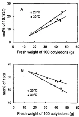
Relationship between cotyledon fresh weight and the content of 16:l(3t) and 16:0—The comparison of Fig. 2
and 4 reveals that the growth curve and the corresponding changes in 16:1(30 displayed about the same pattern. Fig. 5 shows the correlation existing between the cotyledon fresh
0 2 4 6 8 Seedling growth time (days)
Fig. 4 Effect of two growth temperatures on the cotyledon fresh weight during the cotyledon development. Plants were germi-nated and grown at 30 or 20°C. The photoperiod was 12 h and the PFD 100^molm"2s~'. Visible greening appeared after 2 h of
light and progressed rapidly. Data are reported as the mean values ±SD (n = 3).
0 10 20 30 40 50 60
Fresh weight of 100 cotyledons (g)
o 16 : E t U 60 50 40 B o 20°C < > \ . 3 0 ° C i i i i 0 10 20 30 40 50 60
Fresh weight of 100 cotyledons (g)
Fig. 5 Effect of temperature on the relationship between cotyle-don fresh weight and the relative molar content of 16:1(3?) (A) and of 16:0 (B) in phosphatidylglycerol of thylakoid mem-branes. Plants were grown and cotyledons harvested under the conditions shown in Fig. 1. Light intensity was 100/miol m~2
s"1. In A, the equation of the straight lines was y=0.58x—3.52 at
20°C and y=0.44x-1.72 at 30°C. In B, the equations were y= -0.45x + 70.65 at 20°C and y=-0.27x+68.36 at 30°C.
weight and either 16:1(30 or 16:0 level at two growth tem-peratures but at the same low PFD (lOO^umol m~2 s~'). An
increasing linear correlation (r=0.99 at 20°C and 0.98 at 30°C) was found between the fresh weight and 16:1(30 as shown in Fig. 5A. The slope of the straight line was higher for low temperature samples (a = 0.58) than for the high ones (a = 0.44). In contrast, a decreasing linear relationship (r= - 0 . 9 9 at 20°C and - 0 . 9 5 at 30°C) was found between the fresh weight and 16:0 (Fig. 5B). The slope at low tem-perature (a= —0.45) was lower than that at high tempera-ture (a =—0.27).
Discussion
The changes in fatty acid composition are associated with the cotyledon development—In the present
investiga-tion, we show clearly that the changes in the lipid fatty acid composition of thylakoid membranes which are induced by different temperature and light growth conditions take place only during the development of cotyledons but not when these latter have reached their maturity. Indeed, when cotyledons were mature, i.e., when the fresh weight remained constant (after 4 days of growth as shown in
Fig. 4), the composition of their lipid fatty acids was differ-ent and depended on the growth conditions (temperature and light) which preceeded cotyledon maturity. The main differences observed in PG were an increase of 18:3 and 16:1(30 when the growth temperature was lower and an in-crease of 16:1(30 accompanied by a dein-crease in 16:0 when the light intensity of growth was higher (see Table 3). How-ever, after 5 days of growth at 30°C and 100 ftmol m~2 s~'
of light, a transfer of plants to 20°C conditions resulted in no significant changes in the 18:3 content of PG and only slight changes at the level of 16:0 and 16:1(30 (Table 3, compare lines 2 and 5 of PG). Thus, these results indicate that growth temperature- and light-induced changes in fat-ty acid composition of PG can occur only in developing cot-yledons but not in mature ones.
The chloroplast maturity can be estimated by the I6:l(3t) content—During plant growth, the relative molar
proportion of the 4 main lipid classes encountered in thyla-koids (MGDG, DGDG, SQDG and PG) did not undergo any significant changes. These results are in agreement with previous studies (e.g. Bulder et al. 1991). Similarly, varying the growth conditions (e.g. various temperatures and light intensities) did not affect the relative molar content of these lipid classes (cf. Table 2). However, during cotyledon devel-opment, certain glycerolipid fatty acids underwent signifi-cant changes. The prominent ones were an increase in 16:1(30 and a concomitant decrease in 16:0 content in PG (Fig. 2). The 18:3 level in all lipid classes increased also, though to a lesser extent. It is noteworthy that the increase of 16:1(30 and the fresh weight of cotyledons displayed the same pattern.
Thus, as far as the growth of squash cotyledons in rela-tionship with its thylakoid lipid and fatty acid content is concerned, two phases can be distinguished. The first one, corresponding to the development of cotyledons, is charac-terized by an increase of both the fresh weight and 16:1(30 level (see Fig. 2, 4). The second phase, corresponding to the maturity of cotyledons, displayed no change in fresh weight, constant levels of 16:1(30 and of the 16:1(30/16:0 molar ratio. Since the 16:1(30 fatty acid is present only in chloroplast membranes, we propose that the increase of 16:1(30 'S associated with the chloroplast development whereas a constant level of this fatty acid indicates that the chloroplast has reached its maturity. This proposal is substantiated by the increasing linear correlation found between the fresh weight of cotyledons and the 16:1(30 content (see Fig. 5A). It is of interest that a decreasing linear correlation existed between the fresh weight and 16:0. Assuming that a constant level of 16:1(30 is a good criterion for defining the chloroplast maturity, one can estimate the degree of maturity of chloroplasts (probably also of thylakoids) during its development. For instance, when plants were grown under 20°C and 350 ^mol m~~2 s~'
conditions, the thylakoid PG contained 8, 18 and 26 mol%
of 16:1(30 after one, two and three days of growth, which corresponded to 27, 60 and 87% of chloroplast maturity. The fact that the growth temperature and light in-fluence the lipid fatty acid composition only during the development phase but not when the cotyledons (or chloro-plasts) have reached their maturity, underline the impor-tance of defining the development stage of cotyledons (or leaf) which are submitted to temperature and light stress. This is probably why conflicting data about the effect of growth temperature on the lipid fatty acid composition are found in the literature (de la Roche 1979, Chapman et al. 1983, Vigh et al. 1985, Orr and Raison 1987, Krupa et al. 1987, Huner et al. 1989).
High PFD induces an increase in 16:l(3t) fatty acid—
The effect of two light conditions (350 and 100/vmol m"2
s~') during the development of cotyledons resulted in no differences of the relative content of galactolipid and SQDG classes and of their fatty acids. In contrast, at high PFD, the content of 16:l(30PG was higher and the 16:0PG lower than those at low PFD (Table 3). Earlier reports es-tablished a close correlation between the level of 16: l(30PG and the extent of granal stacking during the greening of etiolated leaves. This correlation extends to the accumula-tion of chlorophyll b and LHCII, especially of its oligo-meric form, in developing leaves (Lemoine et al. 1982, Dubacq and Treolieres 1983). Moreover, Hobe et al. (1995), have found that the trimer formation of LHCII is stimulated by adding PG to the reconstitution incubation mixture containing LHCII monomers. Altogether, these data suggest that high PFD enhances the formation of 16:1(3OPG molecular species in order to build up func-tional LHCII trimers.
Low temperatures induce an increase in both 18:3 and 16:l(3t) fatty acids—It is well known that low-temperature
injury can be decreased by a pretreatment of plants at low, non-injurious temperatures. This low temperature acclima-tion is a general phenomenon which has been described in several plants such as maize (Anderson et al. 1994), wheat (de la Roche 1979) and potato (Chen and Li 1980) and results in an increase of the unsaturation degree of mem-brane acyl lipids, and consequently in a higher memmem-brane fluidity (Graham and Patterson 1982 and ref. therein). These two combined phenomena are thought to insure the survival of plants at chilling temperatures whilst a high con-tent of saturated fatty acids in membrane lipids (corsponding to a decrease of the membrane fluidity) may be re-sponsible for chilling injury (Murata et al. 1982). Thus, low temperature conditions appear to result in both an increase of polyunsaturated fatty acids (e.g. 18:3) and a decrease of saturated fatty acids. In agreement with the above concept, our data show that the growth of squash plants at a low temperature compared to a higher one, induced an increase of 18:3 and a decrease of 16:0 levels in the three glycolipid classes (see Table 3). However, these lipid classes contain
such a high level of unsaturated fatty acids that it is highly improbable that they form a solid phase at chilling tempera-ture (Murata and Yamaya 1984). Moreover, even at 30°C growth temperature, the fatty acid composition of MGDG, DGDG and SQDG in thylakoids of squash (a chilling-sensi-tive plant) is very similar (see Table 3) to that of chilling-re-sistant plants such as Arabidopsis (Kunst et al. 1989), pea (Chapman et al. 1983) and spinach (Murata and Yamaya 1984). Thus, despite the fact that growth at 20cC induced a
significant increase of 18:3 in MGDG, DGDG and SQDG in comparison with growth at 30°C, we do not think that these changes contribute to the modulation of chilling sensi-tivity.
In contrast to the glycolipid classes, phosphatidylglyc-erol seems to play a crucial role in chilling sensitivity of plants (Murata et al. 1982, Murata and Yamaya 1984). In squash cotyledons, the PG of thylakoid membranes con-tain 80% of saturated fatty acids [16:0, 18:0 and 16:1(30]. The fatty acid 16:1(30 has a phase transition temperature very similar to that of 16:0 (Bishop and Kenrick 1987). Since the glycerol sn-2 position of PG is occupied only by 16:0 or 16:1(30, while the sn-l position is occupied by either 16:0 or Ci8 fatty acids, PG can form desaturated
mo-lecular species like 16:0/16:0, 16:0/16:1(30, 18:0/16:0 and 18:0/16:1(30 (Xu and Siegenthaler 1996). These species are thought to be responsible for chilling injury (Murata et al. 1982). However, our results show that a lower temperature of growth (compare results at 20 and 30cC in Table 3)
resulted in an increase of the 18:3 level in PG at the expense of 18:1 as well as a concomitant slight decrease of 18:0, but in no change of the sum of 16:0 and 16:1(30 fatty acids. Though this sum remained constant, it is noteworthy that lower growth temperature induced also a decrease in 16:0 and a concomitant increase in 16:1(30- As discussed above, these changes have no major effects on the fluidity of the membrane.
All lipids derived from the prokaryotic pathway con-tain only 18:1 and 16:0 fatty acids when they are synthe-sized in the chloroplast envelope (Browse and Somerville 1991). In PG, the 16:0 present at the sn-2 position is con-verted to 16:1(30 whilst the 18:1 at the sn-l position is desaturated stepwise, first to 18:2, then to 18:3 in the chlo-roplast. In galactolipids, the desaturation occurs either by the prokaryotic pathway in the chloroplast or via the eukaryotic pathway via the endoplasmic reticulum as in squash plants. In PG, the end products of the desaturation process are 18:3 at thesn-l position and 16:1(30 at thesn-2 position whereas in galactolipids the end products are 18:3 at both the sn-l and sn-2 positions. In conclusion, our results show that low temperature conditions of growth in-duce an increase of the end acyl products in both PG and galactolipids.
Possible mechanisms—In order to explain our results,
we have to consider that the effects of temperature on the
composition of fatty acids should depend on the relative rates of the enzyme activities involved in fatty acid synthe-sis and desaturation. Indeed, in safflower seed, Browse and Slack (1983) have found that as the temperature is lowered, the rates of both fatty acid synthesis and desaturation di-minish. However, it is noteworthy that the rate of desatura-tion relative to fatty acid synthesis is increased. Further-more, in plasma membrane of leek cells, Moreau et al. (1994) have observed that the synthesis of unsaturated C)8
-fatty acids is significantly less affected by low temperature than that of C16-fatty acids. The proposed mechanism is
further substantiated by the data of Orr and Raison (1987) who studied the changes in the proportion of the molecular species of PG and SQDG of Nerium oleander L. thylakoids when the plants are grown at two non-injurious tempera-tures. As an example, they found that when the growth tem-perature of this plant is decreased from (45°C, day/35°C, night) to (20°C, day/15CC, night), the major PG molecular
species containing 16:1(30 increase, in particular the PG 18:1/16:1(30 content increases from 25 to 51 mol%. Final-ly, another parameter which has to be considered is that temperature also alters the lipid assembly process and the export of the lipids from their site of synthesis (the inner en-velope membrane) to their final destination (the thylakoid membrane) as found recently (Rawyler et al. 1995).
In conclusion, based on our results, we propose that the mechanism involved in the formation of highly unsatu-rated chloroplast lipids at low temperature is likely to be due to faster fatty acid desaturation compared to fatty acid synthesis reactions. Moreover, the mechanisms involved in the changes of the fatty composition of lipids induced by temperature and light are distinct ones.
We thank Prof. N. Murata (Okazaki, Japan) for providing us generously with seeds of Cucurbita moschata Durch. cv Shirakikuza. This research project was supported by the Swiss National Science Foundation (Grants Nr. 3100.33.693.92 and 31.432.97.95). This work is part of a doctoral program which is be-ing carried out by Y.N. Xu in the Laboratoire de Physiologie veg6tale, Universite de NeuchStel.
References
Anderson, M.D., Prasad, T.K., Martin, B.A. and Stewart, C.R. (1994) Differential gene expression in chilling-acclimated maize seedlings and evidence for the involvement of abscisic acid in chilling tolerance. Plant
Physiol. 105: 331-339.
Bishop, D.G. and Kenrick, J.R. (1987) Thermal properties of 1-hexadeca-noyl-2-fnjn.s-3-hexadecenoyl phosphatidylglycerol. Phytochemistry 26: 3065-3067.
Browse, J. and Slack, C.R. (1983) The effects of temperature and oxygen on the rates of fatty acid synthesis and oleate desaturation in safflower
(Carthamus tinctorius) seed. Biochim. Biophys. Acta 753: 145-152.
Browse, J.A. and Somerville, C.R. (1991) Glycerolipid synthesis: biochem-istry and regulation. Annu. Rev. Plant Mol. Biol. 42: 467-506. Bruinsma, J. (1961) A comment on the spectrophotometric determination
of chlorophyll. Biochim. Biophys. Acta 53: 576-578.
Growth temperature and lipid composition of cucumber genotypes differ-ing in adaptation to low energy conditions. J. Plant Physiol. 138: 655-660.
Chapman, D.J., De-Felice, J. and Barber, J. (1983) Influence of winter and summer growth conditions on leaf membrane lipids of Pisum
sativum L. Planta 157: 218-223.
Chen, H.-H. and Li, P.H. (1980) Characteristics of cold acclimation and deacclimation in tuber-bearing Solarium species. Plant Physiol. 65:
1146-1148.
De La Roche, A.I. (1979) Increase in linolenic acid is not a prerequisite for development of freezing tolerance in wheat. Plant Physiol. 63: 5-8. Dubacq, J.P. and Trdmolieres, A. (1983) Occurrence and function of
phos-phatidylglycerol containing J3-frans-hexadecenoic acid in
photosynthet-ic lamellae. Physiol. Vig. 21: 293-312.
Graham, D. and Patterson, B.D. (1982) Responses of plants to low, non freezing temperatures: proteins, metabolism, and acclimation. Annu.
Rev. Plant Physiol. 33: 347-372.
Hobe, S., Kuttkat, A., Foster, R. and Paulsen, H. (1995) Assembly of trimeric light-harvesting chlorophyll A/B complex in vitro. In Photosyn-thesis: from Light to Biosphere. Edited by Mathis, P. Vol. I. pp. 47-52. Kluwer Academic Publishers.
Huner, N.P.A., Williams, J.P., Maissan, E.E., Myscich, E.G., Krol, M., Laroche, A. and Singh, J. (1989) Low temperature-induced decrease in
trans-A 3-hexadecenoic acid content is correlated with freezing tolerance
in cereals. Plant Physiol. 89: 144-150.
Kenrick, J.R. and Bishop, D.G. (1986) The fatty acid composition of phos-phatidylglycerol and sulfoquinovosyldiacylglycerol of higher plants in relation to chilling sensitivity. Plant Physiol. 81: 946-949.
Kodama, H., Horiguchi, G., Nishiuchi, T., Nishimura, M. and Iba, K. (1995) Fatty acid desaturation during chilling acclimation is one of the factors involved in conferring low-temperature tolerance to young tobac-co leaves. Plant Physiol. 107: 1177-1185.
Krupa, Z., Huner, N.P.A., Williams, J.P., Maissan, E. and James, D.R. (1987) Development at cold-hardening temperatures. The structure and composition of purified rye light harvesting complex II. Plant Physiol. 84: 19-24.
Kunst, L., Browse, J. and Somerville, C. (1989) A mutant of Arabidopsis deficient in desaturation of palmitic acid in leaf lipids. Plant Physiol. 90: 943-947.
Lemoine, Y., Dubacq, J.P. and Zabulon, G. (1982) Changes in light-har-vesting capacities and Zl3-/ra/i5-hexadecenoic acid content in dark- and
light-grown Picea abies. Physiol. Vig. 20: 487-503.
Levitt, J. (1980) Responses of Plants to Environmental Stresses: Vol. 1.
Chilling, Freezing and High Temperature Stresses. 2nd ed. pp. 497.
Academic Press, New York.
Lyons, J.M. (1973) Chilling injury in plants. Annu. Rev. Plant Physiol. 24: 445-466.
Moreau, P., Sturbois, B., Morr6, D.J. and Cassagne, C. (1994) Effect of low temperatures on the transfer of phospholipids with various acyl-chain lengths to the plasma membrane of leek cells. Biochim. Biophys.
Ada 1194: 239-246.
Murata, N. (1983) Molecular species composition of phosphatidylglycerols from chilling-sensitive and chilling-resistant plants. Plant Cell Physiol. 24: 81-86.
Murata, N., Ishizaki-Nishizawa, O., Higashi, S., Hayashi, H., Tasaka, Y. and Nishida, I. (1992) Genetically engineered alteration in the chilling sensitivity of plants. Nature 356: 710-713.
Murata, N. and Nishida, I. (1990) Lipids in relation to chilling sensitivity of plants. In Chilling Injury of Horticultural Crops. Edited by Wang, C.Y. pp. 181-199. CRC Press, Boca Raton, FL.
Murata, N., Sato, N., Takahashi, N. and Hamazaki, Y. (1982) Composi-tions and positional distribuComposi-tions of fatty acids in phospholipids from leaves of chilling-sensitive and chilling-resistant plants. Plant Cell
Physiol. 23: 1071-1079.
Murata, N. and Yamaya, J. (1984) Temperature-dependent phase behavior of phosphatidylglycerols from chilling-sensitive and chilling-resistant plants. Plant Physiol. 74: 1016-1024.
Orr, G.R. and Raison, J.K. (1987) Compositional and thermal properties of thylakoid polar lipids of Nerium oleander L. in relation to chilling sen-sitivity. Plant Physiol. 84: 88-92.
Raison, J.K. and Wright, L.C. (1983) Thermal phase transitions in the polar lipids of plant membranes. Their induction by disaturated phos-pholipids and their possible relation to chilling injury. Biochim.
Bio-phys. Ada 731: 69-78.
Rawyler, A., Meylan Bettex, M. and Siegenthaler, P.A. (1995) (Galac-to)lipid export from envelope to thylakoid membranes in intact chloro-plasts. II. A general process with a key role for the envelope in the estab-lishment of lipid asymmetry in thylakoid membranes. Biochim.
Biophys. Ada 1233: 123-133.
Roughan, P.G. (1985) Phosphatidylglycerol and chilling sensitivity in plants. Plant Physiol. 77: 740-746.
Siegenthaler, P.A., Rawyler, A. and Smutny, J. (1989) The phospholipid population which sustains the uncoupled non-cyclic electron flow activ-ity is localized in the inner monolayer of the thylakoid membrane.
Bio-chim. Biophys. Acta91S: 104-111.
Somerville, C. (1995) Direct tests of the role of membrane lipid composi-tion in low-temperature-induced photoinhibicomposi-tion and chilling sensitivity in plants and cyanobacteria. Proc. Natl. Acad. Sci. USA 92: 6215-6218. Vigh, L., Horvath, I., van Hasselt, P.R. and Kuiper, P.J.C. (1985) Effect of frost hardening on lipid and fatty acid composition of chloroplast thy-lakoid membranes in two wheat varieties of contrasting hardiness. Plant
Physiol. 79: 756-759.
Wu, J. and Browse, J. (1995) Elevated levels of high-melting-point phos-phatidylglycerols do not induce chilling sensitivity in an Arabidopsis mu-tant. Plant Cell!: 17-27.
Xu, Y.N., Rawyler, A. and Siegenthaler, P.A. (1992) Influence of light and temperature on the fatty acid composition of phosphatidylglycerol in squash cotyledons. Experientia 48: A12.
Xu, Y.N. and Siegenthaler, P.A. (1996) Phosphatidylglycerol molecular species of photosynthetic membranes analyzed by HPLC: theoretical considerations. Lipids 31: 223-229.