HAL Id: hal-01636802
https://hal.uca.fr/hal-01636802
Submitted on 4 Mar 2021
HAL is a multi-disciplinary open access
archive for the deposit and dissemination of
sci-entific research documents, whether they are
pub-lished or not. The documents may come from
teaching and research institutions in France or
abroad, or from public or private research centers.
L’archive ouverte pluridisciplinaire HAL, est
destinée au dépôt et à la diffusion de documents
scientifiques de niveau recherche, publiés ou non,
émanant des établissements d’enseignement et de
recherche français ou étrangers, des laboratoires
publics ou privés.
Bioactive glass coating on gelatin scaffolds at ambient
temperature: easy route to make polymer scaffolds
become bioactive
Jonathan Lao, Xavier Dieudonné, Mhammed Benbakkar, Edouard Jallot
To cite this version:
Jonathan Lao, Xavier Dieudonné, Mhammed Benbakkar, Edouard Jallot. Bioactive glass coating on
gelatin scaffolds at ambient temperature: easy route to make polymer scaffolds become bioactive.
Journal of Materials Science, Springer Verlag, 2017, 52 (15), pp.9129-9139.
�10.1007/s10853-017-0781-7�. �hal-01636802�
Bioactive glass coating on gelatin scaffolds at ambient
temperature: easy route to make polymer scaffolds
become bioactive
Jonathan Lao1,* , Xavier Dieudonne´1, Mhammed Benbakkar2, and E´ douard Jallot1 1
CNRS/IN2P3, Laboratoire de Physique Corpusculaire, Clermont Université, Université Clermont Auvergne, BP 80026, 63171 Aubière Cedex, France
2Laboratoire Magmas et Volcans, CNRS-OPGC-IRD, Université Clermont Auvergne, Clermont-Ferrand, France
Increasing the bioactivity of polymeric materials used for bone repair is a con-cern that can be achieved by loading growth factors or using in vitro tissue engineering approach. However, these techniques may have to address regu-latory issues as the implants are shifted from the medical device class to the more constraining drug delivery systems. Alternatively, implants can be coated with bioceramics to achieve bioactivity, but existing coating processes can hardly be applied to polymers because they usually involve thermal treatments or sintering. Here we report an efficient way of coating a bioactive glass phase onto a complex polymeric substrate, namely gelatin scaffolds with controlled spherical porosity, at ambient temperature through a dip-coating process. A multiscale analysis of the bioactive glass-coated gelatin scaffolds properties has been carried out. Homogeneous and remarkably uniform layer of SiO2–CaO bioactive glass is obtained. The bioactive glass
coating exhibits a very high and fast apatite-forming ability, with full mineralization of the coating being achieved in less than 24 h contact with body fluids. Importantly, the mineral-ization takes place homogeneously throughout the scaffold while the remark-able uniformity and thickness regularity of the coating are preserved. These features should enhance the in vivo behaviour of polymer scaffolds and make reconsider the interest of non-bioactive polymers for tissue engineering.
Introduction
Accelerating bone healing at early implantation time is one of the main concerns when developing new biomaterials. [1]. As an implant first interacts through
surface reactions, adequate modifications of its sur-face can enhance or introduce new implant proper-ties regarding, e.g. bioactivity, osteoconduction, biocompatibility and mechanical response [2]. A large number of techniques have been developed to
improve the surface compatibility of medical devices. Among them, surface coating with bioceramics is an efficient way of conferring bioactivity to the implant, i.e. the ability to bond with host tissues. For example, a biocompatible calcium phosphate coating can be applied to the stem of hip endoprostheses or to dental root implants, in order to support osseointegration [3,4]. Due to its similarity with the inorganic content of bone, this coating will be an adequate site for the adhesion and further proliferation of bone cells and will finally result in a strong and lasting bond between the implant and surrounding host tissues [3–5]. Since bioactive glasses (BG) are known to demonstrate the highest bioactivity among synthetic materials [6], it can be of great benefit to take advantage of their superior properties to coat implants. Metallic prostheses have been coated with BG with the aim of improving the implants stability by bonding them to host tissues [7–9] and preventing fibrous encapsulation usually induced by metal implants. For instance titanium implants were dip-coated with a SiO2–CaO–Na2O–P2O5-K2O–MgO–B2O
glass before implantation in rabbit femurs and showed superior bone formation in vivo compared to non-coated implants [10]. A clinical trial demon-strated that glass-coated titanium implants behaved as well as hydroxyapatite-coated implants after 1-year implantation [11].
However, there is only a scarce literature about bioactive coating—and in particular BG coating—on polymer matrices. This is a challenging task since thermal treatments cannot be handled, to avoid the polymer thermal degradation. Kokubo, Tanahashi et al. [12–15] put a polymer substrate in direct contact with SiO2-CaO BG particles soaked in simulated
body fluid (SBF). This allowed the formation of apa-tite nuclei at the polymer surface, that were further grown into a dense, uniform apatite layer by immersing the polymer again into another solution with ion concentrations 1.5 times those of SBF. In addition to being slow (about 1 week required to obtain a uniform coating), this 2-steps process can hardly be used to uniformly coat the inner walls of porous matrices or complex shapes like scaffolds, since the apatite nuclei are only formed on the material surface that is directly facing the BG parti-cles. Miyaji et al. [16], followed by Oliveira et al. [17,18], soaked polymer matrices into a sodium sili-cate gel in order to obtain a SiO2-Na2O glass coating.
They demonstrated this coating to be an effective
initiator of apatite nucleation on polymer substrates with complex shapes like scaffolds [17]. However, conducting the process remains heavy, since an incubation period of 7 days in SBF is first needed to induce the formation of apatite nuclei, followed by 7–14 days soaking in concentrated SBF solutions (1.5 9 to 2 x SBF ionic concentration) for efficient growth of the apatite layer. Moreover, there is only little data about the in vivo behaviour and corrosion of sodium silicate [19]. There have been finally very few attempts to coat BG directly onto polymeric materials. Leach et al. [20], Day et al. [21] and Li et al. [22,23] made slurries consisting of micron-sized BG particles to coat PLGA, PGA or PET, respectively. However, they reported either a non-uniform coating to be obtained, or a loss of porosity inside the mate-rial [21], or insufficient concentrations of BG particles in the coating unable to induce a robust osteogenic action [20]. Another drawback of this technique is that the BG particles are only deposited onto the polymeric substrate and are likely to be released once in contact with body fluids. Stamboulis et al. [24], Niiranen et al. [25] proposed a variant where the BG particles were ‘‘implanted’’ into a Vicryl polymer matrix by pressing the coating using a uniaxial press at 2–160 MPa. Of course, it is unconceivable to apply this method to polymer scaffolds since they would be flattened.
Here we propose a convenient way to coat polymer scaffolds with BG. BG usually need to be sintered, either as a result of their method of production, which derives from the fusion process or the sol–gel process, or for the coating to adhere [6]; but here our process is fully conducted at ambient temperature. Indeed using calcium alkoxide as a calcium precursor in the sol–gel route makes it possible to obtain silicate glasses with calcium ions well incorporated into the inorganic network without the need of thermal treatments otherwise necessary [26–29]. The polymer scaffolds here coated were based on gelatin, since gelatin is obtained from collagen and, therefore, naturally contains biologically relevant functional groups [30]. It shows good cell viability without any antigenicity and has long been used in pharmaceu-tics, wound dressing and adhesives [31–34]. Exten-sive in vivo data on the suitability of gelatin-based scaffolds for bone reconstruction are available [35–39]. In this study bovine gelatin scaffolds with controlled porosity were fabricated using the micro-sphere leaching technique [40, 41]; they were then
dip-coated with a BG alkoxide sol. The physico-chemical properties and potential towards bone mineral formation of these BG-coated gelatin scaf-folds are investigated.
Materials and methods
Gelatin scaffolds synthesis
3-D interconnected macroporous gelatin structures are obtained using the microspheres-leaching tech-nique [40, 42]. A 12.7 wt% gelatin (type B, 225 g bloom number, Aldrich) aqueous solution was first prepared in a thermostated bath at 37 °C. After full completion of gelatin dissolution, the gelatin sol was infiltrated into a compact stack of PMMA micro-spheres (100–300 lm diameter, Kisker Biotech) in cylindrical polyethylene moulds and centrifugated at 6000 rpm. Gelation and ageing of the blend was performed at room temperature for 24 h. The obtained cylinders were immersed in acetone for 24 h to dissolve the PMMA porogen spheres. This opera-tion was renewed two times. Macroporous gelatin scaffolds with controlled porosity were obtained and further dried in an oven at 40 °C for 24 h. Finally, the gelatin scaffolds were crosslinked in a 1 wt% glu-taraldehyde/ethanol solution for 24 h. The reticula-tion of the gelatin was indeed required to prevent its dissolution in the BG sol during the dip-coating process, and to prevent premature degradation of the scaffolds after implantation or when interacting with body fluids. The crosslinked gelatin scaffolds were rinsed in ethanol and dried at room temperature.
SiO
2-CaO BG sol–gel synthesis and
dip-coating of gelatin scaffolds
Hydrolysis of tetraethylorthosilicate (TEOS, Aldrich, 99% purity) was performed in ethanol (absolute 99.8%, Aldrich) containing 2 M HCl (obtained from 37% fuming, Aldrich), following a volume ratio EtOH:HCl = 6:1 and molar ratio EtOH:TEOS = 6:1. In parallel, calcium ethoxide (ABCR) was dispersed in absolute ethanol. Then the two solutions were mixed in stoichiometric proportions, in order to obtain a 75–25 wt% SiO2-CaO glass at a 12.7 wt%
concentration in the sol. A translucent and yellowish sol was obtained and left for condensation for a few hours.
The cylindrical gelatin scaffolds were then simply dipped into the BG sol for 1 min and left for drying for 15 min. These operations were repeated again 1 time.
SEM observations
The scaffolds were carbon-coated using a carbon thread prior to analysis. Observations were con-ducted on a Hirox SH-3000 mini-SEM operating at 10 kV voltage.
Porosity calculation
Pore diameters and interconnections were extracted from SEM pictures thanks to the Image J software. This method of measurement is here preferable to traditional mercury intrusion porosimetry which is limited to the characterization of pores under 250 lm [43]. Total porosity of the scaffolds was deduced from apparent density of cylindrical scaffolds of measured weights and dimensions and from gas pycnometry measurements (1.5 g/cm3 scaffold skeletal density) using the formula: %porosity = (1 - dapparent/
dskeletal).
Apatite-forming ability test in SBF
The ISO-23317 standard procedure was followed. Briefly, c- SBF2 [44], a protein-free solution of inor-ganic composition close to human blood plasma, was prepared following recommendations of Bohner et al. [45]. Scaffolds were immersed in SBF at a 1 mg/mL ratio for up to 7 days at a constant temperature of 37 °C. After interaction, aliquots of the solution are used for determination of the fluids composition by ICP-AES, while the scaffolds are carefully rinsed with pure ethanol and dried to avoid further mineraliza-tion reacmineraliza-tions.
TEM observations
Prior to observation, the scaffolds were embedded in resin (AGAR, Essex, England). 100-nm ultrathin cross sections of materials were cut using a LEICA EM UC6 with diamond knives. A Phillips CM 20 microscope (LaB6thermoelectronic gun) operating at 200 kV was
used to study the microstructure and morphology of hybrid scaffolds before and after soaking in SBF. The images were recorded with a Keenview CCD camera
with 18.67 lm pixel size and processed with the analysis software.
FTIR analysis
The FTIR was conducted on a Nicolet spectrometer 380. Prior to analysis the materials were grinded into a fine powder and then mixed with KBr (Aldrich, IR grade) at a 3 wt% concentration. They are finally pressed into pellets that are ready for FTIR analysis.
PIXE ion beam analysis
Particle-Induced X-ray Emission (PIXE) quantitative microanalysis is very similar to SEM–EDS or electron
microprobe analysis, but provides a deeply increased sensitivity due to limited bremsstrahlung back-ground radiation of incident heavy charged particles. It allows visualizing the chemical changes occurring inside BG-coated scaffolds during interaction with SBF. After interaction with SBF, the scaffolds are dried and embedded in resin (AGAR, Essex, Eng-land). Cross sections of materials (40 lm thick) were cut using a LEICA RM 2145 microtome. PIXE microanalysis of the cross sections was carried out at the AIFIRA platform (CENBG, France) using a 3-MeV incident proton beam (beam diameter of 1 lm). An 80 mm2Si(Li) detector (equipped with a 12-lm-thick beryllium window and an aluminium funny filter
(a) (b) (c) (d) (e) (f)
0
Max
Si
Ca
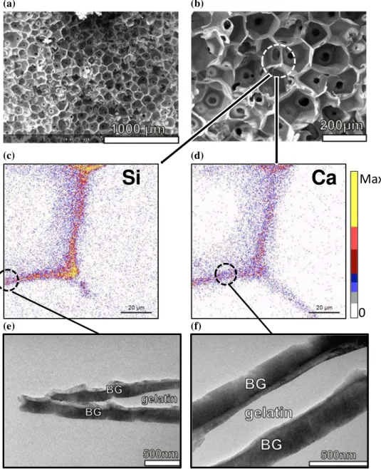
Figure 1 a–b SEM micrographs of bioactive glass-coated gelatin scaffold, c–d PIXE chemical maps showing the cross-sectional distribution of silicon and calcium inside the scaffold.e– f TEM magnification of previous images revealing the morphology of BG coated onto the gelatin struts.
with a tiny hole of 2 mm) orientated at 135° with respect to the incident beam axis was used for X-ray detection,. Quantification was done using the Gupixwin software after calibration against NIST 620 (soda-lime glass) standard reference material.
ICP-AES
Five 0 to 50 ppm solutions containing the elements to analyse (P, Si, Mg, Ca) have been prepared for cali-bration. A ULTIMA-C spectrometer (Horiba scien-tific, Jobin–Yvon) was used. This instrument combines two spectrometers to measure emission lines from elements excited in a single plasma torch: one polychromator and one scanning monochroma-tor. The polychromator was used for the simultane-ous measurement of emission lines from Si, Ca and Mg. The scanning high-resolution monochromator was used for sequential determination of emission line from P. The ICP-AES operating conditions were the following: incident power 1.1 kW; reflected power \15 W; plasma gas flow rate 16 l/min; permanent sheath gas flow rate 0.2 l/min; carrier gas flow rate 0.8 l/min; and solution uptake 0.9 l/min. The ana-lytical lines used were 213.618 nm (P), 288.158 (Si), 279.553 (Mg) and 334.940 (Ca).
Compression tests
Mechanical properties of the scaffolds were mea-sured under compression on cylindrical samples (diameter = height = 10 mm) using a UTS testing machine, equipped with a 50 kN-load cell and cir-cular plates, at a crosshead speed of 0.5 mm/min.
Results and discussion
To elaborate a BG coating without thermal treatment, it is necessary to use a 100% alkoxide route, which implies using a calcium alkoxide as calcium precur-sor [29]. Any other calcium source, such as calcium salts, involve thermal stabilization at high tempera-ture ([400 °C) required to make calcium enter the silicate network [26, 46]. On the contrary, calcium alkoxides can readily be incorporated into the silicate network at room temperature, as a result of the hydrolysis/condensation reactions. Yet the known instability of calcium alkoxides has restricted their use due to the difficult processability of obtained sols [47]. To limit this instability, we previously reported that limiting the amount of water in the sol was the key as calcium alkoxides are very sensitive to water hydrolysis [48]. In our synthesis, tetraethylorthosili-cate (TEOS) is first hydrolysed in a slightly acidified alcoholic solution and then calcium ethoxide Ca(OEt)2is added.
Figure1 shows the multiscale characterization of as obtained BG-coated gelatin scaffolds, from the macroscopic down to the submicron scale. On SEM micrographs (Fig. 1a, b) it can be seen that the scaf-folds own a well-ordered porosity with highly interconnected pores. The diameters of pores lie in the 100–200 lm range, with interconnections 20–50 lm. The scaffold walls are very thin, with struts less than a few microns thick. Figure1c, d displays the chemical distribution of silicon and cal-cium inside a pore strut. Si and Ca are homoge-neously distributed, demonstrating both the uniformity of the BG coating over the gelatin sub-strate and the intrinsic homogeneity of the SiO2–CaO
glass. Figure1e, f is magnification of the struts as observed in TEM. Here enough contrast is provided to allow distinguishing between the organic gelatin substrate and the SiO2–CaO BG coating. The dark
regions correspond to areas of higher density or higher average atomic number, leading to high absorption of the electron beam, such as the inorganic SiO2–CaO BG coating. On the contrary, gelatin
domains appear as bright areas since the organic matrix is associated to both lower density and Z atomic number. TEM observation of ultrathin cross sections of BG-coated gelatin scaffolds reveals our dip-coating process is successful to yield a uniform glass layer around 200 nm thickness that surrounds the gelatin struts of the scaffold. The BG layer is
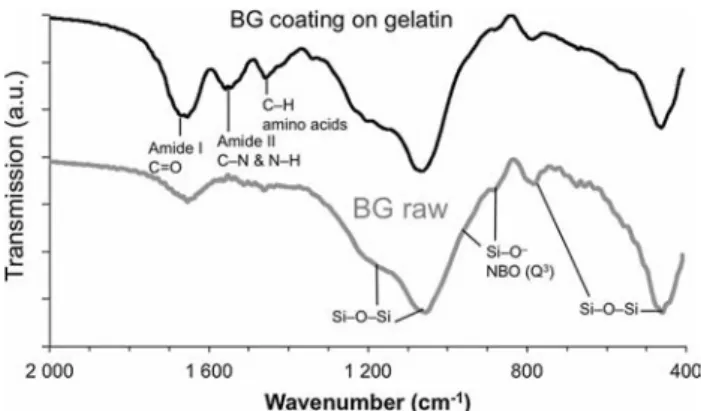
Figure 2 FTIR spectra of SiO2–CaO bioactive glass coated on the
dense, as expected from the acid-catalysed sol–gel route we employed, which leads to polymeric and dense silicate gels [49].
The atomic structure of SiO2–CaO BG coatings was
investigated through FTIR spectroscopy and com-pared to raw SiO2–CaO glass derived from the same
synthesis but not coated onto gelatin scaffolds. Fig-ure2 evidences the characteristic absorption bands attributed to the transverse optical (TO) modes of siloxane Si–O–Si bridges: at *450 cm-1 a low fre-quency mode is assigned to the TO rocking motions of the oxygen bridging two adjacent Si atoms [50]; near 800 cm-1is observed (weak band) the bending
Si–O vibration characteristic of ring structures in the glass matrix [51–53]. TO antisymmetric Si–O-Si stretching modes, resulting from the motion of the O atom back and forth along a line parallel to the Si–Si axis [54], are visible at 1050 cm-1(intense shoulder)
and *1170 cm-1.
Importantly, the shoulder located around 940 cm-1
and the peak at 890 cm-1are assigned to the Si–O
-non-bridging oxygen (NBO)16-17 stretching mode with one NBO involved per SiO4 tetrahedra (Q3
groups, calling for 3 bridging O and 1 NBO per SiO4
tetrahedra). The presence of NBO can result from calcium ions disrupting the siloxane bridges, as a
result of their successful incorporation into the sili-cate network. It can also result from the presence of silanols SiOH, which yields Q3groups as well. In a previous work [48], we had characterized the SiO2–
CaO BG through 29Si–1H cross-polarization (CP) MAS (magic-angle spinning) NMR experiments and we demonstrated the incorporation of Ca into the silicate network, being able to distinguish between the contribution of QH3 and QCa3 units. Since the same
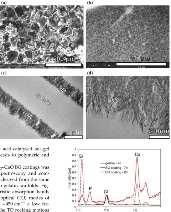
Figure 3 Images of BG-coated gelatin scaffolds after 7 days soaking in simulated body fluid,a–b SEM, and c– d TEM observations
highlighting the formation of a uniform bone-like
hydroxyapatite (HA) layer.
Figure 4 EDXS spectra showing the elemental composition of non-coated gelatin scaffold and BG-coated gelatin scaffold after 7 days of immersion in SBF. The EDXS spectrum of BG-coated gelatin scaffold before immersion is also shown.
SiO2-CaO BG is here considered, and because from
Fig.2 the FTIR fingerprint of silicate networks is identical for the SiO2-CaO BG and the SiO2–CaO
coating, we assume we can interpret the shoulder at 940 cm-1and the band at 890 cm-1as an evidence of
Ca incorporation.
Moreover, gelatin yielded 3 characteristic bands between 1400 and 1700 cm-1. The 1540 cm-1peak is associated with amide II absorption arising from N– H bending and C–N stretching vibrations [55]. The band at 1440 cm-1is due to the gelatin amino acids (C–H groups) [56]. The 1650 cm-1band attribution is
more ambiguous since it results both from the SiOH bonded with molecularly adsorbed water
Si Ca P 0d 6h 1d 7d 0 Max
Figure 5 PIXE chemical maps of silicon, calcium and phosphorus inside cross sections of SiO2–CaO-coated
gelatin scaffolds, as a function of increasing interaction time with simulated body fluid. Scale bar is 100 lm. 0.0% 0.2% 0.4% 0.6% 0.8% 1.0% 1.2% 1.4% 1.6% 1.8% 2.0% 0% 10% 20% 30% 40% 50% 0 24 48 72 96 120 144 168 Mg wt.% Si, Ca, P wt.% Time (h) Ca P Si Mg
Figure 6 Evolution of Si, Ca, P, Mg concentrations in the mineralized areas of the scaffolds with increasing time of interaction with SBF. Starting composition of the BG coating is 75 wt% SiO2—25 wt% CaO.
(*1630 cm-1) already present on the raw BG spectra
[54], and from the C=O stretch vibrations (*1650 cm-1) of the peptide linkages of gelatin’s
amide I [57–59], but its intensity is clearly increased on the BG-coated gelatin spectra compared to raw BG.
The apatite-forming ability of BG-coated gelatin scaffolds was investigated at different scales as visi-ble on Figs.3,4,5 and6. SEM observations (Fig.3a) show the macroscopic porous structure is preserved even after 7 days soaking in SBF. Higher magnifica-tion (Fig.3b) highlights the presence of small pre-cipitates uniformly spread over the scaffold’s surface. The ability to induce the homogeneous nucleation of apatite nuclei over the substrate is key for the coating to confer optimum biocompatibility. Images of cross sections of the coating as observed in TEM show it has endured deep structural and morphological changes. As visible on Fig.3c, d, the coating now consists of nanocrystalline needle-like aggregates typical of apatites. Importantly, the coating still pre-sents a uniform thickness all along the gelatin scaf-fold walls, but its thickness has increased up to *500 nm as a result of the apatite crystal growth. Pure crosslinked gelatin scaffolds, i.e. without BG coating, were also immersed in SBF for comparison. SEM observations did not show the formation of any precipitate after 7 days soaking. Figure4 is an EDXS measurement of the elemental composition of non-coated (pure gelatin) scaffolds compared to BG-coated gelatin scaffolds. Non-BG-coated scaffolds do not show any change in composition, apart from the deposition of chlorides coming from NaCl salts dis-solved in the SBF. On the contrary, BG-coated gelatin scaffolds composition is changed from calcium sili-cate to calcium phosphate after 7 days soaking in SBF. Therefore, this composition change can be unambiguously attributed to the presence of BG as a bioactive layer coated over the gelatin scaffold.
Figures5 and 6 investigate in detail the chemical changes endured by the BG-coated gelatin scaffolds during their progressive mineralization as observed through PIXE quantitative chemical imaging. The homogeneity of mineralization throughout the scaf-fold is well deduced from Fig.5, demonstrating the efficient circulation of fluids due to the good inter-connected structure of the BG-coated scaffolds. The BG coating reacts extremely rapidly; right after 6 h, the SiO2–CaO original phase is changed into a
cal-cium phosphate whose composition (see Fig.5)
stabilizes after 24 h interaction with SBF. The mea-sured Ca/P weight ratio is very close to the 2.2 value (equivalent to 1.67 atomic ratio) of stoichiometric apatite. Interestingly, small amounts (*1 wt%) of magnesium are found to be substituted for calcium in the apatite crystals. As Mg2?is known for stimulation of bone formation and promotion of cellular adhesion and stability due to interaction with integrins [60–62], its incorporation into the coating layer is an attractive feature. From Figs.6and7, which show the evolution of ion concentrations in the coating layer and in the SBF, respectively, we observe the dissolution of the silicate network is very fast, being achieved in the first 24 h of interaction with SBF. Phosphate ions are depleted from SBF, and all concentrations reach equilibrium right after 24 h interaction.
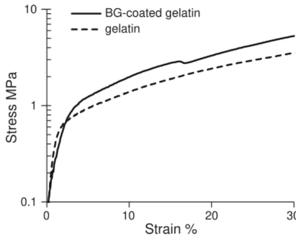
Finally, the mechanical properties of BG-coated gelatin scaffolds were tested in compression. Figure8
7.40 7.50 7.60 7.70 0 20 40 60 80 100 120 140 160 0 24 48 72 96 120 144 168 pH SBF concentration (ppm) Time (h) Ca P Si pH
Figure 7 Evolution of SBF composition as determined by ICP-AES measurements, with increasing time of inetraction with BG-coated gelatin scaffodls.
0.1 1 10 0 10 20 30 Stress MPa Strain % BG-coated gelatin gelatin
Figure 8 Stress–strain compression curves compared for gelatin scaffolds with 80% porosity: BG-coated versus non-coated gelatin scaffolds.
reports the stress–strain curves compared for gelatin scaffolds coated with BG vs non-coated, both having the same 80% porosity. A slight increase in the yield strength is observed for BG-coated scaffolds. The yield strength is near 1 MPa in both cases, lying in the low range of reported values for trabecular bone [63, 64]. Because only a thin BG coating layer of 200 nm has been applied here, the mechanical prop-erties are mainly dependent upon the nature of the polymer chosen to build the scaffold, the toughness being here tightly bound by the gelatin properties.
Conclusion
Coating a substrate with a bioceramics is a well-known and efficient way of making it become bioactive. However, only very few methods can be applied to polymer scaffolds due to their limited processability post-synthesis. We have employed here a dip-coating process involving a BG sol obtained from an alkoxide route. It was successful to yield a bioactive coating with remarkable homogeneity without the need of thermal treatments, preserving the polymer integrity. We believe it can be applied to any other complex shape or substrate. Gelatin scaffolds are coated with layers of BG with a noticeably uniform 200 nm thickness. The coating is highly reactive towards SBF and demon-strates a quick apatite-forming ability. Importantly, mineralization takes place homogeneously through-out the scaffold and the coating, while being chemi-cally changed into an apatite phase, keeps its remarkable uniformity and thickness regularity. A slight increase in the mechanical response to com-pression tests is also observed. These features should enhance the in vivo behaviour of polymer scaffolds and make reconsider the interest of polymers that are non-bioactive for tissue engineering, as both the glass dissolution products from the coating and the newly formed apatite layer help attracting, recruiting and stimulating bone cells. Alternatively, this method can be considered as an efficient way of obtaining a uni-form apatite coating on complex polymer shapes at ambient temperature.
Acknowledgements
The Conseil Re´gional d’Auvergne is acknowledged for funding (‘‘New Researcher’’ Grant). The Centre
d’Etudes Nucle´aires de Bordeaux-Gradignan and the AIFIRA staff are acknowledged for allowing the PIXE experiments and for technical support.
References
[1] Ramakrishna S, Mayer J, Wintermantel E, Leong KW (2001) Biomedical applications of polymer-composite materials: a review. Compos Sci Technol 61(9):1189–1224
[2] Surmenev RA, Surmeneva MA, Ivanova AA (2014) Sig-nificance of calcium phosphate coatings for the enhancement of new bone osteogenesis: a review. Acta Biomater 10(2):557–579
[3] Heimann RB (2016) The challenge and promise of low-temperature bioceramic coatings: an editorial. Surf Coat Technol 301:1–5
[4] Creugers N (2002) The survival of hydroxyapatite-coated implants is questioned. Evid Based Dent 3(3):77–78 [5] Coathup MJ, Blackburn J, Goodship AE, Cunningham JL,
Smith T, Blunn GW (2005) Role of hydroxyapatite coating in resisting wear particle migration and osteolysis around acetabular components. Biomaterials 26(19):4161–4169 [6] Jones JR (2013) Review of bioactive glass: from Hench to
hybrids. Acta Biomater 9(1):4457–4486
[7] Gomez-Vega JM, Saiz E, Tomsia AP, Oku T, Suganuma K, Marshall GW, Marshall SJ (2000) Novel bioactive func-tionally graded coatings on Ti6Al4V. Adv Mater 12(12):894–898
[8] Fathi MH, Doost A (2008) Mohammadi, preparation and characterization of sol–gel bioactive glass coating for improvement of biocompatibility of human body implant. Mater Sci Eng A 474(1–2):128–133
[9] Garcia C, Cere´ S, Dura¨n A (2004) Bioactive coatings pre-pared by sol–gel on stainless steel 316L. J Non Cryst Solids 348:218–224
[10] Moritz N, Rossi S, Vedel E, Tirri T, Yla¨nen H, Aro H, Na¨rhi T (2004) Implants coated with bioactive glass by CO2-laser, an in vivo study. J Mater Sci Mater Med 15(7):795–802 [11] Mistry S, Kundu D, Datta S, Basu D (2011) Comparison of
bioactive glass coated and hydroxyapatite coated titanium dental implants in the human jaw bone. Aust Dent J 56(1):68–75
[12] Kokubo T, Kim HM, Miyaji F, Takadama H, Miyazaki T (1999) Ceramic-metal and ceramic-polymer composites prepared by a biomimetic process. Compos A 30(4):405–409
[13] Kokubo T, Kim H-M, Kawashita M (2003) Novel bioactive materials with different mechanical properties. Biomaterials 24(13):2161–2175
[14] Kokubo T (1996) Formation of biologically active bone-like apatite on metals and polymers by a biomimetic process. Thermochim Acta 280–281:479–490
[15] Tanahashi M, Yao T, Kokubo T, Minoda M, Miyamoto T, Naka-mura T, Yamamuro T (1994) Apatite coating on organic polymers by a biomimetic process. J Am Ceram Soc 77(11):2805–2808 [16] Miyaji F, Kim H-M, Handa S, Kokubo T, Nakamura T
(1999) Bonelike apatite coating on organic polymers: novel nucleation process using sodium silicate solution. Biomate-rials 20(10):913–919
[17] Oliveira AL, Costa SA, Sousa RA, Reis RL (2009) Nucle-ation and growth of biomimetic apatite layers on 3D plotted biodegradable polymeric scaffolds: effect of static and dynamic coating conditions. Acta Biomater 5(5):1626–1638 [18] Oliveira AL, Malafaya PB, Reis RL (2003) Sodium silicate gel as a precursor for the in vitro nucleation and growth of a bone-like apatite coating in compact and porous polymeric structures. Biomaterials 24(15):2575–2584
[19] Pernot F, Zarzycki J, Baldet P, Bonnel F, Rabischong P (1985) In vivo corrosion of sodium silicate glasses. J Biomed Mater Res 19(3):293–301
[20] Kent Leach J, Kaigler D, Wang Z, Krebsbach PH, Mooney DJ (2006) Coating of VEGF-releasing scaffolds with bioactive glass for angiogenesis and bone regeneration. Biomaterials 27(17):3249–3255
[21] Day RM, Boccaccini AR, Shurey S, Roether JA, Forbes A, Hench LL, Gabe SM (2004) Assessment of polyglycolic acid mesh and bioactive glass for soft-tissue engineering scaf-folds. Biomaterials 25(27):5857–5866
[22] Li H, Chen S, Wu Y, Jiang J, Ge Y, Gao K, Zhang P, Wu L (2012) Enhancement of the osseointegration of a poly-ethylene terephthalate artificial ligament graft in a bone tunnel using 58S bioglass. Int Orthop 36(1):191–197 [23] Li H, Wu Y, Ge Y, Jiang J, Gao K, Zhang P, Wu L, Chen S
(2011) Composite coating of 58S bioglass and hydroxyap-atite on a polyethylene terepthalate artificial ligament graft for the graft osseointegration in a bone tunnel. Appl Surf Sci 257(22):9371–9376
[24] Stamboulis A, Hench LL, Boccaccini AR (2002) Mechanical properties of biodegradable polymer sutures coated with bioactive glass. J Mater Sci Mater Med 13(9):843–848 [25] Niiranen H, To¨rma¨la¨ P (1999) Bioabsorbable polymer plates
coated with bioactive glass spheres. J Mater Sci Mater Med 10(12):707–710
[26] Lin S, Ionescu C, Pike KJ, Smith ME, Jones JR (2009) Nanostructure evolution and calcium distribution in sol–gel derived bioactive glass. J Mater Chem 19(9):1276–1282 [27] Pereira MM, Clark AE, Hench LL (1994) Calcium
phos-phate formation on sol–gel-derived bioactive glasses in vitro. J Biomed Mater Res 28(6):693–698
[28] Ramila A, Balas F, Vallet-Regi M (2002) Synthesis routes for bioactive Sol–Gel glasses: alkoxides versus nitrates. Chem Mater 14(2):542–548
[29] Yu B, Turdean-Ionescu CA, Martin RA, Newport RJ, Hanna JV, Smith ME, Jones JR (2012) Effect of calcium source on structure and properties of sol–gel derived bioactive glasses. Langmuir 28(50):17465–17476
[30] Balian G, Bowes JH (1977) The structure and properties of collagen. Academic Press, London
[31] Cortesi R, Nastruzzi C, Davis SS (1998) Sugar cross-linked gelatin for controlled release: microspheres and disks. Bio-materials 19(18):1641–1649
[32] Okino H, Manabe T, Tanaka M, Matsuda T (2003) Novel therapeutic strategy for prevention of malignant tumor recur-rence after surgery: local delivery and prolonged release of adenovirus immobilized in photocured, tissue-adhesive gelati-nous matrix. J Biomed Mater Res Part A 66A(3):643–651 [33] Tao X, Shaolin L, Yaoting Y (2003) Preparation and culture
of hepatocyte on gelatin microcarriers. J Biomed Mater Res Part A 65A(2):306–310
[34] Elisabettacenni, Ciapetti G, Stea S, Corradini A, Carozzi F (2000) Biocompatibility and performance in vitro of a hemostatic gelatin sponge. J Biomater Sci Polym Ed 11(7):685–699
[35] Chen K-Y, Shyu P-C, Dong G-C, Chen Y-S, Kuo W-W, Yao C-H (2009) Reconstruction of calvarial defect using a tri-calcium phosphate-oligomeric proanthocyanidins cross-linked gelatin composite. Biomaterials 30(9):1682–1688 [36] Chen T-M, Yao C-H, Wang H-J, Chou G-H, Lee T-W, Lin
F-H (1998) Evaluation of a novel malleable, biodegradable osteoconductive composite in a rabbit cranial defect model. Mater Chem Phys 55(1):44–50
[37] Yao C-H, Liu B-S, Hsu S-H, Chen Y-S (2005) Calvarial bone response to a tricalcium phosphate-genipin crosslinked gelatin composite. Biomaterials 26(16):3065–3074 [38] Handa T, Anada T, Honda Y, Yamazaki H, Kobayashi K,
Kanda N, Kamakura S, Echigo S, Suzuki O (2012) The effect of an octacalcium phosphate co-precipitated gelatin composite on the repair of critical-sized rat calvarial defects. Acta Biomater 8(3):1190–1200
[39] Lien S-M, Chien C-H, Huang T-J (2009) A novel osteo-chondral scaffold of ceramic-gelatin assembly for articular cartilage repair. Mater Sci Eng C 29(1):315–321
[40] Descamps M, Duhoo T, Monchau F, Lu J, Hardouin P, Hornez JC, Leriche A (2008) Manufacture of macroporous beta-tricalcium phosphate bioceramics. J Eur Ceram Soc 28(1):149–157
[41] Draghi L, Resta S, Pirozzolo M, Tanzi M (2005) Micro-spheres leaching for scaffold porosity control. J Mater Sci Mater Med 1612:1093–1097
[42] Descamps M, Richart O, Hardouin P, Hornez JC, Leriche A (2008) Synthesis of macroporous beta-tricalcium phosphate with controlled porous architectural. Ceram Int 34(5):1131–1137
[43] Callcut S, Knowles JC (2002) Correlation between structure and compressive strength in a reticulated glass-reinforced hydroxyapatite foam. J Mater Sci Mater Med 13(5):485–489 [44] Kokubo T, Takadama H (2006) How useful is SBF in pre-dicting in vivo bone bioactivity? Biomaterials 27(15):2907– 2915
[45] Bohner M, Lemaitre J (2009) Can bioactivity be tested in vitro with SBF solution? Biomaterials 30(12):2175–2179 [46] Skipper LJ, Sowrey FE, Pickup DM, Drake KO, Smith ME, Saravanapavan P, Hench LL, Newport RJ (2005) The structure of a bioactive calcia-silica sol–gel glass. J Mater Chem 15(24):2369–2374
[47] Poologasundarampillai G, Yu B, Jones JR, Kasuga T (2011) Electrospun silica/PLLA hybrid materials for skeletal regeneration. Soft Matter 7(21):10241
[48] Dieudonne´ X, Montouillout V, Jallot E, Fayon F, Lao J (2014) Bioactive glass hybrids: a simple route towards the gelatin-SiO2-CaO system. Chem Commun 50(63):8701– 8704
[49] Brinker CJ (1988) Hydrolysis and condensation of silicates: effects on structure. J Non Cryst Solids 100(1):31–50 [50] Serra J, Gonza`lez P, Liste S, Serra C, Chiussi S, Leon B,
Pe´rez-Amor M, Yla¨nen HO, Hupa M (2003) FTIR and XPS studies of bioactive silica based glasses. J Non Cryst Solids 332(1):20–27
[51] Aguiar H, Serra J, Gonza`lez P, Leon B (2009) Structural study of sol–gel silicate glasses by IR and Raman spectro-scopies. J Non Cryst Solids 355(8):475–480
[52] Brinker CJ, Scherer GW (1990) Sol-gel science: the physics and chemistry of sol–gel processing. Academic press, New York
[53] Sitarz M, Handke M, Mozgawa W (1999) Calculations of silicooxygen ring vibration frequencies, spectrochim. Acta Part A Mol Biomol Spectrosc 55(14):2831–2837
[54] Innocenzi P (2003) Infrared spectroscopy of sol–gel derived silica-based films: a spectra-microstructure overview. J Non Cryst Solids 316(2–3):309–319
[55] Cebi N, Durak MZ, Toker OS, Sagdic O, Arici M (2016) An evaluation of Fourier transforms infrared spectroscopy method for the classification and discrimination of bovine, porcine and fish gelatins. Food Chem 190:1109–1115 [56] Kim H-W, Knowles JC, Kim H-E (2005) Porous scaffolds of
gelatin–hydroxyapatite nanocomposites obtained by biomi-metic approach: characterization and antibiotic drug release. J Biomed Mater Res 74B(2):686–698
[57] Bandekar J (1992) Amide modes and protein conformation. Biochim et Biophys Acta (BBA) Protein Struct Mol Enzy-mol 1120(2):123–143
[58] Jackson M, Mantsch HH (1995) The use and misuse of FTIR spectroscopy in the determination of protein structure. Crit Rev Biochem Mol Biol 30(2):95–120
[59] Birshtein VY, Tul’chinskii VM (1982) A study of gelatin by IR spectroscopy. Chem Nat Compd 18(6):697–700 [60] Hoppe A, Gu¨ldal NS, Boccaccini AR (2011) A review of the
biological response to ionic dissolution products from bioactive glasses and glass-ceramics. Biomaterials 32(11):2757–2774
[61] Zreiqat H, Howlett CR, Zannettino A, Evans P, Schulze-Tanzil G, Knabe C, Shakibaei M (2002) Mechanisms of magnesium-stimulated adhesion of osteoblastic cells to commonly used orthopaedic implants. J Biomed Mater Res 62(2):175–184
[62] Yamasaki Y, Yoshida Y, Okazaki M, Shimazu A, Uchida T, Kubo T, Akagawa Y, Hamada Y, Takahashi J, Matsuura N (2002) Synthesis of functionally graded MgCO3 apatite accelerating osteoblast adhesion. J Biomed Mater Res 62(1):99–105
[63] Keaveny TM, Morgan EF, Yeh OC (2003) Bone biome-chanics. In: Kutz M (ed) Standard handbook of biomedical engineering and design. McGraw-Hill Professional, New York, pp 221–243
[64] Kopperdahl DL, Keaveny TM (1998) Yield strain behavior of trabecular bone. J Biomech 31(7):601–608