REVIEW
Refined sensory measures of neural repair in human spinal
cord injury: bridging preclinical findings to clinical value
Jenny Haefeli&Armin CurtReceived: 4 October 2011 / Accepted: 14 February 2012 / Published online: 20 March 2012 # Springer-Verlag 2012
Abstract Sensory input from the periphery to the brain can be severely compromised or completely abolished after an injury to the spinal cord. Evidence from animal models suggests that endogenous repair processes in the spinal cord mediate extensive sprouting and that this might be further attenuated by targeted therapeutic interventions. However, the extent to which sprouting can contribute to spontaneous recovery after human spinal cord injury (SCI) remains large-ly unknown, in part because few measurement tools are available in order to non-invasively detect subtle changes in neurophysiology. The proposed application of segmental sensory evoked potentials (e.g., dermatomal contact heat evoked potentials and somatosensory evoked potentials) to assess conduction in ascending pathways (i.e., spinothala-mic and dorsal column, respectively) differs from conven-tional approaches in that individual spinal segments adjacent to the level of lesion are examined. The adoption of these approaches into clinical research might provide improved resolution for measuring changes in sensory impairments and might determine the extent by which spon-taneous recovery after SCI is mediated by similar endoge-nous repair mechanisms in humans as in animal models. Keywords Spinal cord injury . Segmental sensory assessment . Neural repair . Somatosensory evoked potentials . Contact heat evoked potentials
Abbreviations
AIS ASIA impairment scale
ASIA American Spinal Injury Association dCHEPs Dermatomal contact heat evoked potentials dSSEPs Dermatomal somatosensory evoked potentials EPT Electrical perception threshold
QST Quantitative sensory testing SCI Spinal cord injury
TRP Transient receptor potential
Introduction
Basic science research in neural repair and regeneration has revealed profound insights into a variety of potential thera-peutic targets that might ameliorate the neurological conse-quences of spinal cord injury (SCI) at some point in the future (Bradbury and McMahon2006). Whereas the under-lying changes in neuroanatomy and physiology can be read-ily disclosed during the course of a preclinical study, the translation of these findings into clinical trials involving humans is still fraught with limitations. In particular, the translation of potential novel therapeutic strategies from “bench to bedside” in order to treat SCI is challenged by a lack of sensitive and valid outcome measures to detect a clinically meaningful change (Steeves et al.2007). Funda-mentally, the outcomes employed as endpoints to assess efficacy in clinical trials should be sufficiently sensitive to reveal changes within the nervous system, while being related to a clinically important difference. However, the focus initially should be on devising methods that can measure even extremely subtle changes in animal models, thus confirming the existence of similar recovery mechanisms in humans. This would represent an important first step in bridging the gap between preclinical and clinical research. This research was funded by the National Center of Competence in
Research (NCCR) and the Swiss National Science Foundation (SNF). J. Haefeli
:
A. Curt (*)Spinal Cord Injury Center, University Hospital Balgrist, University of Zurich,
Forchstrasse 340, 8008 Zürich, Switzerland e-mail: armin.curt@balgrist.ch
Sensory deficits are among the most challenging of neurological consequences to describe after SCI. In the case of these deficits, neither the underlying changes in neuronal function nor the affective component can be readily translated from the animal model to the human experience. Comparatively, motor deficits can be observed in both preclinical animal and clinical human studies. In the interest of improving our understanding of sensory function in human SCI, novel sensory assessments have been developed that target changes occurring within indi-vidual spinal segments near the level of SCI and that are specifically designed to assess the damage in distinct sensory pathways.
Translational research
In order to enhance the translation of preclinical findings to humans with SCI, the International Campaign for a Cure of SCI Paralysis (or the ICCP) have developed guidelines for the conduction of forthcoming clinical trials (Stevees et al.
2007). Additionally, the European Multicenter Study about SCI (www.emsci.org) was initiated in 2001 in order to improve our understanding of the course and trajectory of spontaneous recovery (Curt et al.2004). An important con-tribution of this database to translational research has been to improve the prediction of long-term outcomes based on acute findings (van Middendorp et al.2011).
The anticipated effect of most early interventions applied in humans with SCI is expected to be modest. Combinatory treatments, including cell-based therapies that promote neuro-protection during the acute phase of injury and, in later phases, neural repair (i.e., enhanced sprouting), might be necessary to achieve success in pivotal clinical studies (Bradbury and McMahon 2006). Therefore, the ability to measure small changes attributable to each therapeutic (e.g. cell based ther-apies) included in the combination will be of the utmost importance. Additionally, spontaneous neurological and func-tional recovery, attributable in part to ongoing neural repair and compensation, might make it difficult to discern treatment effects (Curt et al.2008). Thus, a study that aims to treat the neurological consequences of SCI during the acute phase (e.g., by administering a therapeutic agent in the first 72 h) must demonstrate efficacy in a treatment group compared with a control group that is also undergoing considerable recovery. Further, there remains a lack of measurement tools senstive enough to reveal subtle improvements related to changing neu-rophysiology (Ellaway et al. 2004, 2011). Although spinal axons might have the capacity to regenerate over short distances (in the domain of millimeters), in the context of the human spinal cord anatomy, this implies that a similar magnitude of change in man might only be detected over one or two segments adjacent to the lesion side.
We need to bear in mind that, in addition to the beneficial aspects, aberrant sprouting might also lead to detrimental re-wiring, potentially resulting in unfavorable side-effects (Marcol et al.2007; Pezet and McMahon2006). This might include neuropathic pain and autonomic dysreflexia (Bradbury and McMahon2006; Weaver et al.2001).
Recovery of sensory function after SCI
SCI is characterized by the disruption of ascending and descending spinal white matter tracts at variable lesion levels and frequently leads to chronic disability and co-morbidity. The extent of sensorimotor impairment largely depends on the severity of damage in the ascending and descending pathways. Spontaneous functional recovery af-ter SCI depends on a variety of factors, including improve-ments in sensory and motor function. Notably, the recovery and integration of sensory function with regards to the body state and the environment (i.e., proprioception) is crucial for the recovery of motor function (Frigon and Rossignol
2006). Evidence from animal models suggests that extensive sprouting of afferent fibers occurs spontaneously and can be enhanced by specific therapeutic interventions (Blesch and Tuszynski 2009). However, the translation from bench to bedside remains challenging and no current standard treat-ments are available for SCI. In comparison with animal models, the demonstration of sprouting in humans can only be measured indirectly by the employment of non-invasive measurement techniques. The non-invasive assessment of sensory function in animal models is generally limited to observations of a behavioral response to a stimulus. These include hot-plate and cold sensitivity tests, von Frey fila-ments, the withdrawal reflex and the paw compression test (Šedý et al.2008). The motor paralysis incurred during the course of injuring the spinal cord probably also confounds the accurate assessment of sensory deficits in animals. The drawback of electrophysiological and functional magnetic resonance imaging, which can also be performed in humans, is that these measurements need to be performed in anes-thetized animals and hence the cognitive and attentive component is lost (Beydoun et al. 1993). In conjunction with objective measurements of sensory impairment, ratings of perceived intensity and threshold measurements can be assessed in response to standardized stimulus in humans.
Assessment of sensory function in human SCI
The International Standards for the Neurological Classifica-tion of SCI (ISNCSCI) has become a standard clinical tool with which to assess sensory function after SCI (Maynard et al.1997). Light touch (i.e., epicritic sensation) and pinprick
sensation (i.e., protopathic sensation, which includes sharp-dull discrimination) are assessed in cervical, thoracic, lum-bar and sacral dermatomes rostral and caudal to the level of injury (i.e., segmental approach). This detailed neurological assessment permits the spinal segmental location of any preserved sensory function (i.e., sensory level of injury), with the quality of the sensation scored by a three-point ordinal scale (normal, impaired, absent). This scale has obvious problems in terms of tracking changes in sensation, particularly in those that are impaired. Further, these meas-urements of sensory function have shown minor and less consistent changes attributable to spontaneous recovery in comparison with measurements of motor function, which are graded on a six-point scale (0–5, i.e., complete paralysis to normal muscle strength; Curt et al.2008; Zariffa et al.2011; Fig.1). Quantitative sensory testing (QST) has been proposed in order to refine sensory measurements after SCI (Hayes et al.
2002). QST attempts objectively to measure sensory thresh-olds to different stimuli modalities (e.g., vibration, heat, cold and pain; Shy et al.2003). At present, QST still needs further
investigation to demonstrate responsiveness to changes in impaired sensation. The use of electrical perception threshold (EPT) has been proposed as a complement to light touch and pinprick findings in chronic SCI patients (Ellaway et al.2011). However, a more recent systematic investigation has failed to support the earlier findings (Van Hedel et al.2011).
Regardless of the limitations of QST with regards to responsiveness, sensory thresholds do not disclose specific insights into the mechanisms underlying changes in sensa-tion and the readouts remain subjective based on a patient report.
Clinical electrophysiological assessments
Neurophysiological assessments are employed in order to provide complementary and objective information regarding sensory and motor deficits after SCI based on estimates of conduction (e.g., latency) in defined ascending (i.e., dorsal columns) and descending pathways (i.e., corticospinal Fig. 1 a, b Course of recovery
from very acute (2 weeks post injury) to a chronic stage (48 weeks post injury) based on sensory-motor scores (American Spinal Injury Association [ASIA] motor and light touch scores) in ASIA impairment scale (AIS) A-D patients (spinal cord injury [SCI] patients from the data set of the European Multicenter Study about SCI: AIS A 211, AIS B 86, AIS C 114, AIS D 305). In comparison with the motor scores, which reveal a trend to spontaneous recovery, the recovery disclosed by the sensory light touch score is minor. c Delta spontaneous recovery between the chronic and the acute phase after SCI for both the sensory (ligh touch) and motor score in AIS A–D patients
tract). Generally speaking, neurophysiological approaches aim to assess longitudinal or segmental pathways (Fig.2). Conventional somatosensory evoked potential (SSEPs) are recorded in response to electrical stimulation of mixed nerves in the periphery. Following SCI, SSEPs (e.g., tibial nerve) provide an objective measure of the severity of damage through the epicenter of the lesion. Based on these findings, the overall or“global” deficits as measured in the dorsal column are revealed. These methods have shown limited responsiveness in the detection of spontaneous recovery after SCI (Curt et al. 2004; Spiess et al. 2008). Furthermore, conventional SSEPs do not indicate the sever-ity of disrupted ascending pathways in the more ventral spinal cord.
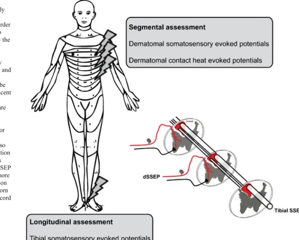
Improved assessment of segmental sensory function Whether conventional longitudinal SSEPs can also detect subtle changes in neurophysiology occurring at or near the level of SCI is not clear. This might be important for inter-ventions that aim to improve function in adjacent spinal segments. In order to understand the extent and complete-ness of SCI, a segmental approach along the spinal axis is required that evaluates several dermatomes close to the area of
cord damage. Dermatomal contact heat evoked poten-tials (dCHEPs) and dermatomal somatosensory evoked potentials (dSSEP) represent potential solutions for assessing changes in sensory function in individual spi-nal segments. Stimulation of thinly myelinated Aδ fibers and myelinated Aβ fibers (contact heat and electrical, respectively) allows a distinct assessment of different sensory pathways. In combination, these techniques increase the cross-sectional area of the spinal cord under investigation and thus provide a more complete picture of the damaged area (Fig. 3). For both methods, stimuli are applied to the defined sensory key points of the International Clinical Standards for Examination of Sensory Function after SCI (i.e., ISNCSCI; Alexander et al.2009). An additional advantage of a segmental approach is that the results can be directly compared with clinical light touch and pinprick findings.
dSSEP are evoked by electrical stimulation of the dermatome to be assessed. In a recent study, dSSEPs have been employed in conjunction with EPT to assess impairments in individual cervical spinal segments (Kramer et al. 2008). In a follow-up study, dSSEPs have been shown to recover toward normal latency values during spontaneous recovery in spinal segments affected by SCI (Kramer et al. 2009b).
Fig. 2 Neurophysiological measurements are commonly performed by assessing longitudinal pathways. In order to enhance the resolution to changes happening close to the level of lesion, segmental assessments with electrical (dermatomal somatosensory evoked potentials [dSSEP]) and heat (contact heat evoked potentials) stimulation can be applied to dermatomes adjacent to the lesion side (flashes indicate where the stimuli are applied). In the segmental assessment, thoracic dermatomes are indicated for display purposes; however, cervical dermatomes can also be assessed. The representation of the spinal cord compares tibial SSEP with dSSEP. dSSEP in SCI are assumed to be more sensitive to the level of lesion and damage to the dorsal horn and central gray area of the cord than tibial SSEP
dCHEPs have a demonstrated value for assessing the integrity of conduction in the spinothalamic tract and alter-nations of thermal sensitivity (Chen et al.2001; Wydenkeller et al. 2008). Contact heat as a noxious stimulus induces depolarization at nociceptive free nerve endings of Aδ fibers and C fibers. Unlike radiant heat stimulation (i.e., lasers), which have also been employed to investigate conduction in the spinothalamic tract (Cruccu et al. 2000), contact heat indirectly activates low threshold mechanosensitive Aβ fibers via pressure applied to the skin surface. Although Aδ, C and Aβ fibers can all be activated by contact heat, the resulting evoked potentials are primarily considered to be a reflection of Aδ fibers (Mouraux and Plaghki 2007). Transient receptor potential (TRP) ion channels located on primary afferent neurons play a crucial role in cutaneous thermal sensation. TRP ion channels can be activated by distinct temperature ranges (Schepers and Ringkamp2009; Willis 2009). TRP vanilloid 1 and 2 are transducers of noxious heat and are activated by temperatures equal to or higher than 42°C and 52°C, respectively (Willis2009). The action potentials generated in the periphery ascend from the peripheral tissue nociceptors and enter the spinal cord via the dorsal root. Aδ fibers project primarily to laminae I and V in the dorsal horn, whereas C fibers terminate in laminae I and II (Craig2003; Zeilhofer2005). The primary
projections of Aδ fibers decussate on the segmental level by the central gray matter and project to the spinothalamic tract of the contralateral side. A proportion of these fibers also ascends in the tract of Lissaeur, one to two segments before decussating in adjacent segments and ascending in the spi-nothalamic tract (Denny-Brown et al. 1973). Since lesions often affect the central gray matter (McDonald and Sadow-sky2002), dCHEPs might be more likely to indicate spinal cord pathology in a dermatomal approach compared with dSSEPs.
dSSEP and dCHEPs can give further and distinct informa-tion about spinal cord pathology. dSSEP have the advantage of occurring as a highly synchronized afferent volley, because of conduction along the fast-conducting Aβ fibers. However, the dermatomal map of small diameter fibers has been pro-posed to be less overlapping than that of larger diameter fibers and thus, dCHEPs might be better suited to investigating individual segments (Lee et al.2008). Furthermore, the de-cussation of Aδ fibers within the segment or adjacent seg-ments results in higher resolution for detecting spinal cord pathology compared with dSSEPs, which are limited to only the posterior spinal cord. Even though evidence exists that sensory segmental assessments are responsive enough to de-tect recovery, this needs to be verified in a longitudinal study in acute SCI subjects (Kramer et al.2009a).
Fig. 3 a–c Structural magnetic resonance imaging (MRI; a) of a patient example (incomplete tetraplegia, AIS D, snake-eye syndrome) with the corresponding dermatomal somatosensory evoked potentials (dSSEP; b) and dermatomal contact heat evoked potentials (dCHEP; c) showing sensory threshold (STh) and pain threshold (PTh). For the dSSEP, two consecutive runs are displayed in gray (first run) and black (second run). d Distinct location of the dorsal column from the
spinothalamic tract in a representation of the spinal cord with the affected regions indicated by shading. This example clearly shows that dSSEP can assess an anatomically distinct part of the spinal cord compared with dCHEP. Whereas dSSEP values are in a normal range, dCHEPs show clear impaired and abolished evoked responses in C6 and C8. Threshold measurements lack the sensitivity to display this impairment
In addition to reduced or completely lost sensation, SCI might result in complex sensory problems, including spon-taneous neuropathic pain, allodynia and paraesthesia. The underlying neurophysiological basis of these sensory com-plications has not yet been thoroughly characterized. Pres-ervation of some spinothalamic tract function seems to play a crucial part in the development of central pain after SCI (Finnerup et al.2007; Wasner et al.2008), although this has largely only been confirmed by using QST approaches. In order to define the sensory deficits more precisely and to improve our understanding of its relationship to sensory complications, novel stimulation paradigms may have to be designed. For example, sensori-sensory interaction between Aβ fibers and Aδ fibers might demonstrate sprouting in the dorsal horn area of the cord after SCI. This could be achieved by concomitant segmental electric and thermal stimulation being applied to the same dermatome while recording evoked potentials (Fig.4). Kupers et al. (2011) have shown, in a small number of subjects, that laser evoked potentials in neuropathic pain patients are modulated by concomitant peripheral nerve stimulation. The conditioning of Aβ fibers during dCHEPs might provide a test to estimate and quantify
the sensori-sensory interaction and might serve as a mean to disclose sensory complications at the level of lesion in SCI subjects.
Concluding remarks
To date, sensitive measurement methods to assess changes close to the level of SCI are clearly lacking. A more detailed assessment of segmental sensory deficits by adopting seg-mental neurophysiological approaches might improve clinical trial protocols by addressing the limitations of clinical sensory testing, which has failed to be suffi-ciently sensitive and responsive for these purposes. dSSEP and dCHEP might serve as complementary as-sessment tools to detect changes attributable to sponta-neous recovery and further serve as important outcome measurements to detect the efficacy of therapeutic interventions.
Acknowledgment We thank J.L.K. Kramer for editorial help and scientific input.
Fig. 4 Conditioning stimulation of contact heat evoked potentials (dCHEPs) by electrical Aβ fiber stimulation (intensity below 3 mA) revealed (b) inhibitory effects at the dermatome C5 that clinically presented an allodynic pain syndrome. a No obvious effect at C4 segment above the injury level with normal sensation (dotted line conditioning stimulation, i.e., electrical and contact heat stimulation, continuous line contact heat stimulation). c Corresponding structural MRI of the patient (recordings of a 53-year-old male subject with traumatic SCI C4 AIS C)
References
Alexander MS, Biering-Sorensen F, Bodner D, Brackett NL, Cardenas D, Charlifue S, Creasey G, Dietz V, Ditunno J, Donovan W, Elliott SL, Estores I, Graves DE, Green B, Gousse A, Jackson AB, Kennelly M, Karlsson AK, Krassioukov A, Krogh K, Linsenmeyer T, Marino R, Mathias CJ, Perkash I, Sheel AW, Schilero G, Schurch B, Sonksen J, Stiens S, Wecht J, Wuermser LA, Wyndaele JJ (2009) International standards to document remaining autonomic function after spinal cord injury. Spinal Cord 47:36–43
Beydoun A, Morrow TJ, Shen JF, Casey KL (1993) Variability of laser-evoked potentials: attention, arousal and lateralized differ-ences. Electroencephalogr Clin Neurophysiol 88:173–181 Blesch A, Tuszynski MH (2009) Spinal cord injury: plasticity,
regen-eration and the challenge of translational drug development. Trends Neurosci 32:41–47
Bradbury EJ, McMahon SB (2006) Spinal cord repair strategies: why do they work? Nat Rev Neurosci 7:644–653
Chen ACN, Niddam DM, Arendt-Nielsen L (2001) Contact heat evoked potentials as a valid means to study nociceptive pathways in human subjects. Neurosci Lett 316:79–82
Craig AD (2003) Pain mechanisms: labeled lines versus convergence in central processing. Annu Rev Neurosci 26:1–30
Cruccu G, Iannetti GD, Agostino R, Romaniello A, Truini A, Manfredi M (2000) Conduction velocity of the human spinothalamic tract as assessed by laser evoked potentials. Neuroreport 11:3029–3032 Curt A, Schwab ME, Dietz V (2004) Providing the clinical basis for new interventional therapies: refined diagnosis and assessment of recovery after spinal cord injury. Spinal Cord 42:1–6
Curt A, Van Hedel HJA, Klaus D, Dietz V (2008) Recovery from a spinal cord injury: significance of compensation, neural plasticity, and repair. J Neurotrauma 25:677–685
Denny-Brown D, Kirk EJ, Yanagisawa N (1973) The tract of Lissauer in relation to sensory transmission in the dorsal horn of spinal cord in the macaque monkey. J Comp Neurol 151:175–199 Ellaway PH, Anand P, Bergstrom EM, Catley M, Davey NJ, Frankel HL,
Jamous A, Mathias C, Nicotra A, Savic G, Short D, Theodorou S (2004) Towards improved clinical and physiological assessments of recovery in spinal cord injury: a clinical initiative. Spinal Cord 42:325–337
Ellaway PH, Kuppuswamy A, Balasubramaniam AV, Maksimovic R, Gall A, Craggs MD, Mathias CJ, Bacon M, Prochazka A, Kowalczewski J, Conway BA, Galen S, Catton CJ, Allan DB, Curt A, Wirth B, Hedel HJ van (2011) Development of quantita-tive and sensiquantita-tive assessments of physiological and functional out-come during recovery from spinal cord injury: a clinical initiative. Brain Res Bull 84:343–357
Finnerup NB, Sørensen L, Biering-Sørensen F, Johannesen IL, Jensen TS (2007) Segmental hypersensitivity and spinothalamic function in spinal cord injury pain. Exp Neurol 207:139–149
Frigon A, Rossignol S (2006) Functional plasticity following spinal cord lesions. Prog Brain Res 157:231–260
Hayes KC, Wolfe DL, Hsieh JT, Potter PJ, Krassioukov A, Durham CE (2002) Clinical and electrophysiologic correlates of quantitative sensory testing in patients with incomplete spinal cord injury. Arch Phys Med Rehabil 83:1612–1619
Kramer JLK, Moss AJ, Taylor P, Curt A (2008) Assessment of poste-rior spinal cord function with electrical perception threshold in spinal cord injury. J Neurotrauma 25:1019–1026
Kramer J, Steeves J, Curt A (2009a) Sensory segmental assessments following spinal cord injury. Top Spinal Cord Inj Rehabil 14:23–33 Kramer JK, Taylor P, Steeves JD, Curt A (2009b) Dermatomal somato-sensory evoked potentials and electrical perception thresholds during recovery from cervical spinal cord injury. Neurorehabil Neural Repair 24:309–317
Kupers R, Laere KV, Calenbergh FV, Gybels J, Dupont P, Baeck A, Plaghki L (2011) Multimodal therapeutic assessment of peripheral nerve stimulation in neuropathic pain: five case reports with a 20-year follow-up. Eur J Pain 15:161.e161– 161.e169
Lee MW, McPhee RW, Stringer MD (2008) An evidence-based ap-proach to human dermatomes. Clin Anat 21:363–373
Marcol W, Kotulska K, Larysz-Brysz M, Kowalik J (2007) BDNF contributes to animal model neuropathic pain after peripheral nerve transection. Neurosurg Rev 30:235–243
Maynard FM Jr, Bracken MB, Creasey G, Ditunno JF Jr, Donovan WH, Ducker TB, Garber SL, Marino RJ, Stover SL, Tator CH, Waters RL, Wilberger JE, Young W (1997) International stand-ards for neurological and functional classification of spinal cord injury. American Spinal Injury Association. Spinal Cord 35:266– 274
McDonald JW, Sadowsky C (2002) Spinal-cord injury. Lancet 359:417–425
Middendorp JJ van, Hosman AJF, Donders ART, Pouw MH, Ditunno JF Jr, Curt A, Geurts ACH, Van de Meent H (2011) A clinical prediction rule for ambulation outcomes after traumatic spinal cord injury: a longitudinal cohort study. Lancet 377:1004–1010
Mouraux A, Plaghki L (2007) Cortical interactions and integration of nociceptive and non-nociceptive somatosensory inputs in humans. Neuroscience 150:72–81
Pezet S, McMahon SB (2006) Neurotrophins: mediators and modula-tors of pain. Annu Rev Neurosci 29:507–538
Schepers RJ, Ringkamp M (2009) Thermoreceptors and thermosensi-tive afferents. Neurosci Biobehav Rev 33:205–212
Šedý J, Urdzíková L, Jendelová P, Syková E (2008) Methods for behavioral testing of spinal cord injured rats. Neurosci Biobehav Rev 32:550–580
Shy ME, Frohman EM, So YT, Arezzo JC, Cornblath DR, Giuliani MJ, Kincaid JC, Ochoa JL, Parry GJ, Weimer LH, Therapeutics and Technology Assessment Subcommittee of the American Academy of Neurology (2003) Quantitative sensory testing: report of the Therapeutics and Technology Assessment Subcommittee of the American Academy of Neurology. Neurology 60:898–904
Spiess M, Schubert M, Kliesch U, Halder P (2008) Evolution of tibial SSEP after traumatic spinal cord injury: baseline for clinical trials. Clin Neurophysiol 119:1051–1061
Steeves JD, Lammertse D, Curt A, Fawcett JW, Tuszynski MH, Ditunno JF, Ellaway PH, Fehlings MG, Guest JD, Kleitman N, Bartlett PF, Blight AR, Dietz V, Dobkin BH, Grossman R, Short D, Nakamura M, Coleman WP, Gaviria M, Privat A (2007) Guidelines for the conduct of clinical trials for spinal cord injury (SCI) as developed by the ICCP panel: clinical trial outcome measures. Spinal Cord 45:206–221
Van Hedel HJ, Kumru H, Röhrich F, Galen S (2011) Changes in electrical perception threshold within the first 6 months after traumatic spinal cord injury: a multicenter responsiveness study. Neurorehabil Neural Repair (in press)
Wasner G, Lee BB, Engel S, McLachlan E (2008) Residual spinotha-lamic tract pathways predict development of central pain after spinal cord injury. Brain 131:2387–2400
Weaver LC, Verghese P, Bruce JC, Fehlings MG, Krenz NR, Marsh DR (2001) Autonomic dysreflexia and primary afferent sprouting after clip-compression injury of the rat spinal cord. J Neurotrauma 18:1107–1119
Willis W (2009) The role of TRPV1 receptors in pain evoked by noxious thermal and chemical stimuli. Exp Brain Res 196:5– 11
Wydenkeller S, Wirz R, Halder P (2008) Spinothalamic tract conduction velocity estimated using contact heat evoked
potentials: what needs to be considered. Clin Neurophysiol 119:812–821
Zariffa J, Kramer JLK, Fawcett JW, Lammertse DP, Blight AR, Guest J, Jones L, Burns S, Schubert M, Bolliger M, Curt A, Steeves JD (2011) Characterization of neurological recovery following trau-matic sensorimotor complete thoracic spinal cord injury. Spinal Cord 49:463–471
Zeilhofer HU (2005) Synaptic modulation in pain pathways. Reviews of physiology, biochemistry and pharmacology. In: Amara SG, Bamberg E, Grinstein S, Hebert SC, Jahn R, Lederer WJ, Lill R, Miyajima A, Murer H, Offermanns S, Schultz G, Schweiger M (eds) Ergebnisse der physiologie biologischen chemie und experimentellen pharmakologie, vol 154. Springer Berlin, pp 73–100