Characterization of a Mammalian Exchange Factor,
ralGDS
by
Barton William Giddings
Honors Bachelor of Science, Mathematics
University of Utah, 1987
Submitted to the Department of Biology
in Partial Fulfillment of the Requirements for the Degree of
DOCTOR OF PHILOSOPHY
at the
Massachusetts Institute of Technology
September 1994
01994 Barton William Giddings. All rights reserved.
The author hereby grants to MIT permission to reproduce and to distribute
publicly paper and electronic copies of the thesis document in whole or part.
Signature of Author
Department of Biology 18 July 19948 13 /94
Certified by Robert A. Weinberg, Ph.D.Professor of Biology & Member, Whitehead Institute for Biomedical Research
Thesis Advisor
Accepted by
-
-Frank Solomon, Ph.D. Professor of Biology
Chairman of the Graduate Committee
MASSACHUSETTS UlSnTUTE
OF TECHNOLOGY
CHARACTERIZATION OF A MAMMALIAN EXCHANGE FACTOR, ralGDS by
BARTON WILLIAM GIDDINGS
Submitted to the Department of Biology on 18 July 1994 in partial fulfillment
of the requirements for the Degree of Doctor of Philosophy in Biology ABSTRACT
The low molecular mass GTPases are important regulators of many
cellular processes, including cell growth and differentiation, cytoskeletal
organization, and protein trafficking. The cycling of these proteins between
their inactive GDP-bound and active GTP-bound forms is controlled by at
least two classes of regulators: GTPase activating proteins (GAPs) and
guanine nucleotide dissociation stimulators (GDSs). In this thesis, I describe
the cloning and characterization of a mammalian guanine nucleotide
dissociation stimulator, ralGDS, that was cloned by virtue of its similarity to
yeast genes that regulate Ras. ralGDS catalyzes nucleotide release from the
RalA and RalB GTPases, but not from closely related gene products. I also
present data that suggest that the specificity of interaction between GTPase
and GDS is regulated by residues in theswitch II region of the GTPase
structure.
Thesis Supervisor: Dr. Robert A. Weinberg
Title: Professor of Biology and Member, Whitehead Institute for
Think I'm just Happy
-Kurt Cobain
ACKNOWLEDGEMENTS
The best part of my scientific training was interacting with the interesting,
talented, and diverse group of people who flock to work with Dr. Robert Weinberg. I am grateful to all of you! I have especially benefited from the
training and direction of Charlie Albright, Sean Egan, and Dennis
Templeton. Many thanks, also, for the constant feedback and attention from
the other members of the Weinberg Ras group: Jeff Settleman, Sang-Ho
Park, Joana Liu, Lauren Foster, Mary Brooks, Peter Sicinski, and David
Shaywitz. I am also grateful to the Weinberg lab's collection of former Ras scientists: Andrew Sizeland, Riki Perlman, and Rene Medema.
Thank you to the members of my thesis committee: David Housman, Paul
Matsudaira, Iswar Hariharan, Peter Kim, and my advisor, Bob Weinberg. I
have greatly appreciated your interest and your guidance through the years.
My early scientific training was guided by Paul Nicholes, Stanley Marcus,
Ted and Tucker Gurney, and Simon Tavar6. Thank you.
Thanks also to my friends, Ed and Laura Wall, Eric and Becky Rebentisch,
Luther and Kit Giddings, Sean Becker, Mark Edlund, David McKean, Greg
Beitel and Annette Elton, and Les Dethlefsen, for helping me get away from
the science once in a while, for listening, and for giving words of
encouragement and wisdom.
Special thanks are due the former inhabitants of what is now known as
Grand Gulch for leaving something beautiful and spiritually redeeming.
Most of all, I am grateful to Susan, Rachel, David, and my parents, Lu and
Bonnie. For everything.
My doctoral work was supported by a National Science Foundation Graduate
Fellowship and by NIH Training Grant 5T32-CA0941.
CONTENTS
Abstract
Acknowledgements
Chapter One
Chapter Two
Chapter Three
Chapter Four
Appendix
Introduction: The regulation of the Ras proteins
and their relatives
Characterization of a guanine nucleotide
dissociation stimulator for a ras-related GTPase
Residues in the switch II region of the Ral GTPase
are required for interaction with its exchange factor,
ralGDS
Discussion: Exchange factors in signal
transduction
Association of Sos Ras exchange protein with
Grb2 is implicated in tyrosine kinase signal
transduction and transformation
Biographical Note
Page
5 7 11 37 49 91 101 111CHAPTER
1
Introduction:
Introduction: The regulation of Ras proteins and their relatives
The ras genes have been extensively studied since the identification of transforming alleles in 1982 (Tabin et al., 1982; Reddy et al., 1982). Since
then, numerous researchers have tried to understand how ras oncogenes
disrupt normal proliferative signaling (reviewed in Barbacid, 1987). The work
described in this thesis is a small part of that very large effort to understand
how ras (and its relatives) function in cellular signaling.
The study of Ras has been an exciting and dynamic field in recent years.
Literally hundreds of reports have been written on Ras and numerous recent
reviews cover every imaginable aspect of its function, including proteins
related to Ras (Hall, 1990a; Valencia et al., 1991), Ras structure/function
(Bourne et al., 1991); Wittinghofer and Pai, 1991); Polakis and McCormick,
1993), and regulation (Bourne et al., 1990; Evans et al., 1991; Lowy et al., 1991; Downward, 1992a; Downward, 1992b; Downward, 1992c; Satoh et al., 1992; McCormick, 1993; Feig, 1993; Medema and Bos, 1993; Lowy and Willumsen, 1993; Boguski and McCormick, 1993).
This thesis focuses on ralGDS, a guanine nucleotide dissociation
stimulator (GDS; also called an exchange factor) for the Ras-related RalA and RalB GTPases. To set the stage for this work, I will briefly describe the
identification and characterization of ras genes. Then I will describe the many
related proteins that have been identified. Finally, I will review what we know
about proteins that regulate the GTPase cycle.
The identification of the ras genes
The study of ras genes began with the identification of the H-ras and
K-ras genes as the transforming factors of the Harvey and Kirsten murine
sarcoma viruses (Harvey, 1964; Kirsten and Mayer, 1967; DeFeo et al., 1981;
Ellis et al., 1981). Scientists interested in thp etiology of cancer originally
turned their attention to viruses because, they reasoned, if they could discover
how a virus with only a few genes caused the development of a tumor, they
might discover important determinants of normal and malignant cell growth
(Logan and Cairns, 1982). This reasoning proved essentially correct, and the
study of tumor viruses yielded numerous genes that regulate the proliferation
of cells (Varmus, 1989).
Transforming ras genes were subsequently isolated from human tumors
(Chang et al., 1982; Shih and Weinberg, 1982; Parada et al., 1982). These
oncogenic alleles were shown to differ from their normal cellular counterparts by the alteration of a single amino acid (Tabin et al., 1982; Reddy et al., 1982).
Mutant ras genes have been identified in many human tumors (reviewed in
Bos, 1989).
These highly conserved 21 kDa proteins tightly bind GTP and GDP (Scolnick et al., 1979; Shih et al., 1980), possess intrinsic GTP hydrolytic
activity (McGrath et al., 1984; Sweet et al., 1984), and are associated with the
plasma membrane (Willingham et al., 1980; Willumsen and Christensen,
1984). By analogy to G proteins, ras gene products were thought to act as molecular switches: biologically active when GTP-bound and inactive when GDP-bound (Gibbs et al., 1985).
Ras-related proteins
The identification of mutated ras genes in human tumors aroused
considerable interest. In the dozen years since, at least fifty related proteins
have been identified. These low molecular mass GTPases regulate numerous
cellular functions, including proliferation, differentiation, cytoskeletal
organization, protein transport, and secretion (Hall, 1990a; Bourne et al., 1990;
Valencia et al., 1991; Boguski and McCormick, 1993).
The low molecular mass GTPases form four families: Ras, Rho, Rab, and Ran (see Fig. 1). Some workers also include the Arf (ADP-ribosylation
factor) family members in the Ras superfamily. Proteins in the Ras family
(described further below) appear to regulate growth control and differentiation. Rho (ras homologous) family members, including RhoA, RhoB, RhoC, Racl, Rac2, CDC42Hs (also known as G25K), and TC10, regulate cytoskeletal
organization. The genes in the Rab family regulate vesicular transport and
secretion. At least twenty-four members of the Rab family have been
identified, including the mammalian RablA, RablB, Rab2, Rab3A, Rab3B,
Rab3C, Rab3D, Rab4, Rab5, Rab6, and Rab7 proteins (reviewed in Valencia et al., 1991). Members of this family have very different effector loop sequences,
suggesting that these proteins interact with a diverse set of downstream
agents (Valencia et al., 1991). Ran and TC4 are related nuclear GTPases
involved in regulating mitosis (Bischoff and Ponstingl, 1991). The
Schizosaccharomyces
pombe gene spil is a member of this family and is
involved in coordination of the completion of DNA synthesis with onset of cell division (Matsumoto andBeach, 1991).
The Ras superfamily has been discussed in several reviews (see, for
UA
E-4i I-Cd k %cu
Cu4a
-a4 a) a) EHa c ) . I' aq Q ItMu
* C C Cd a 9 ~ A ,4 0 C,, I edwork in this thesis focuses on Ras and two of its immediate family members,
RaIA and RalB, I will further describe only the members of the Ras family. These closely related proteins (50 - 60% identical to Ras) comprise the Ras
(H-ras, K-(H-ras, and N-ras), Rap (RaplA, RaplB, Rap2A, Rap2B), R-ras (R-ras and
TC21), and Ral (RalA and RalB) subfamilies.
Ras. The discovery of the H-ras and K-ras genes is described above. N-ras
was isolated from a neuroblastoma cell line (Hall et al., 1983; Shimizu et al., 1983). Oncogenic forms of these proteins display similar biological effects.
ras genes have been identified in organisms other than mammals. In
the yeast Saccharomyces cerevisiae, the RASi and RAS2 genes activate
adenylate cyclase (Broek et al., 1985; Uno et al., 1985). In the fission yeast
S. pombe, the rasl gene (also known as ste5) is not required for viability but is
essential for mating (Fukui et al., 1986; Nadin-Davis et al., 1986). In
nematodes, the let-60 ras gene is required for viability and functions in the
inductive signaling pathway that initiates vulva formation (Han and
Sternberg, 1990; Beitel et al., 1990). In Drosophila, the Drasl gene appears to
mediate signaling by the Sevenless receptor tyrosine kinase (Simon et al.,
1991). Studies of Ras function in these organisms have made critical
contributions to our understanding of Ras regulation and signal transduction.
Rap. The first gene identified in this family was the Drosophila ras3 (Dras3)
gene (Neuman-Silberberg et al., 1984). This gene has since been renamed
Rapi (Hariharan et al., 1991). Human raplA and rap2A were cloned by Pizon
and coworkers (1988) by screening a library with the Drosophila Rapl gene.
The human and fly genes are 88% identical (Hariharan et al., 1991). raplA
was subsequently identified as the v-K-ras reverting gene, K-revl (Kitayama et
al., 1988). The Rap proteins share approximately 50% amino acid sequence
identity with the Ras proteins (Pizon et al., 1988).
In Drosophila, the Roughened mutation affects the Rapl gene
(Hariharan et al., 1991). Flies heterozygous for this mutation have rough eyes,
and a substantial fraction (54%) of the ommatidia in these rough eyes are
missing the R7 photoreceptor (Hariharan et al., 1991). Since Ras signaling is
required for the differentiation of the R7 cell (Simon et al., 1991), these
observations suggest that Rap is an antagonist of Ras function in R7
development.
In S. cerevisiae, the rap homologBUDi (also known as RSR1) functions
in bud site selection (Bender and Pringle, 1989; Chant and Herskowitz, 1991).
Mutations in BUD1 lead to random budding. BUD1 and CDC24 gene products
interact functionally and possibly physically. The Cdc24 protein is a putative
exchange factor for Cdc42, a member of the Rho family (Hart et al., 1991).
CDC24 and CDC42 function in the formation of the bud (Chant et al., 1991).
Thus, in S. cerevisiae, a Rap protein may regulate the action of a Rho protein,
CDC42.
R-ras. The human R-ras gene was cloned by low-stringency screening of
a library with a v-Ha-ras probe (Lowe et al., 1987). R-ras is 55% identical to
H-ras (Lowe et al., 1987) and is stimulated by rasGAP (Garrett et al., 1989).
Mutant R-ras does not transform cells (Lowe and Goeddel, 1987). R-ras was
recently to shown to interact via its C-terminus with Bc12, a protein that
regulates apoptosis (Fernandez-Sarabia and Bischoff, 1993). The mammalian
TC21 and Drosophila Dras2 genes are also members of the R-ras subfamily
Ral. ralA, like raplA, was cloned by screening a library with a ras
oligonucleotide
(Chardin and Tavitian, 1986). Polakis et al. (1989)
subsequently cloned ral while purifying GTP-biding proteins. Human ralA and
a new ral gene, ralB, were identified by screening a human library with the
simian ralA probe (Chardin and Tavitian, 1989). RalA and RalB proteins are
85% identical. The function of the ral genes is unknown. Mutant ral genes
corresponding to activated ras alleles do not transform fibroblasts (Frech et al.,
1990).
A possible clue to Ral regulation comes from recent work on Ras. Several groups using a yeast two-hybrid system to screen for genes whose
products interact with the Ras protein unexpectedly found ralGDS and a
ralGDS-like gene (S. Demo and L. Williams, personal communication; the
original cloning and characterization of the ralGDS gene is described in
Chapter 2 of this thesis). At least one of these groups has shown that the
affinity of ralGDS is greater for the GTP-bound form of Ras than for the
GDP-bound form and that this interaction requires an intact Ras effector domain (S.
Demo and L. Williams, personal communication), consistent with the idea that
Ras regulates ralGDS, which would activate Ral. The significance of the
interaction between Ras and ralGDS is unknown.
Clearly, much work will be required before we can understand the
function and regulation of Ral. How will this be done? For Ras, the effort took
numerous workers and more than a decade of weaving together studies of
mammalian cells and tumors, yeast, flies and worms. Unfortunately, ral genes
have not been cloned or detected in non-mammalian organisms. Thus, without
the benefit of genetic systems to help dissect Ral signaling, I expect that the
most successful approaches in this field will focus on identifying proteins that
interact with Ral and its regulators (ralGDS and ralGAP).
Regulatory proteins
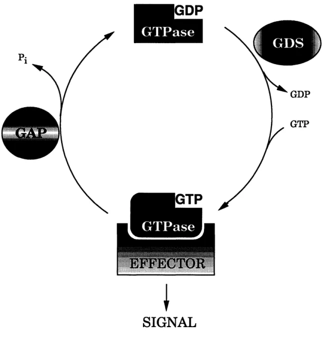
The low molecular mass GTPases act as molecular switches, cycling
between their active GTP-bound and inactive GDP-bound forms (Fig. 2). This cycling is regulated by at least two classes of enzymes. GTPase activating
proteins (GAPs) inactivate the GTPase by stimulating GTP hydrolysis.
Exchange factors, which are also known as guanine nucleotide dissociation
stimulators (GDSs), activate GTPases by accelerating the release of bound
GDP, thereby allowing GTP to bind since it is in molar excess in the cell.
Changes in both exchange factor and GAP activity have been observed in cells
stimulated in a variety of ways (Downward et al., 1990a; Gibbs et al., 1990; Li et al., 1992; Zhang et al., 1992; Medema et al., 1993; Buday and Downward,
1993b). I will briefly describe these two classes of regulators.
GTPase Activating Proteins (GAPs). The study of Ras regulators
began as an effort to understand the consequences of oncogenic mutations.
Using the DNA transfection assay, researchers identified a mutated H-ras
gene in the EJ cell line. ras genes mutated at positions 12, 59, and 61 were
subsequently identified in other human tumors (reviewed in Barbacid, 1987).
The identification of these mutant alleles raised two important questions.
What effect did these single amino acid changes have on Ras function? How
did these changes in Ras function contribute to oncogenesis?
Early analyses of oncogenic forms of Ras revealed that these mutants
possessed reduced rates of GTP hydrolysis in vitro (McGrath et al., 1984;
Pi
GDP GTP
SIGNAL
Fig. 2. The GTPase cycle. The cycling of GTPases between
their active GTP-bound and inactive GDP-bound forms is
regulated by GAPs and GDSs.
lead to a model in which the GTP-bound form of the Ras protein is active and
oncogenic mutations, which impair the GTPase activity, render a greater
fraction a cell's Ras molecules GTP-bound and active.
Subsequent biochemical studies of mutant Ras proteins, however,
undermined the tight correlation between GTPase rate and transforming
potential (Der et al., 1986; Lacal et al., 1986; Trahey et al., 1987). In one study, no correlation was found between GTPase activity and transforming
potential in seventeen mutant proteins with substitutions at amino acid 61
(Der et al., 1986). All of the mutant proteins displayed similarly lower rates of GTP hydrolysis (eight- to ten-fold lower) than wild-type Ras but the
transforming potential of these mutants varied by over 1000-fold. These data
suggest that decreased GTPase activity is not sufficient to explain the
oncogenic potential of Ras mutants.
Similarly, Lacal and coworkers (1986) reported that the Thr-59 Ras
mutant possesses a GTPase activity comparable to the wild-type protein, but
has a potent transforming activity, comparable to the Lys-12 mutant. These
data suggest that decreasing the intrinsic rate of GTP hydrolysis of Ras
proteins is not necessary for oncogenic transformation.
Trahey et al. (1987) reported similar discrepancies: while Val-12 and Asp-12 mutants differ in GTPase activity (12% and 43% of wild-type,
respectively), the mutant gene products possess comparable transforming
potential. Thus, by 1987 the failure to correlate GTPase activity and
transforming potential led some researchers to conclude that the properties of
Ras proteins measured in vitro did not adequately account for their effects in
substantially increased rates of nucleotide release that allow quick
reactivation of the GDP-bound form of Ras (Feig and Cooper, 1988).
Clearly, activating mutations should increase the amount of active
(GTP-bound) Ras in the cell. One group (Gibbs et al., 1987) tested whether
activating mutations actually increased the amount of GTP-bound Ras in vivo
by expressing normal and oncogenic Ras in yeast and determining the relative
proportions of GDP and GTP bound. They reported that wild-type yeast Rasl
and Ras2 proteins were bound entirely to GDP, while an increased proportion of
the activated Rasl and Ras2 mutant proteins were GTP-bound (16% + 4% and
33% ± 10%, respectively). Surprisingly, a substantial fraction (38% ± 10 %) of
the wild-type mammalian Ras expressed in yeast was GTP-bound, comparable
to the fraction (33% ± 10 %) of GTP bound to activated yeast Ras2 (Gibbs et
al., 1987). These observations were the first demonstration that activating
mutations increased the amount of GTP-bound Ras in vivo, although it was
unclear why so much mammalian Ras was GTP-bound.
Trahey and McCormick (1987) solved this puzzle by identifying GAP, a
protein that interacts with Ras to determine the amount of Ras.GTP in the
cell. GAP stimulates GTP hydrolysis by normal but not mutant Ras proteins.
rasGAP activity was also demonstrated in mammalian cell extracts (Trahey
and McCormick, 1987) but not yeast cells (Sigal, 1988), explaining why so
much of the mammalian Ras in yeast cells was GTP-bound.
The identification of rasGAP activity answered the question of what
oncogenic mutations do to ras gene products. Trahey and McCormick (1987)
concluded their seminal Science paper with the observation that "the major
role of Aspl2 and Vall2 mutations is to prevent interaction with GAP, and
that the effects of these mutations on intrinsic GTPase activities measured in
vitro are not biologically significant." The discovery of GAP also inaugurated
the study of Ras-regulating proteins.
rasGAP's ability to convert Ras to its inactive, GDP-bound form
suggests that rasGAP is an upstream, negative regulator of Ras (Trahey and
McCormick, 1987). rasGAP may also function downstream of Ras since it
binds the Ras effector domain (Adari et al., 1988; Cales et al., 1988).
Currently, the status of rasGAP as a biologically relevant Ras effector is
controversial (for reviews of the conflicting evidence, see Hall, 1990b; Wigler, 1990; Medema and Bos, 1993).
Numerous GAPs for Ras proteins have been identified. In mammalian
cells, rasGAP (also called p120GAP) and neurofibromin, the product of the NF1 gene, stimulate the GTPase activity of Ras (Boguski and McCormick, 1993). S. cerevisiae contains two rasGAPs, IRA1 and IRA2, which have catalytic
domains homologous to mammalian rasGAP (Tanaka et al., 1990). In
Drosophila, Gapl appears to negatively regulate Rasl activity in R7
photoreceptor development (Gaul et al., 1992).
GAPs for other GTPases have also been identified. A rapGAP has been identified (Rubinfeld et al., 1991). This protein is not related to rasGAP.
ralGAP activity has been identified in brain lysates but the protein responsible
for this activity has not been identified (Emkey et al., 1991).
A group of related proteins, including Bcr, n-chimaerin, p190, Grbl/p85,
3BP-1, and a Drosophila protein whose gene lies in the Rotund locus, appear to
act as rhoGAPs (reviewed in Boguski and McCormick, 1993). rabGAP
activities have been identified for Yptl (Tan et al., 1991) and Sec4 (Walworth
This protein, called GYP6, is not related to other known GAPs (Strom et al., 1993).
Exchange Factors. The deactiavating effects of GAPs on GTPases in the cell
are counterbalanced by the activating effects of guanine nucleotide
dissociation stimulators (GDSs). GDS proteins, also known as exchange
factors, stimulate the release of GDP from Ras-related proteins, thereby
allowing GTP to bind. These proteins appear to mediate upstream signals to
the GTPase.
Guanine nucleotide exchange activities were first demonstrated for
factors in mammalian cell extracts (West et al., 1990; Wolfman and Macara,
1990; Downward et al., 1990b). It was later shown that yeast SDC25 and
CDC25 gene products, which activate Ras in vivo, could stimulate guanine
nucleotide release from Ras (Cr6chet et al., 1990; Jones et al., 1991). A
Drosophila rasGDS, known as Son of sevenless (Sos), was identified by its role
in receptor tyrosine kinase signaling (Simon et al., 1991; Bonfini et al., 1992).
Sos also has been shown to have GDS activity (Egan et al., 1993; Chardin et
al., 1993; Buday and Downward, 1993a; Liu et al., 1993). Several other
exchange factors have been isolated by virtue of their amino acid similarity to
known GDSs, such as Cdc25 and Sos (Shou et al., 1992; Wei et al., 1992; Bowtell et al., 1992; Albright et al., 1993).
Many of the exchange factors identified to date are related to Cdc25 (see
Chapter 2 of this thesis). This family of GDS proteins now includes at least ten
members: the S. cerevisiae proteins Cdc25 (Camonis et al., 1986; Broek et al., 1987; Robinson et al., 1987), Sdc25 (Boy-Marcotte et al., 1989), Bud5 (Chant
et al., 1991; Powers et al., 1991), and Ltel (Wickner et al., 1987); the S. pombe
protein Ste6 (Hughes et al., 1990), the Drosophila protein Sos (Simon et al.,
1991); (Bonfini et al., 1992), and the mammalian proteins RasGRF (also known as CDC25Mm ; Martegani et al., 1992; Shou et al., 1992; Wei et al.,
1992), ralGDS (Albright et al., 1993), and Sosl and Sos2 (Bowtell et al., 1992).
Each of these Cdc25-like proteins is specific for the GTPases in a subfamily of
the Ras family. Cdc25, Ste6, Sos, and RasGRF are GDSs for Ras proteins.
Sdc25 acts on Ras in vitro and, when activated by removing the N-terminus,
can replace Cdc25 in vivo. Bud5 is the exchange factor for the S. cerevisiae Rap homolog, Budl. ralGDS acts on Ral proteins. The GTPase target for Ltel
is unknown.
A second group of posssible exchange factors, the Dbl family, has also been identified. This family includes Dbl (Eva et al., 1988), Vav (Adams et al.,
1992), Bcr (Hariharan and Adams, 1987), RasGRF (Shou et al., 1992), Ect2 (Miki et al., 1993), and the S. cerevisiae protein Cdc24 (Miyamoto et al., 1987).
Dbl is not related to members of the Cdc25 family of exchange factors but has
been shown to catalyze nucleotide exchange from a Rho protein, CDC42Hs
(Hart et al., 1991). Because Dbl acts on a Rho protein, it was originally
postulated that this unique group of proteins might be the exchange factors for
the Rho and Rac GTPases (for review, see Boguski and McCormick, 1993).
Recently, however, Vav has been shown to act as a GDS for Ras (Gulbins et
al., 1993). It is currently unkown whether proteins in this group other than Dbl
and Vav actually possess GDS activity, and possible GTPase targets have not
been identified.
Another exchange factor, smgGDS, is not related to either Cdc25 or Dbl. Unlike the Cdc25-related GDSs, which stimulate nucleotide release from
non-prenylated forms of their targets, smgGDS only reacts with isonon-prenylated
Cdc25-related GDSs, which are specific for GTPases within a subfamily, in its broad
specificity. smgGDS acts on specific targets from several families: RaplA, RaplB, K-Ras, RhoA, and Racl (Kaibuchi et al., 1991; Mizuno et al., 1991; Hiraoka et al., 1992).
Another class of GDS proteins acts on members of the Rab family. Two rabGDSs have been cloned. The S. cerevisiae protein Dss4 is a GDS for Sec4
and, to a lesser extent, Yptl (Moya et al., 1993). Dss4 does not act on the
related GTPase, Rab3A. The mammalian Mss4 protein stimulates nucleotide
release from Sec4, Yptl, and Rab3A (Burton et al., 1993). These proteins are
not related to other GDS proteins, but they do share two short but highly
conserved stretches of amino acid sequence (Burton et al., 1993).
The final class of GDSs is specific for members of the Ran/TC4 family.
The mammalian Rccl protein is a GDS for Ran (Bischoff and Ponstingl, 1991)
Rccl was originally identified by its role in regulating chromosome
condensation (Ohtsubo et al., 1987). The S. pombe protein Piml is homologous
to Rccl and is involved in the coupling the end of DNA synthesis with the onset
of mitosis (Matsumoto and Beach, 1991). The Rccl/Piml GDSs are not
related to other classes of GDS proteins.
Concluding remarks
The ras genes, which were first identified as retroviral oncogenes, have
been studied extensively. Numerous Ras and Ras-related proteins have been
identified in a variety of species. These small GTPases act as switches in
regulating many important cellular processes.
The study of Ras and related proteins led to the identification of several
proteins that regulate GTPase cycling. These regulatory proteins include
GAPs and GDSs. GAPs deactivate GTPases by stimulating GTP hydrolysis. GDSs activate GTPases by stimulating nucleotide exchange.
In this thesis, I explore the function and regulation of GDS proteins.
Chapter 2 describes the cloning and characterization of a mammalian
exchange factor, ralGDS. In Chapter 3, I examine the features of a GTPase
that allow specific activation by its GDS protein. In the Appendix, I present
work describing how the Ras exchange factor, Sos, relays information from the
Acknowledgments
I would like to thank Susan Demo for sharing her unpublished
observations on the interaction between Ras and ralGDS.
References
Adams, J. M., Houston, H., Allen, J., Lints, T., and Harvey, R. (1992). Oncogene
7, 611-618
Adari, H., Lowy, D. R., Willumsen, B. M., Der, C. J., and McCormick, F. (1988).
Science 240, 518-521.
Albright, C. F., Giddings, B. W., Liu, J., M. Vito, M., and Weinberg, R. A. (1993).
EMBO J. 12, 339-347.
Barbacid, M. (1987). Ann. Rev. Biochem. 56, 779-827.
Beitel, G. J., Clark, S. G., and Horvitz, H. R. (1990). Nature 348, 503-509.
Bender, A., and Pringle, J. R. (1989). Proc. Natl. Acad. Sci. USA 86, 9976-9980.
Bischoff, F. R., and Ponstingl, H. (1991). Nature 354, 80-82. Boguski, M. S., and McCormick, F. (1993). Nature 366, 643-654.
Bonfini, L., Karlovich, C. A., Dasgupta, C., and Banerjee, U. (1992). Science
255, 603-606.
Bos, J. L. (1989). Cancer Res. 49, 4682-4689.
Bourne, H. R., Sanders, D. A., and McCormick, F. (1990). Nature 348, 125-132. Bourne, H. R., Sanders, D. A., and McCormick, F. (1991). Nature 349, 117-127. Bowtell, D., Fu, P., Simon, M., and Senior, P. (1992). Proc. Natl. Acad. Sci. USA 89, 6511-6515.
Boy-Marcotte, E., Damak, F., Camonis, J., Garreau, H., and Jacquet, M. (1989). Gene 77, 21-30.
Broek, D., Samiy, N., Fasano, O., Fujiyama, A., Tamanoi, F., Northup, J., and Wigler, M. (1985). Cell 41, 763-769.
Broek, D., Toda, T., Michaeli, T., Levin, L., Birchmeier, C., Zoller, M., Powers, S., and Wigler, M. (1987). Cell 48, 789-799.
Buday, L., and Downward, J. (1993a). Cell 73, 611-620.
Buday, L., and Downward, J. (1993b). Mol. Cell. Biol. 13, 1903-1910. Burton, J., Roberts, D., Montaldi, M., Novick, P., and De Camilli, P. (1993). Nature 361, 464-467.
Cales, C., Hancock, J. F., Marshall, C. J., and Hall, A. (1988). Nature 332, 548-551.
Camonis, J. H., Kalkine, M., Gondre, B., Garreau, H., Boy, M. E., and Jacquet, M. (1986). EMBO J. 5, 375-380.
Chang, E. H., Furth, M. E., Scolnick, E. M., and Lowy, D. R. (1982). Nature 297, 479-483.
Chant, J., Corrado, K., Pringle, J. R., and Herskowitz, I. (1991). Cell 65, 1213-1224.
Chant, J., and Herskowitz, I. (1991). Cell 65, 1203-1212.
Chardin, P., Camonis, J. H., Gale, N. W., Van Aelst, L., Schlessinger, J., Wigler, M. H., and Bar-Sagi, D. (1993). Science 260, 1338-1343.
Chardin, P., and Tavitian, A. (1986). EMBO J. 5, 2203-2208.
Chardin, P., and Tavitian, A. (1989). Nucleic Acids Res. 17, 4380.
Colby, W. W., Hayflick, J. S., Clark, S. G., and Levinson, A. D. (1986). Mol.
Cell. Biol. 6, 730-734.
CrOchet, J.-B., Poullet, P., Mistou, M.-Y., Parmeggiani, A., Camonis, J., Boy-Marcotte, E., Damak, F., and Jacquet, M. (1990). Science 248, 866-868. Damak, F., Boy-Marcotte, E., Le-Roscouet, D., Guilbaud, R., and Jacquet, M. (1991). Mol. Cell. Biol. 11, 202-212.
DeFeo, D., Gonda, M. A., Young, H. A., Chang, E. H., Lowy, D. R., Scolnick, E. M., and Ellis, R. W. (1981). Proc. Natl. Acad. Sci. USA 78, 3328-3332.
Der, C. J., Finkel, T., and Cooper, G. M. (1986). Cell 44, 167-176. Downward, J. (1992a). Current Biology 2, 329-331.
Downward, J. (1992b). Curr. Opin. Genet. Dev. 2, 13-18. Downward, J. (1992c). Bioessays 14, 177-184.
-Downward, J., Graves, J. D., Warne, P. H., Rayter, S., and Cantrell, D. A. (1990a). Nature 346, 719-723.
Downward, J., Riehl, R., Wu, L., and Weinberg, R. A. (1990b). Proc. Natl. Acad. Sci. USA 87, 5998-6002.
Egan, S. E., Giddings, B. W., Brooks, M. W., Buday, L., Sizeland, A. M., and Weinberg, R. A. (1993). Nature 363, 45-51.
Ellis, R. W., DeFeo, D., Shih, T. Y., Gonda, M. A., Young, H. A., Tsuchida, N., Lowy, D. R., and Scolnick, E. M. (1981). Nature 292, 506-511.
Emkey, R., Freedman, S., and Feig, L. A. (1991). J. Biol. Chem. 266, 9703-9706.
Eva, A., Vecchio, G., Rao, C. D., Tronick, S. R., and Aaronson, S. A. (1988).
Proc. Natl. Acad. Sci. USA 85, 2061-2065.
Evans, T., Hart, M. J., and Cerione, R. A. (1991). Curr. Opin. Cell Biol. 3, 185-191.
Feig, L. A. (1993). Science 260, 767-768.
Feig, L. A., and Cooper, G. M. (1988). Mol. Cell. Biol. 8, 2472-2478.
Fernandez-Sarabia, M. J., and Bischoff, J. R. (1993). Nature 366, 274-275.
Frech, M., Schlichting, I., Wittinghofer, A., and Chardin, P. (1990). J. Biol.
Chem. 265, 6353-6359.
Fukui, Y., Kozasa, T., Kaziro, Y., Takeda, T., and Yamamoto, M. (1986). Cell 44, 329-336.
Garrett, M. D., Self, A. J., van Oers C., and Hall, A. (1989). J. Biol. Chem. 264, 10-13.
Gaul, U., Mardon, G., and Rubin, G. M. (1992). Cell 68, 1007-1019.
Gibbs, J. B., Marshall, M. S., Scolnick, E. M., Dixon, R. A. F., and Vogel, U. S. (1990). J. Biol. Chem. 265, 20437-20442.
Gibbs, J. B., Schaber, M. D., Marshall, M. S., Scolnick, E. M., and Sigal, I. S. (1987). J. Biol. Chem. 262, 10426-10429.
Gibbs, J. B., Sigal, I. S., and Scolnick, E. M. (1985). TIBS 10, 350-353.
Gulbins, E., Coggeshall, K. M., Baler, G., Katzav, S., Burn, P., and Altman, A. (1993). Science 260, 822-825.
Hall, A. (1990a). Science 249, 635-640. Hall, A. (1990b). Cell 61, 921-923.
Hall, A., Marshall, C. J., Spurr, N. K., and Weiss, R. A. (1983). Nature 303, 396-400.
Han, M., and Sternberg, P. W. (1990). Cell 63, 921-931.
Hariharan, I. K, and Adams, J. M. (1987). EMBO J. 6, 115-119.
Hariharan, I. K, Carthew, R. W., and Rubin, G. M. (1991). Cell 67, 717-722.
Hart, M. J., Eva, A., Evans, T., Aaronson, S. A., and Cerione, R. A. (1991).
Nature 354, 311-314.
Harvey, J. J. (1964). Nature 204, 1104-1105.
Hiraoka, K, Kaibuchi, K., Ando, S., Musha, T., Takaishi, K, Mizuno, T., Asada, M., Menard, L., Tomhave, E., Didsbury, J., et al. (1992). Biochem. Biophys. Res. Commun. 182, 921-930.
Hughes, D. A., Fukui, Y., and Yamamoto, M. (1990). Nature 344, 355-357. Jones, S., Vignais, M. L., and Broach, J. R. (1991). Mol. Cell. Biol. 11, 2641-2646.
Kaibuchi, K., Mizuno, T., Fujioka, H., Yamamoto, T., Kishi, K, Fukumoto, Y., Hori, Y., and Takai, Y. (1991). Mol. Cell. Biol. 11, 2873-2880.
Kawata, M., Matsui, Y., Kondo, J., Hishida, T., Teranishi, Y., and Takai, Y. (1988). J. Biol. Chem. 263, 18965-18971.
Kirsten, W. H., and Mayer, L. A. (1967). J. Natl. Cancer Inst. 39, 311-335. Kitayama, H., Sugimoto, Y., Matsuzaki, T., Ikawa, Y., and Noda, M. (1989). Cell 56, 77-84.
Lacal, J. C., Srivastava, S. D., Anderson, O. S., and Aaronson, S. A. (1986). Cell 44, 609-617.
Li, B.-Q., Kaplan, D., Kung, H.-f., and Kamata, T. (1992). Science 256, 1456-1459.
Liu, B. X., Wei, W., and Broek, D. (1993). Oncogene 8, 3081-3084.
Lowe, D. G., Capon, D. J., Delwart, E., Sakaguchi, A. Y., Naylor, S. L., and
Goeddel, D. V. (1987). Cell 48, 137-146.
Lowe, D. G., and Goeddel, D. V. (1987). Mol. Cell. Biol. 7, 2845-2856.
Lowy, D. R., and Willumsen, B. M. (1993). Annu. Rev. Biochem. 62, 851-891. Lowy, D. R., Zhang, K., DeClue, J. E., and Willumsen, B. M. (1991). Trends Genet. 7, 346-351.
Martegani, E., Vanoni, M., Zippel, R., Coccetti, P., Brambilla, R., Ferrari, C., Sturani, E., and Alberghina, L. (1992). EMBO J. 11, 2151-2157.
Matsumoto, T., and Beach, D. (1991). Cell 66, 347-360. McCormick, F. (1993). Nature 363, 15-16.
McGrath, J. P., Capon, D. J., Goeddel, D. V., and Levinson, A. D. (1984). Nature 310, 644-649.
Medema, R. H., and Bos, J. L. (1993). Crit. Rev. Oncog. 4, 615-661.
Medema, R. H., de Vries-Smits, A. M. M., van der Zon, G. C. M., Maassen, J. A.,
and Bos, J. L. (1993). Mol. Cell. Biol. 13, 155-162.
Mild, T., Smith, C. L., Long, J. E., Eva, A., and Fleming, T. P. (1993). Nature
362, 462-465.
Miyamoto, S., Ohya, Y., Ohsumi, Y., and Anraky, Y. (1987). Gene 54, 125-132. Mizuno, T., Kaibuchi, K, Yamamoto, T., Kawamura, M., Sakoda, T., Fujioka,
H., Matsuura, Y., and Takai, Y. (1991). Proc. Natl. Acad. Sci. USA 88, 6442-6.
Moya, M., Roberts, D., and Novick, P. (1993). Nature 361, 460-463.
Nadin-Davis, S. A., Nasim, A., and Beach, A. (1986). EMBO J. 5, 2963-2971. Neuman-Silberberg, F. S., Schejter, E., Hoffmann, F. M., and Shilo, B.-Z. (1984). Cell 37, 1027-1033.
Ohtsubo, M., Kai, R., Furuno, N., Sekiguchi, T., Sekiguchi, M., Hayashida, H., Kuma, K, Miyata, T., Fukushige, S., Murotsu, T., Matsubara, K., and
Nishimoto, T. (1987). Genes Dev. 1, 585-593.
Parada, L. F., Tabin, C. J., Shih, C., and Weinberg, R. A. (1982). Nature 297, 474-478.
Pizon, V., Chardin, P., Lerosey, I., Olofsson, B., and Tavitian, A. (1988). Oncogene 3, 201-204.
Polakis, P., and McCormick, F. (1993). J. Biol. Chem. 268, 9157-9160.
Polakis, P. G., Weber, R. F., Nevins, B., Didsbury, J. R., Evans, T., and
Snyderman, R. (1989). J. Biol. Chem. 264, 16383-16389.
Powers, S., Gonzales, E., Christensen, T., Cubert, J., and Broek, D. (1991). Cell 65, 1225-1231.
Reddy, E. P., Reynolds, R. K., Santos, E., and Barbacid, M. (1982). Nature 300, 149-152.
Robinson, L. C., Gibbs, J. B., Marshall, M. S., Sigal, I. S., and Tatchell, K. (1987). Science 235, 1218-1221.
Rubinfeld, B., Munemitsu, S., Clark, R., Conroy, L., Watt, KI, Crosier, W. J., McCormick, F., and Polakis, P. (1991). Cell 65, 1033-1042.
Satoh, T., Nakafuku, M., and Kaziro, Y. (1992). J. Biol. Chem. 267, 24149-24152.
Scolnick, E. M., Papageorge, A. G., and Shih, T. Y. (1979). Proc. Acad. Natl. Sci. USA 76, 5355-5359.
Shih, C., and Weinberg, R. A. (1982). Cell 29, 161-169.
Shih, T. Y., Papageorge, A. G., Stokes, P. E., Weeks, M. 0., and Scolnick, E. M. (1980). Nature 287, 686-691.
Shimizu, K., Goldfarb, M., Perucho, M., and Wigler, M. (1983). Proc. Natl. Acad. Sci. 80, 383-387.
Shou, C., Farnsworth, C. L., Neel, B. G., and Feig, L. A. (1992). Nature 358, 351-354.
Sigal, I. S. (1988). Nature 332, 485-486.
Simon, M. A., Bowtell, D. D. L., Dodson, G. S., Laverty, T. R., and Rubin, G. M.
(1991). Cell 67, 701-716.
Strom, M., Vollmer, P., Tan, T. J., and Gallwitz, D. (1993). Nature 361, 736-739.
Sweet, R. W., Yokoyama, S., Kamata, T., Feramisco, J. R., Rosenberg, M., and Gross, M. (1984). Nature 311, 276-275.
Tabin, C. J., Bradley, S. M., Bargmann, C. I., Weinberg, R. A., Papageorge, A. G., Scolnick, E. M., Dhar, R., Lowy, D. R., and Chang, E. H. (1982). Nature 300, 143-149.
Tan, T., Vollmer, P., and Gallwitz, D. (1991). FEBS Lett. 291, 322-326.
Tanaka, K., Nakafuku, M., Satoh, T., Marshall, M. S., Gibbs, J. B., Matsumoto, K, Kaziro, Y., and Toh-e, A. (1990). Cell 60, 803-807.
Trahey, M., and McCormick, F. (1987). Science 238, 542-545.
Trahey, M., Milley, R. J., Cole, G. E., Innis, M., Paterson, H., Marshall, C. J., Hall, A., and McCormick, F. (1987). Mol. Cell. Biol. 7, 541-544.
Uno, I., Mitsuzawa, H., Matsumoto, K., Tanaka, KI, Oshima, T., and Ishikawa, T. (1985). Proc. Natl. Acad. Sci. USA 82, 7855-7859.
Valencia, A., Chardin, P., Wittinghofer, A., and Sander, C. (1991). Biochemistry 30, 4637-4648.
Varmus, H. (1989). In Oncogenes
and the Molecular Origins of Cancer, R. A.
Weinberg, ed. (New York: Cold Spring Harbor Laboratory Press), 3-44. Walworth, N. C., Brennwald, P., Kabcenell, A. K., Garrett, M., and Novick, P.
(1992). Mol. Cell. Biol. 12, 2017-2028.
Wei, W., Mosteller, R. D., Sanyal, P., Gonzales, E., McKinney, D., Dasgupta, C., Li, P., Liu, B.-X, and Broek, D. (1992). Proc. Natl. Acad. Sci. USA 89, 7100-7104.
West, M., Kung, H. F., and Kamata, T. (1990). FEBS Lett. 259, 245-248. Wickner, R. B., Koh, T. J., Crowley, J. C., O'Neil, J., and Kaback, D. B. (1987).
Yeast 3, 51-57.
Wigler, M. H. (1990). Nature 346, 696-697.
Willingham, M. C., Pastan, I., Shih, T. Y., and Scolnick, E. M. (1980). Cell 19,
1005-1014.
Willumsen, B. M., and Christensen, A. (1984). Nature 310, 583-586. Wittinghofer, A., and Pai, E. F. (1991). TIBS 16, 382-387.
Wolfman, A., and Macara, I. G. (1990). Science 248, 67-69.
CHAPTER 2
Characterization of a guanine nucleotide dissociation
Preface
This chapter represents work I did with Dr. Charles F. Albright, Joana Liu, and Maja Vito in the laboratory of Professor Robert A. Weinberg. I was involved in all phases of this work. My contributions to this study include
cloning and sequencing the ralGDS PCR fragment, assisting in the screening of
libraries to isolate cDNA clones, performing the sequence comparisons of
Cdc25 family members, assisting in immunoprecipitation studies of the
ralGDS protein, generating and characterizing ralGDS baculoviruses using
transfer vectors prepared by Dr. Albright, and working out the purification of recombinant ralGDS with Dr. Albright. I was involved in critically interpreting
data and assisted in the preparation of the manuscript and figures.
This chapter was previously published in the January 1993 issue of the
The EMBO Journal vol.12 no.1 pp.339-347, 1993
Characterization of a guanine nucleotide dissociation stimulator for a ras-related GTPase
Chades F.Albright, Barton W.Giddings, Joana Liu, Maja Vito and Robert A.Weinbergt Whitehead Institute for Biomedical Research and Department of Biology, Massachusetts Institute of Technology, 9 Cambridge Center, Cambridge, MA 02142, USA
'Corresponding author Communicated by P.A.Sharp
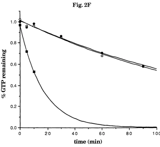
ras-related GTPases participate in signaling for a variety of cellular processes. The GTPases cycle between a GTP-bound active state and a GDP-GTP-bound inactive state. This cycling is partially controlled by guanine nucleotide dissociation stimulators (GDS, also known as exchange factors). We report on the molecular cloning of cDNAs
encoding a new mammalian GDS protein, using
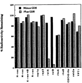
sequences derived from the yeast ras GDS proteins as probes. The encoded protein stimulates the dissociation of guanine nucleotides from the ms-related raL4 and ralB GTPases at a rate at least 30-fold faster than the intrinsic nucleotide dissociation rate. This new GDS, ralGDS, is at least 20-fold more active on the raA and ralB GTPases than on any other GTPase tested, including other members of the ms family (H-rs, N-ras, K-ras, R-ras,
rapla and rap2), members of the rho family (rhoA, rwoB and CDC42-IIs) and members of the rab family (rab3a
and yptl). While the ralGDS protein is phosphorylated on serine residues, we find no evidence that
phosphoryla-tion affects the activity of insect cell-expressed raIGDS towards the rA or ralB GTPase. The 3600 nucleotide
ralGDS mRNA and the 115 kDa protein were found in
all tissues and cell lines examined. Key words: CDC2SlrarallraIGDSras
Introduction
Low molecular mass GTPases constitute a family of proteins that participate in signaling for many cellular functions including cell cycle progression, differentiation, cytoskeletal organization, protein transport and secretion (Bourne et al., 1990; Hall, 1990). The most well known of these are the proteins encoded by the ras gene family. In performing their various cellular functions, GTPases cycle between a GTP-bound form and a GDP-GTP-bound form (Bourne t atl., 1991). The ratio of these two forms is largely controlled by two classes of enzymes: guanine nucleotide dissociation stimulators (GDS, also known as exchange proteins), which accelerate the release of the bound GDP allowing CGTP binding (Lowy et al., 1991) and GTPase activating proteins (GAP), which stimulate the hydrolysis of the bound GTP by GTPases. We focus here on the GDS proteins. It is thought that by stimulating GTP binding, the GDS proteins convey an incoming, activating signal to the GTPases.
Most GDS genes can be assigned to one of two gene
families. The CDC25 family consists of the Saccharomyces cerevisiae genes CDC25 (Camonis et al., 1986; Broek et al., 1987; Robinson et al., 1987), SDC25 (Boy-Marcotte et al., 1989), BUDS (Chant et al., 1991; Powers et al., 1991) and LTE1 (Wickner et al., 1987), the Schizosaccharomyces pombe gene stec6 (Hughes ct al., 1990), the Drosophila mnelanogaster gene Sos (Simon et 41., 1991; Bonfini et al.,
1992) and the mammalian genes CDC2w' (Martegani
et al., 1992), Ras-GRF (Shou et al., 1992), mSos-1 and mSmoa-2 (Bowtell et al., 1992). The proteins encoded by thesc genes are known or suspected to interact with the ras GTPase, with the exception of BUDS and LTEI. The most likely target of BUDS is RSRI (Chant et al., 1991), the probable S. cereviiae homologue of rap] (Bcnder and Pringle, 1989).
A second class of GDS proteins consists of the mammalian genes dbl (Eva et al., 1988), vav (Adams et al., 1992), bcr (Hariharan and Adams, 1987), Ras-GRF (Shou et al., 1992) and the S.cerevisiae gene CDC24 (Miyamoto et al., 1987). The dbl protein has GDS activity for the CDC42-Hs GTPase (Hart et al., 1991) while the target GTPases of the related proteins are still unknown. Two other GDS genes do not fit in one of these two families. One of these proteins, smgGDS, has GDS activity for rhoA, K-rar4b, rapla, raplb and racl GTPases (Kaibuchi t al., 1991; Mizuno et al., 1991; Hiraoka et al., 1992). A second protein, rccl, has GDS activity for the ran GTPase (Bischoff and Ponstingl, 1991).
The various GDS proteins appear to be important regulators of GTPase function. They and GAPs are potential targets for receiving signals that increase the GTP-bound fraction of the GTPases. Thus, increasing GDS activity or decreasing GAP activity can increase the amount of the GTPae binding CGTP. There is evidence from the ras-signaling pathway that both mechanisms are used. In T-cell signaling, it appears that inhibition of GAP activity is largely responsible for ras activation (Downward et al., 1990). In fibroblasts, rat activation by growth factors appears due to increased GDS activity (Gibbs et al., 1990; Zhang et al., 1992). In PC-12 phcochromocytoma cells, both an increase in GDS activity and an increase in GAP activity are observed in response to NGF treatment (Li ct al., 1992):
In vitro assays and genetic analysis also suggest that GDS proteins are themselves subject to regulation. In one case where rasGDS activity was demonstrated in vitro, this activity depended on the presence of phosphatase inhibitors, suggesting that phosphorylation of the GDS protein or a regulatory factor was needed for activity (Wolfrman and Macara, 1990). The phenotypes of various mutant ODS genes also suggest that the activity of GDS proteins is regulated. Deletions and point mutations in the CDC25 gene (Broek et al., 1987; Kim and Powers, 1991), and in the SDC25 gene (Damak et at., 1991), deletions in the dbl gene (Eva et al., 1988; Ron et al., 1988) and a mutation in the Sos gene (Rogge et al., 1991; Bonfini etal., 1992) all
C.F.Albright et o/.
display phenotypes consistent with increased GDS activity. respectively. Among the clones isolated in this fashion was As a consequence, GDSs appear to serve as signal one that encodes a manunmmalian protein having great similarity transducrs that link still poorly characterized upstream with the yeast exchange proteins and able to catalyze
signals to the various GTPases. nucleotide dissociation on. a mammalian GTPase.
Our goal is to understand how mammalian GDS proteins
are regulated by extracellular signals. Toward this end, we Results decided to isolate genes encoding mammalian GDS protcins.
To do so, we exploited a polymerase chain reaction (PCR) Isolation of a mammalian GDS cDNA
strategy to generate GDS probes using regions conserved Degenerate primers corresponding to conserved regions of between the CDC25 and ste6 proteins, two GDSs known CDC25 and ste6 were designed and used to PCR-amplify
to regulate ras proteins in S.cerevisiae and S.pombe, total cDNA from 3T3/AXB, a mouse cell line. These genes
GGCGADGCOC'GCGGCOCGCG GCr -124
Agln.Al.CdA(r, CGrCCA.'cCcC(CAnCoCrCctcr't .,Cr.,r. CWr.Crr.,A.ruSS2 agcc :r.C,A(C .-45
Cl'G XrC(CT(=G.-'TG TGMCGGCC ACC.' .'-'GTA·.ATTC GAGCTCCAC·-.TG= 40
H 14 V C S 8 T 0 I 14 ~~~~~n~ e l r~O 124 AGGACGAT~ACG~TCA*ICACTCCATCTCCC?GL~J7CCAGCIACAMAGAC 124 L I N V Y I S L V L 0 G A K 0 R 1. 42 TAGGGG.CGTOGGCFCTCGR GT GC3CCTCTA~TGC.AGICTK G 206 G C S 1 E A L L Y C 1 V R V K A 0 7 K L V 70
OO~r.D1,1,FRE V CACC7G 1 GGCCracC CAGCAG7eI. . cnenA~TASI A ] IC2CrCCCC7c a¥C¢I'- AcWhI) 7CACTVA 125ICGcAT 24
OCCACAGCWAGACeCA:ZTAA062W3267A7GTRAICCA7CGL.'AGAAG71ACLCtC 37
a V L L K e L i a c c I 3 126 a F C P p a F P C K L A V L N P G D L 162 TGGAcCZC.Crt¢CsCe CC TC7GGaCCCGACGCGCCA CAG1r 6 CT 628
R R A H L L L A L E O L P S A S A L P A P 21C
CACGCGCTC~AG CCCTCAC.GCTAGNkC.GCC7=CCCCAC:C GAC .CAA-~C 712!
V I, S 1. K P A I a . K P A L I. L. .' a V V '7 7 I' V 4 228
GAGACCGCGCGGCC CACCcAaCGOCCTCCAOC1CAGCACCAGCTCCTAGCrMCCACCC 796
P A A A P V P V L A 5 P V V A P A P P V P P 266
CACTCAACGGCCACCTATC1CAGT~/ GGCCGCCGTCTCf. ACCC'G A~NTGGAGT AGCTC ellO
P 0 E E P L A L A P e L P A V S S L I A 294
CTt`~`W`C CT~7(~:CCI(~~K+~:TIA~i~nC1~77(C~~n~.~L~f A '(rTF~TAGM CTTWC..(L.C~`GL`CrAAG~cACGGArAA.C~TAAGrAGAMC~.~C-r'CrC 9964
V T P A L P K S L P A T L N G L T K P H L . 322
?GT7CCC7C TAC7'GOGCTGAACGT AC'C ?GA?CGCAAC??CAMAGAs 1-CT~ACCC7CCIG 1046
P " LI L V & % F. I" L ~ D A E L.:" " K K V V ~ Y iS . G , : C
GCCA'CTGCCCAAC0GCMAGAGCAAGCCCGMCTAACCCCACCMCAACAG 1132 0 I 8 S C I A K G K E H L A P I P A V A F N V 378 TGe(m'AACr AW~A( rG6116C1rA AM AtX11CK '.AeN1rAS l0:xrG¶P 1116
A 14 C V I 7 T C L a D C S 6 KC A P 6 16 A K V V E .4 W I S 40
AXCVTCJ.GOCS~gPDPARVVE~4W!S 40~
AG7`JCACMAO*CAOAGmTCA/AA?.7,CCTcTCCTGC=CA`CC7C7CTC'C7'CAO`ACCAA~AcCC 1309 V A C A L K S L Y A I 1I. A . N A I 434 GCC Ar~ AACGIGAG ' .GiCG 1384 LKI K gvs £ V D S F v F Vr5 0 K I E I .S FS D E N 462
~V~PN J~:j2~` AIqC~n`~t 1(.A.ut!Y-'AA'f]LA .r. C A 1468
s I S K K L L I K E 6 7 S K r A T L 1 44 I P R 16 A a R 490
CRO K G£E7 GV S GT VPY G?71 F 1 7 D 1L V 4 6 7 L A 18
7AF~kekrATCAXF77GMAseAPGAGA1C6 CA7Or.CAA7CAACT70C 166 13 0 ¥ I. G R L, I N t' K R R K E E V I A 0 I K L 546 ACAGTCAGOC=TAwrICAeCACCTCTGAAMCM7G6A(TCCAAT CAzChG 1720 Q S A C N I A P H F 3 ? V F A R L A 574 CT GAGCT.A C'G TC'GT CGCC'GT AA CA CAAAAAAAA L1804 S 7 C I, P S A N R K K T A 1 602
?TC ,CGG(I:C C COAdCGZACAACCAaT AAGrGCCCACC 1089
V K R 1 5 0 8 Q A F 5 ' . 1 5 I 5 S A H 2 K 5 C ') I. 6:30
.T.CGT'f~CA CCTTIC AC ACA':AC C~,"CIC AGTGCAC'[ T CTA GTGG T C AC T GA T 19';
C S P L C : T D A L. 5 V 4 S A G S S S V S 6.8
MNtW?': A:A',,A(]L: TJ 'lI:('uqrI'C':Y&'A :t :C;G u;l ;~*F'(:r(c; ~ r ~:.~(1: t :A ~T:3' : ('C (.; LK)
I. ! 16 14 S ? V P F. S r, I) C K 1 = 86 S A 1 0 £ 1 F : 686
AGA~TcGGC&7CPMc cGG CrCGT:C CcA..*..TGGCA:~A 2142
' s . 1 7 S A . S S " : 0 S :i A '1 P P 'V S 'r i r 4 14 RGCCTCCG7CCAGGGTCTGCAG AGC.CT.CA CCTC7CACVCCAG7 G= A~7CAT G 2224
S V S G V C S Y S S S L P L N C C V C C C I R V 742 SLCVCNCNKYKSIIVTSQ6KAP7VI INA 7
TCU G~Ciid-.c~AA:.A'l .T Ma` Mr1~ -A:C&~ Z;:A~rC GA[A A:I:~G 2UG
S L V N C P Y S I l, V T S P A P T V I R A 77O
CCA7CGCAACWCACGACIGAGCC AGAOATATCG7GC7]CAGACATCTGAGCC0CAMCTC~AP 2392
N 3 K 4 L D E P D Y L V I S E H L. I 4
.'TCCA.GAPCCCA!GCT-'CTA'r(G(XAT r Ax TA'L'tC 1 CCA'I.TAwACAC IA i7cATAmRX4 2476
P N A V6 V F V A 5 T A Y I L K K R 7 F T K C 826 .GCAAAC'AACJCAGCAG(.CeA6=ACCCCT=ATGAAG AGA)PGG ACGAG.:TCC OAGAT CAA 2*(0 A V K H G A S T P R g 6 K C. I R A K G I F · 52 EACeTCC.C1GGGACGGMCGCG AGAACs'GCAC TTACAC"CTAGCACTeGIGPCAAC: TAC1CC CC 2644
crrdr..PAtrcPC:CCIr.~~'cc PccwrCACT?111rT7.A?.': cr01rPnccPr.A7rc.AcActgrrACG~CaU*FcC.r ?'1;
ACC7O6CTCGGCm6oCACI'CCCTCGmcC TCT1.TC T,cA.cCA2AGcTA c7 Tr'AC~T TICAG.WTC 211i? 7LT'C 'GCAGGGACCTOCr TGCACATCCACCAC GCAC CC,'CCCAG0CACATCACTOCC 2096 ACCCATCCG'GATiCmcCseGACACACC'CAAWGCGL'7LCG'ATrcTC:r¶'0G7.CACNIACA 291)
CCflr Td I C*[TA&(.G'AtGC ,AGC(.CTTTC 7CACACrTAGir:TTrC.ACfGCAC-TMCAC. AT1CCMCI' C 144
7ACM'AGTAGGACcCOCAMCAmACCCAAGCCCCcC7TGATCAGGAGCAGAGAAC:TC7C3tACICCCCG 2144
GG7A??GAAICC7CACCfbACCeCCALr CAT7A7CCAC7GC'7.. G 7?'ACAC.A7CAC C,'.GACGTG..T a 2 GiXUAGAG .CCM C T,'IVaATI'UATWACCTAAAATrI/ .'TGrl CFGGAO6ATATT&A7TGr":TN2 T 331
AGVATCCTACWrAAATWArTAAA-~C 7 T?7-AC 3361
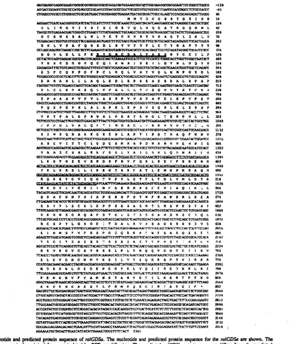
Fig. 1. Nucleotide and predicted protein sequence of raGDSa. The nucleotide and predicted protein sequence for the ralGDSa are shown. The
underlined sequence is the original fragment isolated by PCR amplification. The predicted ralGCDSb protein sequence differs from the predicted ralGDSa protein sequence in several respects: the amino terminus contains an additional 55 amino acids of sequence
MVQRMWAEASGPIGGPEPL-FPGSRRSRSVWDAVRLEVGVPDSCPVVLHSFIQLDPDLPRLE (SSTQ...), thdie 12 amino acids doubly underlined are deleted, amino acids 241 and 242 are deleted, the dipeptide PA is added after amino acid 221 and several conservative substitution: are made.
Characterization of rlGODS
derive from two species of yeast that arc as related to each other as yeast are to mammals (Moreno etal., 1991). Consequently, we expected sequences conserved between these proteins to be functionally important and therefore conserved in their mammnalian homologues. When the major product of this reaction was cloned and sequenced, it was found to have significant similarity to a number of known or suspected GDS proteins (data not shown). TIhis cDNA product was used as a probe to isolate the entire cDNA from a 3T3 library. Its sequence is shown in Figure 1. We have designated this cDNA as ralGDSa for reasons that will be discussed.
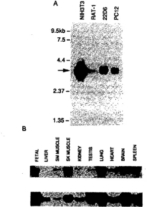
Northern blot analysis revealed a single mRNA of - 3600 nucleotides (Figure 2A). This size is in good agreement with the size of the cDNA isolated by us. Northern blot analysis of total RNA from rat tissues revealed that the gene was transcribed in all tissues examined (Figurc 2B). Furthermore, this mRNA has also been found in all of the 21 mammalian cell lincs examined (data not shown).
The structure of the cDNA is unusual in that the first ATG codon in the long open reading frame is preceded by three ATG codons in other reading frames (Figure 1). Each of the three out-of-frame ATG codons is followed by a stop
A rM 9. 2 1 B I w u > 2 X U. : (a LO z
i'
co C8 oFig. 2. Northern blot analysis of rlGDS. (A) Poly(A) mRNA froml
the stable cell lines NIH3T3, Rat-i, 22D6 (Alt et al., 1981) and PC12
were analyzed by Northem blot analysis. The arrow indicates the
location of the 3600 nucleotide mRNA found in all cell lines. (B) Total RNA from the indicated rat tissues were analyzed by Northern
blot analysis. The upper panel was hybridized with a probe for
ralGDSa and the lower panel was hybridized with a probe for a-tubulin to estimate the relative amount of ralGDS mRNA in each sample. A signal corresponding to the 3600 tlucleotide ItkRNA with the
codon before the in-frame ATG is reached. In addition, the 5' non-coding region is unusually long (212 nuclcotidcs) and has a high percentage of G-C base pairs (80%). Most mammalian mRNAs have 5' non-coding regions between 20 and 100 nucleotides and < 10% have upstream ATG codons (Kozak, 1987).
A second cDNA clone from a rat fibroblast cDNA library was isolated using the coding region of ralGDSa as a probe. While the reading frame encoded a sequence of 895 amino acids, which was 99% homologous with the protein encoded by the previously cloned mouse cDNA, this rat cDNA had an unrelated 5' non-coding region of 201 nucleotides. The rat cDNA also encoded an additional 55 amino acids at the N-terminus, an internal addition of two amino acids, internal deletions of two and 12 amino acids and several conservative substitutions. We have designated this cDNA as ralGDSb. To test the possibility that the two cDNAs represent alternatively spliced transcripts from the same gene, Northern blot analysis was performed with probes specific to the mouse 5' non-coding region and the rat 5' non-coding region. This analysis revealed that both regions are part of an - 3600 nucleotide mRNA and both are found in the same cell line (data not shown).
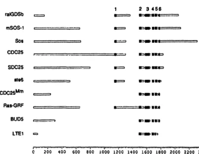
Relationship of rlGDS to other members of the CDC25 family
When the predicted GDS protein sequence was used to search the protein sequence database, we found that the sequence of ralGDS was similar to those of proteins belonging to the CDC25 family of GDS proteins and to several proteins rich in glut;unate, serine and proline. Regions of proteins rich in proline, glutamate, serine and threonine residues, known as PEST sequences, have been found in several proteins with short half-lives (Rogers et al., 1986). PEST sequences are clustered in two regions of the ralGDS sequence: amino acids 196-318 contain 56% PEST residues and amino acids 557-728 contain 51% PEST residues.
To explore further the similarity of ralGDS with other members of the CDC25 family, we used the MACAW computer program (Schuler ct al., 1991), which finds statistically significant blocks of continuous amino acids shared between multiple protein sequences. We found six blocks of sequence similarity present in most members of the CDC25 family (Figure 3). Block I was missing from CDC25n', BUDS and LTEI. LTEJ was also missing block 6 and contained only part of block 5. Previous work with CDC25 (Camrnonis et ., 1986; Broek et al., 1987), SDC25 (Boy-Marcotte et al., 1989), CDC25m (Martegani et al., 1992) and Ras-GRF(Shou et al., 1992) suggests that blocks 2 through 6 are necessary and sufficient for ODS activity. Interestingly, only ralGDS, Sos, mSOS-I and mSos-2 have extensive coding regions to the C-terminal side of these similar regions.
Sizing of the ralGDS gene product
To determine whether our cDNA would produce a protein of the same size as that produced in vivo, anti-ralGDS sera were produced. A portion of the ralGDSa cDNA including conserved blocks 1-6 was introduced into a bacterialt expression vector and the resulting protein was used. to immunize rabbits. When serum from an immunized rabbit. I .
ViII110,00-01% 21,W52-M-1