Publisher’s version / Version de l'éditeur:
Mediators of Inflammation, 2008, pp. 1-6, 2008-08-21
READ THESE TERMS AND CONDITIONS CAREFULLY BEFORE USING THIS WEBSITE. https://nrc-publications.canada.ca/eng/copyright
Vous avez des questions? Nous pouvons vous aider. Pour communiquer directement avec un auteur, consultez la première page de la revue dans laquelle son article a été publié afin de trouver ses coordonnées. Si vous n’arrivez pas à les repérer, communiquez avec nous à PublicationsArchive-ArchivesPublications@nrc-cnrc.gc.ca.
Questions? Contact the NRC Publications Archive team at
PublicationsArchive-ArchivesPublications@nrc-cnrc.gc.ca. If you wish to email the authors directly, please see the first page of the publication for their contact information.
NRC Publications Archive
Archives des publications du CNRC
This publication could be one of several versions: author’s original, accepted manuscript or the publisher’s version. / La version de cette publication peut être l’une des suivantes : la version prépublication de l’auteur, la version acceptée du manuscrit ou la version de l’éditeur.
For the publisher’s version, please access the DOI link below./ Pour consulter la version de l’éditeur, utilisez le lien DOI ci-dessous.
https://doi.org/10.1155/2008/239740
Access and use of this website and the material on it are subject to the Terms and Conditions set forth at
Lymphotoxin-α plays only a minor role in host resistance to respiratory
infection with virulent type A Francisella tularensis in mice
Zhang, Deng; Kuolee, Rhonda; Harris, Greg; Zhang, Qinxian; Conlan, J.
Wayne; Chen, Wangxue
https://publications-cnrc.canada.ca/fra/droits
L’accès à ce site Web et l’utilisation de son contenu sont assujettis aux conditions présentées dans le site LISEZ CES CONDITIONS ATTENTIVEMENT AVANT D’UTILISER CE SITE WEB.
NRC Publications Record / Notice d'Archives des publications de CNRC:
https://nrc-publications.canada.ca/eng/view/object/?id=3bbc4300-63c8-4efd-b971-7a41308e2e20 https://publications-cnrc.canada.ca/fra/voir/objet/?id=3bbc4300-63c8-4efd-b971-7a41308e2e20Volume 2008, Article ID 239740,6pages doi:10.1155/2008/239740
Research Article
Lymphotoxin-α Plays Only a Minor Role in Host
Resistance to Respiratory Infection with Virulent Type A
Francisella tularensis in Mice
Deng Zhang,1, 2Rhonda KuoLee,1Greg Harris,1Qinxian Zhang,2J. Wayne Conlan,1and Wangxue Chen1
1National Research Council Canada, Institute for Biological Sciences, 100 Sussex Drive, Room 3100, Ottawa, ON, Canada K1A 0R6 2Basic Medical College of Zhengzhou University, 100 Science Road, Zhengzhou, Henan 450001, China
Correspondence should be addressed to Wangxue Chen,wangxue.chen@nrc.gc.ca
Received 4 June 2008; Accepted 24 July 2008 Recommended by V. L. Petricevich
This study examined the role of lymphotoxin (LT)-α in host defense against airborne infection with Francisella tularensis, a gram-negative facultative intracellular bacterium and the causative agent of tularemia. Following a low-dose aerosol infection with the highly virulent type A strain of F. tularensis, mice deficient in LTα (LTα−/−) consistently harbored approximately 10-fold fewer
bacteria in their spleens at day 2 and 10-fold more bacteria in their lungs at day 4 than LTα+/+ mice. However, the mortality and median time to death were indistinguishable between the two mouse strains. In addition, the inflammatory responses to the infection, as reflected by the cytokine levels and leukocyte influx in the bronchoalveolar lavage fluid and histopathological analysis, were generally similar between LTα−/−and LTα+/+ mice. These data suggest that although LTα does not contribute significantly to the resistance and host responses of mice to airborne type A F. tularensis infection, it does play a subtle role in the multiplication/dissemination of F. tularensis.
Copyright © 2008 Deng Zhang et al. This is an open access article distributed under the Creative Commons Attribution License, which permits unrestricted use, distribution, and reproduction in any medium, provided the original work is properly cited.
1. INTRODUCTION
Francisella tularensis is a gram-negative facultative
intra-cellular bacterium and the causative agent of tularemia, a systemic infection of many mammals including humans. Left untreated, the virulent type A subspecies of F. tularensis routinely caused lethal infection in people particularly after aerosol exposure to the pathogen; as few as 10 virulent type A bacilli can initiate severe disease [1]. Consequently,
F. tularensis is considered a Category A biological warfare
agent. Despite its clinical and biosecurity importance, the molecular basis for the immunopathogenesis of F. tularensis infection, particularly when initiated through the respiratory tract, remains largely unknown.
Lymphotoxin-α (LTα) is a member of the tumor necrosis factor (TNF) superfamily of cytokines and has two distinct roles: as a membrane-bound heterotrimer in combination with LTβ, it binds the LTβ receptor and is critical in the development and maintenance of organized secondary lymphoid organs [2], and as a soluble homotrimer, it signals
through the TNF receptor pathway and leads to activation of various inflammatory cytokines and chemokines [3,4]. Indeed, LTs (LTα and LTβ), together with TNF and LIGHT (LT-related inducible ligand that competes for glycoprotein D binding to herpesvirus entry mediator on T cells), form an integrated signaling network which is important for the regulation of both innate and adaptive immune responses [4]. In this regard, LTα has been implicated in the host defense against several different bacterial, viral, and parasitic pathogens (reviewed in [3]). For instance, several studies have shown that after infection with Mycobacterium
tuberculosis, mice genetically defective in LTα (LTα−/−mice)
harbour increased bacterial burdens and exhibited a shorter median time to death when compared to LTα+/+ mice [5–7]. Similarly, mice deficient in LTα, LTβ, and the LTβ receptor (LTβR) are more susceptible to Listeria monocytogenes infec-tion than wild-type mice [6,7]. Given that LTα is important in the control of these intracellular bacterial pathogens, in the present study we sought to determine whether it also plays
2 Mediators of Inflammation a role in host defense against low-dose aerosol infection with
a virulent type A strain of F. tularensis.
2. MATERIALS AND METHODS
2.1. Mice
Eight- to twelve-week old, age-matched B6.129S2-Ltαtm1Dch/
J (LTα−/−), and wild-type C57BL/6J (LTα+/+) mice were
used in this study. The foundation breeding pairs of LTα−/−
mice were purchased from Jackson Laboratories (Bar Har-bor, Me, USA). Mice were bred and housed under specific-pathogen-free conditions in a federally licensed animal biosafety level-3 facility and given free access to sterile water and certified mouse chow. The animals were maintained and used in accordance with the recommendations of the Canadian Council on Animal Care Guide to the Care and Use of Experimental Animals.
2.2. F. tularensis and experimental infections
Stocks of type A F. tularensis strain FSC33/snMF, originally isolated from a squirrel in Georgia, USA [8]. For low-dose aerosol exposure, thawed F. tularensis stocks were diluted in Mueller Hinton broth containing 20% (v/v) glycerol to maintain infectivity at the high-relative humidity employed. Aerosols of F. tularensis strains were generated with a Lovelace nebulizer operating at a pressure of 40 p.s.i. to produce particles in the 4–6 µm range required for inhalation and retention in the alveoli [8]. Mice were exposed to these aerosols for 7 minutes (inhaled dose of∼10 organisms) using
a customized commercial nose only exposure apparatus (In-tox Products, Albuquerque, NM) resulting in the implanta-tion of 10–20 organisms into the lungs [8].
2.3. Quantitative bacteriology and histopathology
Groups of five mice of each strain were sacrificed at 2 and 4 days after inoculation (dpi) by CO2 asphyxiation.
Blood samples were collected for the determination of serum cytokine levels. The phenotype of the LTα−/−mice
was confirmed by visual inspection at necropsy to confirm the absence of peripheral lymph nodes. The lungs and spleens were removed aseptically, cut into small pieces, and then homogenized using an aerosol-proof homogenizer for quantitative bacteriology or fixed immediately by immersion in 10% neutral buffered formalin for histopathology. For quantitative bacteriology, ten-fold serial dilutions of the tissue homogenates were plated on cysteine heart agar sup-plemented with 1% (w/v) hemoglobin and sulfamethoxazole and trimethoprim. Colonies were counted after 72 hours of incubation at 37◦C [8]. For histopathology, the tissues were
processed by standard paraffin embedding methods (Depart-ment of Pathology and Laboratory Medicine, University of Ottawa, Ottawa, Ontario). Sections were cut 4 µm thick, stained with haematoxylin-eosin (HE) and examined by light microscopy.
2.4. Bronchoalveolar lavage (BAL) and cytokine measurements
In some experiments, the lungs were lavaged with five 1.0-ml aliquots of PBS supplemented with 3 mM EDTA [9], and the total lavage cell numbers were counted on a haemocytometer, and differential cell counts were car-ried out on cytospin preparations stained with Hema3 Stain Set (Fisher Scientific, Middletown, Va, USA). The bronchoalveolar lavage (BAL) fluid was centrifuged at 3000 xg for 7 minutes, supernatants were removed, sterile-filtered, and stored at –80◦C. Serum and BAL levels of
cytokines and chemokines were determined using Beadlyte Mouse 21-Plex Cytokine Detection System (Upstate, Teme-cuta, Calif, USA) on a Luminex 100IS system (Luminex, Austin, Tex, USA) [10]. Undiluted BAL and 1 : 2 diluted serum samples (50 µl) were analyzed as specified by the manufacturer (http://www.millipore.com/userguides/tech1/ proto mpxmcyto-70k). The analysis was done in duplicate, and the cytokine/chemokine concentrations were calculated against the standards using Beadview software (ver 1.03, Upstate).
2.5. Statistical analysis
All data are presented as mean±standard deviation (SD) for each group. Differences between groups were analyzed by two-way ANOVA followed by the Bonferroni multiple comparison test (GraphPad Prism 4.0, GraphPad Software, Inc., San Diego, USA). P < .05 was considered to be statistically significant.
3. RESULTS AND DISCUSSION
Initially, a total of 22 LTα−/− and 14 LTα+/+ mice were
challenged by low-dose aerosol with virulent type A F.
tularensis and their survival monitored. With the exception
of two LTα−/− mice and one LTα+/+ mouse, all mice
succumbed to infection between day 4 and 7 with a median time to death of 5 days (range 4–6 days for LTα−/−mice and
5–7 days for LTα+/+ mice, P > .05 by Kaplan-Meier survival analysis) (Figure 1), indicating that LTα−/− mice are no
more susceptible to low-dose aerosol challenge with this strain of the pathogen than control LTα+/+ mice. It has been previously reported that increased susceptibility of certain immunocompromised miceto intradermal infection with the live vaccine strain (LVS) of F. tularensis [11] and oral type A
F. tularensis infection [10] is only apparent when using a very high inoculum. To examine the possible effect of inoculum size on the need for LTα expression to control respiratory infection with type A F. tularensis, groups of LTα−/−and
LTα+/+ mice were intranasally challenged with 10, 100, and 1000 cfu type A F. tularensis and their survival monitored. This study revealed that the LD100of type A F. tularensis for
LTα−/−and LTα+/+ mice was comparable in that all mice
died of the infection by dpi 5 (data not shown). These results indicate that LTα does not appear to play a significant role in determining the clinical outcome of respiratory infection with various doses of type A F. tularensis in mice.
0 1 2 3 4 5 6 7 8 9 10 Days post infection
0 10 20 30 40 50 60 70 80 90 100 Su rv iv al (% )
Figure1: Comparison of the survival rates of LTα−/−(open cir-cles) and LTα+/+ (closed circir-cles) mice following aerosol inoculation with a low-dose of virulent type A F. tularensis. Groups of LTα−/−
(n=22) and LTα+/+ (n=14) mice were challenged by aerosol with
type A F. tularensis strain FSC033 (inhaled dose of∼10 organisms) and their survival was monitored daily.
Since type A strains of F. tularensis are extremely virulent for mice even at the minimum challenge dose [8], it remained possible that subtle effects of LTα expression were overlooked by the above relatively crude survival experiments. Therefore, we next examined whether LTα contributes to the control of F. tularensis replication and systemic dissemination by comparing the bacterial burdens in the lungs and spleens of LTα−/− and LTα+/+ mice at
dpi 2 and 4 following aerosol challenge (Figure 2). There was no difference in the bacterial burdens in the lungs, the primary site of infection, between LTα−/−and LTα+/+ mice
at dpi 2. However, the bacterial burdens in the spleens of LTα−/−mice were about 1 to 1.5 log, but not statistically
significant, lower than those in LTα+/+ mice at this time point. By dpi 4, LTα−/− mice had approximately 10-fold
more bacteria in their lungs than did LTα+/+ mice (P < .01), and the bacterial burdens in the spleens of LTα−/− mice
were also higher, although not statistically significant, than those in LTα+/+ mice. The subtle differences in bacterial burdens were consistently observed in three independent experiments. Hence, these data imply that although LTα is not sufficient to control virulent F. tularensis infection, it may play a minor role in the initial dissemination of F. tularensis from the lung to spleen and subsequent multiplication of the pathogen in the lungs and spleen.
Histopathologically, both LTα−/− and LTα+/+ mice
showed moderate inflammatory infiltrations in the livers and spleens and mild, focal bronchopneumonia at dpi 2, and by dpi 4 moderately severe necrotic hepatitis, lymphoid follicle destruction in the spleen, and bronchopneumonia. However, as would be expected from the quantitative bacteriology and survival data, no overt differences in tissue histopathology or blood clinical chemistry (data not shown) were observed between LTα−/−and LTα+/+ mice following
aerosol exposure to type A F. tularensis.
2 4
Days post infection 0 1 2 3 4 5 6 7 8 9 10 Lo g10 CFU/organ Lung ∗∗ 2 4
Days post infection 0 1 2 3 4 5 6 7 8 9 10 Lo g10 CFU/organ Spleen
Figure2: Bacterial burdens in the lungs and spleens of LTα−/− (open bars) and LTα+/+ (filled bars) mice on days 2 and 4 after aerosol inoculation with a low-dose of type A F. tularensis strain, FSC033. The data shown are compiled from two independent experiments with similar results and expressed as mean±standard
deviation (n=8).∗∗P < .01 versus LTα+/+ mice.
Early pulmonary recruitment of inflammatory cells and local and systemic production of proinflammatory cytokines are considered important characteristics of innate host responses against respiratory infections including F.
tularen-sis [12]. Previous studies have shown that respiratory infec-tion of mice with type A and attenuated live vaccine strain of F. tularensis upregulates a number of proinflammatory cytokines, which play important roles in host defense against
F. tularensis infection [13–15]. Therefore, we determined total and differential leukocyte counts in the BAL fluid to identify the inflammatory cell influx into the lungs on dpi 2 and 4. As can be seen in Table 1, low-dose aerosol infection of mice with type A F. tularensis induced no significant difference in either the total cell number or the composition of cell populations (macrophage, neutrophil, and lymphocyte) in the lavage fluids of LTα+/+ and LTα−/−
mice with the exception ofa small but not significant increase in lymphocytes in LTα−/−mice on both dpi 2 and 4. This
is likely due to a higher baseline number of lymphocytes in the lungs of LTα−/− mice [16] rather than a result of F.
4 Mediators of Inflammation
0 2 4
Days post inoculation 0.001 0.01 0.1 1 10 100 1000 ×103 (pg/ml) IL-6 0 2 4
Days post inoculation 0.001 0.01 0.1 1 10 100 1000 ×103 (pg/ml) KC 0 2 4
Days post inoculation 0.01 0.1 1 10 100 1000 ×103 (pg/ml) MCP-1 0 2 4
Days post inoculation 0.001 0.01 0.1 1 10 100 1000 ×103 (pg/ml) IFN-γ (a) 0 2 4
Days post inoculation 1 10 100 1000 10000 (pg/ml) IL-6 ∗ 0 2 4
Days post inoculation 1 10 100 1000 10000 (pg/ml) KC 0 2 4
Days post inoculation 1 10 100 1000 10000 (pg/ml) MCP-1 0 2 4
Days post inoculation 1 10 100 1000 10000 (pg/ml) IFN-γ (b)
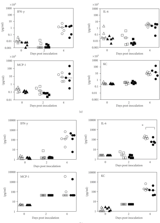
Figure3: Cytokine and chemokine levels in sera (a) and bronchoalveolar lavage (BAL) fluid (b) of mice inoculated by aerosol with type A F. tularensis. Groups of LTα−/−(open symbols) and LTα+/+ (closed symbols) mice (n=5) were challenged by aerosol with low-dose
type A F. tularensis strain, FSC033 (inhaled dose of∼10 organisms) on day 0, and blood samples and bronchoalveolar lavage fluid samples
were collected at dpi 0, 2, and 4. Cytokine and chemokine levels in the serum and BAL fluid were determined using the Beadlyte Mouse 21-Plex Cytokine Detection System on a Luminex 100 IS instrument. Each symbol represents the corresponding cytokine concentration of an individual mouse. Horizontal lines indicate the median of each group of mice on the indicated post-inoculation days. The detection limits of the assays were <5 pg/ml for both sera and BAL fluid.∗P < .05 versus LTα+/+ mice.
Table1: Comparison of cell populations in the bronchoalveolar lavage fluid of LTα−/−and LTα+/+ mice on day 2 and 4 following low-dose aerosol inoculation with type A F. tularensis.
Days post-inoculation Mouse strain Total cell count (×105)(a) Differential counts (%)
Macrophages Lymphocytes Neutrophils 2 LTα−/− 2.91±1.59 97.60±0.89 2.00±1.00 0.40±0.55 LTα+/+ 1.96±0.31 98.80±0.84 0.40±0.55 0.80±0.84
4 LTα−/− 3.02±0.76 95.20±3.27 2.40±1.52 2.40±1.82 LTα+/+ 2.70±0.55 94.80±6.76 0.20±0.45 5.00±6.82 (a)
The total leukocyte counts are expressed as absolute numbers, and differential counts are expressed as percentages. All data are mean±standard deviation (n = 5) in each group at each time point. No significant differences were observed between LTα−/−and LTα+/+ mice at any time point.
F. tularensis-induced cytokine responses following aerosol
challenge with the pathogen, levels of a panel of 21 cytokines and chemokines, including IFN-γ, IL-6, KC, and MCP-1, in the BAL and the sera of LTα−/−and LTα+/+ mice killed at
dpi 2 and 4 were measured. Overall, there was little change in the levels of the majority of assayed cytokines in either the BAL or the sera at dpi 2 or 4 in either mouse strain (data not shown). However, F. tularensis infection resulted in a substantial increase of MCP-1 and a moderate increase of KC in BAL fluid at dpi 2 (Figure 3(b)) and a substantial increase of IFN-γ, IL-6, KC, and MCP-1 in both BAL and sera at dpi 4 (Figures3(a)and3(b)), but again no differences were observed between the two mouse strains with the exception of IL-6, which was significantly higher in BAL fluid of LTα−/−mice than that of LTα+/+ mice (Figure 3(b), P < .05).
Recent studies have established that, in addition to its role in the organogenesis of secondary lymphoid organs, LTα plays an important role in host defenses against microbial infections (reviewed in [3]). However, the role of LTα in host defenses against infection appears to be complex and varies from pathogen to pathogen. In this study, we utilized LTα−/−mice and performed some preliminary studies to
examine the potential role of LTα in the host resistance to respiratory infection with virulent type A F. tularensis. Our results showed that LTα−/−mice had lower bacterial
burdens in their spleens on dpi 2 and higher bacterial burdens in their lungs on dpi 4 when compared to LTα+/+ mice but showed no overt differences in clinical outcome, tissue damage, or host immune responses to the infection.
Although our data suggest that LTα exerts some subtle influence over the course of aerosol-initiated tularemia, its mechanism of action remains unknown. The possible reasons are as follows: (1) LT-α is not crucial in host defense against this pathogen; (2) the role of LT-α can be compen-sated by other cytokines/chemokines in this infection model; and (3) the pathogen is too virulent and even immuno-competent hosts have little resistance to the infection. In this regard, we have previously shown that a number of immunodeficient mice show similar clinical outcome to the immunocompetent mice [17]. The lower bacterial burdens in the spleen of LTα−/−mice at dpi 2 could be explained
by a delay/reduction in the dissemination of bacteria from the lung since LTα−/− mice lack tracheobronchial lymph
nodes which normally are the major draining lymph nodes
for the lung. Once F. tularensis reached the spleens, however, bacteria quickly multiplied and by dpi 4, bacterial burdens were no longer significantly different in the spleens of LTα−/−and LTα+/+ mice. In fact, LTα−/− mice actually
seem to have slightly higher burdens in their spleens at this time point despite starting on dpi 2 with substantially lower bacterial burdens (seeFigure 2). Also by this time, LTα−/−
mice harbored significantly more bacteria in their lungs than LTα+/+ mice (P < .01), suggesting that LTα may play a role in host defense against F. tularensis infection which is distinct from its role in lymphoid organogenesis. Alternatively, the lack of draining lymph nodes in LTα−/−mice may simply
cause a delay in antigen presentation leading to a delayed or otherwise impaired antibacterial host response.
In summary, following a low-dose aerosol infection with the highly virulent type A strain of F. tularensis, LTα−/−mice
consistently harbored approximately 10-fold fewer bacteria in their spleens at dpi 2- and 10-fold more bacteria in their lungs at dpi 4 than LTα+/+ mice. However, the mortality and median time to death were indistinguishable between the two mouse strains. In addition, the inflammatory responses to the infection, as reflected by the cytokine levels and leukocyte influx in BAL fluid and histopathological analysis, were generally similar between LTα−/−and LTα+/+ mice.
These data suggest that although LTα does not contribute significantly to the resistance and host responses of mice to airborne type A F. tularensis infection; it does play a subtle role in the multiplication/dissemination of F. tularensis. ACKNOWLEDGMENT
This work was partially supported by Grants R01AI 48474 and R21AI59064 from the National Institutes of Health, USA and by the National Research Council, Canada (A-base). REFERENCES
[1] S. Saslaw, H. T. Eigelsbach, J. A. Prior, H. E. Wilson, and S. Carhart, “Tularemia vaccine study. II. Respiratory challenge,”
Archives of Internal Medicine, vol. 107, pp. 702–714, 1961.
[2] Y.-X. Fu, G. Huang, M. Matsumoto, H. Molina, and D. D. Chaplin, “Independent signals regulate development of pri-mary and secondary follicle structure in spleen and mesenteric lymph node,” Proceedings of the National Academy of Sciences
of the United States of America, vol. 94, no. 11, pp. 5739–5743,
6 Mediators of Inflammation
[3] T. W. Spahn, H.-P. Eugster, A. Fontana, W. Domschke, and T. Kucharzik, “Role of lymphotoxin in experimental models of infectious diseases: potential benefits and risks of a therapeutic inhibition of the lymphotoxin-β receptor pathway,” Infection
and Immunity, vol. 73, no. 11, pp. 7077–7088, 2005.
[4] C. F. Ware, “Network communications: lymphotoxins, LIGHT, and TNF,” Annual Review of Immunology, vol. 23, pp. 787–819, 2005.
[5] D. R. Roach, H. Briscoe, B. Saunders, M. P. France, S. Riminton, and W. J. Britton, “Secreted lymphotoxin-α is essential for the control of an intracellular bacterial infection,”
The Journal of Experimental Medicine, vol. 193, no. 2, pp. 239–
246, 2001.
[6] D. R. Roach, H. Briscoe, B. M. Saunders, and W. J. Britton, “Independent protective effects for tumor necrosis factor and lymphotoxin alpha in the host response to Listeria
monocytogenes infection,” Infection and Immunity, vol. 73, no.
8, pp. 4787–4792, 2005.
[7] S. Ehlers, C. H”olscher, S. Scheu, et al., “The lymphotoxin β receptor is critically involved in controlling infections with the intracellular pathogens Mycobacterium tuberculosis and
Listeria monocytogenes,” The Journal of Immunology, vol. 170,
no. 10, pp. 5210–5218, 2003.
[8] J. W. Conlan, W. Chen, H. Shen, A. Webb, and R. KuoLee, “Experimental tularemia in mice challenged by aerosol or intradermally with virulent strains of Francisella tularensis: bacteriologic and histopathologic studies,” Microbial
Patho-genesis, vol. 34, no. 5, pp. 239–248, 2003.
[9] W. Chen, E. A. Havell, L. L. Moldawer, K. W. McIntyre, R. A. Chizzonite, and A. G. Harmsen, “Interleukin 1: an important mediator of host resistance against Pneumocystis carinii,” The
Journal of Experimental Medicine, vol. 176, no. 3, pp. 713–718,
1992.
[10] R. KuoLee, X. Zhao, J. Austin, G. Harris, J. W. Conlan, and W. Chen, “Mouse model of oral infection with virulent type A
Francisella tularensis,” Infection and Immunity, vol. 75, no. 4,
pp. 1651–1660, 2007.
[11] W. Chen, R. KuoLee, H. Shen, M. B `usa, and J. W. Conlan, “Toll-like receptor 4 (TLR4) plays a relatively minor role in murine defense against primary intradermal infection with
Francisella tularensis LVS,” Immunology Letters, vol. 97, no. 1,
pp. 151–154, 2005.
[12] P. Zhang, W. R. Summer, G. J. Bagby, and S. Nelson, “Innate immunity and pulmonary host defense,”
Immunolog-ical Reviews, vol. 173, no. 1, pp. 39–51, 2000.
[13] K. L. Elkins, S. C. Cowley, and C. M. Bosio, “Innate and adaptive immune responses to an intracellular bacterium,
Francisella tularensis live vaccine strain,” Microbes and Infec-tion, vol. 5, no. 2, pp. 135–142, 2003.
[14] H. Andersson, B. Hartmanov´a, R. KuoLee, et al., “Transcrip-tional profiling of host responses in mouse lungs following aerosol infection with type A Francisella tularensis,” Journal of
Medical Microbiology, vol. 55, no. 3, pp. 263–271, 2006.
[15] A. Macela, J. Stulik, L. Hernychova, M. Kroca, Z. Krocova, and H. Kovarova, “The immune response against Francisella
tularensis live vaccine strain in Lpsn and Lpsdmice,” FEMS
Immunology & Medical Microbiology, vol. 13, no. 3, pp. 235–
238, 1996.
[16] H.-S. Kang, S. E. Blink, R. K. Chin, et al., “Lymphotoxin is required for maintaining physiological levels of serum IgE that minimizes Th1-mediated airway inflammation,” The Journal
of Experimental Medicine, vol. 198, no. 11, pp. 1643–1652,
2003.
[17] W. Chen, R. KuoLee, H. Shen, and J. W. Conlan, “Susceptibil-ity of immunodeficient mice to aerosol and systemic infection with virulent strains of Francisella tularensis,” Microbial
Patho-genesis, vol. 36, no. 6, pp. 311–318, 2004.
View publication stats View publication stats