HAL Id: hal-01579784
https://hal.sorbonne-universite.fr/hal-01579784
Submitted on 31 Aug 2017
HAL is a multi-disciplinary open access archive for the deposit and dissemination of sci-entific research documents, whether they are pub-lished or not. The documents may come from teaching and research institutions in France or abroad, or from public or private research centers.
L’archive ouverte pluridisciplinaire HAL, est destinée au dépôt et à la diffusion de documents scientifiques de niveau recherche, publiés ou non, émanant des établissements d’enseignement et de recherche français ou étrangers, des laboratoires publics ou privés.
oligonucleotides restore dystrophin production in
Duchenne Muscular Dystrophy myotubes
Vanessa Borges Pires, Ricardo Simões, Kamel Mamchaoui, Célia Carvalho,
Maria Carmo-Fonseca
To cite this version:
Vanessa Borges Pires, Ricardo Simões, Kamel Mamchaoui, Célia Carvalho, Maria Carmo-Fonseca. Short (16-mer) locked nucleic acid splice-switching oligonucleotides restore dystrophin production in Duchenne Muscular Dystrophy myotubes. PLoS ONE, Public Library of Science, 2017, 12 (7), pp.e0181065. �10.1371/journal.pone.0181065�. �hal-01579784�
Short (16-mer) locked nucleic acid
splice-switching oligonucleotides restore dystrophin
production in Duchenne Muscular Dystrophy
myotubes
Vanessa Borges Pires1, Ricardo Simões1, Kamel Mamchaoui2, Ce´lia Carvalho1☯*, Maria Carmo-Fonseca1☯
*
1 Instituto de Medicina Molecular, Faculdade de Medicina, Universidade de Lisboa, Lisboa, Portugal, 2 Center for Research in Myology, INSERM UMRS974, CNRS FRE3617, Sorbonne Universite´s, UPMC Univ
Paris 06, Paris, France
☯These authors contributed equally to this work.
*carmo.fonseca@medicina.ulisboa.pt(MCF);hcelia@medicina.ulisboa.pt(CC)
Abstract
Splice-switching antisense oligonucleotides (SSOs) offer great potential for RNA-targeting therapies, and two SSO drugs have been recently approved for treating Duchenne Muscular Dystrophy (DMD) and Spinal Muscular Atrophy (SMA). Despite promising results, new de-velopments are still needed for more efficient chemistries and delivery systems. Locked nucleic acid (LNA) is a chemically modified nucleic acid that presents several attractive properties, such as high melting temperature when bound to RNA, potent biological activity, high stability and low toxicity in vivo. Here, we designed a series of LNA-based SSOs com-plementary to two sequences of the human dystrophin exon 51 that are most evolutionary conserved and evaluated their ability to induce exon skipping upon transfection into myo-blasts derived from a DMD patient. We show that 16-mers with 60% of LNA modification effi-ciently induce exon skipping and restore synthesis of a truncated dystrophin isoform that localizes to the plasma membrane of patient-derived myotubes differentiated in culture. In sum, this study underscores the value of short LNA-modified SSOs for therapeutic applications.
Introduction
Antisense oligonucleotides are powerful tools to modulate gene expression. Namely, antisense oligonucleotides can be used to induce RNA interference or RNase H based mechanisms of gene down-regulation, inhibit the function of microRNAs, or modulate splicing (for a recent review see [1]). Despite early clinical trial failures, new developments are fueling optimism in the antisense field. In particular, the design of novel chemical modifications and delivery sys-tems is improving the potency and efficacy of these drugs in RNA-targeting therapeutic a1111111111 a1111111111 a1111111111 a1111111111 a1111111111 OPEN ACCESS
Citation: Pires VB, Simões R, Mamchaoui K, Carvalho C, Carmo-Fonseca M (2017) Short (16-mer) locked nucleic acid splice-switching oligonucleotides restore dystrophin production in Duchenne Muscular Dystrophy myotubes. PLoS ONE 12(7): e0181065.https://doi.org/10.1371/ journal.pone.0181065
Editor: Emanuele Buratti, International Centre for Genetic Engineering and Biotechnology, ITALY Received: December 16, 2016
Accepted: June 26, 2017 Published: July 24, 2017
Copyright:© 2017 Pires et al. This is an open access article distributed under the terms of the Creative Commons Attribution License, which permits unrestricted use, distribution, and reproduction in any medium, provided the original author and source are credited.
Data Availability Statement: All relevant data are within the paper and its Supporting Information files.
Funding: This work was supported by Fundac¸ão para a Ciência e Tecnologia, Portugal (FCT-ANR/ BIM-ONC/0009/2013),http://www.fct.pt/; Faculdade de Medicina da Universidade de Lisboa, Gabinete de Apoioà Investigac¸ão Cientı´fica, Tecnolo´gica e Inovac¸ão, Portugal (GAPIC 2012009 GAPIC 20130041 (RS)),http://www.medicina.
applications [1]. Recently, antisense oligonucleotide drugs have been approved for treating Duchenne Muscular Dystrophy (DMD) [2] and Spinal Muscular Atrophy [3].
DMD was the first disease shown to benefit from antisense oligonucleotides designed to modulate pre-mRNA splicing. This X-linked genetic disease is mainly caused by frame-shift-ing deletions or nonsense mutations in theDMD gene that result in a loss of functional
dystro-phin protein [4], leading to fatal progressive muscle wasting [5]. A related but milder form of the disease called Becker Muscular Dystrophy (BMD) is caused by in-frame mutations in the
DMD gene that allow expression of an internally truncated but partially functional protein [6]. Antisense oligonucleotides have been extensively used to induce skipping of the exon contain-ing a frame-shift deletion or a nonsense mutation, thus restorcontain-ing the mRNA readcontain-ing frame and producing an internally deleted protein similar to that observed in BMD patients. Due to mutation clustering in hotspot regions, the largest fraction of patients (approximately 13%) would benefit from skipping of exon 51 directed therapy [7].
To date, two distinct splice-switching antisense oligonucleotides (SSOs) complementary to the human dystrophin exon 51 sequence have been tested in clinical trials: drisapersen (Pro-sensa/GlaxoSmithKlein, presently BioMarin) and eteplirsen (Sarepta Therapeutics). Drisaper-sen is a 24-mer based on phosphorothioated 2’-O-methyl RNA chemistry and eteplirDrisaper-sen is a 30-nucleotide phosphorodiamidate morpholino. The promising results obtained prompted the accelerated FDA approval of Eteplirsen (Exondys 51). However, the levels of dystrophin restoration reported in these trials are still low [8–13]. Thus, much attention is devoted to find-ing new strategies that might improve the pharmacological properties of SSOs.
An alternative chemical modification with attractive properties for clinical applications is locked nucleic acid (LNA) [14]. LNA is a nucleotide analog carrying an altered ribose in which a methylene bridge connects the 2’-O with the 4’-C atoms in the furanose ring [15,16]. This bridge enables LNA to form a strictlyN-type conformation that enhances the binding affinity
against complementary RNA [17–19]. Several studies have shown that introducing LNA into antisense oligonucleotides is advantageous for a variety of gene silencing techniques [20,21] and recent clinical trials with an LNA-modified oligonucleotide targeting miRNA-122 (mira-virsen) suggest that this may be an effective and safe strategy for patients chronically infected with hepatitis C virus [22]. LNA-based SSOs have also been tested in cellular and mouse mod-els with encouraging results [23–28]. Inspired by a recent study that systematically addressed the optimal design of LNA-based SSOs targeting the human dystrophin exon 58 sequence [29], here we designed a series of LNA-modified antisense oligonucleotides and evaluated their ability to induce skipping of the dystrophin exon 51 in myoblast cells derived from a DMD patient.
Materials and methods
Databases
The sequence ofDMD gene orthologues was analyzed on Ensembl Genome Browser [30]. Sequence alignment was performed on CLUSTALW (GenomeNet). UCSC Genome Browser [31] was used for visualization of the humanDMD gene.
Oligonucleotides
Splice-switching antisense oligonucleotides (SSOs) that contain a fully phosphorothioate mod-ified backbone and 60% LNA, with two LNA-modmod-ified nucleotides at the 3’-end and one LNA-modified nucleotide at the 5’-end, were purchased from Exiqon (Vedbaek, Denmark). The AO51 2’-O-methyl-phosphorothioate SSO was gently provided by C. Trollet [32].
ulisboa.pt/investigacao/gapic/. The funders had no role in study design, data collection and analysis, decision to publish, or preparation of the manuscript.
Competing interests: The authors have declared that no competing interests exist.
Cell lines
Immortalized myoblasts derived from a DMD patient (DM8036) and from a control individ-ual (KM155) were previously described [32]. Cells were maintained in Skeletal Muscle Cell Growth Medium (PromoCell) in 5% CO2, at maximum confluence of 60–70%. To induce
dif-ferentiation, cells were plated at a density intended to reach 80% confluence 24 hours later, and then the medium was switched to differentiation medium: DMEM high glucose (Gibco) supplemented with ITS—Insulin, Transferrin, and Sodium Selenite (Sigma). To maintain cells differentiating, half of the differentiation medium was replaced each 2–3 days.
Cell transfection
SSOs were transfected using Lipofectamine RNAimax Reagent (Invitrogen) according to the manufacturer’s instructions. Specifically, for a cell culture area of 1.9 cm2, 1μL of Lipofectamine RNAimax in 15μL of OptiMEM (Gibco) was added to 15 μL of SSO diluted in OptiMEM. For mock transfection, no SSO was used. Transfection was performed either simultaneously with myoblast platting or 3 days after initiating differentiation, as indicated on results.
RNA isolation and reverse transcription-PCR
Total RNA was extracted using PureZol RNA isolation reagent (BioRad) and purified using DNAse I treatment (Roche) and acidic phenol extraction with UltraPureTM Phenol:Cloro-form:Isoamyl Alcohol (25:24:1, v/v; Invitrogen) according to the manufacturer’s instructions. Purity and quantity of purified RNA was analyzed using Nanodrop2000 (ThermoScientific). For cDNA synthesis, 0.6μg of RNA was retrotranscribed in a 20 μL reaction using Transcrip-tor High Fidelity cDNA Synthesis Kit (Roche) and 20 pmol of reverse primerDMD_exon_54
(5’-GGAGAAGTTTCAGGGCCAAG-3’), at 55˚C for 90 min. 0.5μL of cDNA was PCR-ampli-fied in a 10μL reaction with KAPA2GTMFast (Kapa Biosystems), with the primers
DMD_ex-on_47F (5’-ACCCGTGCTTGTAAGTGCTC-3’) andDMD_exon_53R (5’-TGACTCAAGCT
TGGCTCTGG-3’). The cycling conditions were 95˚C, 5 min, followed by 45 cycles of 95˚C, 15 sec; 58˚C, 30 sec and 72˚C, 15 sec. PCR products were separated by electrophoresis on a 2% agarose gel followed by imaging on Chemidoc XRS+system (BioRad) and band intensity quantification using the Image Lab 5.2 software (BioRad). For statistics and EC50
determina-tion, Graphpad Prism 6 (GraphPad Software) was used.
Protein extraction and Western blot
Protein extraction and Western blot methods were adapted from previous descriptions [33,34]. Cells on 3.8 cm2culture plates were washed briefly with phosphate-buffered saline (PBS) at room temperature and then lysed in 40μL of a buffer containing 0.1 M Tris-HCl pH 6.8 and 20% sodium dodecyl sulfate (SDS). Protein concentration was determined using the PierceTMBCA Protein Assay kit (ThermoScientific) according to the manufacturer’s instruc-tions. Before SDS-PAGE, protein samples were treated with 1% Benzonase (Sigma), 14mM MgCl2for 15 min, the homogenate was completed to a final composition of 75 mM Tris-HCl
pH 6.8, 15% sodium dodecyl sulfate, 5% dithiothreitol, 20% glycerol and 0.01% bromophenol blue, and then boiled for 5 min. For Western Blot analysis, 1μg of protein extract was loaded on a 7% polyacrylamide gel and run for 90 min (10 min at 60 V + 80 min at 100 V), with Preci-sion Plus Protein Standards (BioRad) as molecular weight marker. Transfer buffer (0.6% Tris, 2.88% glycine, 0.01%SDS, 4% methanol) was used to transfer proteins to nitrocellulose BA85 membranes (Whatman Protran) with a Tank Transfer System (BioRad Mini Trans-Blot R Cell) for 90 min at 300 mA. Blots were first incubated with 5% non-fat dried milk in
Tris-buffered saline containing 0.05% Tween-20 (TBST), for 1 hour, and then incubated overnight at 4˚C with rabbit polyclonal anti-dystrophin antibody (Abcam ab85302, diluted 1:1000 in 5% non-fat dried milk in TBST). Secondary antibody Goat Anti-Rabbit IgG (H+L)-HRP (BioRad, dilution 1:3000), was incubated for 60 min at room temperature. The polyclonal antibody rab-bit anti-Lamin A/C (Santa Cruz) was used as a loading control (overnight incubation at 4˚C; dilution 1:10000). WesternBright Quantum (Advansta) was used to chemiluminescent detec-tion and imaging with the Chemidoc XRS+system (BioRad).
Immunofluorescence microscopy
For microscopy analysis, cells were cultured onto 0.1% gelatin-coated glass coverslips (10x10 mm2, Normax). Cells were first fixed with 3.7% formaldehyde (freshly prepared from parafor-maldehyde) in phosphate-buffered saline (PBS) for 10 min at room temperature, and then per-meabilized with 0.5%Triton X-100 in PBS for 10 min at room temperature. Next, cells were incubated for 30 min at room temperature in 1%BSA and 0.05%Tween20 in PBS. Cells were then incubated with polyclonal rabbit anti-dystrophin antibody (Abcam ab85302, dilution 1:100) for 60 min at room temperature followed by incubation with tetramethylrhodamine (TRITC) conjugated donkey anti-rabbit antibody (Jackson ImmunoResearch; dilution 1:200) for 60 min. Antibodies were diluted in 1%BSA, 0.05%Tween20 in PBS. To counterstain the nuclei, cells were incubated for 10 min with 1μg/mL 4’,6-diamidino-2-phenylindole (DAPI; Sigma). VECTASHIELD1 Antifade Mounting medium (Vector Laboratories) was used. Cells were imaged with a LSM 710 Confocal Point-Scanning Microscope (ZEISS), using the lasers Diode 405–30 (405 nm) and DPSS 561–10 (561 nm).
Results
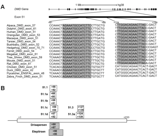
The rational design of an exon skipping oligonucleotide involves the selection of an antisense sequence targeting the exon that is to be skipped. Several parameters influence SSO activity, including the melting temperature, guanine-cytosine content, length of the oligonucleotide, and secondary structures or sequence motifs that correspond to splicing signals of the target RNA [35–38]. Here, we explored sequence conservation as a complementary strategy to design SSOs. A search for the longest evolutionary conserved stretches of DNA that are unique to exon 51 of the humanDMD gene revealed two regions. One region contains a sequence of 15
nucleotides (from +68 to +82;Fig 1A) that is conserved across mammalian species ranging from primates (Human, Orangutan, Macaque, Tarsier, Bushbaby), to Tree-Shrew, Megabat, Hedgehog, Alpaca, Dolphin, Ferret, and rodents (Mouse and Rat), as well as bird species such as Chicken, Duck, Flycatcher, and Zebra-finch. Noteworthy, this highly conserved sequence is included in the region targeted by the drisapersen and eteplirsen SSOs that have been used in clinical trials. To investigate the influence of oligonucleotide length on the ability to induce exon skipping, we synthesized a 15-mer SSO targeting the conserved sequence (51.1), and additional 13-, 14- and 16-mers SSOs shifted by one or two nucleotides (Fig 1B). Another region contains a sequence of 15 nucleotides (from +125 to +139;Fig 1A) conserved across the 13 mammalian species analyzed. We synthesized a 13-mer SSO targeting this sequence (51.3), and an additional 16-mer SSO shifted by three nucleotides (Fig 1B).
To evaluate whether these SSOs modulate the splicing of endogenous human dystrophin pre-mRNA, we used myogenic cells derived from a DMD patient carrying a deletion of exons 48–50 that would be frame-corrected by skipping exon 51 [32]. Cultured myoblasts were trans-fected with each SSO and one day after transfection the cells were induced to differentiate into myotubes.
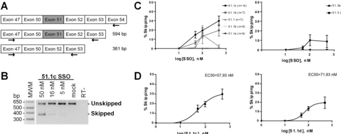
For splice switching evaluation, transcripts were analyzed by RT-PCR. RNA was extracted from differentiated cultures, retrotranscription was performed with a primer specific forDMD
exon 54 (Fig 2A) and PCR reactions with forward and reverse primers on exons 47 and 53, respectively (Fig 2A). In mock transfected cells, we observed a single band of approximately 600 bp that corresponds toDMD transcripts including exon 51 (Fig 2B). In cells transfected with SSO 51.1c, we detected a second band of approximately 360 bp that corresponds toDMD
transcripts excluding exon 51 (Fig 2B). In order to assess the efficiency of exon skipping in-duced by each SSO, we quantified the relative intensity of the bands corresponding to skipped and unskipped transcripts observed in agarose gels from independent experiments (S1 Fig). Skipping percentage was calculated as [skipped transcript/(skipped + unskipped transcripts)] x
100. The skipping activity of the 16-mer 51.1c ranged between 15 and 25% in a concentration-dependent manner (Fig 2C). Shifting the SSO sequence by only two nucleotides (16-mer 51.1d) resulted in slightly lower efficiency while shorter SSOs (15-mer 51.1; 13-mer 51.1a; and 14-mer 51.1b) had very little (<10%) skipping activity (Fig 2C). The 13-mer 51.3 also pre-sented very little skipping activity, whereas the 16-mer 51.3a targeting the same region had an efficiency in the range of 15% (Fig 2C). For SSOs 51.1c and 51.1d, a dose-response curve was generated taking into account the skipping percentage observed after transfection of different oligonucleotide concentrations. From these curves we estimated values for the half maximal effective concentration (EC50) in the range of 50 to 75 nM (Fig 2D).
Fig 1. Oligonucleotide design. (A) Schematic representation of the human dystrophin gene and sequence
alignment of two exon 51 regions across the indicated species. Conserved nucleotide sequences are shadowed. (B) Annealing sites of SSOs designed in this study are indicated by boxes. For comparison, the annealing sites of Eteplirsen and Drisapersen are indicated by shaded boxes. Position +1 corresponds to the first nucleotide in exon 51.
We also observed that the skipping activity tended to be higher in cells analyzed 3 days after transfection (Fig 2B) than in cells analyzed 8 days post-transfection (S1 Fig). Since cells in-duced to differentiate no longer divide, it is unlikely that this effect is due to dilution of the oli-gonucleotides in daughter cells. One possibility is that either the transfection procedure or the presence of SSOs interferes with the differentiation process and/or cell viability, and therefore long-term differentiated cultures become enriched in non-modified cells. Another possibility is that SSOs are not efficiently recycled after annealing to nascent pre-mRNAs.
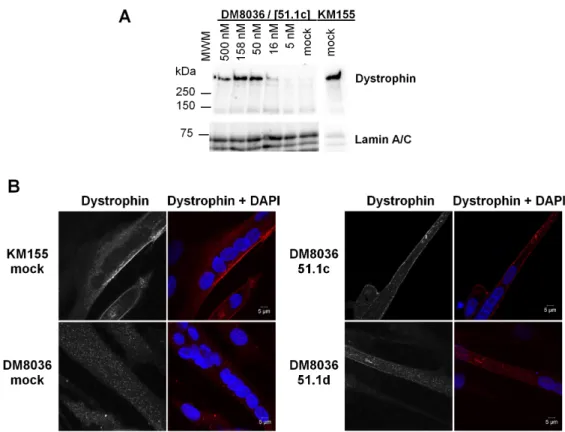
In order to evaluate dystrophin protein synthesis restoration by LNA-SSO 51.1c, we used Western blot analysis. In control cells from a healthy individual (KM155), Western blot analy-sis with an anti-dystrophin antibody revealed a single high molecular weight band (Fig 3A) consistent with the size of wild-type dystrophin (427 kDa). As expected, no dystrophin band was observed in mock transfected patient DM8036 cells (Fig 3A). Transfection with LNA-SSO 51.1c resulted in the appearance of a band corresponding to truncated dystrophin protein with an estimated size of approximately 400 kDa (Fig 3A).
The localization of the rescued truncated protein in differentiated myotubes was assessed by immunofluorescence with anti-dystrophin antibody (Fig 3B). The vast majority of myo-tubes derived from KM155 cells were clearly labeled at the plasma membrane (Fig 3B), consis-tent with the well-established dystrophin localization at the cytoplasmic face of the muscle cell plasma membrane, or sarcolemma [39]. In contrast, practically all myotubes derived from patient DM8036 cells did not show any dystrophin labeling (Fig 3B). Upon transfection of LNA-SSOs 51.1c and 51.1d (Fig 3B), dystrophin labeling at the plasma membrane was observed in a subset of myotubes derived from DM8036 cells.
Next, we compared the results obtained with short LNA-modified SSOs with a previously described oligonucleotide that carries full-length 2’-O-methyl-substituted ribose molecules
and phosphorothioate internucleotide linkages, AO51 [32]. This oligonucleotide is also known as h51AON1 [40] and PRO051 [41]. Following successful results obtained in preclinical studies [42] and clinical trials [41,43], this oligonucleotide has been developed as an experimental drug termed drisapersen. However, the FDA rejected drisapersen in early 2016, due to safety
Fig 2. RT-PCR analysis of exon 51 skipping. (A) Schematic representation of DMD exons with the location of
primers used for retrotranscription and RT-PCR analysis of exon 51 skipping; expected sizes of PCR products are indicated. (B) DM8036 cells were transfected with SSO 51.1c at the indicated concentrations and analyzed 2 days after induction of myotube differentiation. Electrophoresis of PCR products in agarose gels shows non-skipped and skipped transcripts. Mock: mock transfection; -RT: no retrotranscriptase. (C) Percentage of exon skipping induced by each SSO at day 7 post-induction of myotube differentiation. Values were obtained from quantification of the gels shown inS1 Figand represent mean±SEM. (D) Dose-response curves of the two most efficient SSOs. EC50: half maximal effective concentration.
issues [2]. As shown inFig 4andS2 Fig, the 16-mer LNA-modified SSO 51.1c is more effective than AO51 in inducing exon skipping and restoring synthesis of a truncated dystrophin iso-form. We further observed that upon transfection into myoblasts both oligonucleotides have a similar effect on myotube differentiation (S3 Fig).
Fig 3. Analysis of dystrophin protein restoration. (A) Western blot analysis of total proteins extracted from
cells derived from a healthy individual (KM155) and a DMD patient (DM8036). Patient myoblasts were either transfected with SSO 51.1c at the indicated concentrations or mock transfected. Western blotting was carried out at day 2 of differentiation using antibodies to dystrophin and Lamin A/C. The position of molecular weight markers (MWM) is indicated. (B) Immunofluorescence analysis of cells derived from a healthy individual (KM155) and a DMD patient (DM8036) at day 7 of differentiation using anti-dystrophin antibody (red staining). Nuclei were counterstained with DAPI (blue staining). Patient myoblasts were either transfected with the indicated SSOs at 50 nM or mock transfected.
https://doi.org/10.1371/journal.pone.0181065.g003
Fig 4. Comparison of LNA-SSOs and AO51. (A) DM8036 myoblasts were transfected with the indicated SSOs
at 50nM and analyzed at day 2 of differentiation. Electrophoresis of PCR products in agarose gels shows non-skipped and non-skipped transcripts. Mock: mock transfection. (B) Percentage of exon skipping induced by each SSO. Values were obtained from quantification of gels (shown in A andS2 Fig) and represent mean±SD (N = 4). (C) Western blot analysis of total proteins extracted from cells derived from a healthy individual (KM155) and a DMD patient (DM8036). Patient myoblasts were transfected with the indicated SSOs at 50 nM and induced to differentiate. Western blotting was carried out at day 5 of differentiation using antibodies to dystrophin and Lamin A/C.
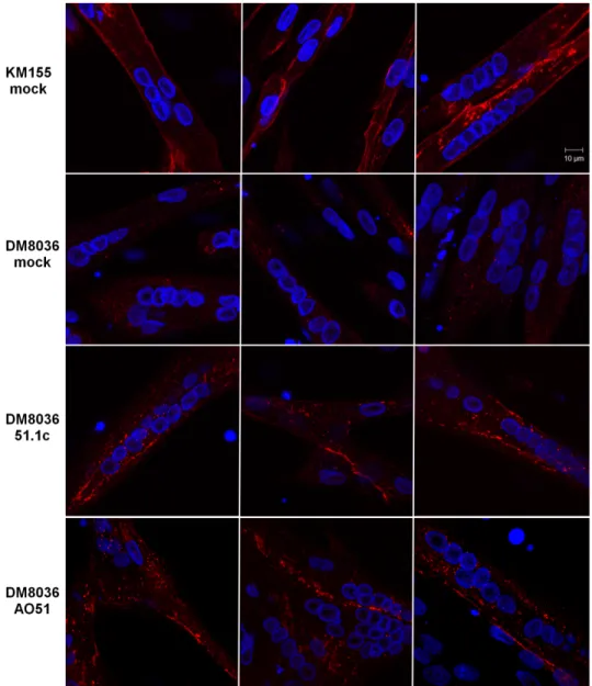
Finally, we tested whether LNA-SSO 51.1c can rescue dystrophin production in differenti-ated myotubes. DM8036 myoblasts were first induced to differentiate for 3 days prior to SSO transfection. As shown inS3 Fig, approximately 70% of all nuclei observed in cultures that were not transfected with SSOs and were induced to differentiate for 3 days are present in mul-tinucleated myotubes indicating that under these culture conditions the majority of myoblasts differentiated into myotubes. Upon transfection with either LNA-SSO 51.1c or AO51, immu-nofluorescence analysis reveals the presence of restored dystrophin localized to the plasma membrane (Fig 5). Thus, as previously shown for AO51, LNA-SSO 51.1c is most likely active in rescuing dystrophin production in differentiated muscle.
In conclusion, we demonstrate that 16-mer LNA-modified SSOs targeting a unique se-quence inDMD exon 51 that has been highly evolutionary conserved effectively induce exon
Fig 5. Dystrophin rescue in myotubes. DM8036 and KM155 myoblasts were induced to differentiate for 3
days. Myotubes were then either mock transfected or transfected with the indicated SSOs at 50 nM and fixed 2 days later. Immunofluorescence was carried out with anti-dystrophin antibody (red staining) and nuclei are highlighted with DAPI (blue staining).
skipping and restore synthesis of a truncated dystrophin protein isoform that localizes to the plasma membrane of patient-derived myotubes differentiated in culture.
Discussion
The selection of an antisense oligonucleotide sequence capable of inducing exon skipping is difficult to predict. Although statistical modeling approaches have been used to derive algo-rithms that predict exon skipping efficacy [44], experimental screening of multiple oligonucle-otides remains the most reliable strategy. Efficient exon skipping requires that the SSO binds tightly to the nascent pre-mRNA and prevents assembly of the spliceosome at the target splice sites. Accordingly, LNA-modified SSOs targeting either the acceptor or donor splice sites of theDMD exon 58 were shown to be effective in inducing exon skipping. In addition, SSOs
tar-geting exonic regions have also been reported to induce exon skipping, most probably through interference with RNA structures and/or binding of regulatory proteins that are critical for spliceosome assembly and splicing [29,38,45,46]. Because sequences important for splicing control tend to be evolutionary conserved [47,48], we designed antisense oligonucleotides tar-geting the two most conserved unique sequences inDMD exon 51 and we show that both are
effective in inducing exon skipping. Thus, evolutionary conservation is probably a good crite-rion for selection of SSOs.
The results reported in this study using oligonucleotides with 60% of LNA-modified bases are in good agreement with previous observations indicating that 15-mer SSOs containing 8 LNA units showed high exon skipping activity [29]. SSOs fully modified with LNA were previ-ously reported to have lower skipping activity, presumably because they form stable self-dimers or fail to recycle after splicing inhibition [20,29,49,50].
Our results further reveal that 16-mer LNA-SSOs effectively induce skipping of dystrophin exon 51. These LNA-SSOs are significantly shorter than drisapersen (24-mer) and eteplirsen (30-mers), which have been used in clinical trials. Shorter oligonucleotides are advantageous because they can bind more specifically [51]. This is because longer oligonucleotides have a higher chance of binding to off-target sequences containing a few mismatches [51]. LNA oligo-nucleotides as short as 8-mers were shown to bind and inhibit microRNA activity without off-target effects [52], and 7-mer LNA-SSOs were reported to modulate splicing [29]. However, for each target exon there are optimal sequences, lengths and number of LNA modifications. For example, a 13-mer LNA-SSO induced skipping ofDMD exon 58 with higher efficacy than
a 7-mer, and SSOs longer than 15 nucleotides showed reduced activity [29].
In addition to assessing the effect of SSOs by RT-PCR, we demonstrate that 16-mer LNA-modified SSOs effectively restore synthesis of a truncated dystrophin protein isoform that localizes to the plasma membrane of patient-derived myotubes differentiated in culture. Thus, our results strengthen the view that introducing LNA modifications in oligonucleotides allows for the use of shorter antisense sequences for therapeutic modulation of splicing.
Supporting information
S1 Fig. RT-PCR analysis of exon 51 skipping. DM8036 cells were transfected with each SSO
at the indicated concentrations and analyzed 7 days after induction of myotube differentiation. Electrophoresis of PCR products in agarose gels shows non-skipped and skipped transcripts in independent biological experiments. Mock: mock transfection; -RT: no retrotranscriptase. (TIF)
S2 Fig. RT-PCR comparison of LNA-SSOs and AO51. DM8036 cells were transfected with
differentiation. Electrophoresis of PCR products in agarose gels shows non-skipped and skipped transcripts in independent biological experiments. Mock: mock transfection; -RT: no retrotranscriptase.
(TIF)
S3 Fig. Effect of SSO transfection on myotube differentiation. DM8036 cells were either
mock transfected or transfected with the indicated SSO at 50 nM. (A) Cells were observed by phase-contrast microscopy 3 and 5 days after induction of myotube differentiation. (B) Fusion index was calculated as the percentage of total nuclei in myotubes relative to the total number of nuclei. At least 200 nuclei were counted in each experiment. Error bars represent standard deviation (N = 3).
(TIF)
Acknowledgments
We are grateful to Annemieke Aartsma-Rus for helpful discussion and advice, the Association Franc¸aise Contre les Myopathies and the Platform for the Immortalization of Human Cells and Collaboration, Institut de Myologie, Paris, for the immortalized human cell lines, and Capucine Trollet (Centre de Recherche en Myologie, Sorbonne Universite´s, Paris) for oligonu-cleotide AO51. We also thank Ana de Jesus and Ana Margarida Nascimento for technical support.
Author Contributions
Conceptualization: Ricardo Simões, Ce´lia Carvalho, Maria Carmo-Fonseca.
Formal analysis: Vanessa Borges Pires. Funding acquisition: Maria Carmo-Fonseca.
Investigation: Vanessa Borges Pires, Ricardo Simões, Ce´lia Carvalho.
Methodology: Vanessa Borges Pires, Ricardo Simões, Kamel Mamchaoui, Ce´lia Carvalho.
Project administration: Maria Carmo-Fonseca. Resources: Kamel Mamchaoui.
Supervision: Ce´lia Carvalho, Maria Carmo-Fonseca. Validation: Ce´lia Carvalho, Maria Carmo-Fonseca. Visualization: Vanessa Borges Pires.
Writing – original draft: Vanessa Borges Pires, Ce´lia Carvalho, Maria Carmo-Fonseca. Writing – review & editing: Ce´lia Carvalho, Maria Carmo-Fonseca.
References
1. McClorey G, Wood MJ. An overview of the clinical application of antisense oligonucleotides for RNA-tar-geting therapies. Curr Opin Pharmacol. 2015; 24: 52–58.https://doi.org/10.1016/j.coph.2015.07.005
PMID:26277332
2. Lim KRQ, Maruyama R, Yokota T. Eteplirsen in the treatment of Duchenne muscular dystrophy. Drug Des Devel Ther. 2017; 533–545.https://doi.org/10.2147/DDDT.S97635PMID:28280301
3. Corey DR. Nusinersen, an antisense oligonucleotide drug for spinal muscular atrophy. Nat Neurosci. 2017; 20:497–499.https://doi.org/10.1038/nn.4508PMID:28192393
4. Hoffman EP, Brown RH, Kunkel LM. Dystrophin: The protein product of the Duchenne muscular dystro-phy locus. Cell. 1987; 51: 919–928. PMID:3319190
5. Flanigan KM. Duchenne and Becker muscular dystrophies. Neurol Clin. 2014; 32:671–688.https://doi. org/10.1016/j.ncl.2014.05.002PMID:25037084
6. Koenig M, Beggs AH, Moyer M, Scherpf S, Heindrich K, Bettecken T, et al. The molecular basis for Duchenne versus Becker muscular dystrophy: correlation of severity with type of deletion. Am J Hum Genet. 1989; 45:498–506. PMID:2491009
7. Aartsma-Rus A, Fokkema I, Verschuuren J, Ginjaar I, Van Deutekom J, Van Ommen GJ, et al. Theo-retic applicability of antisense-mediated exon skipping for Duchenne muscular dystrophy mutations. Hum Mutat. 2009; 30: 293–299.https://doi.org/10.1002/humu.20918PMID:19156838
8. Goemans NM, Tulinius M, van den Akker JT, Burm BE, Ekhart PF, Heuvelmans N, et al. Systemic administration of PRO051 in Duchenne’s muscular dystrophy. The New England journal of medicine. 2011.https://doi.org/10.1056/NEJMoa1011367PMID:21428760
9. Cirak S, Arechavala-Gomeza V, Guglieri M, Feng L, Torelli S, Anthony K, et al. Exon skipping and dys-trophin restoration in patients with Duchenne muscular dystrophy after systemic phosphorodiamidate morpholino oligomer treatment: An open-label, phase 2, dose-escalation study. Lancet. 2011; 378: 595–605.https://doi.org/10.1016/S0140-6736(11)60756-3PMID:21784508
10. Mendell JR, Rodino-Klapac LR, Sahenk Z, Roush K, Bird L, Lowes LP, et al. Eteplirsen for the treatment of Duchenne muscular dystrophy. Ann Neurol. 2013; 74: 637–47.https://doi.org/10.1002/ana.23982
PMID:23907995
11. Voit T, Topaloglu H, Straub V, Muntoni F, Deconinck N, Campion G, et al. Safety and efficacy of drisa-persen for the treatment of Duchenne muscular dystrophy (DEMAND II): An exploratory, randomised, placebo-controlled phase 2 study. Lancet Neurol. 2014; 13: 987–996. https://doi.org/10.1016/S1474-4422(14)70195-4PMID:25209738
12. Kole R, Krieg AM. Exon skipping therapy for Duchenne muscular dystrophy. Adv Drug Deliv Rev. 2015; 87: 104–107.https://doi.org/10.1016/j.addr.2015.05.008PMID:25980936
13. FDA summary review. Exondys 51 injection (eteplirsen). Available at:www.accessdata.fda.gov.
14. Lundin KE, Hansen BR, Persson R, Bramsen JB, Koch T, Wengel J, et al. Biological Activity and Bio-technological Aspects of Locked Nucleic Acids. Adv Genet. 2013; 82:47–107.https://doi.org/10.1016/ B978-0-12-407676-1.00002-0PMID:23721720
15. Singh SK, Nielsen P, Koshkin AA, Wengel J. LNA (locked nucleic acids): synthesis and high-affinity nucleic acid recognition. Chem Commun. 1998; 455–456.
16. Obika S, Nanbu D, Hari Y, Morio K, In Y, Ishida T, et al. Synthesis of 20-O,40-C-methyleneuridine and -cytidine. Novel bicyclic nucleosides having a fixed C3, -endo sugar puckering. Tetrahedron Lett. 1997; 38: 8735–8738.https://doi.org/10.1016/S0040-4039(97)10322-7
17. Braasch DA, Corey DR. Locked nucleic acid (LNA): fine-tuning the recognition of DNA and RNA. Chem Biol. Elsevier; 2001; 8: 1–7.https://doi.org/10.1016/S1074-5521(00)00058-2PMID:11182314
18. Bondensgaard K, Petersen M, Singh SK, Rajwanshi VK, Kumar R, Wengel J, et al. Structural Studies of LNA:RNA Duplexes by NMR: Conformations and Implications for RNase H Activity. Chemistry. 2000; 6: 2687–2695.https://doi.org/10.1002/1521-3765(20000804)6:15<2687::AID-CHEM2687>3.0.CO;2-U
PMID:10985717
19. Vester B, Wengel J. LNA (Locked Nucleic Acid): High-Affinity Targeting of Complementary RNA and DNA. Biochemistry. 2004; 43: 13233–41.https://doi.org/10.1021/bi0485732PMID:15491130
20. Straarup EM, Fisker N, Hedtja¨rn M, Lindholm MW, Rosenbohm C, Aarup V, et al. Short locked nucleic acid antisense oligonucleotides potently reduce apolipoprotein B mRNA and serum cholesterol in mice and non-human primates. Nucleic Acids Res. 2010; 38: 7100–7111.https://doi.org/10.1093/nar/gkq457
PMID:20615897
21. Franck N, Maris M, Nalbandian S, Talukdar S, Schenk S, Hofmann HP, et al. Knock-down of IL-1Ra in obese mice decreases liver inflammation and improves insulin sensitivity. PLoS One. 2014; 9: 1–10.
https://doi.org/10.1371/journal.pone.0107487PMID:25244011
22. van der Ree MH, van der Meer AJ, de Bruijne J, Maan R, van Vliet A, Welzel TM, et al. Long-term safety and efficacy of microRNA-targeted therapy in chronic hepatitis C patients. Antiviral Res. 2014; 111: 53– 59.https://doi.org/10.1016/j.antiviral.2014.08.015PMID:25218783
23. Guterstam P, Lindgren M, Johansson H, Tedebark U, Wengel J, El Andaloussi S, et al. Splice-switching efficiency and specificity for oligonucleotides with locked nucleic acid monomers. Biochem J. 2008; 412: 307–13.https://doi.org/10.1042/BJ20080013PMID:18271753
24. Aartsma-Rus A, Kaman WE, Bremmer-Bout M, Janson AA, den Dunnen JT, van Ommen GJ, et al. Comparative analysis of antisense oligonucleotide analogs for targeted DMD exon 46 skipping in mus-cle cells. Gene Ther. 2004; 11: 1391–1398.https://doi.org/10.1038/sj.gt.3302313PMID:15229633
25. Graziewicz MA, Tarrant TK, Buckley B, Roberts J, Fulton L, Hansen H, et al. An endogenous TNF-alpha antagonist induced by splice-switching oligonucleotides reduces inflammation in hepatitis and
arthritis mouse models. Mol Ther. 2008; 16: 1316–22.https://doi.org/10.1038/mt.2008.85PMID:
18461057
26. Ittig D, Liu S, Renneberg D, Schu¨mperli D, Leumann CJ. Nuclear antisense effects in cyclophilin A pre-mRNA splicing by oligonucleotides: a comparison of tricyclo-DNA with LNA. Nucleic Acids Res. 2004; 32: 346–53.https://doi.org/10.1093/nar/gkh187PMID:14726483
27. Roberts J, Palma E, Sazani P,Ørum H, Cho M, Kole R. Efficient and Persistent Splice Switching by Systemically Delivered LNA Oligonucleotides in Mice. Mol Ther. 2006; 14: 471–475.https://doi.org/10. 1016/j.ymthe.2006.05.017PMID:16854630
28. Yilmaz-Elis AS, Aartsma-Rus A, ‘t Hoen PA, Safdar H, Breukel C, van Vlijmen BJ, et al. Inhibition of IL-1 Signaling by Antisense Oligonucleotide-mediated Exon Skipping of IL-1 Receptor Accessory Protein (IL-1RAcP). Mol Ther Nucleic Acids. 2013; 2:e66.https://doi.org/10.1038/mtna.2012.58PMID:23340324
29. Shimo T, Tachibana K, Saito K, Yoshida T, Tomita E, Waki R, et al. Design and evaluation of locked nucleic acid-based splice-switching oligonucleotides in vitro. Nucleic Acids Res. 2014; 42: 8174–8187.
https://doi.org/10.1093/nar/gku512PMID:24935206
30. Yates A, Akanni W, Amode MR, Barrell D, Billis K, Carvalho-Silva D, et al. Ensembl 2016. Nucleic Acids Res. 2016; 44: D710–D716.https://doi.org/10.1093/nar/gkv1157PMID:26687719
31. Kent WJ, Sugnet CW, Furey TS, Roskin KM. The Human Genome Browser at UCSC W. Genome Res. 2002; 12: 996–1006.https://doi.org/10.1101/gr.229102PMID:12045153
32. Mamchaoui K, Trollet C, Bigot A, Negroni E, Chaouch S, Wolff A, et al. Immortalized pathological human myoblasts: towards a universal tool for the study of neuromuscular disorders. Skelet Muscle. 2011; 1: 34.https://doi.org/10.1186/2044-5040-1-34PMID:22040608
33. Tanganyika-de Winter CL, Heemskerk H, Karnaoukh TG, van Putten M, de Kimpe SJ, van Deutekom J, et al. Long-term Exon Skipping Studies With 2’-O-Methyl Phosphorothioate Antisense Oligonucleotides in Dystrophic Mouse Models. Mol Ther Nucleic Acids Nucleic Acids. 2012; 1: e43.https://doi.org/10. 1038/mtna.2012.38PMID:23344236
34. Anderson L V, Davison K. Multiplex Western blotting system for the analysis of muscular dystrophy pro-teins. Am J Pathol. 1999; 154: 1017–1022.https://doi.org/10.1016/S0002-9440(10)65354-0PMID:
10233840
35. Aartsma-Rus A, van Vliet L, Hirschi M, Janson AA, Heemskerk H, de Winter CL, et al. Guidelines for antisense oligonucleotide design and insight into splice-modulating mechanisms. Mol Ther. 2009; 17: 548–553.https://doi.org/10.1038/mt.2008.205PMID:18813282
36. Popplewell LJ, Trollet C, Dickson G, Graham IR. Design of Phosphorodiamidate Morpholino Oligomers (PMOs) for the Induction of Exon Skipping of the Human DMD Gene. Mol Ther. 2009; 17: 554–561.
https://doi.org/10.1038/mt.2008.287PMID:19142179
37. Pramono ZA, Wee KB, Wang JL, Chen YJ, Xiong QB, Lai PS, et al. A Prospective Study in the Rational Design of Efficient Antisense Oligonucleotides for Exon Skipping in the DMD Gene. Hum Gene Ther. 2012; 23: 781–790.https://doi.org/10.1089/hum.2011.205PMID:22486275
38. Aartsma-Rus A, Houlleberghs H, van Deutekom JCT, van Ommen G-JB, ‘t Hoen PAC. Exonic Sequences Provide Better Targets for Antisense Oligonucleotides Than Splice Site Sequences in the Modulation of Duchenne Muscular Dystrophy Splicing. Oligonucleotides. 2010; 20: 69–77.https://doi. org/10.1089/oli.2009.0215PMID:20377429
39. Zubrzycka-Gaarn EE, Bulman DE, Karpati G, Burghes AH, Belfall B, Klamut HJ, et al. The Duchenne muscular dystrophy gene product is localized in sarcolemma of human skeletal muscle. Nature. 1988; 333: 466–469.https://doi.org/10.1038/333466a0PMID:3287171
40. Aartsma-Rus A, Bremmer-Bout M, Janson AAM, den Dunnen JT, van Ommen GB, van Deutekom JCT. Targeted exon skipping as a potential gene correction therapy for Duchenne muscular dystrophy. Neu-romuscular Disorders. 2002; 12 Suppl 1:S71–S77.
41. van Deutekom JC, Janson AA, Ginjaar IB, Frankhuizen WS, Aartsma-Rus A, Bremmer-Bout M, et al. Local dystrophin restoration with antisense oligonucleotide PRO051. N Engl J Med. 2007; 357: 2677– 86.https://doi.org/10.1056/NEJMoa073108PMID:18160687
42. Aartsma-Rus A, Janson AAM, Kaman WE, Bremmer-Bout M, den Dunnen JT, Baas F, et al. Therapeu-tic antisense-induced exon skipping in cultured muscle cells from six different DMD patients. Hum Mol Genet. 2003; 12(8):907–14.https://doi.org/10.1093/hmg/ddg100PMID:12668614
43. Goemans NM, Tulinius M, van den Akker JT, Burm BE, Ekhart PF, Heuvelmans N, et al. Systemic administration of PRO051 in Duchenne’s muscular dystrophy. N Engl J Med. 2011; 364: 1513–1522.
https://doi.org/10.1056/NEJMoa1011367PMID:21428760
44. Echigoya Y, Mouly V, Garcia L, Yokota T, Duddy W. In silico screening based on predictive algorithms as a design tool for exon skipping oligonucleotides in duchenne muscular dystrophy. PLoS One. 2015; 10: 1–24.https://doi.org/10.1371/journal.pone.0120058PMID:25816009
45. Wilton SD, Fall AM, Harding PL, McClorey G, Coleman C, Fletcher S. Antisense oligonucleotide-induced exon skipping across the human dystrophin gene transcript. Mol Ther. 2007; 15: 1288–96.
https://doi.org/10.1038/sj.mt.6300095PMID:17285139
46. Aartsma-Rus A, De Winter CL, Janson AA, Kaman WE, Van Ommen GJ, Den Dunnen JT, et al. Func-tional analysis of 114 exon-internal AONs for targeted DMD exon skipping: indication for steric hin-drance of SR protein binding sites. Oligonucleotides. 2005; 15: 284–297.https://doi.org/10.1089/oli. 2005.15.284PMID:16396622
47. Caceres EF, Hurst LD, Ca´ceres EF, Hurst LD. The evolution, impact and properties of exonic splice enhancers. Genome Biol. 2013; 14: R143.https://doi.org/10.1186/gb-2013-14-12-r143PMID:
24359918
48. Schu¨ler A, Ghanbarian AT, Hurst LD. Purifying selection on splice-related motifs, not expression level nor RNA folding, explains nearly all constraint on human lincRNAs. Mol Biol Evol. 2014; 31: 3164–3183.
https://doi.org/10.1093/molbev/msu249PMID:25158797
49. Lennox KA, Behlke MA. Chemical modification and design of anti-miRNA oligonucleotides. Gene Ther. 2011; 18: 1111–1120.https://doi.org/10.1038/gt.2011.100PMID:21753793
50. Yamamoto T, Yasuhara H, Wada F, Harada-Shiba M, Imanishi T, Obika S. Superior silencing by 2’,4’-BNA(NC)-based short antisense oligonucleotides compared to 2’,4’-BNA/LNA-based apolipoprotein B antisense inhibitors. J Nucleic Acids. 2012; 2012: Article ID 707323, 7 pages.https://doi.org/10.1155/ 2012/707323PMID:23056920
51. Kandimalla ER, Manning A, Lathan C, Byrn RA, Agrawal S. Design, biochemical, biophysical and bio-logical properties of cooperative antisense oligonucleotides. Nucleic Acids Res. 1995; 23: 3578–3584.
https://doi.org/10.1093/nar/23.17.3578PMID:7567472
52. Obad S, dos Santos CO, Petri A, Heidenblad M, Broom O, Ruse C, et al. Silencing of microRNA families by seed-targeting tiny LNAs. Nat Genet. 2011; 43: 371–378.https://doi.org/10.1038/ng.786PMID: