HAL Id: inserm-00292941
https://www.hal.inserm.fr/inserm-00292941
Submitted on 19 Nov 2009HAL is a multi-disciplinary open access archive for the deposit and dissemination of sci-entific research documents, whether they are pub-lished or not. The documents may come from teaching and research institutions in France or abroad, or from public or private research centers.
L’archive ouverte pluridisciplinaire HAL, est destinée au dépôt et à la diffusion de documents scientifiques de niveau recherche, publiés ou non, émanant des établissements d’enseignement et de recherche français ou étrangers, des laboratoires publics ou privés.
Regulation of EDEN-dependent deadenylation of Aurora
A/Eg2-derived mRNA via phosphorylation and
dephosphorylation in Xenopus laevis egg extracts.
Lenaick Detivaud, Gaetan Pascreau, Anthi Karaiskou, Howard Beverley
Osborne, Jacek Kubiak
To cite this version:
Lenaick Detivaud, Gaetan Pascreau, Anthi Karaiskou, Howard Beverley Osborne, Jacek Kubiak. Regulation of EDEN-dependent deadenylation of Aurora A/Eg2-derived mRNA via phosphorylation and dephosphorylation in Xenopus laevis egg extracts.. Journal of Cell Science, Company of Biologists, 2003, 116 (Pt 13), pp.2697-705. �10.1242/jcs.00477�. �inserm-00292941�
Introduction
Calcium oscillations triggered by fertilization stimulate the release of the oocyte from metaphase II (M II) meiotic arrest (Nuccitelli, 1991). Calcium induces inactivation of cytostatic factor (CSF), thereby enabling inactivation of the M-phase promoting factor (MPF) (Lorca et al., 1993). The molecular mechanism of CSF activity is not well characterized; however, we know that at least the c-mos/…/mitogen-activated protein (MAP) kinase extracellular signal-regulated kinase 2 (ERK2)/p90rsk pathway is involved (Sagata et al., 1989; Haccard et al., 1993; Gross et al., 1999) (reviewed by Masui, 2000). MPF is a complex of p34cdc2protein kinase and cyclin B (Labbé et al., 1989) (reviewed by Dorée and Hunt, 2002). Inactivation of both CSF and MPF is necessary for the successful transition to the first embryonic interphase and the beginning of the developmental programme.
Early development preceding the midblastula transition in Xenopus laevis depends entirely on the maternal information stocked in mRNA and proteins accumulated during oogenesis. Gene expression during early development (as well as during oocyte maturation) depends largely on post-transcriptional modification of maternal mRNAs (reviewed by Richter, 1999). Polyadenylation and deadenylation of mRNA synthesized before oocyte maturation are the most important mechanisms involved in this process (Osborne and Richter, 1997). Different classes of maternal mRNA were described in Xenopus oocytes
and embryos with respect to their pattern of polyadenylation/ deadenylation. The Eg family of mRNA containing Eg1/cdk2 (cyclin-dependent kinase 2), Eg2/Aurora A (kinase), Eg5 (kinesin-like protein) and c-mos are polyadenylated during oocyte maturation due to the presence of a cytoplasmic polyadenylation element (CPE) in the 3′-untranslated region (3′-UTR) (Paris and Richter, 1990; McGrew and Richter, 1990) and deadenylated following fertilization due to the presence of an embryo deadenylation element (EDEN) also in the 3′-UTR (Paillard et al., 1998). Most EDEN-containing mRNAs known to date are involved in cell cycle regulation during early development. For example, c-mos must be degraded and its synthesis/accumulation downregulated to enable the exit from M II-arrest after fertilization, as c-mos is a key component of the CSF activity. Ectopic expression of c-mos following fertilization induces cell cycle alterations and eventually cell cycle arrest (Sagata et al., 1989; Murakami and Van de Woude, 1998; Murakami et al., 1999). Deadenylation of c-mos mRNA (Paillard et al., 1998) after c-mos degradation (Lorca et al., 1993) could play an important role in the elimination of c-mos gene products during early development of Xenopus laevis (Ueno and Sagata, 2002).
The EDEN-dependent deadenylation of maternal mRNA depends on the presence of a trans-factor, EDEN binding protein (EDEN-BP) (Paillard et al., 1998). EDEN-BP is a homologue of human Nab50, or CUG-BP protein (Timchenko
Deadenylation is an intimate part of the post-transcriptional regulation of maternal mRNAs in embryos. EDEN-BP is so far the only known member of a complex regulating the deadenylation of maternal mRNA in
Xenopus laevis embryos in a manner that is dependent
on the 3′-untranslated region called EDEN (embryo
deadenylation element). In this report, we show that calcium activation of cell-free extracts triggers EDEN binding protein (EDEN-BP) dephosphorylation and concomitant deadenylation of a chimeric RNA bearing Aurora A/Eg2 EDEN sequence. Deadenylation of mRNA deprived of EDEN sequence (default deadenylation) does not change with egg activation. Kinase and phosphatase inhibitors downregulate EDEN-dependent deadenylation
but they do not substantially influence default deadenylation. Using indestructible ∆90 cyclin B to revert interphase extracts to the M-phase, we show that modulation of EDEN-dependent deadenylation is independent of M-phase promoting factor (MPF) activity. These results suggest that the increase in EDEN-dependent deadenylation following egg activation is achieved, at least partially, via dephosphorylation and/or phosphorylation
of regulatory proteins, including EDEN-BP
dephosphorylation. This regulation proceeds in a manner independent from MPF inactivation.
Key words: Aurora A, Eg2, EDEN-BP, Protein phosphorylation, MPF, RNA deadenylation
Summary
Regulation of EDEN-dependent deadenylation of
Aurora A/Eg2-derived mRNA via phosphorylation and
dephosphorylation in Xenopus laevis egg extracts
Lénaïck Detivaud1, Gaëtan Pascreau1, Anthi Karaïskou2, Howard B. Osborne1and Jacek Z. Kubiak1,*1UMR 6061 CNRS, University of Rennes 1, Faculty of Medicine, 2 Ave. Prof. Léon Bernard, CS 34317, 35043 Rennes Cedex, France 2UMR 7622 CNRS, University Paris 6, Paris, France
*Author for correspondence (e-mail: jacek.kubiak@univ-rennes1.fr) Accepted 13 March 2003
Journal of Cell Science 116, 2697-2705 © 2003 The Company of Biologists Ltd doi:10.1242/jcs.00477
2698
et al., 1996). This protein was shown to be involved in maturation of mRNA in human somatic cells (Philips et al., 1998). It is a phosphoprotein with a high potential to be hyper- or hypophosphorylated. Phosphorylation and dephosphorylation of CUG-BP was thought to be one of the key mechanisms involved in regulation of the localization (nuclear versus cytoplasmic) and activity of this protein in mRNA maturation (Roberts et al., 1997).
Because EDEN-dependent deadenylation is active in early Xenopus embryos, whereas the quantities of EDEN-BP protein are similar before and after fertilization (Paillard et al., 1998), we investigated whether post-translational modifications, like phosphorylation or dephosphorylation, regulate this sequence-specific deadenylation activity. In this paper we show that EDEN-dependent deadenylation is sensitive to kinase and phosphatase inhibitors. EDEN-BP is phosphorylated during oocyte maturation and dephosphorylated following egg activation. Dephosphorylation of EDEN-BP is calcium dependent and correlates with the increase in the activity of EDEN-dependent deadenylation. It suggests that these post-translational modifications could regulate the efficiency of EDEN-dependent deadenylation. MPF reactivation following egg extract activation does not influence EDEN-dependent deadenylation, suggesting that the regulation does not depend directly on MPF activity. These results link calcium signals and the cell cycle regulatory machinery to the deadenylation of certain maternal mRNAs at the very beginning of the embryonic development.
Materials and Methods
Oocytes and extracts
Oocytes were collected as described by Blot et al. (Blot et al., 2002). The stage VI oocytes were then incubated overnight in Merriam buffer (Merriam, 1971) [NaCl 88 mM, Ca(NO3)2 0.33 mM, KCl 1 mM,
CaCl2 0.41 mM, MgSO4 0.82 mM and HEPES 10 mM, pH 7.5].
Maturation was induced by addition of progesterone to 1 µM final concentration in the buffer. Oocytes were collected at different times, frozen in liquid nitrogen and kept at –70°C.
Unfertilized eggs were dejellied with 2% L-cysteine pH 8.2 in XB buffer (Murray, 1991) (KCl 100 mM, MgCl22 mM, HEPES 10 mM
pH 7.65, sucrose 50 mM) and washed in XB. Some of the eggs were collected and the others were activated using 0.5 µg/ml calcium ionophore A23187 for 2 minutes and then extensively washed in XB. Eggs collected at different times after activation were frozen in liquid nitrogen and kept at –70°C.
Extracts were prepared by the addition of homogenization buffer (β-glycerophosphate 60 mM, p-nitrophenyl phosphate 15 mM, MOPS 25 mM pH 7.2, EGTA 15 mM, MgCl215 mM, dithiothreitol 2 mM,
Na-orthovanadate 1 mM, NaF 1 mM, di-Na-phenylphosphate 1 mM, leupeptin 10 µg/ml, aprotinin 10 µg/ml, chymostatin 10 µg/ml and pepstatin 10 µg/ml; 200 µl for 40 oocytes) to the thawing oocytes and pipetting. After centrifugation at 12,000 g at 4°C the supernatant was used for the experiment.
CSF extracts were prepared using joint protocols previously described (Lohka and Masui, 1984) and Blow (Blow, 1993) with few modifications: additional EGTA up to 7 mM final concentration and sucrose 50 mM in the CSF extraction buffer (Blow et al., 1993) (KCl 50 mM, HEPES 50 mM pH 7.6, MgCl25 mM, EGTA 5 mM and DTT
2 mM) and two centrifugations at 15,000 g. Calcium-activated extracts were prepared from CSF extracts supplemented with CaCl2
(0.8 mM final concentration). To check the integrity of our extracts, 10-20 µl of extract were incubated at 21°C for 1 to 1.5 hours with sperm nuclei. The chromatin condensation (CSF extract) and
chromatin decondensation (activated extract) was visualized by fluorescence and phase contrast microscopy using Hoechst dye in fixing media. In some cases we also measured histone H1 kinase activity or determined the state of phosphorylation of Cdc25 phosphatase by western blotting.
Calcium ionophore-activated embryo extract was prepared as described by Legagneux et al. (Legagneux et al., 1995).
Drugs and recombinant proteins
Extracts were supplemented with recombinant bacterial ∆90 sea urchin cyclin B to 0.2 ng/µl (Murray and Kirschner, 1989). Okadaic acid (OA) (Goris et al., 1989) was used at a concentration of 1 µM, and roscovitine (Meijer et al., 1997) at 100 µM.
Transcripts
Two plasmid constructs 410 (Legagneux et al., 1992) and pEg2-410a (Bouvet et al., 1994) were used. Both were derived from the 3′ -UTR of Eg2/Aurora A mRNA. Eg-410 is a substrate for EDEN-dependent deadenylation. Eg2-410a RNA is a substrate for default deadenylation as it contains neither a CPE nor an EDEN. These plasmids were used to produce capped radiolabelled transcripts as previously described (Legagneux et al., 1992). The radiolabelled transcripts were purified by electrophoresis on a 4% polyacrylamide-urea gel.
In vitro deadenylation and RNA analysis
The deadenylation experiments were realized in vitro as previously described (Legagneux et al., 1995). In brief, 6 µl extracts were incubated with 1 µl capped radiolabelled transcripts Eg2-410 and Eg2-410a at 21°C for 1 to 3 hours. The transcripts were extracted as described by Harland and Misher (Harland and Misher, 1988), then precipitated and analysed by electrophoresis on 4% polyacrylamide-urea gel and phosphorimaging. 0.5 µl of radiolabelled transcripts were loaded directly on the gel without precipitation (lanes T) as a size marker for the input RNA, the starting point of deadenylation.
Immunoprecipitation
Anti-EDEN-BP sepharose beads were prepared with anti-EDEN-BP rabbit serum (Paillard et al., 1998) loaded on protein A-sepharose beads. The IgG were then covalently coupled to the beads using dimethyl pimelimidate coupling agent according to standard procedure.
Oocyte or unfertilized egg extracts were added to anti-EDEN-BP sepharose beads (200 µl of extract for 20 µl of beads) and incubated at 4°C for 40 minutes followed by extensive washing in bead buffer (Tris 50 mM pH 7.4, NaF 5 mM, NaCl 250 mM, EDTA 5 mM, EGTA 5 mM, Nonidet P-40 0.1%, leupeptin 10 µg/ml, aprotinin 10 µg/ml, chymostatin 10 µg/ml and pepstatin 10 µg/ml). The bound material was either used for further experiment or eluted at 90°C in elution buffer (Tris 62.5 mM pH 6.8, 2% sodium dodecyl sulfate) and collected after centrifugation. Concentrated Laemmli denaturating buffer was added to the eluted material. Samples were boiled before analysis.
Phosphatase assays
For these experiments the extracts were made in homogenization buffer without di-Na phenyl phosphate and p-nitrophenyl phosphate. The phosphatase λ (Lambda Protein phosphatase P0753S Biolabs) was incubated according to the manufacturer’s instructions with either nonactivated egg extract or ionophore-activated egg extract (40 units/µl of extract) or with EDEN-BP immunoprecipitated from unfertilized egg extract on sepharose beads (160 Unit/µl of beads).
The incubation was realized at 30°C for 30 minutes to 2 hours. The reaction was stopped by addition of Laemmli denaturating buffer. The samples were boiled before analysis.
Electrophoresis and western blotting
Protein was analysed on a sodium dodecyl sulfate-polyacrylamide gel electrophoresis (SDS/PAGE) gel (Laemmli, 1970); when required the separating gel consisted of 15% acrylamide and 0.13% bisacrylamide (ratio 29.8:0.2) to allow better separation of different forms of EDEN-BP (Anderson et al., 1973). Otherwise, 10% SDS/PAGE gels were used (Laemmli, 1970). Separated proteins were transferred to nitrocellulose membranes (Hybond C; Amersham) according to standard procedures and probed with either purified rabbit polyclonal anti-EDEN-BP antibodies 1/1000 (nonimmunoprecipitated extracts), polyclonal anti-EDEN-BP guinea pig serum 1/3000 (immunoprecipitation experiments) or anti-Cdc25 (gift from Marcel Dorée, CRBM, Montpellier, France) and followed by alkaline phosphatase-coupled secondary antibody (Sigma) 1/30000.
Results
EDEN-dependent deadenylation is accelerated on calcium addition to the CSF extract
The influence of calcium on EDEN-dependant deadenylation was evaluated using CSF extracts prepared as described in Materials and Methods. The extracts were supplemented with CaCl2 to a final concentration of 0.8 mM, as well as with
radiolabelled chimeric mRNAs: Eg2-410 that contains the EDEN sequence and Eg2-410a that does not contain the EDEN sequence. These RNAs are derived from the 3′-UTR of Eg2 mRNA (Bouvet et al., 1994). The functional activity of these extracts was verified by measuring the accumulation of the poly(A)–form of the chimeric RNAs. Samples were collected
at 1, 2 and 3 hours after adding the radiolabelled transcripts. Deadenylation of EDEN-containing RNA is processive (Legagneux et al., 1995), which is shown by the accumulation of the deadenylated form, whereas the initial adenylated form persists (see A– of Eg2-410 in Fig. 1A); default
dedeadenylation is distributive (Legagneux et al., 1995), which is visible as a smear in the gel (see A–of Eg2-410a in Fig. 1A).
The analysis of the transcripts shows a weak EDEN-dependent deadenylation (Eg2-410 RNA) in CSF extract (Fig. 1A, left panel) that is substantially increased in extracts activated with 0.8 mM calcium (Fig. 1A, right panel). Only a slight change in the pattern of accumulation of deadenylated forms of Eg2-410a chimeric RNA was detected on calcium-induced activation of the extracts (Fig. 1A, compare Eg2-410a in left
and right panels). Interphase versus M-phase state of extracts was checked by observing the chromatin condensing or decondensing activity; demembranated sperm nuclei were added followed by Hoechst staining and phase contrast observation (Fig. 1B). In CSF extracts we found condensed chromosomes forming metaphase plates (Fig. 1B, left), whereas interphase nuclei were formed in activated extracts (Fig. 1B, right). Therefore, the increase in calcium ion concentrations that induced activation of CSF extracts triggered a substantial acceleration of the EDEN-dependent deadenylation, whereas only a slight modification in the dynamics of default deadenylation was observed.
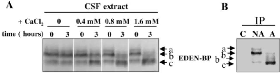
Calcium triggers changes in the electrophoretic mobility of EDEN-BP in CSF extract and M II oocytes
EDEN-BP migrates as a doublet of 55/53 kDa in SDS/PAGE gels (Legagneux et al., 1992). Therefore, we evaluated the effect of increasing the concentration of calcium ions in the CSF extracts on the electrophoretic mobility of EDEN-BP. Using Anderson’s modified SDS/PAGE gels (Anderson et al., 1973), we detected a more complex pattern of EDEN-BP electrophoretic mobility. Three bands (Fig. 2A,B; named a, b, c) were observed. The uppermost weak band (a) is only visible in CSF extracts collected before calcium addition. The second band (b) corresponds to the major band in the CSF extracts and its intensity decreases after calcium activation. This decrease appears sensitive to the amount of calcium added (compare extracts 3 hours after activation with 0.4, 0.8 and 1.6 mM CaCl2, Fig. 2A). The lower major band (c) is clearly observed
in the activated extracts; however, it is also present as a minor band in CSF extracts. The intensity of this band increases in parallel with the increase in calcium concentration (compare extracts 3 hours after activation with 0.4, 0.8 and 1.6 mM CaCl2, Fig. 2A).
To determine whether the calcium-induced changes in the electrophoretic mobility of EDEN-BP occur in vivo, we studied the protein in parthenogenetically activated M II oocytes. The protein was detected by western blotting either directly (data not shown) or after immunoprecipitation (Fig. 2B). In nonactivated eggs (arrested in M II of meiosis), similarly to CSF extracts, three EDEN-BP bands (a, b, c) with a strong upper major band (b) and a very weak lower band (c), were detected (Fig. 2B, NA; compare with untreated CSF extracts, time 0, in Fig. 2A). In activated eggs the uppermost band (a) was absent, the intensity of major band (b)
Fig. 1. Acceleration of EDEN-dependent deadenylation in calcium-activated CSF extract. (A) Capped, radiolabelled transcript Eg2-410 and Eg2-410a were incubated in CSF- or calcium (0.8 mM)-activated extracts for 1, 2 or 3 hours. After extraction and precipitation RNAs were analysed on urea/acrylamide 4% gels. The positions of the different forms of the transcripts are indicated: (A+), fully adenylated (A65) form, and (A–), fully
deadenylated form. The lane (T) corresponds to the fully adenylated radiolabelled transcripts without previous incubation with extract and precipitation. (B) Sperm nuclei chromatin was stained with Hoechst dye and observed in fluorescence and phase contrast. Condensed sperm chromosomes in the CSF extract (left) and decondensed nuclei in calcium activated extract (right).
2700
significantly diminished and the lower major band (c) dramatically increased (Fig. 2B, A). Immunoprecipitation of EDEN-BP enabled us to more clearly visualize the intermediate bands (b′), sometimes visible as a doublet in between the major bands (b) and (c) (Fig. 2B, A). Therefore, on egg activation the electrophoretic mobility of EDEN-BP increases as observed in the calcium-treated CSF extracts.
The electrophoretic mobility changes in EDEN-BP occur during oocyte maturation and following egg activation
To characterize the dynamics of the changes in the electrophoretic mobility of EDEN-BP, we studied the protein in detail during oocyte maturation and following egg activation. Samples of maturing oocytes were collected every hour following progesterone addition (Fig. 3A) and at short intervals following parthogenetic activation of M II oocytes with calcium ionophore (Fig. 3B), and analysed as above. Stage VI prophase I-arrested oocytes contain two forms of EDEN-BP: the major lower band (c) and a smear of minor ones corresponding to the bands (b′). Four hours after the addition of progesterone, when 50% of oocytes had undergone germinal vesicle breakdown (GVBD), as judged by the appearance of the maturation spot, the mobility of EDEN-BP changed and the upper band (b) appeared. The uppermost band (a) was
detected after 6 hours of progesterone treatment. After 7 hours (100% GVBD), the intensity of the upper bands (a) and (b) clearly increased, whereas the intensity of the lower (c) band decreased (Fig. 3A).
Following activation of M II oocytes we observed a rapid decrease in the intensity of the upper bands (a) and (b) (Fig. 3B). The uppermost band (a) disappeared within 15 minutes, whereas the band (b) persisted until 2 hours postactivation. The intensity of lower bands (b′) and (c) increased concomitantly. Thus, EDEN-BP undergoes rapid post-translational modifications at the time of the GVBD during oocyte maturation and following egg activation. The dynamics of these changes in EDEN-BP electrophoretic mobility resemble those observed for other proteins that undergo cell-cycle-dependent phosphorylation during GVBD and dephosphorylation following entry into development (e.g. ERK2 MAP kinase) (Ferrell et al., 1991).
EDEN-BP is post-translationally modified by phosphorylation/dephosphorylation
To verify whether the electrophoretic changes of EDEN-BP were due to changes in the phosphorylation of the protein, extracts were treated with the phage λphosphatase at 30°C for 0, 0.5 or 1 hour. The samples were then analysed by SDS/PAGE and anti-EDEN-BP western blotting. The phage λ phosphatase treatment caused an increase in the electrophoretic mobility of EDEN-BP, whereas without phage λphosphatase treatment, the major upper band (b) of EDEN-BP was still detectable after 1 hour of incubation at 30°C (Fig. 4A). To show the direct effect of the phage λphosphatase on EDEN-BP, we immunoprecipitated EDEN-BP followed by λ phosphatase treatment. In these conditions a similar modification of EDEN-BP was observed (Fig. 4B). However, the conversion of the immunoprecipitated protein appeared to be less complete and residual amounts of bands (a) and (b) were sometimes observed. This reduced conversion to the dephosphorylated form could be due to a masking effect because EDEN-BP is complexed with the antibodies during the phosphatase treatment. The data presented show that the phage
λ phosphatase treatment mimics to a large part the down shift Journal of Cell Science 116 (13)
Fig. 2. Changes in the electrophoretic mobility of EDEN-BP is triggered by calcium. (A) Increasing amounts of CaCl2were added to activate CSF extract. The extracts were
incubated for 3 hours, analysed on 15% Anderson SDS/PAGE gels, blotted and reacted with anti-EDEN-BP antibody. (B) Proteins extracted from nonactivated eggs (NA) and calcium ionophore-activated eggs (A) were immunoprecipitated on proteinA-sepharose beads covalently coupled with anti-EDEN-BP antibodies. Extracts from nonactivated eggs were also loaded on control proteinA-sepharose beads that were not in contact with the antibody (C). The bound material was analysed by electrophoresis and
immunoblotted with anti EDEN-BP antibodies. a, b, b′and c indicate the different electrophoretic forms of EDEN-BP.
Fig. 3. Electrophoretic mobility of EDEN-BP changes during oocyte maturation and activation. (A) Stage VI oocytes were incubated in Merriam buffer and maturated in vitro by addition of 1 µM progesterone. Samples of 20 oocytes were collected at 0, 1, 2, 4 and 5-9 hours after progesterone addition. The GVBD status was estimated as the percentage of oocytes showing the maturation spot (% GVBD). The samples were extracted, analysed by electrophoresis and immunoblotted with anti EDEN-BP antibody. (B) Unfertilized eggs were treated (+) or not (–) with calcium ionophore. Samples of ten parthenogenetic embryos were collected every 15 minutes during the first hour postactivation, and then every 30 minutes. Extracts were analysed as described above. The positions of different electrophoretic forms of EDEN-BP are indicated on the right of the blot.
of EDEN-BP observed after calcium addition to CSF extract or calcium ionophore treatment of M II oocytes. These experiments show that the post-translational modifications of EDEN-BP are, at least in part, due to phosphorylation and dephosphorylation. EDEN-BP is phosphorylated during maturation and dephosphorylated following egg activation.
Kinase and phosphatase inhibitors downregulate EDEN-dependent deadenylation
A large number of kinases are inactivated on egg activation (Karsenti et al., 1987). The temporal correlation between the beginning of EDEN-BP dephosphorylation and the acceleration of EDEN-dependent deadenylation following egg activation suggests that the two processes, which are triggered by free calcium increase, could be due to a change in the equilibrium between kinases and phosphatases activities. Therefore, we determined whether kinase and phosphatase inhibitors could influence the process of EDEN-dependent mRNA deadenylation.
To inhibit a broad range of serine/threonine kinases we used roscovitine at high concentration (100 µM) added to CSF extract activated by the addition of CaCl2 (Fig. 5). At this
concentration roscovitine inhibits a large spectrum of kinases (Meijer et al., 1997). We observed a complete inhibition of the EDEN-dependent deadenylation; no poly(A)–form of Eg2-410
chimeric RNA was detected even after 3 hours of incubation (Fig. 5). Only slight changes (higher accumulation of intermediate forms of Eg2-410a chimeric RNA) in the dynamics of the default deadenylation were observed.
Similar experiments were performed by inhibiting a broad range of phosphatases. A mixture of concentrated phosphatase inhibitors was added to the CSF extract containing chimeric RNAs before CaCl2addition. EDEN-dependent deadenylation
was very efficiently arrested by these inhibitors both in CSF and calcium-activated extracts (data not shown).
Okadaic acid is a potent inhibitor of phosphatases 1 and 2A (Goris et al., 1989). Therefore, using this drug enabled us to identify the spectrum of phosphatases inhibiting EDEN-dependent deadenylation. CSF extracts supplemented with OA were incubated at 21°C for 15 minutes and then activated with CaCl2, and radiolabelled transcripts were added. We observed
that OA inhibited the deadenylation process in
calcium-activated extracts (Fig. 6A). The inhibition of EDEN-dependent deadenylation was correlated with the inhibition of the dephosphorylation of EDEN-BP (Fig. 6B). The dynamics of default deadenylation was only slightly modified by OA, although the accumulation of several partially deadenylated products became visible. From these experiments, we can conclude that EDEN-dependent deadenylation is regulated by a balance between phosphorylation and dephosphorylation activities and that this process is sensitive to OA.
MPF pathway does not inhibit EDEN-dependent deadenylation
MPF is finely regulated via phosphorylation/ dephosphorylation. Cdc25 phosphatase is a major activator of MPF and this phosphatase is itself activated by a series of phosphorylations on the entry into the M-phase (Kumagai and Dunphy, 1991; Gautier et al., 1991). MPF activity depends on the stability of cyclin B (Murray and Kirschner, 1989; Murray et al., 1989). Moreover, OA is known to indirectly activate MPF in Xenopus oocytes and extracts (Goris et al., 1989; Rime et al., 1990; Jessus et al., 1991; Lorca et al., 1991). Therefore, the sensitivity of EDEN-dependent deadenylation to OA could be due, in part, to activation of MPF. To determine whether this activity interferes with the process of EDEN-dependent deadenylation we specifically activated MPF via the addition of indestructible ∆90 cyclin B of sea urchin into CSF extracts followed by calcium treatment (Fig. 7A). Addition of exogenous ∆90 cyclin B into CSF extract treated with Ca2+
maintained condensed chromosomes (Fig. 7A, lower panel, compare left and middle images), indicating that the extract remained in M-phase despite calcium addition, whereas interphase nuclei were present in calcium-activated extract (Fig. 7A, lower panel, right). However, the high MPF activity in the extract had no effect on EDEN-dependent deadenylation (Fig. 7A, upper panel).
Fig. 4. Changes in the electrophoretic mobility of EDEN-BP are due to its phosphorylation and dephosphorylation. (A) Extracts from non-activated eggs and calcium ionophore-activated eggs were incubated at 30°C with (+) or without (–) phophatase λfor the indicated times. The extracts were analysed on 15% Anderson SDS/PAGE and immunoblotted with anti-EDEN-BP antibodies. (B) EDEN-anti-EDEN-BP immunoprecipitated from unfertilized egg extract was incubated with phospatase λand treated as above. The different electrophoretic forms of EDEN-BP are indicated on the left.
Fig. 5. Roscovitine downregulates EDEN-dependent deadenylation. Roscovitine was added (+) or not (–) to calcium-activated CSF extract to a final concentration of 100 µM. Capped, radiolabelled transcript Eg2-410 and Eg2-410a were incubated in the extract for 1, 2 or 3 hours at 21°C. After extraction and precipitation radiolabelled RNAs were analysed on urea/acrylamide 4% gel. The lane (T) corresponds to the fully adenylated radiolabelled transcripts without previous incubation with extract and precipitation.
2702
To check whether MPF activity could affect the deadenylation activity, which had already been accelerated on egg activation, we added ∆90 cyclin B to ionophore-activated embryo extract and followed EDEN-dependent deadenylation (Fig. 7B). In parallel, we checked the inhibitory effect of OA in the same extract, as well as the combined effect of both ∆90 cyclin B and OA (Fig. 7B). As expected, ∆90 cyclin B restored an M-phase state in the extract, which was confirmed in western blot with anti-Cdc25; the disappearance of the fast migrating (dephosphorylated) band is concomitant with the appearance of a new, upshifted band (Fig. 7B, lane 2 in the lower panel). The addition of ∆90 cyclin B did not affect EDEN-dependent deadenylation in this extract when compared with the control, interphase extract (Fig. 7B, upper panel, compare experiments 1 and 2). As already shown for the calcium activated CSF extract, 1 µM OA slowed this process and this reduced rate of deadenylation was not affected when the OA treatment extract was supplemented with ∆90 cyclin B (Fig. 7B, compare experiments 3 and 4). As expected, the combined effect of OA and ∆90 cyclin B treatment induced a more pronounced phosphorylation of Cdc25 (Fig. 7B, lower panel, lane 4). Such a combined treatment seems to have a synergistic effect on the M-phase state of the extract, but does not influence significantly EDEN-dependent deadenylation compared with the OA treatment alone (Fig. 7B, upper panel, compare experiments 3 and 4). From these experiments we conclude that EDEN-dependent deadenylation of RNA is not affected by MPF activity.
Discussion
Calcium signalling upregulates EDEN-dependent deadenylation on egg activation
Calcium plays a major role in egg activation on fertilization or parthenogenetic activation (Stricker, 1999; Runft et al., 2002). Transient rises in intracellular Ca2+are of great importance for
numerous biochemical and cell biological processes necessary for further development. Here, we showed that calcium-mediated activation of CSF extracts greatly enhanced EDEN-dependent deadenylation activity, as well as dephosphorylation of EDEN-BP, the only protein known so far to be involved in this process. Moreover, we showed that the same modifications of EDEN-BP occurred when M II oocytes were parthenogenetically activated via calcium ionophore treatment. Because Xenopus laevis EDEN-containing maternal mRNAs are deadenylated following fertilization (Paris et al., 1988; Paris and Philippe, 1990), we believe that the same calcium-dependent mechanism triggers EDEN-dependent deadenylation of maternal mRNAs on fertilization. Deadenylation of maternal mRNAs that contain EDEN-sequence (Eg family and c-mos) (Bouvet et al., 1994; Paillard et al., 1998) results in their translational arrest (Ezzeddine et al., 2002; Ueno and Sagata, 2002). Therefore, our data provide a link between calcium signalling and translational repression of maternal genes during early development of Xenopus laevis. We also observed that an increase in the amount of calcium added to CSF extract, induced a more pronounced dephosphorylation of EDEN-BP. Fertilization generates transient intracellular calcium waves. Adding supraphysiological amounts of calcium (1.6 mM) to CSF extract may in one step induce rapid modifications of numerous biochemical processes that in concert enhance EDEN-BP dephosphorylation.
Phosphorylation and dephosphorylation modulate EDEN-dependent deadenylation of mRNA
Calcium waves that occur after fertilization provoke a rapid inactivation of CSF and MPF activity, resulting in metaphase to anaphase transition followed by the first embryonic interphase (Lorca et al., 1993) (reviewed by Maller, 1998). Inactivation of MPF via cyclin B degradation (Murray et al., 1989) triggers changes in the phosphorylation/ dephosphorylation equilibrium in favour of dephosphorylation (Karsenti et al., 1987). Inactivation of multiple kinases participates in this process and correlates with dephosphorylation of EDEN-BP. Using nondegradable sea urchin ∆90 cyclin B, which enables the MPF kinase activity to be artificially maintained in calcium-treated CSF extracts, or to activate MPF in interphase extracts, we show that the EDEN-dependent deadenylation is independent of MPF activity. In contrast, OA reduces the activity of EDEN-dependent deadenylation. OA is known to activate MPF and ERK2 MAP kinase besides inactivating phosphatases PP1 and PP2 (Jessus et al., 1991). Whether the effect of OA on EDEN-dependent deadenylation is via ERK2 activation or phosphatase inactivation is not clear at present. However, in preliminary experiments we observed that inactivation of ERK2 by the addition of recombinant phosphatase CL 100 to OA-treated extract restored the EDEN-dependent deadenylation activity (our unpublished results). In addition, Journal of Cell Science 116 (13)
Fig. 6. Okadaic acid downregulates EDEN-dependent deadenylation and inhibits dephosphorylation of EDEN-BP. (A) Okadaic acid (OA) at 1 µM final concentration was added (+) or not (–) to CSF extract and incubated for 15 minutes at 21°C. Both extracts were then supplemented with CaCl2(to final concentration of 0.8 mM) and
incubated at 21°C for 1, 2 or 3 hours with radiolabelled transcripts Eg2-410 and Eg2-410a. RNA were extracted, analysed on urea/polyacrylamide 4% gel and autoradiography. The lane (T) corresponds to the fully adenylated radiolabelled transcripts without previous incubation with extract and precipitation. (B) Proteins were resolved on 15% Anderson SDS/PAGE, transferred to membrane and immunoblotted with anti-EDEN-BP antibody. The different
electrophoretic forms of EDEN-BP are indicated both on the left and on the right.
the timing of MAP kinase ERK2 activation and inactivation during oocyte maturation and after fertilization correlates with phosphorylation and dephosphorylation of EDEN-BP (Ferrell et al., 1991; Abrieu et al., 1996; Chau and Shibuya, 1999). A positive feedback between MPF and ERK2 activation in CSF extracts through the phosphorylation of c-mos, an upstream regulator of the ERK2, was reported recently by Castro et al. (Castro et al., 2001). In some of our experiments in which ∆90 cyclin B was added to CSF extracts followed by calcium addition, we observed an intermediate rate of EDEN-dependent deadenylation compared with CSF extracts and calcium-activated extracts (data not shown). In these extracts we detected the phosphorylated form of ERK2 MAP kinase. This suggests that the feedback loop described by Castro et al. (Castro et al., 2001) is provoking ERK2 MAP kinase activation
in these extracts, supposedly when c-mos was not totally degraded. Further studies are necessary to examine the potential role of ERK2 MAP kinase, and/or a combined role of MPF and MAP kinase pathways, in the regulation of EDEN-dependent deadenylation and, in particular, of EDEN-BP phosphorylation.
The insensitivity of EDEN-dependent deadenylation to MPF contrasts with the recent data by Groisman et al. (Groisman et al., 2002) showing that cyclin B1 mRNA polyadenylation is stimulated via MPF and that its deadenylation is induced by MPF inactivation. There is no proof that cyclin B1 3′-UTR contains an EDEN, suggesting that the deadenylation of this mRNA is independent of EDEN-BP. Therefore, two groups of maternal Xenopus mRNAs can be distinguished on the basis of their behavior regarding deadenylation during early
Fig. 7. MPF activation through ∆90 cyclin B addition to Ca2+, or ioniphore-activated extracts does not influence EDEN-dependent
deadenylation. A. Nondegradable sea urchin ∆90 cyclin B was added (+) or not (–) to CSF extract at final concentration of 0.2 ng/µl and incubated for 15 minutes at 21°C to allow its association with cdk1 and the formation of a pool of stable MPF. Both extracts were then supplemented with CaCl2(to final concentration 0.8 mM) and incubated at 21°C for 1, 2 or 3 hours with radiolabelled transcripts Eg2-410 and
Eg2-410a. RNA. Upper panel: autoradiography of the extracted RNAs separated on a urea/acrylamide 4% gel. The lane (T) corresponds to the fully adenylated radiolabelled transcripts without previous incubation with extract and precipitation. The two extracts deadenylate chimeric RNAs with similar dynamics independently from the presence or absence of ∆90 cyclin B. Lower panel: sperm nuclei were incubated with extracts and stained with Hoechst dye following observation in fluorescence and phase contrast. Condensed sperm chromosomes in the control CSF extract (left) and in the calcium activated CSF extract with ∆90 cyclin B (middle) were found at the end of the experiment, whereas decondensed nuclei were present in calcium activated CSF extract (right). This confirms that ∆90 cyclin B-supplemented extract was in M-phase similarly to the control, untreated CSF extract. B. Nondegradable sea urchin ∆90 cyclin B (final concentration 0.2 ng/µl), OA (final concentration 1 µM), or both, were added to ionophore-activated eggs extract prepared 45 minutes after egg activation. The extracts were incubated for 15 minutes at 21°C to allow association of exogenous cyclin with cdk1 and formation of stable MPF or to inactivate protein phosphatases in the case of OA. Then they were supplemented with radiolabelled Eg2-410 transcript and further incubated at 21°C for 3 hours. RNA and proteins were analysed as indicated in Materials and Methods. Upper panel: autoradiography of the extracted RNAs separated on urea/acrylamide 4% gel. The lane (T) corresponds to the fully adenylated radiolabelled transcripts without previous incubation with extract and precipitation. Untreated, as well as ∆90 cyclin B-supplemented extracts dedenylate Eg2-410 transcript rapidly (experiments 1 and 2), whereas OA- and OA+∆90 cyclin B-supplemented extracts deadenylate only slightly Eg2-410 transcript (experiments 3 and 4). Lower panel: western blot with an anti-Cdc25 antibody. The fast migrating, lowest band represents dephosphorylated (inactive) form of Cdc25. The upshifted forms of Cdc25 produced by different treatments represent phosphorylated (active) forms of the Cdc25 phosphatase. Untreated extract contains inactive Cdc25 (lane 1), whereas ∆90 cyclin B-supplemented extract contains activated form of the phosphatase (lane 2) indicating induction of the M-phase. OA-treated and OA+∆90 cyclin B-treated extracts contain phosphorylated forms of Cdc25 (lanes 3 and 4, respectively). In OA+∆90 cyclin B containing extract (lane 4) Cdc25 is phosphoryletd to higher degree than in OA-treated extract (lane 3), indicating a synergistic effect of ∆90 cyclin B and OA on Cdc25 phosphorylation.
2704
development: namely, MPF- or cell-cycle-dependent deadenylation, like cyclin B1 mRNA (Groisman et al., 2002), and MPF-independent, but activation-dependent deadenylation, like Eg2 (this paper). Because calcium is as an important regulator of the cell cycle, we cannot exclude that mRNA deadenylation is controlled by cytoplasmic calcium in both cases.
EDEN-BP phosphorylation and dephosphorylation seems a very complex process. We could distinguish four different forms of this protein on western blots. The complex pattern of EDEN-BP phosphorylation suggests that multiple phosphorylation sites are modified during oocyte maturation and following egg activation. The inhibitory effects of both kinase and phosphatase inhibitors on EDEN-dependent deadenylation supports the hypothesis of a network of kinases and phosphatases controlling this process.
Could BP phosphorylation regulate EDEN-dependent deadenylation?
EDEN-BP is a good candidate for the regulatory molecule involved in the modulation of EDEN-dependent deadenylation on M II oocyte fertilization or activation. After calcium ionophore-induced activation this protein undergoes a series of dephosphorylations. The hyperphosphorylated band (a) disappears rapidly, whereas the intensity of the dephosphorylated band (c) increases to reach a maximum 2.5 to 3 hours after egg activation. The intensity of the hyperphosphorylated band (b), which is the major band in CSF extracts, decreases gradually and disappears 2 hours after activation. This correlation suggests that phosphorylation of EDEN-BP could be involved in the inhibition of EDEN-dependent deadenylation. However, EDEN-BP is part of a multimeric complex of high molecular weight (Legagneux et al., 1995) that probably contains several other factors required for deadenylation. Therefore, at present we can not exclude that changes in the phosphorylation of one or several of these factors are not involved in the regulation of EDEN-dependent deadenylation. Identification of the amino acids of EDEN-BP that are phosphorylated on oocyte maturation and studies of mutant proteins will permit us to evaluate the exact role of EDEN-BP phosphorylation and dephosphorylation during oocyte maturation and early embryonic development.
We thank Marie-Anne Felix (Institut Jacques Monod, Paris, France) for the generous gift of ∆90 cyclin B, the members of H. B. Osborne lab for helpful discussions, Yann Audic for a critical reading of the manuscript, Erwan Watrin for help with microscopy and Franck Chesnel for help in histone H1 kinase activity assay. This work was supported by grants from the European Union (contract QLRT 2000-00721) and the Ministère de la Recherche (Bio-ingénierie 2001, N° 128) to H.B.O. and from ARC (4298) to J.Z.K. UMR 6061 is a component of IFR 97.
References
Abrieu, A., Lorca, T., Labbe, J. C., Morin, N., Keyse, S. and Dorée, M. (1996). MAP kinase does not inactivate, but rather prevents the cyclin degradation pathway from being turned on in Xenopus egg extracts. J. Cell
Sci. 109, 239-246.
Anderson, C. W., Baum, P. R. and Gesteland, R. F. (1973). Processing of adenovirus 2-induced proteins. J. Virol. 12, 241-252.
Blot, J., Chartrain, I., Roghi, C., Philippe, M. and Tassan, J. P. (2002). Cell cycle regulation of pEg3, a new Xenopus protein kinase of the KIN1/PAR-1/MARK family. Dev. Biol. 241, 327-338.
Blow, J. J. (1993). Preventing re-replication of DNA in a single cell cycle: evidence for a replication licensing factor. J. Cell Biol. 122, 993-1002. Bouvet, P., Omilli, F., Arlot-Bonnemains, Y., Legagneux, V., Roghi, C.,
Bassez, T. and Osborne, H. B. (1994). The deadenylation conferred by the 3′untranslated region of a developmentally controlled mRNA in Xenopus embryos is switched to polyadenylation by deletion of a short sequence element. Mol. Cell. Biol. 14, 1893-1900.
Castro, A., Peter, M., Magnaghi-Jaulin, L., Vigneron, S., Galas, S., Lorca, T. and Labbé, J. C. (2001). Cyclin B/cdc2 induces c-Mos stability by direct phosphorylation in Xenopus oocytes. Mol. Biol. Cell 12, 2660-2671. Chau, A. S. and Shibuya, E. K. (1999). Inactivation of p42 mitogen-activated
protein kinase is required for exit from M-phase after cyclin destruction. J.
Biol. Chem. 274, 32085-32090.
Dorée, M. and Hunt, T. (2002). From Cdc2 to Cdk1: when did the cell cycle kinase join its cyclin partner? J. Cell Sci. 115, 2461-2464.
Ezzeddine, N., Paillard, L., Capri, M., Maniey, D., Bassez, T., Ait-Ahmed, O. and Osborne, H. B. (2002). EDEN-dependent translational repression of maternal mRNAs is conserved between Xenopus and Drosophila. Proc.
Natl. Acad. Sci. USA 99, 257-262.
Ferrell, J. E., Jr, Wu, M., Gerhart, J. C. and Martin, G. S. (1991). Cell cycle tyrosine phosphorylation of p34cdc2 and a microtubule-associated
protein kinase homolog in Xenopus oocytes and eggs. Mol. Cell. Biol. 11, 1965-1971.
Gautier, J., Solomon, M. J., Booher, R. N., Bazan, J. F. and Kirschner, M. W. (1991). cdc25 is a specific tyrosine phosphatase that directly activates p34cdc2. Cell 67, 197-211.
Goris, J., Hermann, J., Hendrix, P., Ozon, R. and Merlevede, W. (1989). Okadaic acid, a specific protein phosphatase inhibitor, induces maturation and MPF formation in Xenopus laevis oocytes. FEBS Lett. 245, 91-94. Groisman, I., Jung, M.-Y., Sarkissian, M., Cao, Q. and Richter, J. D.
(2002). Translational control of the embryonic cell cycle. Cell 109, 473-483. Gross, S. D., Schwab, M. S., Lewellyn, A. L. and Maller, J. L. (1999). Induction of metaphase arrest in cleaving Xenopus embryos by the protein kinase p90Rsk. Science 286, 1365-1367.
Haccard, O., Sarcevic, B., Lewellyn, A., Hartley, R., Roy, L., Izumi, T., Erikson, E. and Maller, J. L. (1993). Induction of metaphase arrest in cleaving Xenopus embryos by MAP kinase. Science 262, 1262-1265. Harland, R. and Misher, L. (1988). Stability of RNA in developing Xenopus
embryos and identification of a destabilizing sequence in TFIIIA messenger RNA. Development 102, 837-852.
Jessus, C., Rime, H., Haccard, O., van Lint, J., Goris, J., Merlevede, W. and Ozon, R. (1991). Tyrosine phosphorylation of p34cdc2 and p42 during meiotic maturation of Xenopus oocyte. Antagonistic action of okadaic acid and 6-DMAP. Development 111, 813-820.
Karsenti, E., Bravo, R. and Kirschner, M. (1987). Phosphorylation changes associated with the early cell cycle in Xenopus eggs. Dev Biol. 119, 442-453. Kumagai, A. and Dunphy, W. G. (1991). The cdc25 protein controls tyrosine dephosphorylation of the cdc2 protein in a cell-free system. Cell 64, 903-914.
Labbé, J. C., Capony, J. P., Caput, D., Cavadore, J. C., Derancourt, J., Kaghad, M., Lelias, J. M., Picard, A. and Dorée, M. (1989). MPF from starfish oocytes at first meiotic metaphase is a heterodimer containing one molecule of cdc2 and one molecule of cyclin B. EMBO J. 8, 3053-3058. Laemmli, U. K. (1970). Cleavage of structural proteins during the assembly
of the head of bacteriophage T4. Nature 227, 680-685.
Legagneux, V., Bouvet, P., Omilli, F., Chevalier, S. and Osborne, H. B. (1992). Identification of RNA-binding proteins specific to Xenopus Eg maternal mRNAs: association with the portion of Eg2 mRNA that promotes deadenylation in embryos. Development 116, 1193-1202.
Legagneux, V., Omilli, F. and Osborne, H. B. (1995). Substrate-specific regulation of RNA deadenylation in Xenopus embryo and activated egg extracts. RNA 1, 1001-1008.
Lohka, M. J. and Masui, Y. (1984). Roles of cytosol and cytoplasmic particles in nuclear envelope assembly and sperm pronuclear formation in cell-free preparations from amphibian eggs. J. Cell Biol. 98, 1222-1230. Lorca, T., Fesquet, D., Zindy, F., le Bouffant, F., Cerruti, M., Brechot, C.,
Devauchelle, G. and Dorée, M. (1991). An okadaic acid-sensitive phosphatase negatively controls the cyclin degradation pathway in amphibian eggs. Mol. Cell. Biol. 11, 1171-1175.
Lorca, T., Cruzalegui, F. H., Fesquet, D., Cavadore, J. C., Mery, J., Means, A. and Dorée, M. (1993). Calmodulin-dependent protein kinase II mediates
inactivation of MPF and CSF upon fertilization of Xenopus eggs. Nature 366, 270-273.
Maller, J. L. (1998). Recurring themes in oocyte maturation. Biol. Cell 90, 453-460.
Masui, Y. (2000). The elusive cytostatic factor in the animal egg. Nat. Rev. Mol. Cell. Biol. 1, 228-232.
McGrew, L. L. and Richter, J. D. (1990). Translational control by cytoplasmic polyadenylation during Xenopus oocyte maturation: characterization of cis and trans elements and regulation by cyclin/MPF.
EMBO J. 9, 3743-3751.
Meijer, L., Borgne, A., Mulner, O., Chong, J. P., Blow, J. J., Inagaki, N., Inagaki, M., Delcros, J. G. and Moulinoux, J. P. (1997). Biochemical and cellular effects of roscovitine, a potent and selective inhibitor of the cyclin-dependent kinases cdc2, cdk2 and cdk5. Eur. J. Biochem. 2432, 527-536. Merriam, R. W. (1971). Progesterone-induced maturational events in oocytes
of Xenopus laevis. I. Continuous necessity for diffusible calcium and magnesium. Exp. Cell Res. 68, 75-80.
Murakami, M. S. and Vande Woude, G. F. (1998). Analysis of the early embryonic cell cycles of Xenopus; regulation of cell cycle length by Xe-wee1 and Mos. Development 125, 237-248.
Murakami, M. S., Copeland, T. D. and Vande Woude, G. F. (1999). Mos positively regulates Xe-Wee1 to lengthen the first mitotic cell cycle of
Xenopus. Genes Dev. 13, 620-631.
Murray, A. W. (1991). Cell cycle extracts. Methods Cell Biol. 36, 581-605. Murray, A. W. and Kirschner, M. W. (1989). Cyclin synthesis drives the
early embryonic cell cycle. Nature 339, 275-280.
Murray, A. W., Solomon, M. J. and Kirschner, M. W. (1989). The role of cyclin synthesis and degradation in the control of maturation promoting factor activity. Nature 339, 280-286.
Nuccitelli, R. (1991). How do sperm activate eggs? Curr. Top. Dev. Biol. 25, 1-16.
Osborne, H. B. and Richter, J. D. (1997). Translational control by polyadenylation during early development. Prog. Mol. Subcell. Biol. 18, 173-198.
Paillard, L., Omilli, F., Legagneux, V., Bassez, T., Maniey, D. and Osborne, H. B. (1998). EDEN and EDEN-BP, a cis element and an associated factor that mediate sequence-specific mRNA deadenylation in Xenopus embryos.
EMBO J. 17, 278-287.
Paris, J. and Philippe, M. (1990). Poly(A) metabolism and polysomal recruitment of maternal mRNAs during early Xenopus development. Dev
Biol. 140, 221-224.
Paris, J. and Richter, J. D. (1990). Maturation-specific polyadenylation and translational control: diversity of cytoplasmic polyadenylation elements, influence of poly(A) tail size, and formation of stable polyadenylation complexes. Mol. Cell. Biol. 10, 5634-5645.
Paris, J., Osborne, H. B., Couturier, A., le Guellec, R. and Philippe, M. (1988). Changes in the polyadenylation of specific stable RNA during the early development of Xenopus laevis. Gene 72, 169-176.
Philips, A. V., Timchenko, L. T. and Cooper, T. A. (1998). Disruption of splicing regulated by a CUG-binding protein in myotonic dystrophy. Science 280, 737-741.
Richter, J. D. (1999). Cytoplasmic polyadenylation in development and beyond. Microbiol. Mol. Biol. Rev. 63, 446-456.
Rime, H., Huchon, D., Jessus, C., Goris, J., Merlevede, W. and Ozon, R. (1990). Characterization of MPF activation by okadaic acid in Xenopus oocyte. Cell Differ. Dev. 29, 47-58.
Roberts, R., Timchenko, N. A., Miller, J. W., Reddy, S., Caskey, C. T., Swanson, M. S. and Timchenko, L. T. (1997). Altered phosphorylation and intracellular distribution of a (CUG)n triplet repeat RNA-binding protein in patients with myotonic dystrophy and in myotonin protein kinase knockout mice. Proc. Natl. Acad. Sci. USA 94, 13221-13226.
Runft, R. R., Jaffe, L. A. and Mehlmann, L. M. (2002). Egg activation at fertilization: where it all begins. Dev. Biol. 245, 237-254.
Sagata, N., Watanabe, N., Vande Woude, G. F. and Ikawa, Y. (1989). The c-mos proto-oncogene product is a cytostatic factor responsible for meiotic
arrest in vertebrate eggs. Nature 342, 512-518.
Stricker, S. A. (1999). Comparative biology of calcium signaling during fertilization and egg activation in animals. Dev. Biol. 211, 157-176. Timchenko, L. T., Miller, J. W., Timchenko, N. A., DeVore, D. R.,
Datar, K. V., Lin, L., Roberts, R., Caskey, C. T. and Swanson, M. S. (1996). Identification of a (CUG)n triplet repeat RNA-binding protein and its expression in myotonic dystrophy. Nucleic Acids Res. 24, 4407-4414.
Ueno, S. and Sagata, N. (2002). Requirement for Both EDEN and AUUUA motifs in translational arrest of Mos mRNA upon fertilization of Xenopus eggs. Dev. Biol. 250, 156-167.