HAL Id: hal-00005893
https://hal.archives-ouvertes.fr/hal-00005893
Submitted on 11 Jul 2005HAL is a multi-disciplinary open access archive for the deposit and dissemination of sci-entific research documents, whether they are pub-lished or not. The documents may come from teaching and research institutions in France or abroad, or from public or private research centers.
L’archive ouverte pluridisciplinaire HAL, est destinée au dépôt et à la diffusion de documents scientifiques de niveau recherche, publiés ou non, émanant des établissements d’enseignement et de recherche français ou étrangers, des laboratoires publics ou privés.
Stéphanie Lebreton, Laurent Boissel, Nathalie Iouzalen, Jacques Moreau
To cite this version:
Stéphanie Lebreton, Laurent Boissel, Nathalie Iouzalen, Jacques Moreau. RLIP mediates downstream signalling from RalB to the actin cytoskeleton during Xenopus early development.. Mechanisms of Development, Elsevier, 2004, 121, pp.1481-1494. �10.1016/j.mod.2004.07.008�. �hal-00005893�
Review Copy
RLIP mediates downstream signalling from RalB to the actin cytoskeleton during
Xenopus early development
Stéphanie Lebreton, Laurent Boissel, Nathalie Iouzalen and Jacques Moreau*
Mécanismes Moléculaires du Développement, Institut Jacques Monod, CNRS, Universités Paris VI et Paris VII,. 2 Place Jussieu, 75251 Paris cedex 05, France.
* To whom correspondence should be addressed
Submitted to: Mechanisms of Development
Running title: Study of the RalB pathway in Xenopus early development
Phone: (033) 01 44 27 82 91 Fax. : (033) 01 44 27 52 65 e-mail : moreauja@ijm.jussieu.fr
Sequence data from this article have been deposited in the EMBL/genbank Data Libraries under accession N° AJ304845 and AJ252165
Review Copy
Abstract
The Ras protein activates at least three different pathways during early development. Two of them regulate mesodermal gene expression and the third is thought to participate in the control of actin cytoskeleton dynamics via the Ral protein. From a yeast two-hybrid screen of a Xenopus maternal cDNA library, we identified the Xenopus orthologue of the Ral interacting protein (RLIP, RIP1 or RalBP1), a putative effector of small G protein Ral. Previously we observed that a constitutively activated form of Ral GTPase (XralB G23V) induced bleaching of the animal hemisphere and disruption of the cortical actin cytoskeleton. To demonstrate that RLIP is the effector of RalB in early development, we show that the artificial targeting of RLIP to the membrane induces a similar phenotype to that of activated RalB. We show that overexpression of the Ral binding domain (RalBD) of XRLIP, which binds to the effector site of Ral, acts in competition with the endogenous effector of Ral and protects against the destructive effect of XralB G23V on the actin cytoskeleton. In contrast, the XRLIP has a synergistic effect on the activated form of XralB, which is dependent on the RalBD of RLIP. We provide evidence for the involvement of RLIP by way of its RalBD on the dynamics of the actin cytoskeleton and propose that signalling from Ral to RLIP is required for gastrulation.
Review Copy
Introduction
The fibroblast growth factor (FGF) family plays an important role in mesodermal induction. eFGF (Isaacs et al., 1992), FGF-3 (Tannahill et al., 1992) and FGF-8 (Christen and Slack, 1997) are expressed at gastrula stages in the marginal zone of Xenopus embryos, where they activate the Ras/MAP kinase pathway (Kristen and Grainger, 2000). A dominant negative form of FGF expressed in embryos inhibits mesodermal gene expression in animal cap experiments and causes gastrulation failure in embryos (Amaya et al., 1991), (Kroll and Amaya, 1996) and (Nutt et al., 2001). FGF signalling is also implicated in morphogenesis of the mesoderm in Drosophila embryos (Wilson and Leptin, 2000). Through many other examples the FGF proteins have also emerged as key mediators of cell migration in vivo. It was thus demonstrated that the FGF receptors FGFR8 and FGFR1 are essential for cell migration and mesodermal patterning during gastrulation of mouse embryos (Yamaguchi et al., 1994). Even though the Ras/MAP kinase pathway is known to be the canonical pathway required to transduce the FGF signal, it is now clear that FGF can activate a number of other pathways. Indeed, the small G protein Ras activated by the FGF signal, interacts with different effectors to direct the signal towards many targets (White et al., 1995). Ras signalizing mediates effects such as proliferation, cytoskeleton organization (Bar-Sagi and Feramisco, 1986), synergises with MAP kinase to transform cells (White et al., 1996). Recently, we demonstrated (Lebreton et al., 2003) that FGF is able to activate the small G protein XralB in Xenopus embryos. Ral is a member of the Ras family whose activation can be Ras dependent (reviewed in (Feig et al., 1996; Wolthuis and Bos, 1999)), and which is found in its active form mainly in the marginal zone of developing Xenopus embryos (Lebreton et al., 2003). We previously showed that one target of the FGF/Ras/Ral pathway is the cortical actin cytoskeleton that requires no new gene expression, and that overexpression of the dominant negative form of XralB in the marginal zone blocks gastrulation movements (Moreau et al., 1999). Like Ras, Ral may utilize multiple effectors to mediate its action, and some of these effectors may be cell type specific. Proteins such as filamin and RalBP1/RLIP76 may be direct effectors of Ral that mediate regulation of either filopodia formation (Ohta et al., 1999) or endocytosis (Jullien-Flores et al., 2000), whereas Sec5 modulates exocyst function as well as filopodia formation (Moskalenko et al., 2002). To characterize the Ral pathway and understand its function during early vertebrate development, we searched for the RalB effector present during early development. Using a two-hybrid
Review Copy
screen, we cloned XRLIP (RIP1 or RalBP1) the putative effector of XralB, and showed that targeting of XRLIP to the plasma membrane induces disruption of the actin cytoskeleton similar to the effect of the dominant positive form of XralB. We also present evidence from rescue experiments that XRLIP is specifically required to induce the phenotype characteristic of the activated form of XralB and that, like XralB, XRLIP is necessary for normal gastrulation.
Review Copy
Results
RLIP is an embryonic target of RalB
Many downstream targets of Ral (RalA or RalB) have been identified in somatic tissues. These include phospholipase D (PLD) (Jiang et al., 1995), which interacts with the N-terminal sequence of Ral independently of its activation state. Other proteins, namely filamin (Ohta et al., 1999), RLIP (Jullien-Flores et al., 1995) (also called RalBP1 (Cantor et al., 1995) (Ikeda et al., 1998) or RIP1 (Park and Weinberg, 1995)) and Sec5 (Moskalenko et al., 2002; Sugihara et al., 2002) have also been reported to interact specifically with the GTP-bound form of Ral. To identify which effector is present during embryonic development, the XralB was used as bait in a yeast two-hybrid screen of a Xenopus maternal cDNA library (Iouzalen et al., 1998). We isolated from three million clones 23 clones that corresponded to 4 different cDNAs coding for proteins that interact specifically with the GTP-RalB form. These proteins correspond to the mammalian Ral effector RLIP and have been designated XRLIP for
Xenopus RLIP. The four XRLIP cDNAs differ in their nucleotide sequence and in the size of
their 3’untranslated sequence comprised between the stop codon and the poly A tail (respectively 182, 263, 289 and 1641 bp for clone 354, 31, 38 and 14). Among the four encoded proteins, XRLIP-DH38 (designated XRLIP1) and XRLIP-DH31 (designated XRLIP2) share 85% and 81.4% identity with the mammalian RLIP76 sequence. The amino acid sequence of the XRLIPs differs from the mammalian sequence by the absence from the
Xenopus sequence of one repetitive peptide (143EEKHK147) from the amino terminal region in the mammalian sequence). The less conserved region, compared to mammalian homologues, is located in the 59 carboxy terminal amino acids (Fig. 1).
The temporal expression of the XRLIP mRNAs was analyzed by Northern blotting of total RNAs from different developmental stages and tissues of Xenopus using the coding sequence of XRLIP1 as a probe. At least four hybridization signals were detected (Fig. 2A) corresponding to four XRLIP transcripts of approximately 5.1 kb, 4.0 kb, 3.2 kb and 2.8 kb. The largest transcript (referred to as the adult or tissue-specific form) prevailed in late development and in adult tissues, whereas the other transcripts (referred to as developmental specific forms), were primarily detected during oogenesis and early development. However, an overlapping expression pattern was observed from gastrula to the tadpole stages (a period
Review Copy
corresponding to the beginning of organogenesis) and in some adult tissues such as the spleen. The presence of 4 different mRNAs as revealed by Northern blot analysis is in agreement with the finding of 4 different cDNAs isolated by the two-hybrid screen (Iouzalen et al., 1998). This diversity of RLIP mRNAs could originate from genome duplication in accordance with the hypothesis of total genome duplication that occurred thirty million years ago (Bisbee et al., 1977).
In the yeast two-hybrid system, all these XRLIP isoforms bound specifically to wild type XralB and GTP–bound XralB (XralB G23V) but did not interact with GDP-bound XralB (XralB S28NMCT) (Fig. 2C). However, as opposed to previously described results (Bauer et al., 1999), the mutants XralB G23VD49N and XralB D49NMCT exhibited the same interaction with XRLIP as did XralB G23V.
The temporal expression of the XRLIP protein was also analyzed by Western blotting using antibodies against rat RLIP (RalBP1). The specificity of the rat antibodies against XRLIP had previously been tested on XRLIP2 and on a deletion mutant overexpressed in embryos (data not presented). As shown in Fig. 2B, two predominant bands of 95 kD and 83 kD were detected. These results are in agreement with the size of the RLIP protein observed by Cantor et al. (Cantor et al., 1995). The 95 kD band was detected from unfertilized egg to the gastrula stages. This expression pattern was comparable to the expression profile of the embryonic XRLIP mRNA. Beyond the gastrula stage, however, the pattern changed. The 95 kD band disappeared and a smaller one of 83 kD appeared. Analysis of different parts of embryos at stage 10.5 of gastrulation revealed the same level of expression of XRLIP protein in the whole embryo (data not shown). Taken together, these results show, first, that XRLIP is present during the early development of Xenopus without any specific localization, and, secondly, that it interacts specifically with either the native or the activated form of XralB. In embryos, RLIP interacts with the activated form of Ral.
Using the two-hybrid assay, we showed that RLIP interacts with the activated form of Ral. To determine whether this interaction occurs in the embryo, we investigated whether Ral co-immunoprecipitates with myc-tagged RLIP. Ral protein is known to be associated to the plasma membrane. The co-immunoprecipation assay was performed with XralB mutated in the CAAX box (XralB C203S), which is ineffective in membrane targetting. As shown in Fig 3A, XralB G23V interacted with Myc-RLIP (lane 3) whereas XralB G23V injected alone
Review Copy
(lane 2) or with Myc-RLIP deleted of its Ral interacting domain (RLIP MRalBD) (lane 4) could not be detected by Western blot.
Since XralB interacts in vivo with RLIP, we postulated that XralB is competent to target RLIP to the plasma membrane. To investigate this possibility, the cellular localization of the XRLIP2 protein was examined by confocal screening laser microscopy using anti-myc antibody. When XRLIP2 mRNA was injected alone, the protein was distributed in the cytosol (Fig. 3C left), whereas when XRLIP2 mRNA was coinjected with XralB G23V mRNA, it was clearly located in the cortical region of the blastomere (Fig. 3C middle). We confirmed the causal localization of RLIP by RalG23V by coexpression of XralB G23V mRNA and XRLIP2 mRNA deleted of its RalB interacting domain (Fig. 3B). In this case, RLIP is exclusively located in the cytosol (Fig. 3C right), as when XRLIP mRNA is expressed in the absence of Ral.
Previously, we showed that Ral is activated by FGF. Therefore, we examined whether FGF is able to trigger the recruitment of RLIP into the plasma membrane. To this end, we incubated in the absence or presence of FGF, animal cap explants from embryos microinjected with RLIP2 and Ral wild type mRNAs, and examined the cellular localization of RLIP by confocal microscopy. When animal cap explants were incubated in the absence of FGF, Myc-RLIP was localized in the cytosol of 72.6% (n=62) of cells examined (Fig 3D); when animal cap explants were incubated for 90 min in the presence of 125 ng / ml of FGF, Myc-RLIP was located in the periphery in the subcortical area of 88,3% (n=68) of cells examined. In the majority of cells originating from animal cap explants incubated with FGF, Myc-RLIP was not homogeneously arranged at the periphery of the cell, but preferentially arranged into certain characteristic domains. If we compare the localization of Myc-RLIP in animal caps incubated in the presence of FGF with the localization of Myc-RLIP targeted to the plasma membrane by Ral G23V (Fig 3C middle), the signal was more diffuse near the plasma membrane with FGF. Taken together, these results indicate that RLIP interacts with the Ral activated form and its localization in the plasma membrane is directed by FGF.
The RalBD of RLIP is necessary for gastrulation
The Ral binding domain (RalBD) of RLIP interacts specifically with Ral and is used to measure Ral activation (Wolthuis et al MCB 1998 18, 2486-2491). Previously we showed that the overexpression of RalBD in the marginal zone is able to block gastrulation at stage 10.5-11 (Lebreton et al., 2003) as does a dominant negative form of Ral. Since many effectors of
Review Copy
Ral have been characterized, it was necessary to confirm that, in early development, RLIP was indeed the Ral effector involved in this phenotype. To investigate this, we examined whether the overexpression of RLIP could compete with the RalBD and rescue the wild type phenotype. Embryos were injected at the four-cell stage into the marginal zone with RalBD (4 x 300 pg) or co-injected with XRLIP2 (4 x 1.5 ng) mRNAs. The molar ratio between the RalBD and XRLIP mRNAs was 1:1. While an arrest of development at the gastrulation stage (stage 10.5) occurred in 87% of the embryos injected with RalBD mRNA, development of co-injected embryos was delayed compared to unco-injected controls (Fig. 4) up to stage 12 (n= 70). To examine whether the rescue effect is specific of RLIP, RalBD mRNA was co-injected with mRNA coding for RLIP deleted of its RalBD (XRLIPMRalBD). Embryos injected with 4 x 1.5 ng of XRLIPMRalBD mRNA developed normally (Fig. 4), whereas embryos co-injected with RalBD (4 x 300 pg) and XRLIPMRalBD (4 x 1.5 ng) in a 1:1 ratio exhibited the same severe defect (Fig. 4) characterized by a blocking of blastopore closure at stage 10.5 (90%; n = 45) induced by RalBD. At times, failure of gastrulation of embryos injected with RalBD alone or with XRLIPMRalBD (40-50%) was accompanied by a leakage of vitellus around the blastopore lip. This effect, observed in only 10% of the embryos co-injected with XRLIP (data not shown), could be related to membrane fragility of the mesendodermal cell due to inhibition of the RalB pathway. These data clearly show that the RalBD, like the dominant negative form of Ral, inhibits the gastrulation process, and that, by a specific rescue with XRLIP but not with XRLIPMRalBD, this effect is dependent on the RalBD.
RLIP, the putative RalB effector, is necessary for the RalB pathway to act on the actin cytoskeleton
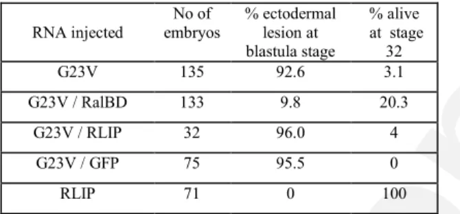
The depigmented phenotype observed in XralB G23V injected embryos has been correlated with F-actin disruption (Moreau et al., 1999). We sought to determine whether RLIP is involved in actin disruption and whether there is a functional link between Ral and the actin cytoskeleton. To test this hypothesis, we examined whether RalBD can compete, as in gastrulation, with endogenous RLIP to interfere in the depigmentation phenotype induced by the constitutively activated form of XralB G23V. Embryos were injected with XralB G23V RNA (300 pg) alone or together with GST-RalBD RNA (5 ng). This corresponds to a 1:2 ratio of XralB G23V mRNA : GST-RalBD mRNA. In these conditions, XralB G23V alone induced depigmentation in 92.6% of the embryos at the blastula stage. Later, the animal
Review Copy
hemisphere of the injected embryos developed ectodermal lesions, and the embryos did not survive beyond the blastula stage. When the GST-RalBD and XralB G23V mRNAs were co-injected, the depigmented phenotype was significantly reduced (Fig. 5A) and survival of embryos was dramatically improved, since only 9.8% of the embryos presented extensive ectodermal lesions and died before gastrulation, whereas 20.3% reached stage 32 (Table 1). Nevertheless, all surviving embryos exhibited gastrulation abnormalities including an open blastopore and a shortened antero-posterior body axis. These phenotypes relate to RalBD overexpression. To check if the rescue effect could be correlated with expression of GST-RalBD, proteins synthesized from the injected mRNAs were monitored by Western blot. The amount of XralB protein produced was comparable (data not shown) whether XralB G23V mRNA was injected alone or with RalBD and confirmed that the rescue effect of XralB G23V can be attributed to the presence of the GST-RalBD protein. This rescue effect was also analyzed by observing the actin cytoskeleton by confocal microscopy. When XralB G23V mRNA was co-injected with GST-RalBD mRNA, the cortical actin cytoskeleton was partially present, whereas it was absent from embryos injected with XralB G23V mRNA alone (Fig. 5B). To show that rescue of XralB G23V by expression of RalBD was not due to competitive association of the RalB catalytic domain with another effector, XralB G23V was co-injected with XRLIP2 mRNA (Fig. 6A). Coexpression did not diminish the depigmentation induced by XralB G23V, but enhanced it. When XralB G23V mRNA was co-injected with mRNA coding for the Green Fluorescent Protein (GFP from the GFP.RN3 plasmid kindly provided by Dr J.B. Gurdon), no significant alteration of depigmentation, either positive or negative, was observed. To confirm the enhancer effect of XRLIP2 expression, XralB G23V mRNA was injected at a lower concentration (100 pg /blastomere) that did not induce depigmentation in early embryos, or was co-injected with either the XRLIP2 (3 ng) or GFP (2 ng) mRNAs. Co-injection of all embryos with the XralB G23V and XRLIP2 mRNAs (n=40) led to depigmentation starting from the 64-cell stage (Fig. 6A), whereas embryos injected with XralB G23V mRNA alone or together with GFP mRNA (2 ng) developed normally, without any alteration of pigmentation.
These results clearly show an enhancing effect of XRLIP2 on the XralB activated form. To confirm that this synergistic effect was due to interaction of the RalBD of XRLIP2 with XralB, we examined whether overexpression of XRLIP2 deleted of its Ral binding domain (XRLIP2 MRalBD) also affected XralB G23V-induced depigmentation. XRLIP2 MRalBD mRNA (3 ng) was injected alone or with XralB G23V mRNA (100 pg) into both animal
Review Copy
blastomeres at the 2-cell stage. Whether these two mRNAs were injected individually or jointly, no depigmentation was observed (Fig. 6B).
Consequently, these data show that the RalBD of XRLIP2 is involved in the interaction with XralB in vivo, that XralB directs RLIP to the plasma membrane and that XRLIP2 is most likely required for the Ral pathway to act on pigmentation distribution.
Docking of RLIP to the membrane induces actin disruption
Since expression of the constitutively active XralB mutant requires XRLIP2 to induce depigmentation, we next examined whether the Ral protein acts by recruiting XRLIP2 to the membrane and disturbs the actin cytoskeleton. Previously, it was demonstrated that the carboxy terminal CAAX sequence of Ras is sufficient to promote localization of Raf to the membrane (Mineo et al., 1997), and that expression of the RalGEF Rlf-CAAX or of Raf1-CAAX (Verheijen et al., 1999) is sufficient to induce differentiation of F9 embryonal carcinoma cells. Therefore, the 10 terminal amino acids of XralB were fused to the C-terminal end of XRLIP2 to determine if localization of RLIP to the membrane can mimic the XralB G23V effect. Embryos were injected with 1.5 ng of XRLIP2-CAAX mRNA into the two animal blastomeres at the 2-cell stage. Depigmentation of the animal blastomeres in all embryos (n= 110) was observed from the 32-cell stage onwards (Fig. 7A). However, unlike the effect of the activated form of XralB, XRLIP2-CAAX induced a rounded appearance of the blastomeres during the cleavage stage, which could correspond to loss of adhesion. Moreover, whereas the external surface of depigmented blastomeres injected with XRalB G23V was smooth, XRLIP2-CAAX-injected blastomeres were irregular with dynamic membrane ruffling and finger-like protrusions that changed extremely rapidly (Fig. 7D). Although these phenotypes were more dramatic than the phenotype induced by the expression of XralB G23V, later in development the blastomeres of embryos injected with XRLIP2-CAAX mRNA seemed to revert to normal, and the embryos survived beyond the gastrula stage. To determine whether XRLIP2-CAAX was present in the plasma membrane, the cellular localization of the protein was examined by confocal screening laser microscopy using anti-myc antibody. In contrast to XRLIP2, which was mainly detected inside the cytosol and only faintly on the plasma membrane, XRLIP2-CAAX was clearly located in the cortical region of the blastomere (Fig. 7B). As the depigmentation of blastomeres expressing XralB G23V is always associated with alteration of the cortical actin cytoskeleton, we investigated
Review Copy
whether XRLIP-CAAX also affects the integrity of the actin cytoskeleton. Figure 7C shows the confocal analysis of the cortical actin cytoskeleton of animal blastomeres from uninjected embryos and from embryos injected with XRLIP2, XRLIP2-CAAX or XralB G23V mRNA. Only the embryos injected with either the XRLIP-CAAX or the XralB G23V mRNA exhibited severe disruption of the cortical actin cytoskeleton.
These results demonstrate clearly that XRLIP partly phenocopies the XralB G23V effect when the protein is translocated to the plasma membrane. Taken together, these data suggest that RLIP is recruited to the plasma membrane through its interaction with XralB to affect the integrity of the actin cytoskeleton. However, the different appearance of blastomeres induced by overexpression of XralB G23V or XRLIP-CAAX and the relative reversibility of the depigmented phenotype induced by XRLIP compared with the stable phenotype induced by XralB G23V, suggest that another Ral effector could be present in embryos and could cooperate with RLIP.
Docking of RLIP to the membrane is required to transduce the Ral signal during gastrulation
We showed previously that both the RalBD of RLIP and the dominant negative form of Ral (XralB S28N) blocked gastrulation movements when specifically injected into the marginal zone of embryos (Lebreton, 2002) and (Lebreton et al., 2003), but have no effect when injected into the animal or vegetative pole. However, it cannot be excluded that another effector, not identified by the two-hybrid screen, interacts with XralB in Xenopus embryos. The overexpression of myc- or GST-RalBD could therefore compete with such an effector for binding with the effector domain of XralB. To clarify this point, XRLIP2 was overexpressed in embryos to saturate the effector site of endogenous Xral, and thereby suppress the potential recruitment of another putative effector. Injection of 1 ng XRLIP mRNA into the marginal zone of each blastomere at the 4-cell stage did not alter the development of the embryos, and more than 90% (n=42) reached the tailbud stage without exhibiting any abnormal phenotype. This result favours the hypothesis that the effect of RalBD is specific of the endogenous XRLIP protein and is not due to possible competition with another effector.
Since activated forms of XralB and XRLIP produce similar effects on cortical actin integrity in blastomeres of the animal hemisphere, and since XRLIP did not compete with another putative Ral effector, we sought to confirm that RLIP is a part of the RalB pathway in
Review Copy
the morphogenetic events affecting the marginal zone mesoderm during gastrulation. Hence, we tested whether the inhibitory effect of XralB S28N on gastrulation could be rescued by co-injection with RNA encoding XRLIP2-CAAX. To this end, the amount of XRLIP2-CAAX mRNA that did not induce depigmentation of the embryo or have deleterious effects on early development was determined. Based on the observation that 500 pg of XRLIP2-CAAX altered neither pigmentation nor gastrulation, 750 pg of XralB S28N mRNA alone or together with either XRLIP2-CAAX mRNA (500 pg) or -galactosidase mRNA (500 pg) as control were microinjected into the marginal zone of embryos. Whereas XralB S28N mRNA alone or co-injected with -galactosidase mRNA blocked gastrulation of 90% of the embryos between stage 10.5 and 11 (Fig. 8A), co-injection of XralB S28N with XRLIP2-CAAX allowed all the embryos (n= 35) to develop to the end of gastrulation (stage 12; Fig. 8A). Compared with uninjected siblings, co-injected embryos presented a delay in development, yet 50% of the embryos (n = 42) closed their blastopore completely and 15% developed up to the early tailbud stage (stage 21). Protein expression in embryos injected with the XralB S28N and XRLIP2-CAAX mRNAs was monitored by Western blot (Fig. 8B) and the expression of the dominant negative form of XralB in rescued embryos was confirmed. Thus, XRLIP-CAAX rescues inhibition of gastrulation caused by overexpression of XralB S28N and partly extends normal development of the embryos compared to siblings injected with XralB S28N alone. Hence 500 pg of XRLIP2-CAAX mRNA was sufficient to rescue Xral S28N. These results indicate that XRLIP is required for the gastrulation process and that it functions in the Ral pathway in vivo.
Review Copy
Discussion
RLIP is the downstream effector of RalB
I is known that signalling pathways do not correspond to a simple linear signalling cascade but include many branchings that crosstalk with other signalling pathways in order to direct signals towards multiple targets or to initiate self-assembly of active multiprotein complexes. The Ras protein activated by FGF signalling interacts with at least three effectors. Each effector has different targets that control processes such as gene expression and actin cytoskeleton dynamics (reviewed in (Vojtek and Der, 1998) and (Reuther and Der, 2000)). Thus, the FGF signal transduction pathway exemplifies the nature of complex biological responses. It is clear today, that during embryonic development, FGFs play an essential role in both the morphogenesis and patterning of mesoderm, in particular during gastrulation. During gastrulation in Xenopus, FGFs are expressed in the marginal zone whereby they activate MAP kinases and are concordant with Xbra expression. The aim of the present study was to characterize the Ral pathway and its involvement in the molecular mechanisms of early vertebrate development. To date, several target molecules of Ral have been identified. All previous data concerning the identification of Ral-binding proteins were derived from experiments carried out in adult mammalian tissues or cell cultures. Therefore, we investigated which effector could be identified in early embryonic development. Using a two-hybrid screen of a maternal Xenopus library, we isolated cDNAs encoding proteins that specifically interact with Ral-GTP. These proteins correspond to at least two orthologues of mammalian RLIP, one preferentially expressed in adult tissues (XRLIP1) and the second preferentially expressed during embryonic development (XRLIP2). For the first time, the interaction of RLIP with Ral is demonstrated in vivo by the co-imunoprecipitation of Xral with XRLIP2. Thus we suggest that RLIP is the effector protein of Ral. We previously demonstrated, by animal cap explant experiments, that FGF activates Ral (Lebreton et al., 2003). Our current study shows that cytosolic RLIP is relocated to the cortical area in a FGF-dependent manner. Indeed, overexpression of the RalBD of XRLIP in the marginal zone of the embryo blocked gastrulation as does the constitutively inactivated form of XralB. Both mRNAs arrest gastrulation between stage 10.5 and 11 in a dose-dependent manner. In contrast, overexpression of XRLIP2 in the same region does not alter gastrulation. The overexpression of XRLIP2 has a synergistic effect on the activity of XralB G23V on cortical
Review Copy
actin. This synergistic effect of XRLIP requires its RalBD. Moreover targeting of XRLIP to the plasma membrane through a XralB CAAX sequence phenocopies the effect of XralB G23V on pigmentation and actin cytoskeleton integrity. The putative Ral effector function of RLIP was reinforced by the rescue experiment in which XRLIP2-CAAX was able to revert the arrest of gastrulation induced by XralB S28N. As no enzymatic activity can be associated with the activation of XRLIP by RalB, it is likely that GTP-bound Ral acts on RLIP by its recruitment to the plasma membrane where it in turn recruits other proteins. So far, four protein domains involved in different functions have been identified in RLIP. The N-terminal O2 binding domain (Jullien-Flores et al., 2000) and potentially also the C-terminal Reps1 binding domain (Nakashima et al., 1999) are involved in endocytosis. A third domain that joins the C-terminal Reps1 binding domain, interacts with the GTP-bound Ral protein, and a fourth domain, between the O2 domain and the RalBD contains a GTPase-activating protein activity (GAP) that acts on the Rac and Cdc42 proteins (Cantor et al., 1995) (Jullien-Flores et al., 1995) (Park and Weinberg, 1995). Rac and Cdc42 are members of the Rho family of GTPases. These proteins regulate diverse cellular processes and in turn control reorganization the actin cytoskeleton (Ridley et al., 1992), (Kozma et al., 1995). This suggests that Ral can participate in the control of polymerisation-depolymerisation of actin filaments by activation or inactivation of the Rac and Cdc42 activity through the RLIP GAP activity. During early
Xenopus development, overexpression of a dominant negative Cdc42 inhibits convergent
extension (Djiane et al., 2000), while Rac inhibits gastrulation (unpublished results), and Rho (Wunnenberg-Stapleton et al., 1999) like Rac is important for cell-cell adhesion. These three closely related proteins, Rho, Rac1 and Cdc42, are indispensable for fibroblast and epithelial cell motility, due to their ability to regulate actin cytoskeletal dynamics, such as filopodia and lamellipodia extensions (Hall, 1994), (Zigmond, 1996). Consequently, the GAP activity of RLIP could be a possible candidate involved in depigmentation induced by targeting RLIP to the plasma membrane. Today, few examples exist in which the Ras-Ral pathway is involved in cell migration as in chemotactic migration of myoblasts (Suzuki et al., 2000) or in activation of invasiveness of fibroblasts (Ward et al., 2001).
While our data suggest that Ral and RLIP are involved in the control of actin rearrangement, other studies (Nakashima et al., 1999) (Matsuzaki et al., 2002) (Moskalenko et al., 2002) suggest a role for these two proteins in endocytosis and exocytosis. These two mechanisms are coordinated and could be necessary for cell migration (Lauffenburger and Horwitz, 1996). For example they may signal to recycle integrin by endocytosis in the trailing edge region of
Review Copy
cell, to promote detachment from the substrate, and to create new adhesions at the leading edge (Bretscher, 1992) .
Finally, it has been shown that during gastrulation FGF is required for cell convergent extension movements by inducing mesodermal genes such as brachyury, which in turn activates targets such as Wnt11 (Tada and Smith, 2000). Previously (Lebreton et al., 2003) we demonstrated that FGF controls morphogenetic movements of gastrulation by a different pathway than MAP Kinase and gene activation. Here, our studies show that RalB activated by FGF recruites RLIP to the plasma membrane and locally regulates the dynamics of the actin cytoskeleton to allow gastrulation. These results make it possible to establish a parallel between the function of the RLIP protein on one hand, and one the other hand the involvement of the dishevelled protein in the non-canonical Wnt/Fz/Dvl/Daam1/Rho and Wnt/Fz/Dvl/Rac pathway (Habas et al., 2003). In both cases, Wnt and FGF, through dishevelled and RLIP respectively, locally regulate the small G protein family Rho/Rac/Cdc42 to control actin cytoskeletal dynamics and thus morphogenetic movements. The next challenge will be to investigate whether the GAP domain of RLIP is required for actin cytoskeleton dynamics and gastrulation, and to define the cellular behaviour dependent on the Ral/RLIP pathway.
Review Copy
Experimental Procedures
Two-hybrid Screen and Two-hybrid Assays
Screening of the library and controlling the binding of XRLIP sequences selected from the
Xenopus library with Ral mutants have been described (Iouzalen et al., 1998).
Northern Blot
Isolation of RNA from embryos, electrophoresis and hybridization were carried out as described (Iouzalen et al., 1998).
Embryos and Microinjections
Xenopus laevis were imported from South Africa (South Africa Farms, Fish Hoek) or from
the CNRS frog colony (Rennes). Animals were housed and fed as described (Gurdon et al. 1984). Embryos were fertilized in vitro and chemically dejellied with 0.3 X modified Barth’s solution pH 7.6 (MBS) with 2% cysteine-HCl, pH 7.8-7.9, and then maintained in 1 X MBS (Gurdon and Wickens, 1983) until used. Microinjection was performed in 1 X MBS with 3% Ficoll, followed by incubation of the embryos overnight at 16° C in 0.1 X MBS with 3% Ficoll, and then at 15-22°C until they reached the appropriate stages. Embryonic stages were determined according to Nieuwkoop and Faber (Nieuwkoop and Faber, 1956). In vitro transcribed mRNAs (4.6 nl of the appropriate dilutions) were microinjected into the animal hemisphere of 2-cell stage embryos, or into the marginal zone of 4-cell stage embryos.
Western Blots and binding assay of RLIP and XralB
Proteins were extracted by lysing embryos in 20 Ol of buffer A (50 mM Tris-HCl pH 7.5, 100 mM NaCl, 0.1% Triton X-100, 5 mM EDTA, 2 mM PMSF) per embryo, followed by Freon extraction of the vitellus. Protein extracts were separated by SDS-PAGE on a 7.5% acrylamide gel and transferred to a Hybond C membrane (Amersham). The membrane was probed overnight at 4°C with goat anti-human RLIP antibodies (Sc-1948, Santa Cruz) diluted 1/250 in 1 X PBS, 0.1% Tween 20 and 10% milk. The second antibodies were peroxidase – coupled rabbit antibodies specific to goat immunoglobulins and diluted 1/2500 (Chemicon). The signals were detected by chemiluminescence (ECL, Amersham).
Translation of myc-RalBD and myc-XRLIP2-CAAX after microinjection of the mRNAs was checked by probing the membrane with a monoclonal antibody directed against the myc epitope (9E10, Santa-Cruz, Sc-40) at a dilution of 1/1000 in 1 X PBS, 0.1% Tween 20 and
Review Copy
5% BSA. Antibodies against -tubulin (N357, Amersham) were used at a dilution of 1/4000 in 1 X PBS, 0.1% Tween 20 and 5% BSA to monitor the amount of proteins loaded on the gels.
To show the interaction of RLIP with Ral, embryos expressing Myc-RLIP (full lengh or deleted of Ral binding domain) were suspended in lysis buffer (Tris 50 mM pH 7.5, NaCl 100 mM, Triton X100 0.5% and 1 mM PMSF). The lysate was centrifugated at 10000 rpm for 30 min and supernatant was mixed twice with 1,1,2-trichlorofluoroethan and aqueous phase incubated over night with 6 Ol of Myc (9E10) antibody bound to agarose (Santa-Cruz, Sc-40-AC).
cDNA cloning and in vitro transcription of RNA for injection
pRN3-myc-XRLIP2 was constructed by inserting the EcoRI fragment of the pGAD-XRLIP-DH31 selected from the two-hybrid screen (Iouzalen et al., 1998) into the EcoRI site of the pRN3-myc vector. To obtain pRN3-XRLIP2-CAAX, the membrane-targeting CAAX signal of XralB was added to the 3’ end of the XRLIP2 coding sequence by PCR using pRN3-myc-XRLIP2 as template, the primer (5’–AACCGTGTCCTGTATGTG-3’) matching a sequence upstream of an internal Sac I restriction site in the XRLIP2 coding sequence, and the reverse primer (5‘– CGAGTTAGCGGCCGCTCAGAGTAAACAGCAGCGTTGTTTGAAGCCCTTGATGAGA GTTTCAGAGG- 3’) containing the CAAX sequence and a Not I restriction site (Not I site underlined). The amplified fragment coding for XRLIP2-CAAX was digested with SacI and NotI and exchanged with the SacI-NotI fragment of XRLIP2 in pRN3-myc-XRLIP2.
pRN3-XRLIP2-MRalBD was constructed by PCR amplification of the sequence coding for the N-terminal region of the protein starting at amino acid 473, using RLIP as template. The primer (5’-GCGGAACAGGAGCTCCTTGTGGCAATGGAGCAG-3’), introducing a Sac I restriction site (double underline), was used with the reverse primer (5’-GGAGCAGATACGAATGGCTAC-3’) located within the pRN3 vector downstream of the 3’ Not I integration site. The Sac I-Not I fragment of XRLIP2 was then exchanged with the Sac I-Not I digested fragment of the PCR amplification product, thereby deleting the region delimited by amino acids 343 and 473, that corresponds to the entire RalBD.
RNA transcripts were produced using T3 RNA polymerase. Capped mRNAs were obtained with the mMessage Machine System (Ambion) according to the manufacturer’s instructions.
Review Copy
Immunostaining of Embryos and Confocal Microscopy Analysis
Xenopus embryos at the appropriate stage of development were incubated in a
permeabilization buffer (80 mM Hepes pH 6.8, 1 mM MgCl2, 100 mM EDTA, 30 % glycerol,
0.1 % Triton X-100) for 15 min at room temperature before being fixed in 1 X PBS and 1.4% formaldehyde overnight, or their vitelline membrane was manually removed under a binocular microscope before being fixed in 1 X PBS and 3.5% formaldehyde for 1 hour at room temperature. Embryos were then treated as described (Moreau et al., 1999). The 9E10 anti-myc antibody was diluted 1/1000 in 1 X PBS, 1% BSA and 0.1% Triton X-100. Confocal observations were made as described (Moreau et al., 1999), using a Leica SB2 AOBS confocal imaging system (Leica Instruments, Heidelberg, Germany).
Review Copy
Acknowledgments
We thank Gerard Geraud for the confocal analyses and Anne-Lise Haenni and Mike Jones for critical reading of the manuscript. This work was supported by the CNRS, “Association pour la Recherche sur le Cancer” and the “Fondation pour la Recherche Médicale ”.
Review Copy
References
Amaya, E., Musci, T.J. and Kirschner, M.W. (1991) Expression of dominant negative mutant of the FGF receptor disrupts mesoderm formation in Xenopus embryos. Cell 66, 257-270.
Bar-Sagi, D. and Feramisco, J.R. (1986) Induction of membrane ruffling and fluid-phase pinocytosis in quiescent fibroblasts by ras protein. Science 233, 1061-1068. Bauer, B., Mirey, G., Vetter, I.R., Garcia-Ranea, J.A., Valencia, A., Wittinghofer, A.,
Camonis, J.H. and Cool, R.H. (1999) Effector recognition by the small GTP-binding proteins Ras and Ral. The Journal of Biological Chemistry 274, 17763-17770.
Bisbee, C.A., Baker, M.A., Wilson, A.C., Hadji-Azimi, I. and Fischberg, M. (1977) Albumin phylogeny for clawed frogs (Xenopus). Science 195, 785-787.
Bretscher, M.S. (1992) Circulating integrins: 5ß1, 6ß4 and Mac-1 but not 3ß1, 4ß1 or LFA-1. EMBO J. 11, 405-410.
Cantor, S.B., Urano, T. and Feig, L.A. (1995) Identification and characterization of Ral-binding protein 1, a potential downstream target of Ral GTPases. Molecular and Cellular Biology 15, 4578-4584.
Christen, B. and Slack, J.M.W. (1997) FGF-8 is associated with anteroposterior patterning and limb regeneartion in Xenopus laevis. Developmental biology 192, 455-466. Djiane, A., Riou, J.F., Umbhauer, U.M., Boucaut, J.C. and Shi, D.L. (2000) Role of Frizzled7
in the regulation of convergence extension movements during gastrulation in Xenopus
laevis. Development 127, 3091-3100.
Feig, L.A., Urano, T. and Cantor, S. (1996) Evidence for a Ras/Ral signaling cascade. Trends in Biochemical Sciences 21, 438-441.
Gurdon, J.B. and Wickens, M.P. (1983) The use of Xenopus oocytes for the expression of cloned genes. In Press, A. (ed.), Methods in EnzymologyVol. 101, pp. 370-382. Habas, R., Dawid, I.B. and He, X. (2003) Coactivation of Rac and Rho by Wnt/frizzled
signaling is required for vertebrate gastrulation. Genes and Development 17, 295-309. Hall, A. (1994) Small GTP-binding proteins and the regulation of the actin cytoskeleton.
Annual Review of Cell Biology 10, 31-34.
Ikeda, M., Ishida, O., Hinoi, T., Kishida, S. and Kikuchi, A. (1998) Identification and characterization of a novel protein interacting with Ral-binding protein 1, a putative effector protein of Ral. Journal of Biological Cemistry 273, 814-821.
Iouzalen, N., Camonis, J. and Moreau, J. (1998) Identification and characterization in Xenopus of XsmgGDS, a RalB binding protein. Biochemical and Biophysical Research Communications 250, 359-363.
Isaacs, H.V., Tannahill, D. and Slack, J.M.W. (1992) Expression of a novel FGF in the Xenopus embryo. A new candidate inducing factor for mesoderm formation and anteroposterior specification. Development 114, 711-720.
Jiang, H., Luo, J.-Q., Urano, T., Frankel, P., Lu, Z., Foster, D.A. and Feig, L.A. (1995) Involvelment of ral GTPase in v-Src-induced phospholipase D activation. Nature 378, 409-412.
Jullien-Flores, V., Dorseuil, O., Romero, F., Letourneur, F., Sragosti, S., Berger, R., Tavitian, A., Gacon, G. and Camonis, J.H. (1995) Bridging Ral GTPase to Rho pathways. The Journal of Biological Chemistry 270, 22473-22477.
Jullien-Flores, V., Mahé, Y., Mirey, G., Leprince, C., Meunier-Bisceuil, B., Sorkin, A. and Camonis, J.H. (2000) RLIP76, an effector of the GTPase Ral, interacts with the AP2 complex: involvement of the Ral pathway in receptor endoocytosis. Journal of Cell Science 113, 2837-2844.
Review Copy
Kozma, R., Ahmed, S., Best, A. and Lim, L. (1995) The Ras-related protein Cdc42Hs and Bradykinin promote formation of peripherical actin microspikes and filopodia in swiss 3T3 fibroblasts. Mol. Cell. Biol. 15, 1942-1952.
Kristen, C.L. and Grainger, R.M. (2000) Expression of activated MAP Kinase in Xenopus
laevis embryos: Evaluating the roles of FGF and other signaling pathways in early
induction and patterning. Development Biology 228, 41-56.
Kroll, K.L. and Amaya, E. (1996) Transgenic Xenopus embryos from sperm nuclear
transplantations reveal FGF signaling requirements during gastrulation. development 122, 3173-3183.
Lauffenburger, A.D. and Horwitz, A.F. (1996) Cell migration: A physically integrated molecular process. Cell 84, 359-369.
Lebreton, S. (2002) Etude de la petite protéine G XralB au cours du développement
embryonnaire de Xenopus laevis. ph D thesis. Université Pierre et Marie Curie. Paris. Lebreton, S., Boissel, L. and Moreau, J. (2003) Control of embryonic Xenopus
morphogenesis by a Ral-GDS/Xral branch of the signalling pathway. J. Cell Sci. 116, 4651-4662.
Matsuzaki, T., Hanai, S., Kishi, H., Liu, Z.H., Bao, Y.L., Kukuchi, A., Tsuchida, K. and Sugino, H. (2002) Regulation of endocytosis of activin type II receptors by a novel PDZ protein through Ral/Ral-binding protein 1-dependent. The Journal of Biological Chemistry 277, 19008-19018.
Mineo, C., Anderson, R.G.W. and White, M.A. (1997) Physical association with ras enhances activation of membrane-bound raf (RafCAAX). The Journal of Biological Chemistry 272, 10345-10348.
Moreau, J., Lebreton, S., Iouzalen , N. and Mechali, M. (1999) Characterization of Xenopus RalB and its involvement in F-actin control during early development. Developmental Biology 209, 268-281.
Moskalenko, S., Henry, D.O., Rosse, C., Mirey, G., Camonis, J.H. and White, M.A. (2002) The exocyst is a Ral effector complex. Nature Cell Biology 4, 66-72.
Nakashima, S., Morinaka, K., Koyama, S., Ikeda, M., Kishida, M., Okawa, K., Iwamatsu, A., Kishida, S. and Kikuchi, A. (1999) Small G protein Ral and its downstream molecules regulate endocytosis of EGF and insulin receptors. EMBO J. 18, 3629-3642.
Nieuwkoop, P.D. and Faber, J. (1956) Normal table of Xenopus laevis. North-Holland, Amsterdam.
Nutt, S.L., Dingwell, D.S., Holt, C.E. and Amaya, E. (2001) Xenopus Sprouty2 inhibits FGF-mediated gastrulation movements but does not affect mesoderm induction and
patterning. Genes and Development 15, 1152-1166.
Ohta, Y., Suzuki, N., Nakamura, S., Hartwig, J.H. and Stossel, T.P. (1999) The small GTPase RalA targets filamin to induce filopodia. Proc. Natl. Acad. Sci. USA 96, 2122-2128. Park, S.H. and Weinberg, R.A. (1995) A putative effector of Ral has homology to Rho/Rac
GTPase activating proteins. Oncogene 11, 2349-2355.
Reuther, G.W. and Der, C.J. (2000) The Ras branch of small GTPases: Ras family members don't fall far from the tree. Current Opinions in Cell Biology 12.
Ridley, A.J., Paterson, H.F., Johnston, C.L., Diekmann, D. and Hall, A. (1992) The small GTP-binding protein rac regulates growth factor-induced membrane ruffling. Cell 70, 401-410.
Sugihara, K., Asano, S., Tanaka, K., Iwamatsu, A., Okawa, K. and Ohta, Y. (2002) The exocyst complex binds the small GTPase RalA to mediate filopodia formation. Nature Cell Biology 4, 73-78.
Suzuki, J., Yamazaki, Y., Guang, L., Karziro, Y. and Koide, H. (2000) Involvement of Ras and Ral in chemotactic miration of skeletal myoblasts. Mol. Cell. Biol. 20, 4658-4665.
Review Copy
Tada, M. and Smith, J.C. (2000) Xwnt11 is a target of Xenopus brachyury: Regulation of gastrulation movements via dishevelled, but not through the canonial Wnt pathway. Development 127, 2227-2238.
Tannahill, D., Isaacs, H.V., Close, M.J., Peters, G. and Slack, J.M. (1992) Developmental expression of tthe Xenopus int-2 (FRF-3) gene: Activation by mesodermal and neural induction. Development 115, 695-702.
Verheijen, M.H., Wolthuis, R.M., Defize, L.H., Hertog, J.d. and Bos, J.L. (1999) Interdependent action of RalGEF and Erk in Ras-induced primitive endoderm differentiation of F9 embryonal carcinoma cells. Oncogene 18, 4435-4439.
Vojtek, A.B. and Der, C.J. (1998) Increasing complexity of the Ras signaling pathway. The Journal of Biological Chemistry 273, 19925-19928.
Ward, Y., Wang, W., Woodhouse, E., linnoloila, I., Liotta, L. and Kelly, K. (2001) Signal pathways which promote invasion and metastasis: Critical and distinct contributions of extracellular signal-regulated kinase and ral-specific guanine exchange factor
pathways. Mol. Cell. Biol. 21, 5958-5969.
White, M.A., Nicolette, C., Minden, A., Polverino, A., Aelst, L.V., Karin, M. and Wigler, M.H. (1995) Multiple Ras functions can contribute to mammalian cell transformation. Cell 80, 533-541.
White, M.A., Vale, T., Camonis, J.H., Schaefer, E. and Wigler, M.H. (1996) A role for the Ral guanine nucleotide dissociation stimulator in mediating Ras-induced
transformation. J. Biol. Chem. 271, 16439-16442.
Wilson, R. and Leptin, M. (2000) Fibroblast growth factor receptor-dependent morphogenesis of the Drosophila mesoderm. Phil. Trans. R. Soc. Lond. 355, 891-895.
Wolthuis, R.M.F. and Bos, J.L. (1999) Ras caught in another affair: the exchange factors for Ral. Current Opinion in Genetics and Development 9, 112-117.
Wunnenberg-Stapleton, K., Blitz, I.L., Hashimoto, C. and Cho, K.W. (1999) Involvement of the small GTPases XRhoA and XRnd1 in cell adhesion and head formation in early
Xenopus developement. Development 126, 5339-5351.
Yamaguchi, T.P., Harpal, K., Henkemeyer, M. and Rossant, J. (1994) Fgfr-1 is required for embryonic growth and mesodermal patterning during mouse gastrulation. Genes and Development 8, 3032-3044.
Zigmond, S.H. (1996) Signal transduction and actin filament organization. Current Opinion in Cell Biology 8, 66-73.
Review Copy
Table I - Arrest of development caused by constitutively activated RalB (XralB G23V) and rescued by the RalBD mRNA.
RNA injected No of embryos % ectodermal lesion at blastula stage % alive at stage 32 G23V 135 92.6 3.1 G23V / RalBD 133 9.8 20.3 G23V / RLIP 32 96.0 4 G23V / GFP 75 95.5 0 RLIP 71 0 100
Note. Each blastomere of two-cell embryos was injected with the amount of mRNA indicated in the legend of Fig. 4. The data of at least three separate experiments were pooled.
Review Copy
Figure legends Figure 1
Amino acid sequence alignments between human RLIP (Q15311) and Xenopus XRLIP1 (AJ252165) and XRLIP2 (AJ304845) proteins. Dashes represent gaps inserted to maximize alignment.
Figure 2
A - Temporal expression of Xenopus XRLIP mRNAs during oogenesis, development and in some adult tissues analyzed by Northern blot. Ten Og of RNA of each stage were loaded, except for oogenesis stage I/II where 2 Og were loaded. RNAs were separated on an agarose gel, blotted and hybridized with the XRLIP DH14 probe. EF1- [Krieg, 1989 #94] was used as loading control on the same Northern blots (the zygotic transcription of EF1- mRNA begins at the mid-blastula transition (MBT). B – Temporal expression of the Xenopus XRLIP proteins during development. Protein extracts corresponding to one embryo for each of the different developmental stages were run on an SDS-polyacrylamide gel and immunoblotted with antiserum against RLIP. Protein loading on each lane was monitored with anti-tubulin antibodies (Ab tubulin). C - XRLIP interacts specifically with RalB in the two-hybrid system. Interactions of both Xenopus XRLIP, XRLIP DH31 (amino acids 19 to 655) and XRLIP DH14 (amino acids 30 to 655), were tested with either the inactivated form (XralB S28NMCT), both activated forms, (XralB G23V, XralB G23VD49N), or wild type XralB. Human lamin served as control.
Figure 3
In vivo interaction of Ral with RLIP. Embryos at the two cell stage were microinjected with
XralS28NC203S (lane2) mRNA or co-injected with XRLIP2 (lane 3) or XRLIP2MRalBD (lane 4) proteins were extracted at stage 10.5. XRLIP and Myc-XRLIP2MRalBD were immuno retained on agarose and the co-immuno retained Ral proteins were assessed with anti-RalB antibodies. Panel A shows the RLIP retained by anti–Myc agarose and displays the associated RalB (IP:RalB). B - Schematic representation of XRLIP2 indicates the position of the RalBD in XRLIP2 and the deletion of this domain in XRLIP2MRalBD. Numbers refer to amino acids. C - Localization of XRLIP was visualized
Review Copy
with the monoclonal c-myc antibody 9E10 and secondary antibodies conjugated to FITC. D – Localization of XRLIP in cells of animal cap incubated with FGF. Scale bars represent 50 Om, each picture corresponds to the stacking of 3 confocal optical sections of 1 Om thickness.
Figure 4
Rescue of RalBD-injected embryos is dependent on the RalBD of RLIP. All embryos correspond to the same developmental stage (stage 12.5) as the sibling controls (top row, left). Each blastomere of four-cell-stage embryos was injected in the marginal zone. Embryos injected with RLIP (middle left) or RLIP2MRalBD (bottom left) mRNAs developed normally with a short delay in development compared with controls. RalBD mRNA injection (400 pg) arrested development at stage 10.5 (top right). RLIP mRNA rescued the effect of RalBD mRNA (middle right). However, when RalBD mRNA was co-injected with XRLIP2MRalBD mRNA (bottom right), embryos were blocked at the beginning of gastrulation (stage 10.5) as were embryos injected with only RalBD mRNA (top right).
Figure 5
RLIP acts on the actin cytoskeleton by its RalBD. A - View of the animal hemisphere of embryos at the large-cell blastula stage. Embryos were injected into each blastomere at the 2-cell stage with 300 pg of XralB G23V mRNA (G23V), or 5 ng of GST-RalBD mRNA, or co-injected with 300 pg of XralB G23VmRNA and 5 ng of GST-RalBD mRNA. B - Rescue of the effect of XralB G23V mRNA on cortical actin cytoskeleton by co-injection with GST-RalBD mRNA. Analysis by confocal scanning microscopy of actin cables stained by rhodamine-phalloidin (7.5 Og/ml). Embryos injected in each blastomere with 300 pg XralB G23V mRNA, or co-injected with 5 ng of GST-RalBD mRNA, cultured until the midblastula stage. Arrows show the partially reconstituted cortical actin cables. Scale bars represent 50 Om, confocal optical sections are 1 Om thick.
Figure 6
Synergistic effect of XRLIP2 and RalG23V. A - Embryos were injected in the animal hemisphere with XralB G23V mRNA (300 pg) alone, or co-injected with 3 ng of either XRLIP2 or GFP mRNAs. B – XRLIP2, but not XRLIP2MRalBD enhances the XralB G23V
Review Copy
effect. Embryos were injected with 100 pg of XralB G23V mRNA, 3 ng XRLIP2 mRNA, or 3 ng of XRLIP2MRalBD mRNA, or co-injected with XralB G23V and XRLIP2 or XRLIP2MRalBD mRNAs. Depigmentation was observed only when XralB G23V mRNA was co-injected with XRLIP.
Figure 7
XRLIP2-CAAX phenocopies the XralB G23V effect. A – Three nanograms of XRLIP2 CAAX mRNA, XRLIP mRNA, or 500 pg of XralB G23V mRNA were injected into the animal hemisphere of 2-cell stage embryos. Only XRLIP2-CAAX and XralB G23V induced depigmentation of the embryos. B - Targeting of XRLIP2 to the plasma membrane. All XRLIP constructs contain a c-myc tag at the N terminus. XRLIP2 or XRLIP2-CAAX (1 ng each) mRNA were microinjected into embryos at the 2-cell stage. Localization of XRLIP was visualized with the monoclonal c-myc antibody 9E10 and secondary antibodies conjugated to FITC. C – Confocal microscope analysis of actin cytoskeleton cables stained by rhodamine-phalloidin at the 500 cell stage in animal pole explants. The cortical actin cytoskeleton is visible only in uninjected control embryos and embryos injected with wild type XRLIP. Each picture corresponds to stacking of 3 confocal optical sections of 1 Om thickness. D - Magnification of animal blastomeres in an embryo injected with 0.75 ng of XRLIP2-CAAX mRNA shows dynamic finger-like protusions on the cell surface (black arrowheads). Cell division proceeds normally (white arrowheads). The time lapse between each picture is 15 mn. Scale bars represent 50 Om.
Figure 8
Rescue of the Xral S28N induces gastrulation defect by XRLIP2-CAAX expression. A - Embryos at the 4-cell stage were microinjected in the marginal zone with 750 pg/blastomere of XralB S28N mRNA or 500 pg/blastomere of XRLIP2-CAAX mRNA, or coinjected with both mRNAs. The figure shows a vegetal view of embryos at the same developmental time after fertilization, as control siblings. Only embryos in which XralB S28N mRNA has been injected alone exhibit a very wide blastopore compared to control uninjected embryos. Embryos co-injected with XralB S28N and RLIP2-CAAX mRNAs are delayed in blastopore closure. B - Analysis by Western blot of protein expression in rescue experiments using
Review Copy
appropriate antibodies (anti-RalB, and anti-myc for the XRLIP constructs). Each lane was loaded with protein extracts corresponding to 1 embryo.