HAL Id: inserm-00484284
https://www.hal.inserm.fr/inserm-00484284
Submitted on 28 Dec 2010
HAL is a multi-disciplinary open access
archive for the deposit and dissemination of
sci-entific research documents, whether they are
pub-lished or not. The documents may come from
teaching and research institutions in France or
abroad, or from public or private research centers.
L’archive ouverte pluridisciplinaire HAL, est
destinée au dépôt et à la diffusion de documents
scientifiques de niveau recherche, publiés ou non,
émanant des établissements d’enseignement et de
recherche français ou étrangers, des laboratoires
publics ou privés.
plasmacytoid dendritic cell maturation: potential
implications in inflammatory diseases.
Fanny Angelot, Estelle Seillès, Sabeha Biichlé, Yael Berda, Béatrice Gaugler,
Joël Plumas, Laurence Chaperot, Francoise Dignat-George, Pierre Tiberghien,
Philippe Saas, et al.
To cite this version:
Fanny Angelot, Estelle Seillès, Sabeha Biichlé, Yael Berda, Béatrice Gaugler, et al..
Endothe-lial cell-derived microparticles induce plasmacytoid dendritic cell maturation: potential implications
in inflammatory diseases.. Haematologica, Ferrata Storti Foundation, 2009, 94 (11), pp.1502-12.
�10.3324/haematol.2009.010934�. �inserm-00484284�
Acknowledgments: the authors would like to thank Sylvain Perruche, Francis Bonnefoy, Claire Latruffe, Guillaume Aranda, Evelyne Racadot, Christian Naegelen, Jean-Paul Remy-Martin, Emilie Gaiffe, Jean-Luc Pretet and Jean-Marie Certoux for helpful discussion, manuscript editing and technical support. Funding: this work was supported by the University of Franche-Comté (BQR 2006 to FGO, BQR 2008 to ES), the Fondation Transplantation (ET-051222, ET-060934, ET-070636 to FA), the Fondation Recherche Médicale (DEA20050904854 to FA), the Conseil Régional de Franche-Comté (confocal microscope) and the Fondation de France (Appel d’offre "Recherche sur les maladies cardiovasculaires 2007" to PS).
Manuscript received on May 6, 2009. Revised version arrived on May 29, 2009. Manuscript accepted on May 29, 2009. Correspondence: Francine Garnache-Ottou, 1 Bd A. Fleming, BP1937, F25020 Besançon cedex, France. E-mail: [email protected] Background
Increased circulating endothelial microparticles, resulting from vascular endothelium dys-function, and plasmacytoid dendritic cell activation are both encountered in common inflammatory disorders. The aim of our study was to determine whether interactions between endothelial microparticles and plasmacytoid dendritic cells could contribute to such pathologies.
Design and Methods
Microparticles generated from endothelial cell lines, platelets or activated T cells were incubated with human plasmacytoid dendritic cells sorted from healthy donor blood or with monocyte-derived dendritic cells. Dendritic cell maturation was evaluated by flow cytometry, cytokine secretion as well as naive T-cell activation and polarization. Labeled microparticles were also used to study cellular interactions.
Results
Endothelial microparticles induced plasmacytoid dendritic cell maturation. In contrast, conventional dendritic cells were resistant to endothelial microparticle-induced matura-tion. In addition to upregulation of co-stimulatory molecules, endothelial microparticle-matured plasmacytoid dendritic cells secreted inflammatory cytokines (interleukins 6 and 8, but no interferon-α) and also induced allogeneic naive CD4+T cells to proliferate and to
produce type 1 cytokines such as interferon-γ and tumor necrosis factor-α. Endothelial microparticle endocytosis by plasmacytoid dendritic cells appeared to be required for plas-macytoid dendritic cell maturation. Importantly, the ability of endothelial microparticles to induce plasmacytoid dendritic cells to mature was specific as microparticles derived from activated T cells or platelets (the major source of circulating microparticules in healthy subjects) did not induce such plasmacytoid dendritic cell maturation.
Conclusions
Our data show that endothelial microparticles specifically induce plasmacytoid dendritic cell maturation and production of inflammatory cytokines. This novel activation pathway may be implicated in various inflammatory disorders and endothelial microparticles could be an important immunmodulatory therapeutic target.
Key words: endothelial cell-derived microparticles, plasmacytoid, inflammatory diseases, dendritic cells.
Citation: Angelot F, Seillès E, Biichlé S, Berda Y, Gaugler B, Plumas J, Chaperot L, Dignat-George F, Tiberghien P, Saas P, and Garnache-Ottou F. Endothelial cell-derived microparticles induce plas-macytoid dendritic cell maturation: potential implications in inflammatory diseases. Haematologica 2009;94:1502-1512. doi:10.3324/haematol.2009.010934
©2009 Ferrata Storti Foundation. This is an open-access paper.
Endothelial cell-derived microparticles induce plasmacytoid dendritic cell
maturation: potential implications in inflammatory diseases
Fanny Angelot,1 Estelle Seillès,1 Sabeha Biichlé,1 Yael Berda,2 Béatrice Gaugler,1 Joel Plumas,3 Laurence Chaperot,3
Françoise Dignat-George,2Pierre Tiberghien,1Philippe Saas,1 and Francine Garnache-Ottou1
1Inserm UMR645, University of Franche-Comté, EFS Bourgogne Franche-Comté, IFR133, Besançon, France; 2Inserm U608,
Laboratoire d'Hématologie et d'Immunologie, UFR de Pharmacie, Université de la Méditerranée, Marseille, France, and 3Inserm
U823, EFS du sang Rhône Alpes, Université Joseph Fourier, La Tronche, France
Introduction
Membrane vesiculation is a general physiological process that leads to the release of cell plasma frag-ments, called microparticles.1-3These microparticles are defined by their size (0.1-1 µm) and can be generated from nearly every type of cell during activation, injury or apoptotic processes.1 All microparticles, whatever their cell origin, have negatively charged phospholipids – such as phosphatidylserine – in their outer membrane leaflet, accounting for their procoagulant properties.1-3 They also express proteins, characteristic of their cellu-lar origin, on their surface and carry proteins packaged from numerous cellular compartments,4,5 as well as mRNA.6 Microparticles are different from exosomes, since these latter are smaller (30-90 nm) and derived from endocytic compartments leading to an enrichment of tetraspanin molecules.7Microparticles can participate in the maintenance of homeostasis under physiological conditions. Among the various circulating microparti-cles, platelet-derived microparticles are the most abun-dant in the bloodstream, representing between 70 to 90% of circulating microparticles in healthy subjects.8 As shown for apoptotic bodies9– another form of cell
dust or debris, tumor- or fibroblast-derived
microparti-cles captured by monocytes may induce regulatory cytokine secretion.10 In addition, microparticles from activated T cells can deliver differentiation signals to stem cells.11In contrast, microparticles can initiate dele-terious processes if produced in excess1or when carry-ing pathogenic constituents or inflammatory signals.12,13 Vascular endothelium aggression may lead to the vesic-ulation and shedding of endothelial microparticles (EMP). Increased levels of EMP have been reported in a variety of pathological situations including thrombo-sis,3,8atherosclerosis,14renal failure,15,16 diabetes,17 graft-versus-host disease after hematopoietic cell transplanta-tion18,19 and systemic lupus erythematosus.1,20,21 These data emphasize the link between endothelial damage, the release of EMP and the modulation of inflammato-ry and/or immune responses.
Dendritic cells play a major role in immune respons-es. They are specialized to capture and present antigens to T cells.22Two major subsets of dendritic cells have been described in humans: conventional dendritic cells (previously called myeloid dendritic cells) and plasma-cytoid dendritic cells (PDC).23These latter represent a particular population of dendritic cells that were first identified as the principal cells secreting interferon α (IFN-α) in response to viral or bacterial stimulation.23As such, PDC contribute to the innate viral and anti-bacterial defense system. Alterations of PDC homeosta-sis and function with increased production of IFN-α have been implicated in various autoimmune or inflam-matory diseases including type I diabetes, psoriasis, multiple sclerosis, and systemic lupus erythematosus. 23-25In addition, PDC have been found in the atheroscle-rotic plaques.26-28 However, a growing body of data shows that PDC can also be involved in tolerance induc-tion.29-31On the other hand in vitro activation of imma-ture human PDC with ligands for Toll-like receptor
(TLR) 7 or TLR9 (e.g., R848 or type A CpG motifs, respectively) leads to proinflammatory cytokine pro-duction and to increased co-stimulatory molecule expression, subsequently inducing naive T-cell activa-tion.23Other factors, such as damage-associated molec-ular patterns, also induce PDC activation.23,32Nothing is known about the capacity of EMP to activate PDC. The common finding of endothelial damage – associated with increased EMP release – and PDC activation in sev-eral inflammatory diseases32 led us to investigate whether EMP could provide signals that promote phe-notypic and functional maturation of PDC in vitro.
Design and Methods
Flow cytometry and antibodies
Flow cytometry was performed with a CANTO A cytometer (BD Biosciences, Le Pont de Claix, France) using DIVA 6.1 software (BD Biosciences). The following monoclonal antibodies were used: fluorescein isothio-cyanate-conjugated BDCA-2 (AC144, Miltenyi Biotech, Paris, France), CD14 (MYA-4), HLA-DR (B-F1), phyco-erythrin-conjugated CD40 (mAb89), CD31 (1F11), phy-coerythrin-Texas red-x (ECD)-conjugated CD41 (P2), phycoerythrin-cyanin-5 (PC5) CD146 (TEA 1/34), phy-coerythrin cyanin-7-conjugated CD3 (UCHT1), CD4 (SFCI12T4D11) (Beckman Coulter Immunotech, Villepinte, France), phycoerythrin-conjugated CD123 (9F5), CCR7 (CD197, clone 2H4), CD83 (HB15e), fluo-rescein isothiocyanate-conjugated CD80 (MAB104), CD86 (2331FUN1) and allophycocyanin-conjugated CD1a (HI149), CD62E (68-5H11) (BD Biosciences). Fluorescent-conjugated isotype control monoclonal anti-bodies from the different monoclonal antibody providers were used. The mean fluorescence intensity ratio was obtained by dividing the mean fluorescence intensity for a given marker by the mean fluorescence intensity of the respective isotype control monoclonal antibody.
Generation and flow cytometry quantification of
microparticles
EMP were prepared from a human microvascular der-mal endothelial cell line (HMEC-1) as previously described.33,34Briefly, confluent HMEC-1 cells were incu-bated for 24 h with 50 ng/mL TNF-α (Sigma Aldrich, Saint-Quentin Fallavier, France). Culture supernatants were collected and cleared from detached cells and cell fragments by centrifugation at 1200g for 5 min. The supernatant was then centrifuged twice at 15000g for 90 min (Cambrex, Verviers, Belgium) at 4°C. Pelleted EMP were resuspended in culture media and used immediate-ly. The absence of residual TNF-α in EMP samples was confirmed using ELISA (Diaclone). EMP were also pre-pared from quiescent HMEC-1 (without TNF-α) as well as from the human umbilical vein cell line, EAY926 (ATCC CRL-2922) using the same protocol. EMP prepa-rations were checked for endotoxin contamination using the Limulus amebocyte lysate assay. Endotoxin content was always less than 0.05 ng/mL. In some experiments, the supernatant resulting from the last wash was used as a control. This supernatant was free of EMP, as
demon-strated by cytometric analysis. The absence of mycoplas-ma was confirmed for all cultures using the Mycolasmycoplas-ma PCR detection kit (Venor® GeM, BioValley, Marne la Vallée, France).
Platelet-derived microparticles were isolated from acid-citrate-dextrose anticoagulated human blood from healthy volunteers obtained after written informed con-sent (Etablissement Français du Sang Bourgogne Franche-Comté, EFS B/FC, Besançon, France). The platelet-rich plasma was obtained by centrifugation at 100g for 15 min. This plasma was spun at 1000g for 10 min, the supernatant was then collected and the platelet-derived microparticles were pelleted by centrifugation at 20000g for 2 h at 4°C.12The purity of platelet-derived microparti-cle isolation (~95%) was assessed using CD31 and CD41 monoclonal antibody staining. Microparticles derived from activated T cells were prepared, using the same pro-tocol as that for EMP, from CD3/CD28 activated periph-eral blood mononuclear cells isolated from healthy donors. After isolation on Ficoll-Hypaque (Pharmacia, Uppsala, Sweden) centrifugation, the peripheral blood mononuclear cells were activated with CD3/CD28 Dynabeads (Invitrogen, Cergy Pontoise, France) and 600 UI/mL of interleukin (IL)-2 (Proleukine, Sanofi, Paris, France) for 48 h. Supernatants were collected and T-cell-microparticles were isolated.
In order to quantify the microparticles, an aliquot of 10 µL of microparticles was labeled using fluorescein isoth-iocyanate-conjugated annexin V (Annexin V apoptosis detection kit I, BD Biosciences) according to manufactur-er’s instructions. In each experiment, control labeling was performed by incubating annexin V with the supernatant resulting from the last wash of the microparticle prepara-tion. A defined number of calibrated 3-µm latex beads (FlowcountTMbeads, Beckman Coulter) was added to the sample, as an internal standard. Consequently, micropar-ticles derived from endothelial cells, platelets and activat-ed T cells were definactivat-ed as elements with a size comprisactivat-ed between 0.1-1 µm and positively labeled by fluorescein isothiocyanate-annexin V.
Generation and isolation of dendritic cells
The GEN 2.2 cell line derived from a PDC leukemia patient was cultured on MS-5 irradiated feeder cells in RPMI 1640 Glutamax 1 (InVitrogen, Cergy Pontoise, France) containing 1 mM sodium pyruvate (Sigma), 20 µg/mL gentamicin (InVitrogen), non-essential amino acids (InVitrogen) (hereafter referred to as RPMI complete medium) and 10% heat inactivated fetal calf serum (Life Technologies, Gaithersburg, MD, USA). Peripheral blood mononuclear cells from healthy donors were obtained after written informed consent and isolated by Ficoll-Hypaque (Pharmacia) centrifugation. Circulating normal PDC were isolated using immunomagnetic cell sorting (BDCA-4 cell isolation Kit, Miltenyi Biotec) according to the manufacturer’s recommendations. After isolation, PDC purity was controlled by cytometry using CD123/BDCA-2 monoclonal antibody staining and was between 94-98%. Monocyte-derived dendritic cells (Mo-DC) were generated from blood monocytes – isolated by negative depletion (Negative Monocyte Isolation Kit, Dynal, InVitrogen) – by a 6-day culture in RPMI 1640,
containing 10% heat inactivated fetal calf serum, 50 ng/mL granulocyte-monocyte colony-stimulating factor and 20 ng/mL IL-4 (PeproTech, London, UK). At the end of the culture, the generation of Mo-DC was attested by a CD1a+CD14–phenotype.
Evaluation of dendritic cell maturation
Maturation of freshly isolated PDC was induced by culture for 18 h in the presence of 10 ng/mL IL-3 (PeproTech) and R848 (1 µg/mL, Invivogen, Toulouse, France), or CpGA (2 µmol/L, ODN2216, Invivogen), or recombinant CD40L (1 µg/L, COGER, Paris, France) or by addition of microparticles (ratio 10 microparticles:1 den-dritic cell; this corresponds to 5000 microparticles/µL, an EMP concentration found in vivo in several pathological situations including myocardial infarction35and thrombo-cytopenic purpura36). Mo-DC (6-day culture) were incu-bated with either lipopolysaccharide from Escherichia coli (LPS, 2 µg/mL, Sigma) or EMP (10 EMP:1 Mo-DC) for 48 h. To assess the involvement of phosphatidylserine-medi-ated signaling in Mo-DC maturation, cells were incubat-ed with EMP or apoptotic cells, as a positive control, (cor-responding to murine thymocytes submitted to 40 Gray γ-irradiation- with a ratio of 5 apoptotic cells:1 dendritic cell), and stimulated 24 h later by lipopolysaccharide. IL-12(p70) was measured in supernatants (ELISA, BD OptEIA, BD Biosciences).
Confocal microscopy analyses
PDC were incubated with anti-BDCA-2 monoclonal antibody (AC144 Miltenyi Biotech) for 30 min at +4°C, washed and labeled with cyanin 5-conjugated goat anti-mouse Ig (BD Biosciences). PDC were incubated with EMP previously labeled with CFSE (5 mM) (Molecular Probes, Leiden, Netherland) for 2 h at 37°C in a Labtek II culture chamber (VWR International, Fontenay-sous-Bois, France). After nuclear staining using DAPI (Mol-ecular Probes), cells were washed in phosphate-buffered saline, fixed in 4% paraformaldehyde and mounted (Fluorescent Mounting Medium, Dako, Trappes, France). Fluorescent images were acquired on an FV1000 confocal microscope (Olympus, Rungis, France).
Cytokine production
Culture supernatants were collected from PDC or Mo-DC incubated with or without TLR ligands or EMP for 18 h. The following cytokines: IL-12p70 (only for Mo-DC), TNF-α, IL1-β, IL-10, IL-6, IL-8 and transforming growth factor (TGF)-β were measured in the culture supernatants using Luminex Technology kits (HCYTO-60 K, Linco, Millipore, Saint-Quentin en Yvelines, France) on a Luminex 100® analyzer (Luminex, Austin, TX, USA), according to the manufacturer’s instructions. Interferon (IFN)-α was measured by using a Luminex Technology kit (Biosource, Clinisciences, Montrouge, France).The minimal detectable concentrations were as follows: 1 pg/mL for 12(p70), TNF-α and 1β; 2 pg/mL for IL-10, IL-6 and IL-8; 15 pg/mL for IFN-α; 20 pg/mL for TGF-β. Intracellular expression of IFN-α by PDC was deter-mined by flow cytometry using anti-IFN-α monoclonal antibody (clone LT27:295, Miltenyi Biotec). Brefeldin A (10 µg/mL, Sigma) was added 3 h before the end of the
activation with TLR ligands or EMP. Staining was per-formed according to the manufacturer’s recommenda-tions for fixed and permeabilized cells (Cytofix/cytop-erm Plus kit, BD Biosciences).
Capacity of dendritic cells to induce naive CD4
+T-cell proliferation and cytokine production
Mixed leukocyte reactions were performed by cultur-ing naive CD4+T cells as responding cells and allogene-ic dendritallogene-ic cells stimulated with or without TLR ligands or EMP as stimulating cells. CD4+ CD45RA+ naive T cells were isolated from peripheral blood by negative selection using two sorting kits (CD4 Negative Isolation Kit, Dynal, InVitrogen and CD45 microbeads, Miltenyi Biotec) in two steps according to the manufacturers’ instruction. CD4+CD45RA+ T-cell purity evaluated by cytometry was between 90 to 94%. Naive CD4+T cells were then incubated with allogeneic dendritic cells in RPMI complete medium containing 10% human serum (EFS BFC, Besançon, France) for 6 days. T-cell prolifera-tion was measured using the DELFIA Cell Proliferaprolifera-tion Kit (AD0077P-1, Perkin Elmer, Boston, MA, USA) on a DELFIA-type reader (DELFIA Envision, 2102 Multilabel Reader, Perkin Elmer). Five-bromo-2-deoxyuridine (10 µL/mL, Perkin Elmer) was added 18 h before the end of the mixed lymphocyte reaction. Cytokine secretion by T cells after mixed lymphocyte reactions was assayed with a Luminex Kit (HCYTO-60 K, Linco, Millipore). After 7 days, culture supernatants were collected and cytokines (IFN-γ, IL-5, IL-4, IL-2, IL-10, IL-17, TNF-α and TGF-β) of primed T cells were evaluated. CD45RA+ CD4+naive T cells cultured in complete medium alone were used as controls. The minimal detectable concen-trations were the same as described above plus as fol-lows: 1 pg/mL for IL-5, and IL-17; 2 pg/mL for IFN-γ and IL-2; 4 pg/mL for IL-4.
Analysis of endothelial microparticle-plasmacytoid
dendritic cell interactions
EMP were added to PDC and incubated at 37°C or 4°C for 15 to 90 min. After incubation, PDC were washed three times to remove non-adherent EMP. Then, transfer of phosphatidylserine or endothelial-specific antigens (CD146 and CD62E) to PDC was analyzed by cytome-try. Alternatively, CFSE-labeled EMP were incubated at different EMP:PDC ratios (1:1, 10:1, 100:1) for 4 h and PDC were analyzed by cytometry. Sorted CD4+T cells were used as a negative control for endocytosis. The role of sodium-proton exchange and an intact PDC cytoskeleton for EMP capture was evaluated by pretreat-ment of PDC for 1 h with either dimethyl amiloride (100 µM) or cytochaline D (20 µM) (Sigma-Aldrich).37
Statistical analysis
Statistical analyses were performed using Sigma Stat 2.0 software (SPSS Inc., Jandel Scientific, Erkrath, Germany). Group comparisons of parametric or non-parametric data were performed using the Student’s t test or Mann Whitney test, respectively. Data were tested for mean ± S.E.M. A p value less than 0.05 was considered statistically significant.
Results
Endothelial microparticles increase co-stimulatory
molecule expression on plasmacytoid dendritic cells,
but not on conventional dendritic cells
Interactions between PDC and EMP in vitro have never been evaluated to date. Since PDC activation and endothelial damage associated with EMP production are encountered in similar diseases, we decided to explore these interactions, as well as the capacity of EMP to modulate PDC maturation. We first used TNF-α-induced EMP as representative of inflammatory conditions. Expression of co-stimulatory (CD80, CD86, CD40) and activation (HLA-DR, CD83, CCR7) molecules on freshly isolated PDC or a PDC cell line (GEN2.2) incubated with EMP was compared to expression of these molecules on immature PDC or GEN2.2. Incubation of TNF-induced EMP with freshly isolated PDC for 18 h, induced PDC maturation, as evidenced by upregulation of the expres-sion of co-stimulatory molecules, the increase of HLA-DR molecules, as well as of CD83 and CCR7 as com-pared to levels in unstimulated immature PDC (Figure 1A-C). The effects of EMP on PDC maturation were comparable to those induced by classical maturation agents such as the TLR7 ligand, R848 (Figure 1A-C) and the TLR9 ligand, CpGA (data not shown). Similar results were obtained using the GEN2.2 PDC cell line (Figure 1D) or using the endothelial cell line EAY926 as the source of EMP (data not shown). As shown in Figure 1E, PDC maturation was still observed for a lower (1:80) PDC:EMP ratio. In contrast, PDC incubated with the supernatant resulting from the last wash of EMP, but not containing EMP, did not induce PDC or GEN2.2 matura-tion (Figure 1C-D). Moreover, we checked for the absence of residual TNF-α, bacterial endotoxins or mycoplasms which could affect dendritic cells matura-tion. These factors were not detected in any of the cul-ture conditions (data not shown). We also verified that EMP derived from TNF-α-stimulated HMEC-1 did not express the membrane markers HLA-DR, CD83 or CD123 on their surface and that the increase of such markers on PDC did not result from EMP uptake by PDC (data not shown). PDC maturation was also confirmed by morphological analysis, since EMP-stimulated PDC acquired dendrites (Figure 1F), a morphological feature of mature dendritic cells.
To determine whether EMP-induced maturation was restricted to PDC or was also observed for conventional DC, we studied the interactions between EMP and Mo-DC using the same design experiment as described for PDC. We did not observe a significant increase of expres-sion of co-stimulatory molecules on the surface of Mo-DC exposed to EMP at a ratio of 1 Mo-Mo-DC to 10 EMP (Figure 1G). Only a slight increase of HLA-DR on the sur-face of Mo-DC was observed in the presence of EMP (Figure 1G). The same results were observed with a high-er numbhigh-er of EMP (Mo-DC:EMP ratio 1:80, data not
shown). The TLR4 ligand, lipopolysaccharide, was used
as a control for Mo-DC maturation (Figure 1G). Altogether, these data showed that EMP generated in the presence of TNF-α induced PDC maturation, but not
Mo-DC maturation.
Endothelial microparticle-induced mature
plasmacytoid dendritic cells are functional
and secrete inflammatory cytokines
In order to confirm PDC maturation after EMP interac-tions using a functional assay, we determined cytokines secreted in the supernatants after culture of PDC with EMP and by comparison with PDC cultures in the pres-ence of the TLR7 ligand, R848 or the TLR9 ligand, CpGA. Immature PDC alone did not produce significant levels of IL-1β, IL-6, IL-10 or TNF-α (Figure 2A). As expected, PDC in the presence of R848 or CpGA secreted high levels of IL-6 and IL-8, and low levels of IL-10 (Figure 2A), while TGF-β, IL-1α and TNF-β were not detected (data not
shown). In the presence of EMP, PDC also secreted IL-6
and IL-8, although at lower levels than in the presence of R848 or of CpGA (Figure 2A). The same experiments were performed using Mo-DC matured in the presence of lipopolysaccharide or incubated with EMP. In the pres-ence of EMP, Mo-DC did not secrete significant levels of IL-6 and exhibited a slight secretion of IL-8 that did not reach statistical significance (Figure 2B). Lipopoly-saccharide induced the secretion of IL-12 by Mo-DC (Figure 2B). In contrast, incubation of Mo-DC with EMP did not increase IL-12 secretion (Figure 2B). These func-tional data further confirm that EMP induced PDC matu-ration, but not Mo-DC maturation.
PDC are the major cells producing IFN-α, as confirmed
in vitro after stimulation with the TLR9 ligand, CpGA
(Figure 2C). However, PDC did not secrete IFN-α when stimulated by EMP, nor in the presence of R848 (Figure
Figure 1. EMP induce morphological and phe-notypic maturation of PDC but not of conven-tional dendritic cells. PDC or the GEN 2.2 PDC cell line (GEN) were incubated with classical maturation agents R848 (PDC R848, GEN R848) or with medium used to generate EMP but not containing EMP (Med) or with TNF-induced EMP (PDC EMP, GEN EMP) at the ratio of 1 dendritic cell to 10 EMP (corresponding to 5000 EMP/µL in vivo)35,36for 18 h.
Co-stimula-tory and maturation marker expression was then determined by flow cytometry and com-pared with the immature unstimulated PDC (iPDC) or GEN2.2 cell (iGEN) phenotype. Expression of the PDC-specific marker CD123 did not vary in any of the conditions tested (A). (A) Histograms from one representative exper-iment out of eight are shown. (B) Histograms from one representative experiment out of two are shown. On each histogram (A,B), values represent the mean fluorescent intensity (MFI) ratio (MFIR) obtained by dividing the MFI for a given marker by the MFI of the respective iso-type control monoclonal antibody. (C,D) Histograms represent the mean±S.E.M of HLA-DR and CD83 expression (expressed as MFIR) from three independent experiments. *p<0.05. (E) PDC were incubated for 18 h with TNF-induced EMP at different PDC:EMP ratios: 1 PDC for 0 (corresponding to medium stimulat-ed PDC), 1, 5, 10, 20, 40, and 80 EMP and mat-uration was determined by cytometry. HLA-DR (P) and CD83 (M) expression (assessed by MFIR) on PDC incubated with EMP was com-pared with R848-activated PDC (PDC R848). Results from two independent experiments are shown. (F) Morphological analysis of PDC incu-bated with EMP (PDC EMP) or not (iPDC) con-firmed PDC maturation as attested by dendrite acquisition (white arrows) using confocal microscopy, which also enabled the purity of the PDC after immunomagnetic cell sorting to be assessed. PDC are identified here by BDCA-2 staining (red fluorescence) and nuclear stain-ing (blue fluorescence).
Before incubation with PDC, EMP were labeled with CSFE (green fluorescence). Results from one experiment out of two are shown. (G) Conventional dendritic cells were activated with the TLR4 ligand lipopolysaccharide (Mo-DC LPS) or incubated with EMP (Mo-DC EMP) at the ratio of 1 Mo-DC to 10 EMP for 48 h. Co-stimulation and maturation marker expression was then determined by flow cytometry and compared with the immature unstimulated Mo-DC (iMo-DC) phenotype. Expression of CD83, CD86, CD80, CD40 and HLA-DR mol-ecules increased only in response to lipopolysaccharide stimulation (Mo-DC LPS), but not after EMP incubation (Mo-DC EMP). Histograms from one representative experiment out of five are shown. Values on each histogram represent MFIR.
A C F B D E G HLA-DR CD83 CD86 CD80 CD40 HLA-DR CD83 CD86 CD80 CD40 CD123 80 3 10 3 74 74 6 22 8 101 59 2 13 3 72 110 0,8 1 0,7 7 293 369 8 14 24 136 260 708 14 4 10 55 180 iMO-DC iPDC PDC R848 EMPMed iGEN GEN R848 EMPMed PDC R848 PDC:EMP ratio 1/0 1/1 1/5 1/10 1/20 1/40 1/80 iPDC PDC R848 PDC EMP 35 20 10 0 10 6 2 0 100 800 600 400 200 0 300 200 100 0 500 300 100 0 30 20 10 0 CCR7 27 29 11 PDC EMP PDC R848 iPDC Mo-DC LPS Mo-DC EMP
Relative cell number
Relative cell number
CD83
HLA-DR HLA-DR
HLA-DR (MFIR) CD83 (MFIR)
CD83
2C). In addition, significant amounts of IFN-α were not found in the supernatant of EMP- or R848-stimulated PDC (data not shown). The same results were observed whatever the PDC:EMP ratio used (data not shown). Overall, these data demonstrate that exposure of PDC to TNF-α-induced EMP increases the secretion of inflam-matory cytokines by PDC with one major exception, IFN-α.
EMP express high levels of phosphatidylserine33 and phosphatidylserine signaling in conventional dendritic cells after interactions with apoptotic cells is known to interfere with dendritic cell maturation.38We, therefore, incubated Mo-DC with EMP or apoptotic cells then stimulated with lipopolysaccharide for 24 h. Supernatants were collected and IL-12p70 was meas-ured. Pre-incubation of EMP with Mo-DC reduced lipopolysaccharide-induced IL-12p70 secretion, such as observed with apoptotic cell pre-incubation (Figure 2D). This suggests that Mo-DC respond to an EMP-induced inhibitory signal.
Endothelial microparticle-induced mature
plasmacytoid dendritic cells stimulate naive
CD4 T-cell proliferation
To further explore the maturation of PDC by EMP,
EMP-stimulated PDC were cultured with naive allogene-ic CD45RA+CD4+T cells. As shown in Figure 3A, naive CD4+T cells proliferated significantly in the presence of EMP-induced mature PDC whereas no proliferation was observed using unstimulated immature PDC (p=0.025). The rate of proliferation in the presence of EMP-induced mature PDC was similar to that observed in the presence of R848-induced mature PDC (p=0.72) (Figure 3A). Similar results were obtained using the GEN2.2 cell line as stimulating cells (Figure 3B). In contrast, Mo-DC stim-ulated by EMP did not induce significant naive CD4+ T-cell proliferation in comparison to that induced by unstimulated Mo-DC (Figure 3B). IL-2 secretion at the end of the mixed leukocyte reaction was also assessed as a marker of naive CD4+T-cell proliferation. While signif-icant amounts of IL-2 were not detected in CD4+T cells co-cultured with immature PDC (mean + S.E.M, 7±4 pg/mL, n=5), increased levels of IL-2 were found in CD4+ T cells cultured with R848-stimulated PDC (1574±436 pg/mL, n=5, p=0.007 vs. immature PDC) or EMP-incu-bated PDC (590±362 pg/mL, n=5, p=0.008 vs. immature PDC).
Endothelial microparticle-induced mature
plasmacytoid dendritic cells stimulate naive
Figure 2. EMP induce cytokine secretion by PDC, but not by conventional dendritic cells. (A) Culture supernatants collected from imma-ture PDC (iPDC), PDC stimulated with the TLR7 ligand R848 (PDC R848), the TLR9 lig-and CpGA (PDC CPGA) or with TNF-induced EMP (PDC EMP, ratio of 1 PDC to 10 EMP) for 18 h were assayed for IL-6, IL-8, and IL-10 using Luminex Technology. EMP-activated PDC secreted the same cytokine profile as PDC R848 or PDC CpGA. Data, expressed in pg/mL, represent the mean±S.E.M of four independent experiments; *p<0.05; **p<0.001. Data with PDC CPGA are from one experiment. (B) Culture supernatants collected from immature Mo-DC (iMo-DC), Mo-DC stimu-lated with lipopolysaccharide (Mo-DC LPS) or with EMP (Mo-DC EMP, ratio 1:10) for 18 h were assayed for IL-12, IL-6 and IL-8 using Luminex Technology. Data, expressed in pg/mL, represent the mean±S.E.M of five experiments: *p<0.05; **p<0.001. (C) Percentage of IFN-α+
cells after stimulation of PDC with R848 (PDC R848), CpGA (PDC CPGA) or with EMP (PDC EMP, ratio 1:10) for 18 h were assayed for intracellular IFN-α measured by flow cytometry. Data were com-pared with unstimulated immature PDC (iPDC). Dot plot histograms represent expres-sion of IFN-α on BDCA2+
CD123+
cells. Histograms from one representative experi-ment out of three are shown. Similar data were obtained in cell supernatants (data not
shown). (D) Culture supernatants collected from immature (iMo-DC), Mo-DC stimulated with TNF-induced EMP (Mo-DC EMP, ratio of 1 Mo-DC to 10 EMP) or with apoptotic cells (APO) (Mo-DC APO, ratio: 1 Mo-DC to 5 APO) for 24 h and then stimulated for 24 h with the TLR4 ligand lipopolysaccharide (LPS), were assayed for IL-12(p70) using ELISA. Data, expressed in pg/mL, represent the mean±S.E.M. of three independent experi-ments. A B C D EMP APO IL-12 pg/mL (Mo-DC) LPS PDC CPGA PDC R848 PDC EMP iPDC iMo-CD IL-12 IL-8 IL-8 IL-10 IL-6 IL-6 Mo-DC
iPDC PDC iPDC PDC iPDC PDC
Mo-DC
EMP LPS EMP LPS
EMP R848 CPGA EMP R848 CPGA EMP R848 CPGA
p=0.057 p=0.057 EMP LPS Mo-DC iMo-CD iMo-CD 8360 817 31341634 IFN-α 5% 0% 0% 0% BDCA2 pg/mL pg/mL 350 250 150 50 0 400 300 200 100 0 820 400 200 0 100 75 50 25 0 3100 400 200 0 10000 6000 2000 0 9500 2000 1000 0
CD4 T cells to produce Th1 cytokines
In order to explore CD4+T-cell polarization after acti-vation by EMP-induced mature PDC, cytokine secretion by co-cultured CD4+ T cells was examined and com-pared with that of CD4+T cells incubated with imma-ture PDC and TLR7-stimulated PDC as well as with Th1 or Th2 well-defined conditions (lipopolysaccharide-stimulated Mo-DC and CD40 plus IL-3 (lipopolysaccharide-stimulated-PDC, respectively). Cytokine production was measured directly in the supernatants after the mixed lymphocyte reactions. As shown in Figure 3C, naive CD4+ T cells stimulated by EMP-induced mature PDC secreted Th2-related cytokines, IL-5 and IL-4, but at non-significant and variable levels according to the experiments consid-ered. However, CD4+ T cells primed by EMP-induced PDC secreted significant levels of Th1-related cytokines, IFN-γ and TNF-α (Figure 3C). Despite the high levels of IL-6 produced by PDC after EMP interactions and the role of IL-6 in Th17 polarization, we did not observe either IL-17-secreting CD4+T cells after co-culture with EMP-induced mature PDC or TGF-β production in any of the culture conditions (data not shown). The cytokine profile observed after EMP stimulation was similar to that of CD4+ T cells stimulated with TLR7-activated PDC (Figure 3C). Naive CD4+ T cells stimulated by immature PDC did not secrete these cytokines (Figure 3C). None of these cytokines was detected in the super-natants from EMP or T cells cultured in medium alone (data not shown). Comparison with Th1 or Th2 control conditions (Figure 3C) suggests that EMP-induced mature PDC favor Th1 polarization, as evidenced by IFN-γ and TNF-α secretion.
In contrast to endothelial microparticles,
plasmacytoid dendritic cells do not respond to
platelet- or T-cell-derived microparticles
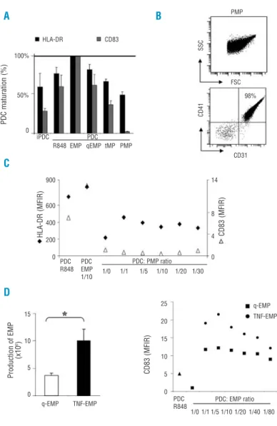
In order to appreciate the relevance of EMP-induced PDC maturation, we compared the effects of TNF-α-induced EMP to the effects of quiescent EMP (i.e., EMP produced in the absence of TNF-α), of platelet-derived microparticles (the major form of microparticles found in the bloodstream of healthy subjects) and of normal T-cell-derived microparticles. As shown in Figure 4A, quiescent EMP were as potent as TNF-induced EMP at inducing PDC maturation, as assessed by co-stimulatory mole-cule expression (Figure 4A) and naive CD4+T-cell activa-tion (data not shown). In contrast, microparticles derived from platelets or activated T cells did not significantly induce PDC maturation (Figure 4A). We also studied dif-ferent ratios of PDC to platelet-derived microparticles, but whatever the ratio used, the platelet-derived microparticles did not induce PDC maturation (Figure 4B-C). This suggests that the origin of microparticles – and particularly the endothelial origin – influences PDC maturation.
Concerning the effect of TNF on EMP production, we observed that TNF-α stimulation of endothelial cells sig-nificantly increased the number of EMP produced (Figure 4D). We, therefore, analyzed whether a dose-effect relationship exists for PDC maturation. As shown in Figure 4E, we observed a dose-effect of EMP on PDC maturation. Altogether, this suggests that PDC respond
Figure 3. EMP-induced mature PDC induce allogeneic naive CD4 T-cell proliferation and Th1 cytokine secretion. (A) Proliferation of naive CD4+CD45RA+T cells in response to unstimulated
imma-ture PDC (iPDC), PDC activated by R848 (PDC R848) or to PDC incubated with TNF-induced EMP at a ratio of 1:10 (PDC EMP) was analyzed as described in the Design and Methods section. Co-cul-ture conditions were defined by preliminary experiments and the adequate ratio is 20,000 PDC to 50,000 naive CD4+T cells. Basal
CD4+ T-cell proliferation was also measured (Med). Histograms
represent the mean±S.E.M of the four independent experiments. Results are expressed in europium count (EU count). *p<0.05. (B) Proliferation of naive CD4+CD45RA+T cells in response to
unstim-ulated immature Mo-DC (iMo-DC), Mo-DC activated by lipopolysac-charide (MO-DC LPS), Mo-DC incubated with EMP at a ratio of 1:10 (Mo-DC EMP), or to GEN2.2 PDC cell line (unstimulated, iGEN; acti-vated with R848, GEN R848 or incubated with EMP, GEN EMP) was analyzed as described in the Design and Methods section. Data are from one representative experiment out of three for GEN2.2 and of two for Mo-DC. Results are expressed in europium counts (EU count, mean±S.E.M.). *p<0.05; **p<0.001. (C) Culture super-natants collected from CD4+T cells co-cultured with immature
(iPDC), with R848-activated PDC (PDC R848), or with PDC incubat-ed with TNF-inducincubat-ed EMP (PDC EMP) were assessincubat-ed for Th2-relat-ed cytokines (IL-4 and IL-5, left panels) and for Th1-relatTh2-relat-ed cytokines (IFN-γ and TNF-α, right panels) using Luminex Technology. Histograms represent the mean±S.E.M. of five inde-pendent experiments. Results are expressed in pg/mL for each of the cytokines analyzed. In order to help data interpretation, addi-tional controls were performed consisting in Th2 control condi-tions (using PDC stimulated with CD40L and IL-3, as described)49
and Th1 control conditions (using Mo-DC stimulated with lipopolysaccharide). *p<0.05; **p<0.001. A B C iPDC PDC Med EMP R848 iPDC PDC PDC Th2 EMP R848 Ctrl iPDC PDC PDC Th1 EMP R848 Ctrl iGEN GEN iMO Mo-DC
EMP R848 EMP LPS IFN-γ TNF-α IL-4 IL-5 40000 30000 20000 10000 0 40000 30000 20000 10000 0 40000 30000 20000 10000 0 40000 30000 20000 10000 0 1500 1000 500 0 600 400 200 0 EU count pg/mL EU count
to a given number of EMP rather than to inflammatory EMP (i.e., TNF-induced EMP) and, therefore in vivo PDC may be activated in pathological conditions in which EMP production is increased, such as injury to the vas-cular endothelium.
Endothelial microparticle-plasmacytoid dendritic cell
interactions are temperature-dependent and require
sodium-proton exchanges as well as an intact
cytoskeleton
To define the mechanism involved in the maturation process of PDC induced by EMP, we first determined whether EMP adhere to PDC and, if so, whether a fusion between EMP and PDC membranes occurs. In these con-ditions, a transfer of endothelial antigen to PDC would
occur. Incubation of EMP with PDC did not lead to endothelial antigen (CD62E or CD146) or phos-phatidylserine transfer to PDC (data not shown). To fur-ther evaluate a role of an uptake of EMP by PDC, EMP were labeled with CFSE before their incubation with PDC. As shown in Figure 5A-B, significant numbers of CFSE+ PDC were identified when cells were incubated with EMP at 37°C whereas incubation at 4°C significant-ly decreased the percentage of CFSE+PDC. In contrast, significant amounts of CFSE were not detected in CD4+ T cells whatever the incubation temperature used (Figure 5A). This suggests that EMP uptake by PDC is tempera-ture-dependent. To go further, we treated PDC with an inhibitor of sodium-proton exchange, dimethyl amiloride or the F-actin depolymerizing agent,
cytocha-Figure 4.PDC respond to EMP but not to activat-ed T-cell- or platelet-derivactivat-ed microparticles. (A) The effects of TNF-α-stimulated EMP (EMP) on PDC maturation were compared to those of qui-escent EMP (qEMP), T-cell-derived microparticles (tMP) or platelet-derived microparticles (PMP). PDC were activated by R848 (PDC R848) or incu-bated with TNF-α induced EMP (PDC EMP) or qui-escent EMP (PDC qEMP) at a ratio of 1 PDC to 10 EMP for 18 h. PDC were also incubated with tMP and PMP at the same ratio and in the same experimental conditions. Maturation marker expression of HLA-DR and CD83 on stimulated PDC – analyzed by cytometry – was then com-pared with the expression on immature unstimu-lated PDC (iPDC). Histograms represent the per-centage of PDC maturation based on HLA-DR and CD83 expression with expression on PDC EMP arbitrarily considered to indicate 100% mat-uration. Pooled results of four independent exper-iments are shown. (B) Platelet-derived micropar-ticle were isolated and analyzed by flow cytome-try, identified in the FSC vs. SSC gate based on expression of CD41 and CD31. A representative experiment out of four is shown. (C) PDC were incubated for 18 h with PMP at different PDC:PMP ratios: 1 PDC to 0 (corresponding to medium-stimulated PDC), 1, 5, 10, 20, and 30 PMP and maturation was determined by cytome-try. HLA-DR (X) and CD83 (g) expression (assessed by MFIR) on PDC incubated with PMP was compared with expression on R848-activat-ed PDC (PDC R848). Results from one out of two independent experiments are shown. (D) TNF-α
exposure increases EMP production by the der-mal endothelial cell line HMEC-1. EMP were pro-duced as described in the Design and Methods section. The numbers of EMP produced per cul-ture flask (50 mL) in basal conditions (q-EMP) or after TNF-α (50 ng/mL, TNF-EMP) stimulation were quantified by cytometry. Histograms repre-sent the mean + S.E.M of the absolute number of q-EMP or TNF-EMP in a 50 mL culture flask from six independent productions. *p=0.03. (E) PDC were incubated for 18 h with TNF-induced EMP (TNF-EMP,Q) or quiescent EMP (qEMP,L) at dif-ferent PDC:EMP ratios: 1 PDC to 0 (correspon-ding to medium-stimulated PDC), 1, 5, 10, 20, 40, and 80 EMP and maturation was determined by cytometry. CD83 expression (assessed by MFIR) on PDC incubated with EMP was com-pared with expression on R848-activated PDC (PDC R848). Results from two independent experiments are shown. Of note, 1 EMP:1 PDC corresponds to 500 EMP/µL; a higher concentra-tion than that in healthy donors (10 + 10 EMP/µL, personal data).
PDC maturation (%) HLA-DR (MFIR) Production of EMP (x10 6) CD83 (MFIR) CD83 (MFIR) iPDC PDC PDC R848 PDC R848 q-EMP TNF-EMP PDC: PMP ratio PDC: EMP ratio 1/0 1/1 1/5 1/10 1/20 1/30 1/0 1/1 1/5 1/10 1/20 1/40 1/80 q-EMP TNF-EMP PDC EMP 1/10 CD31 FSC SSC CD41 PMP 98% R848 EMP qEMP tMP PMP HLA-DR CD83 100% 50% 0 900 600 400 200 0 15 10 5 0 25 20 15 10 5 0 14 8 4 0 A C D B
lasin D 1 h prior to the addition of EMP. This treatment significantly reduced the number of CFSE+ PDC (Figure 5B) and prevented PDC maturation (assessed by co-sti-mulatory marker expression, data not shown). Taken together, these data indicate that PDC activation by EMP is dependent on temperature and requires sodium-proton exchange and an intact cytoskeleton.
Discussion
Increased circulating EMP related to vascular endothelial dysfunction and PDC activation are encoun-tered in similar pathological situations, including vascu-lar diseases as well as inflammatory diseases.3,8,14-21,23-28 We, therefore, hypothesized that EMP could trigger PDC maturation and that such an interaction could con-tribute to the above mentioned inflammatory disorders. Here, we demonstrate that EMP can indeed induce PDC maturation, as shown by co-stimulatory molecule upregulation, inflammatory cytokine secretion, and by
allogeneic naive CD4+ T-cell proliferation. Moreover, naive CD4+ T cells primed in the presence of EMP-matured PDC produced mainly type 1 cytokines. Despite the high levels of IL-6 produce by PDC after EMP interactions and the role of IL-6 in Th17 polariza-tion,39IL-17-secreting CD4+ T cells were not observed after co-culture with EMP-induced mature PDC. This could be due to the absence of TGF-β – another critical cytokine for Th17 differentiation39 – in PDC cultures after EMP incubation. In contrast to PDC isolated from healthy volunteers or to the GEN2.2 PDC cell line, EMP did not induce significant Mo-DC maturation and, in this setting, reduced lipopolysaccharide-induced IL-12 secretion, as observed after incubation with apoptotic cells.9,38Furthermore, microparticles derived from acti-vated T cells or platelets did not induce PDC matura-tion. Lastly, PDC maturation induced by EMP required an active uptake mechanism. Overall, these data sug-gest a link between vascular endothelial damage and PDC activation. Maturation of PDC by EMP released following damage to the endothelium may, therefore, be implicated in the physiopathology of different inflammatory and vascular diseases.
The in vivo relevance of our observations remains to be explored further. The PDC:EMP ratio used here (1:10) corresponds to 5000 EMP/µL, an EMP concentration found in vivo in pathological situations.35,36Infiltration of mature PDC in injured tissues (such as in atherosclerot-ic plaques,26-28 in the skin in psoriasis23,32 or systemic lupus erythematosus)24,25 has been observed in several chronic inflammatory diseases. Increased expression of the chemokine receptor CCR740– as observed here – has also been reported in trapped dendritic cells found in atherosclerotic plaques.26 Thus, elevated concentrations of EMP in response to vascular endothelial damage and/or endothelial dysfunction15,41 may initiate PDC migration and subsequent maturation.
Chronic inflammatory diseases are often associated with uncontrolled α production by PDC. Such IFN-α secretion is notably found in systemic lupus erythe-matosus,42,43 psoriasis23,32 and in atherosclerotic pla-ques.27,28In our study, EMP did not cause PDC to secrete IFN-α. A first hypothesis to explain this is that PDC acti-vation by EMP may be insufficient to induce IFN-α secretion. Preliminary data show that EMP are able to activate nuclear factor-κB (data not shown). Thus, EMP can be considered, similarly to necrotic cells, as a cell byproduct that may alert the innate immune system (i.e., a damage-associated molecular pattern). Necrotic cells have been reported to activate conventional den-dritic cell maturation through the TLR2 signaling path-way44or CLEC9A.45Since nucleic acids can be packed into microparticles,6one can speculate that EMP-induced PDC maturation may involve endosomal TLR7 or TLR9. These receptors are specialized in sensing foreign nucleic acids. Production of IFN-α is dependent on localization of TLR9-CpGA interactions in endosomal compart-ments; interactions in the transferrin+early endosomes lead to IFN-α secretion while interactions in the LAMP1+ late endosomes (i.e., lysosomes) are responsible for IL-6 and IL-8 production as well as increased expression of co-stimulatory molecules.46,47 This may be an
explana-Figure 5.Interaction between EMP and PDC is an active process dependent on temperature, sodium-proton exchanges and on an intact actin cytoskeleton. (A) EMP were labeled with CFSE and incubated with PDC or CD4+T cells at 37°C or 4°C at the ratio of
1 PDC (or 1 CD4+T cell) to 100 EMP for 4 h. PDC were identified
based on high expression of CD123. CD4+T cells were identified
by CD4 expression. After extensive washes, cells were analyzed by cytometry. Dot plot histograms show the data for one representa-tive experiment out of three. (B) Histograms represent the mean of the percentage of PDC (black) or CD4+T cells (gray) positive for
CFSE after different incubation conditions: 37°C, 4°C, dimethyl amiloride (DMA), or cytochalasin D (CD) treatment. DMA, an inhibitor of sodium-proton exchange, or CD, an F-actin depolymer-izing agent, was incubated with PDC or CD4+T cells for 1 h before
addition of EMP. Histograms from three or two independent exper-iments are shown. Results are expressed as mean ± SEM of the percentage of CFSE+cells.
A B 37°C CD123 CD4 PDC CD4+T cells 37°C 4°C DMA CD CD4+T cells PDC PDC EMP PDC EMP
CD4+T cells EMP CD4+T cells EMP
37°C 4°C DMA CD 10 8 6 4 5 0 CFSE CFSE %
CFSE Positive cells
tion for our observations. In our hands, R848 (a TLR7 ligand) had the same effects as EMP on PDC (namely, increased expression of co-stimulatory molecules, IL-6 and IL-8 secretion, Th1 CD4+T-cell polarization and no secretion of IFN-α). However, pretreatment of EMP by DNAse I did not prevent PDC maturation (data not
shown), excluding a role of nucleic acids packed into
EMP in PDC maturation. Alternatively, EMP-induced PDC maturation may use a distinct mechanism that does not lead to IFN- α secretion. Some C-type lectin receptors, such as DCIR, on the PDC surface have been shown to inhibit IFN-α secretion despite co-stimulatory molecule upregulation.48EMP contain fragments of the endothelial cell plasma membrane and may thus express several potential ligands at their surface that could induce PDC maturation without IFN-α secretion. Nevertheless, the release of IL-8 by EMP-stimulated PDC may lead to neutrophil infiltration and participate in such inflammation. One may also speculate that EMP-induced PDC activation occurs early in chronic inflammatory diseases and that other IFN-α- inducing agents (such as host defense peptide LL-37/DNA com-plexes in psoriasis)32 act in parallel to induce IFN-α secretion. The molecular mechanisms used by EMP to induce PDC maturation remain to be identified. However, we observed here that PDC maturation induced by EMP is an active mechanism resembling endocytosis. Indeed, like endocytosis,37EMP/PDC inter-actions are temperature-dependent and inhibited by dimethyl amiloride or cytochalasin D treatment.
Microparticles can be released during several physio-logical or pathophysio-logical processes from nearly every type of cell. These microparticles contain membrane, cyto-plasmic as well as nuclear components, related to their origin. Circulating microparticles constitute a heteroge-neous population, differing in cellular origin, antigenic composition and functional properties.2 Here we demonstrated that EMP (generated from two different endothelial cell lines) differed from microparticles derived from platelets or activated T cells in their capacity to induce increased expression of co-stimula-tory molecules on PDC. Data obtained with platelet-derived microparticles are particularly relevant, since such microparticles represent the major form of microparticles in the blood stream in healthy subjects.
Therefore the endothelial origin of microparticles influ-ences PDC activation.
PDC maturation is not the hallmark of TNF-induced EMP, since similar maturation was obtained with EMP derived from unstimulated endothelial cells. This sug-gests that factors involved in PDC activation are shared by EMP and quiescent EMP and are not induced by TNF-α. A recent proteomic study comparing EMP gen-erated by different agonists identified 432 common pro-teins in quiescent EMP, plasminogen activator inhibitor type 1-induced EMP and TNF-induced EMP.5Variations in protein abundance in these different EMP were found.5 Our data are rather in favor of an increase of EMP in inflammatory conditions (as observed here after TNF-α stimulation of endothelial cells) being sufficient to induce PDC maturation. Indeed, increased concentra-tions of circulating EMP in comparison with levels in healthy controls have been observed in many inflam-matory and vascular diseases,35,36 suggesting a dose-effect relationship for in vitro PDC maturation.
In summary, we show here for the first time that EMP are able to specifically activate PDC. EMP, resulting from increased membrane vesiculation of endothelial cells, could represent a new activating factor for PDC and thus contribute to inflammation. Control of EMP production in inflammatory disorders may be beneficial in order to avoid excessive and inappropriate PDC acti-vation. EMP may be an important immunmodulatory therapeutic target.
Authorship and Disclosures
FA performed experiments, analyzed data, prepared the figures and tables and drafted the manuscript; SB per-formed experiments; YB and FDG provided expertise on endothelial microparticles, the HMEC-1 cell line and reviewed the manuscript; BG analyzed data and reviewed the manuscript; JP and LC provided the plas-macytoid dendritic cell line GEN2.2 that enabled the ini-tiation of this work; PT reviewed the manuscript; PS supervised the study and wrote the manuscript, FGO and ES designed and supervised the study and con-tributed to writing the manuscript.
The authors reported no potential conflicts of interest.
References
1. Ardoin SP, Shanahan JC, Pisetsky DS. The role of microparticles in inflam-mation and thrombosis. Scand J Immunol 2007;66:159-65.
2. Hugel B, Martinez MC, Kunzelmann C, Freyssinet JM. Membrane microparticles: two sides of the coin. Physiology (Bethesda) 2005;20:22-7. 3. Leroyer AS, Tedgui A, Boulanger
CM. Role of microparticles in atherothrombosis. J Intern Med 2008; 263:528-37.
4. Banfi C, Brioschi M, Wait R, Begum S, Gianazza E, Pirillo A, et al. Proteome of endothelial cell-derived procoagulant microparticles.
Proteo-mics 2005;5:4443-55.
5. Peterson DB, Sander T, Kaul S, Wakim BT, Halligan B, Twigger S, et al. Comparative proteomic analysis of PAI-1 and TNF-α-derived endothelial microparticles. Proteo-mics 2008;8:2430-46.
6. Baj-Krzyworzeka M, Szatanek R, Weglarczyk K, Baran J, Urbanowicz B, Branski P, et al. Tumour-derived microvesicles carry several surface determinants and mRNA of tumour cells and transfer some of these determinants to monocytes. Cancer Immunol Immunother 2006;55:808-18.
7. Thery C, Zitvogel L, Amigorena S. Exosomes: composition, biogenesis and function. Nat Rev Immunol
2002;2:569-79.
8. Leroyer AS, Isobe H, Leseche G, Castier Y, Wassef M, Mallat Z, et al. Cellular origins and thrombogenic activity of microparticles isolated from human atherosclerotic plaques. J Am Coll Cardiol 2007;49:772-7. 9. Saas P, Bonnefoy F, Kury-Paulin S,
Kleinclauss F, Perruche S. Mediators involved in the immunomodulatory effects of apoptotic cells. Trans-plantation 2007;84:S31-4.
10. Koppler B, Cohen C, Schlondorff D, Mack M. Differential mechanisms of microparticle transfer to B cells and monocytes: anti-inflammatory prop-erties of microparticles. Eur J Immunol 2006;36:648-60.
Coulombe J, Debili N, Vainchenker W, et al. Transfer of differentiation signal by membrane microvesicles harboring hedgehog morphogens. Blood 2006;108:3012-20.
12. Sprague DL, Elzey BD, Crist SA, Waldschmidt TJ, Jensen RJ, Ratliff TL. Platelet-mediated modulation of adaptive immunity: unique delivery of CD154 signal by platelet-derived membrane vesicles. Blood 2008; 111:5028-36.
13. MacKenzie A, Wilson HL, Kiss-Toth E, Dower SK, North RA, Surprenant A. Rapid secretion of interleukin-1β by microvesicle shedding. Immunity 2001;15:825-35.
14. Mallat Z, Hugel B, Ohan J, Leseche G, Freyssinet JM, Tedgui A. Shed membrane microparticles with pro-coagulant potential in human athero-sclerotic plaques: a role for apoptosis in plaque thrombogenicity. Circu-lation 1999;99:348-53.
15. Amabile N, Guerin AP, Leroyer A, Mallat Z, Nguyen C, Boddaert J, et al. Circulating endothelial micropar-ticles are associated with vascular dysfunction in patients with end-stage renal failure. J Am Soc Nephrol 2005;16:3381-8.
16. Faure V, Dou L, Sabatier F, Cerini C, Sampol J, Berland Y, et al. Elevation of circulating endothelial microparti-cles in patients with chronic renal failure. J Thromb Haemost 2006; 4:566-73.
17. Sabatier F, Darmon P, Hugel B, Combes V, Sanmarco M, Velut JG, et al. Type 1 and type 2 diabetic patients display different patterns of cellular microparticles. Diabetes 2002;51:2840-5.
18. Pihusch V, Rank A, Steber R, Pihusch M, Pihusch R, Toth B, et al. Endo-thelial cell-derived microparticles in allogeneic hematopoietic stem cell recipients. Transplantation 2006;81: 1405-9.
19. Nomura S, Ishii K, Inami N, Kimura Y, Uoshima N, Ishida H, et al. Evaluation of angiopoietins and cell-derived microparticles after stem cell transplantation. Biol Blood Marrow Transplant 2008;14:766-74.
20. Joseph JE, Harrison P, Mackie IJ, Isenberg DA, Machin SJ. Increased circulating platelet-leucocyte com-plexes and platelet activation in patients with antiphospholipid syn-drome, systemic lupus erythemato-sus and rheumatoid arthritis. Br J Haematol 2001;115:451-9.
21. McNiff JM, Kaplan DH. Plasmacytoid dendritic cells are present in cuta-neous dermatomyositis lesions in a pattern distinct from lupus erythe-matosus. J Cutan Pathol 2008;35:452-6.
22. Fonteneau JF, Gilliet M, Larsson M, Dasilva I, Munz C, Liu YJ, et al. Activation of influenza virus-specific CD4+ and CD8+ T cells: a new role for plasmacytoid dendritic cells in adaptive immunity. Blood 2003; 101: 3520-6.
23. Gilliet M, Cao W, Liu YJ. Plasma-cytoid dendritic cells: sensing nucleic
acids in viral infection and autoim-mune diseases. Nat Rev Immunol 2008;8:594-606.
24. Banchereau J, Pascual V. Type I inter-feron in systemic lupus erythemato-sus and other autoimmune diseases. Immunity 2006;25:383-92.
25. Ronnblom L, Pascual V. The innate immune system in SLE: type I inter-ferons and dendritic cells. Lupus 2008;17:394-9.
26. Doherty TM, Fisher EA, Arditi M. TLR signaling and trapped vascular dendritic cells in the development of atherosclerosis. Trends Immunol 2006;27:222-7.
27. Niessner A, Sato K, Chaikof EL, Colmegna I, Goronzy JJ, Weyand CM. Pathogen-sensing plasmacytoid dendritic cells stimulate cytotoxic T-cell function in the atherosclerotic plaque through interferon-α. Circulation 2006;114:2482-9. 28. Niessner A, Shin MS, Pryshchep O,
Goronzy JJ, Chaikof EL, Weyand CM. Synergistic proinflammatory effects of the antiviral cytokine inter-feron-α and Toll-like receptor 4 lig-ands in the atherosclerotic plaque. Circulation 2007;116:2043-52. 29. Gilliet M, Liu YJ. Generation of
human CD8 T regulatory cells by CD40 ligand-activated plasmacytoid dendritic cells. J Exp Med 2002; 195:695-704.
30. Moseman EA, Liang X, Dawson AJ, Panoskaltsis-Mortari A, Krieg AM, Liu YJ, et al. Human plasmacytoid dendritic cells activated by CpG oligodeoxynucleotides induce the generation of CD4+CD25+ regulato-ry T cells. J Immunol 2004;173:4433-42.
31. Ochando JC, Homma C, Yang Y, Hidalgo A, Garin A, Tacke F, et al. Alloantigen-presenting plasmacytoid dendritic cells mediate tolerance to vascularized grafts. Nat Immunol 2006;7:652-62.
32. Lande R, Gregorio J, Facchinetti V, Chatterjee B, Wang YH, Homey B, et al. Plasmacytoid dendritic cells sense self-DNA coupled with antimicro-bial peptide. Nature 2007;449:564-9. 33. Sabatier F, Roux V, Anfosso F, Camoin L, Sampol J, Dignat-George F. Interaction of endothelial micro-particles with monocytic cells in vitro induces tissue factor-dependent procoagulant activity. Blood 2002; 99:3962-70.
34. Lacroix R, Sabatier F, Mialhe A, Basire A, Pannell R, Borghi H, et al. Activation of plasminogen into plas-min at the surface of endothelial microparticles: a mechanism that modulates angiogenic properties of endothelial progenitor cells in vitro. Blood 2007;110:2432-9.
35. Bernal-Mizrachi L, Jy W, Jimenez JJ, Pastor J, Mauro LM, Horstman LL, et al. High levels of circulating endothe-lial microparticles in patients with acute coronary syndromes. Am Heart J 2003;145:962-70.
36. Jimenez JJ, Jy W, Mauro LM, Horstman LL, Ahn YS. Elevated endothelial microparticles in
throm-botic thrombocytopenic purpura: findings from brain and renal micro-vascular cell culture and patients with active disease. Br J Haematol 2001;112:81-90.
37. Ivanov AI. Pharmacological inhibi-tion of endocytic pathways: is it spe-cific enough to be useful? Methods Mol Biol 2008;440:15-33.
38. Savill J, Dransfield I, Gregory C, Haslett C. A blast from the past: clearance of apoptotic cells regulates immune responses. Nat Rev Immun-ol 2002;2:965-75.
39. Wahl SM. Transforming growth fac-tor-β: innately bipolar. Curr Opin Immunol 2007;19:55-62.
40. Penna G, Sozzani S, Adorini L. Cutting edge: selective usage of chemokine receptors by plasmacy-toid dendritic cells. J Immunol 2001; 167:1862-6.
41. Morel O, Toti F, Hugel B, Bakou-boula B, Camoin-Jau L, Dignat-George F, et al. Procoagulant micro-particles: disrupting the vascular homeostasis equation? Arterioscler Thromb Vasc Biol 2006; 26:2594-604.
42. Blanco P, Palucka AK, Gill M, Pascual V, Banchereau J. Induction of den-dritic cell differentiation by IFN-α in systemic lupus erythematosus. Science 2001;294:1540-3.
43. Pascual V, Farkas L, Banchereau J. Systemic lupus erythematosus: all roads lead to type I interferons. Curr Opin Immunol 2006;18:676-82. 44. Li M, Carpio DF, Zheng Y, Bruzzo P,
Singh V, Ouaaz F, et al. An essential role of the NF-kappa B/Toll-like receptor pathway in induction of inflammatory and tissue-repair gene expression by necrotic cells. J Immunol 2001;166:7128-35. 45. Sancho D, Joffre OP, Keller AM,
Rogers NC, Martinez D, Hernanz-Falcon P, et al. Identification of a den-dritic cell receptor that couples sens-ing of necrosis to immunity. Nature 2009;458:899-903.
46. Guiducci C, Ott G, Chan JH, Damon E, Calacsan C, Matray T, et al. Properties regulating the nature of the plasmacytoid dendritic cell response to Toll-like receptor 9 acti-vation. J Exp Med 2006;203:1999-2008.
47. Moynagh PN. TLR signalling and activation of IRFs: revisiting old friends from the NF-kappaB path-way. Trends Immunol 2005;26:469-76.
48. Meyer-Wentrup F, Benitez-Ribas D, Tacken PJ, Punt CJ, Figdor CG, de Vries IJ, et al. Targeting DCIR on human plasmacytoid dendritic cells results in antigen presentation and inhibits IFN-α production. Blood 2008;111:4245-53.
49. Garnache-Ottou F, Chaperot L, Biichle S, Ferrand C, Remy-Martin JP, Deconinck E, et al. Expression of the myeloid-associated marker CD33 is not an exclusive factor for leukemic plasmacytoid dendritic cells. Blood 2005;105:1256-64.