Linker-scanning analysïs of the HIV-J Integrase protein
par Tan Wang
Département de chimie Faculté des arts et des sciences
Mémoire présenté à la Faculté des Études Supérieures en vue de l’obtention du grade de Maître ès science (M.Sc.)
en Bio-informatique
Avril, 2006
If
50 2006 ©Tan Wang, 2006
Direction des bibliothèques
AVIS
L’auteur a autorisé l’Université de Montréal à reproduire et diffuser, en totalité ou en partie, par quelque moyen que ce soit et sur quelque support que ce soit, et exclusivement à des fins non lucratives d’enseignement et de recherche, des copies de ce mémoire ou de cette thèse.
L’auteur et les coauteurs le cas échéant conservent la propriété du droit d’auteur et des droits moraux qui protègent ce document. Ni la thèse ou le mémoire, ni des extraits substantiels de ce document, ne doivent être imprimés ou autrement reproduits sans l’autorisation de l’auteur.
Afin de se conformer à la Loi canadienne sur la protection des renséignements personnels, quelques formulaires secondaires, coordonnées ou signatures intégrées au texte ont pu être enlevés de ce document. Bien que cela ait pu affecter la pagination, il n’y a aucun contenu manquant. NOTICE
The author of this thesis or dissertation has granted a nonexciusive license allowing Université de Montréal to reproduce and publish the document, in part or in whole, and in any format, solely for noncommercial educational and research purposes.
The author and co-authors if applicable retain copyright ownership and moral rights in this document. Neither the whole thesis or dissertation, nor substantial extracts from it, may be printed or otherwise reproduced without the author’s permission.
In compliance with the Canadian Privacy Act some supporting forms, contact information or signatures may have been removed from the document. While this may affect the document page count. it does not represent any loss of content from the document.
Université de Montréal Faculté des Etudes Supérieures
Ce mémoire intitulé:
Linker-scannïng analysis of the HIV-1 Integrase proteïn
présenté par: Tan Wang
a été évalué par un jury composé des personnes suivantes:
Président du jury : Prof. Andreea R. Schmitzer Directeur de recherche : Prof. Joelle Pelletier
Membre du jury: Prof. Luc DesGroseillers
Résumé
L’intégrase du VIH-1 (IN) est l’enzyme rétrovirale responsable de l’intégration d’une copie d’ADN viral à double brin dans le chromosome hôte. Ceci constitue une étape essentielle au cycle de vie viral. Il n’existe actuellement aucune structure de l’IN du Vifi- 1 de pleine longueur ni aucune structure d’une IN en complexe avec un ADN substrat. En l’absence de ces informations, des études de modélisation moléculaire et des études de mutagenèse, telles celles présentées ici, pourraient constituer une stratégie propice à l’obtention de nouvelles informations structure-fonction sur cette importante cible pharmacologique. Une banque de balayage par insertion avait été préalablement produite dans le gène de l’intégrase du VLH-1. La banque a été produite en utilisant un système de transposase Tn5. Nous avons obtenu une série de vecteurs contenant chacun une insertion de 57 paires de bases disposées aléatoirement dans le gène et donnant lieu à une insertion de 19 acides aminés au cours de l’expression protéique, peu importe le cadre de lecture. Au total, 55 mutants d’insertion uniques ont été analysés: 2 insertions dans le domaine N-terminal, 29 insertions dans le noyau catalytique et 24 insertions dans le domaine C-terminal. Les effets de l’insertion sur l’activité enzymatique ont été déterminés in vitro. Nous avons identifié trois régions qui ont fonctionnellement toléré diverses insertions. Celles-ci correspondent à la jonction entre le domaine N-terminal et le noyau catalytique, à la jonction entre le noyau catalytique et le domaine C-terminal et au domaine C-terminal de l’iN. Ces résultats corrèlent avec des études de délétion délimitant les limites des domaines et des sous-domaines de diverses iNs.
Summary
HIV-1 integrase (IN) is the retroviral protein responsible for the insertion of a double-stranded DNA copy into host chromosome, which is an essential step during the viral life cycle. As of today, there is presently no structure available for any IN complexed with a DNA substrate, and no full-length experimental structure of HW- 1 IN is available. In the absence of this information, molecular modeling studies and mutational studies, such as the ones presented here, might be a good strategy to explore a possible solution for obtaining structure-function information on this pharmacologically important enzyme. A linker-scanning library was previously generated within the HW- 1 integrase gene. The library was generated using a Tn5 transposition system and resulted in a series of vectors each containing a single 57 base pair insertion at random locations. Insertions resulted in 19-amino acid in-frame insertions. A total of 55 unique insertion mutants were analyzed: 2 insertions within the N-terminal domain, 29 insertions within the catalytic core and 24 insertions within the C-terminal domain. The effects of the insertions on enzymatic activity were determined
in vitro. Three regions were identified that functionally tolerated various linker-insertions. These correspond to the N-terminal domainlcatalytic core junction, the junction between the IN catalytic core and the C-terminal domain and the C-terminal domain of IN. These resuits correlate with deletional studies mapping the domain and sub-domain boundaries ofvarious IN.
TABLE 0F CONTENTS
TITLE PAGE I
DENTIFICATION 0FJURY II
RÉSUMÉ III
SUMMARY W
LIST 0F TABLES VIII
LIST 0F FIGURES IX
LIST 0F ABREVIATIONS XI
CHAPTERI 1
INTRODUCTION 1
1.1 HIV- 1 Integrase 2
1.1.1 Structure ofthe catalytic core domain 7
1.1.2 Structure of the N-Terminal Domain 9
1.1.3 Structure ofthe C-Terminal Domain 12
1.1.4 Integrase-DNA interaction 14
1.1.5 Multimeric organization ofHW-1 IN 19
1.2 PurposeandScopeofstudy 20
1.2.1 Research objectives 20
1.2.2 Significance ofthe study 21
CHAPTER II 22
COMPREHENSWE LINKER-SCANNING ANALYSIS 22
2.2 Introduction .23
2.3 Experimental Procedures 24
2.3.1 Materials 24
2.3.2 Mutagenesis 25
2.3.3 Selection of clones with insertions within the HIV-1IN coding region....25
2.3.4 Generating the IN frame 19-codon insertion 26 2.3.5 Expression and purification of mutant and wild-type IN 27 2.3.6 Substrate preparation for in vitro activity assays 27 2.3.7 In vitro integration and disintegration assays 27
2.3.8 Structural model ofHW-1 IN monomer 28
2.4 RESULTS 28
2.3.1 Selection of clones with insertions within the HIV-1ll’1 coding region by PCR 29
2.4.2 Insertion Sites ofHW-1 Individual mutants 32 2.4.3 Expression and purification of the wild type and mutant IN 33 2.4.4 In vitro Analysis oflndividual HIV-1 IN Mutants 35
2.5. DISCUSSION 42
CHAPTER III 47
COMPARISON 0F TWO AVAILABLE TETRAMER MODELS 47
3.1 Introduction 47
3.2 General description ofModels A and B 47
3.3 Detailed comparison oftetramer Models A and B 50 3.3.1 Juxtaposition of the core domain and the C-terminal domain 51 3.3.2 Role ofDNAin construction ofModelsAand B 51
3.3.3 Biological relevance ofthe models .53 3.4 Integrating the data from the insertional mutations into the models 54
CHAPTER W 61
Discussion 61
REFERENCES 70
LIST 0F TABLES
LIST 0F FIGURES
FIGURE 1.1. Overview of the retroviral life cycle 4 FIGURE 1.2. Schematic representation of in vitro analysis of the enzymatic activities
catalyzed by the retroviral IN 5
FIGURE 1.3. A schematic model ofHW-1 IN 6
FIGURE 1.5. Ribbon drawings of N-terminal domain structures 11 fIGURE 1.6. Ribbon drawings of C-terminal domain structures shown in monomeric
form 14
FIGURE 2.1. Generation ofthe HW-1 insertional library 31 FIGURE 2.2. PCR products in the clones with insertion and without insertion within the
IN coding region 32
FIGURE 2.4.A Schematic representation of the enzymatic activities catalyzed by the
retroviral IN in vitro 3$
fIGURE 2.4 B Nucleotide sequences ofHIV-1 LTR substrates used in the assays 38 FIGURE 2.5 A Integration activities ofthe mutant INs 39 FIGURE 2.5 B Disintegration activities ofthe mutant INs 39 FIGURE 2.6. Positions of each insertion and their activity 40 FIGURE 2.7. A three-dimensional structural model ofthe HW-1 monomer 41 FIGURE. 3.1. Comparison of the integrase monomer structure of model A 48 FIGURE. 3.2. Model A ofHIV-1 integrase tetramer complexed with viral DNA 49 FIGURE. 3.3. Model B ofHIV-1 integrase tetramer complexed with DNA 50 fIGURE. 3.4 Insertion N27_L in the context of model A of HW-1 integrase tetramer
FIGURE. 3.5 Insertion N27L, E212 L and G247 A in the context ofmode B ofHW-1 integrase tetramer complexed with viral DNA and host DNA 57 FIGURE. 3.6 Insertion D55_C and P58G in the context of mode! A and model B 58
Figure.4. 1. Positions of each insertion (indicated by arrow) and their activity 64 Figure 4.2. MuLV viable domain mapped onto the HW-1 Core-C-terminus structure ...65
LIST 0F ABREVIATIONS
Â
AngstromADS Acquired Immunodeficiency Syndrome ASV avian sarcoma virus
DTT dithiothreitol
EDTA Ethylene-Diamine-Tetra-acetic-Acid
Env env gene (coding env precursor: gp 160 protein) FW feline Tmmunodeficiency Virus
HTLV human T-cell leukemia virus
HW-1 human immunodeficiency virus type 1 Gag gag gene (coding for HIV p55)
IN integrase
IPTG isopropyl-beta-D-thiogalactopyranoside kDa kilo Dalton
LTR long terminal repeat
NMR Nuclear magnetic resonance pi microliter
micromolar
M-MuLV Moloney murine leukemiavirus
MOPS morpholinepropanesulfonic acid NaC1 sodium chloride
pmole picomole
pol p01 gene (that codes for the protease, reverse transcriptase andintegrase) RSV Rous sarcoma virus
RT reverse transcriptase SH3 Src-homology 3
Chapter I
Introduction
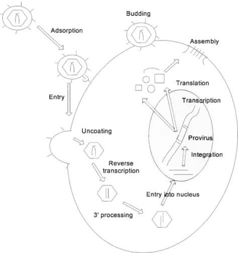
Acquired immunodeficiency syndrome (ADS) is caused by the human immunodeficiency virus (HIV). HW is a retrovirus belonging to the lentivirus family that was unknown until the early 1980’s, but since that time has been spread around the world and has infected millions of persons[l]. The mature HW retrovirus core has two short strands of RNA 9200 nucleotide bases long--along with the enzymes reverse transcriptase, protease, ribonuclease, and integrase (IN). The core of the retrovirus is encased in an outer lipid envelope containing an antigen, gp120, which plays a role in the binding ofthe virus to the target celis with CD4 receptors[2]. The gene content ofthe HW genome, similar to other retroviruses, has three major genes--gag, pol, and env[3]. Afier entering the body, HIV retrovirus enters the target celi via the CD4 receptor. The HIV particle uncoats from the envelope to release its RNA into the cellular cytoplasm. Its RNA genome is transcribed into proviral DNA by reverse transcriptase bound to the HW RNA, which is one of the enzyme products of the p01 gene. HW proviral DNA is then inserted into host cell genomic DNA by the iN enzyme. Once the HIV proviral DNA is within the infected cell’s genome, it becomes part of the host genome. The host cell can replicate the HIV provirus. The infected ceil can undergo lysis with release of new HIV virions, which in tum can infect additional cells. Current antiviral drugs are either inhibitors of HW reverse transcriptase (RT) or protease (PR) but no drugs against IN are available yet. HIV IN is a good target for drug discovery, since IN is essential for retroviral replication and production of new virus; moreover, it has no obvious
functional analogue in the host (Fig.1.1).
In this thesis, the HW-1 IN gene was subjected to random insertion mutagenesis. Jndividual constructs, selected from the library, were assayed for the effects on iN functions in vitro. The effect of individual insertion on enzymatic activity were analyzed in the context of HW-1 monomer, dimer and tetramer model withlwithout DNA to gain insight into the organization of the HW- 1 integrase complex with DNA. One aim of this mutational analysis was to identify sites within the IN protein that may tolerate small insertional tags whose function may alter the target-site selection of the viral integrases. Another aim is to compare available tetramer models with our experimental data to see how they agree with ours and existing biochemical data. Moreover, the domain boundaries defined in this study might be useful in expressing minimized iN constructs for crystallization studies.
1.1 111V-1 Integrase
Integrase catalyzes the integration of a double-stranded DNA copy of the retroviral RNA genome into the genome of a host[4]. This DNA integration reaction requires specific sequences at the 3’ and 5’ termini of the viral DNA (referred to as U3 and U5, respectively, in reference to their unique character) and can be done using purified IN alone in vitro [5]. The integration reaction is carried out in two steps: 3’-end processing, which is a hydrolytic cleavage, occurs two bases from the 3’ end of the U3 and U5 termini, just 3’ of a conserved CA dinucleotide, and strand transfer: the 3’-end processing reaction exposes the free 3’-hydroxyl, which is then used to perform a nucleophilic attack on the target DNA. The sites ofnucleophilic attack on the two strands of the target DNA are separated by 5 bp. The 5’ ends of the viral DNA are left unjoined in the resulting integration intermediate. The removal of the
two unpaired nucleotides at each 5’ end of the viral DNA, gap fihling, and ligation are likely to be carried out by host enzymes[6, 7]. An in vitro reconstructed system using purified IN enzyme and model DNA substrates that consist of short oligodeoxynucleotide duplexes has been employed to explore the details of the biochemistry of IN and its catalytic mechanism (fig. 1.2). This short oligodeoxynucleotide duplex substrate mimics the U5 or U3 ends of the viral DNA [6, 8]. Blunt-ended viral substrates can be properly processed at their 3’ ends in vitro,
and the processed substrates can subsequently be used as a strand transfer substrate and inserted into a target DNA. In addition, HIV IN also can carry out the disintegration reaction, in which a substrate that mimics one end of the viral DNAjoined to the target DNA is cleaved into its viral and target DNA parts [5, 6]. The disintegration reacfion is the reverse of the strand transfer reaction. There is no evidence that disintegration is biologically relevant in vivo but it is a useful assay to test the ability of IN to catalyze polynucleotidyl transfer reactions (fig. 1.2).
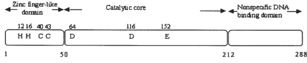
The homooligomeric HIV-1 IN protein is 288 amino acids in length. Structural studies, amino acid sequence alignments, limited proteolysis, site-directed mutagenesis studies and complementation experiments [9-13] have revealed the presence of three distinct domains per monomer: the N-terminal domain, the catalytic core domain, and the C-terminal domain. Each domain can independently fold within each monomer. The first two domains are structurally highly conserved among retroviral and retrotransposon INs. The N-terminal domain (1-50) binds zinc, the core domain (50-212) contains the catalytic triad motif (D, D, 35E), and the C-terminal domain (213-288), binds DNA non-specifically (Fig.3). Insolubility of full-length HW-1 IN has limited structural analyses to individual domains or two contiguous domains.
Budding AdsorPtion N Assembly N ansIation\ Entry //1:\ Trancription
\
]7’
_— Uncoating ‘ 7 - //
Provirus’ ion/1
ntrjto nucleus /N
3 processingfIGURE 1.1. Overview of the retroviral life cycle. Attachment of the viral envelope surface protein to specific receptors on the surface of a host cell resuits in fusion and release of the viral core into the host cell cytoplasm. Reverse transcription then generates a double stranded DNA copy of the RNA genome. The viral DNA undergoes 3’-processing in the cytoplasm and subsequently travels into the nucleus, where strand transfer results in the integration of the viral DNA into the host genome to form the provirus. Transcription generates messenger RNAs as well as viron RNAs. mRNAs are translated in the cytoplasm. Virus proteins and progeny RNA assemble and bud off at the plasma membrane and subsequently mature into infectious particles.
32p 5 3’ 32p 5 3, Disintegration LP 32p 5 3, E F processing CAAG 3’ GTTC 5’ CA 3’ GTTC 5’
f4,
-v
Bc
TC CAAG 3’ GTTC 5’ DFIGURE 1.2. Schematic representation of the 32P-labelled substrates and products ofin
vitro analysis of the enzymatic activities catalyzed by the retroviral IN for a single U5 end. Step 1: 3’-processing; step 2: strand transfer; and step 3: disintegration, which is the reverse of the strand-trans fer reaction.
6
Catalyac care NÛnspei2fic DNA
- bincngdonn
1216 4J43 64 116 152
[HHcc[D D E
][
j
1 SF1 212 28
FIGURE 1.3. A schematic model ofHIV-1 IN. It shows the tbree independently folding domains: the N-terminal domain, the catalytic core and the C-terminal domain. The conserved and catalytically active residues are indicated and the corresponding residue numbers are shown above each residue. Adapted from FIGURE 10.1 of Asante-Appiah and Skalka, 1999 [14].
The structures of individual domain structures have been determined by X-ray diffraction [15-23] or by solution NMR [24-28]. Structures of two contiguous domains, both the N-terminal domain plus the core domain [13] and the core domain plus the C-terminal domain [12, 29, 30] have also been resolved. Both individual domain and two-domain structures are also available for several other retroviral lNs, which are structurally very similar to HW- 1 lNs. However, there is presently no structure available forany IN complexed with DNA substrate. In the absence ofthis information, molecular modeling studies and mutational studies, such as the one presented here, constitute an alternative method to obtain structure-function information on this pharmacologically important enze.
1.1.1 Structure of the catalytic core domain
The catalytic core domain of HW IN is well conserved among retrovirai INs,shares significant structural simiiarity with the transposase proteins and contains the characteristic D, D (35) E motif found in polynucleotidyl transferases [26, 28, 31]. These invariant residues, Asp-64, Asp-116, and Glu-152, are key residues of the active site. Mutagenesis studies show that substitution of any of the three catalytic residues abolishes ail three reactions [10, 11, 32, 33]. The core domain alone can carry out the disintegration reaction [10, 11, 34-36]. However, truncated IN proteins lacking either the N-terminai domain or the C-terminal domain cannot catalyze 3’ processing and strand transfer t33, 34, 36-39]. Anumber of structures ofthe catalytic core domain ofHIV-1 IN exist [12, 13, 15-18, 21, 40]. The overail topology of ail these structures is similar to the structure in (fig. 1.4). It is roughiy spherical in shape and two core domains associate in the crystal to form a two-foid axis-reiated dimer. The dimer interface is quite large with approximately 1300
À2
per monomer excluded from solvent. The interface is quite hydrophobic and the primary contacts between subunits in both structures involve a-helices 1 and 5. As the corresponding helices are not involved in protein—protein contacts in the Tn5 transposase-DNA compiex, which has been recentiy resolved [41], and as the DNA plays an important roie in Tn5 transposase dimerization, one might argue against the bioiogicai relevance of this interface. However, functionai IN has been suggested to be active as a tetramer [14, 42] or even an octamer [43]. This crystaiiized IN dimer and its interface may act as a whoie to bind the same virai end and function as the single Tn5 transposase monomer does.A !i(onuz.er 6 Der
FIGURE 1.4. Stmcture ofthe catalytic core domain ofHIV-1 integrase. Corresponding 13-strands and a-helices are labeled in panel A, Side chains of consewed acidic residues of the active site are shown in a bail and stick representation in panel B. Coordinates ftom a HW-1 IN core domain (accession codes 111G in the Brookhaven Protein Data Bank) was used to generate this Figure; middle panels were created using published coordinates.
Each monomer consists of a five-stranded 13-sheet together with six Œ-helices. The three catalytic residues of the core domain are Ïocated in close proximity in the structure and define the position of the active site (Fig. 1.4). However, the two catalytic sites are on opposite sides of the spherical,crystal dimer and are separated by approximateiy 30À. On the other hand, the integration sites on target DNA are generally separated by 5 bp, which is equivalent to roughly 15
Â
in B-form DNA. Thus, the distance observed in the dimer structure is not compatible with catalysis of the integration event. Either this crystal dimer structure is not biologically relevant or else higher order multimers are formed in vivo based on the dimer structure, such that two active sites could be positioned doser in a higher order muitimer [15].1.1.2 Structure of the N-Terminal Domain
The crystal structure of the N-terminal domain reveals a dimeric structure. Conserved residues His 12, Ris 16, Cys 40 and Cys 43, located in a three-helix bundie, ensure tetrahedral coordination of Zn2. At the monomer level, the structure of the N-terminal domain determined by X-ray diffraction as part of the N-terminal domain plus core structure is very similar to the previously determined NMR structure of the isolated N-terminal domain. However, the dimer interface is entirely different (Fig. 1.5) [13]. Whereas it is dominated by interactions between the third helix in the NMR structure, the dimeric interface in the 1—2I2 crystal structure (N-terminal domain plus
core) comprises the N-termini of the first and third -he1ix. The surface of the dimer interface in the two-domain structure is smaller and more hydrophobic than in the dimer ofthe isolated domain in solution.
The biological relevance of Zn2 and the N-terminal HHCC domain in HW IN has been well documented. Indeed, utilizing a combination of techniques including UV visible absorption, circular dichroism, and fluorescence spectroscopies, it has been demonstrated that metal ions (Zn2, Co2, or Cd2) are bound with equimolar stoichiometry by the isolated N-terminal domain t44]. A mutation in the HHCC motif abolishes zinc-binding capacity of HIV-1 iN. The isolated N-terminal domain is disordered in the absence of zinc [45]. The N-terminal domain is necessary for 3’-processing and strand transfer activity in HIV-1: deletion of this domain or mutation of any of the four conserved HHCC residues abolishes 3’-processing and strand transfer activity ofHW-1 iN [10].
However, in a study of RSV IN, 3’-processing and strand transfer products are detectable in reactions after deletion of the HHCC domain, but is much less efficient
than with the wild type in the presence of Mn2 but flot Mg2. Most strikingly, when a mutant of RSV IN lacking the N-terminal HHCC domain is fused to various short peptides, efficient strand transfer activity can be restored to the level of wild type RSV IN [46]. Similarly, deletion of this domain in Visna virus [47] IN has no effect on 3’-processing. Substitution of Hisl2 and Hisl6 in RSV IN does not significantly impair 3’-processing or strand transfer [48]. When the N-terminal domain of IN expressed independently, it does flot bind DNA [9, 48], but it has been suggested that it interacts with DNA in the context of the intact protein t48-51]. Furthermore, several studies showed that Zn2 promotes multimerization, which should thus occur through the HHCC motif and enhances the catalytic activity of HIV-l IN [45, 52], however, mutants lacking this domaincan stiil form tetramers [53, 54]. Based on the above observations; the exact role of the N-terminal domain in these reactions is not clear. However, the high conservation of this motif and resuits from genetic experiments suggest that this domain is functionally important. Further biochemical studies are needed to conclude on its in
A B C
FIGURE 1.5. Ribbon drawings of N-terminal domain structures. Side chains of conserved HHCC residues of the Zn2-che1ating HHCC motif are shown in a bail and stick representation. The Zn2 cation is shown as CKP. The N-terminus and C-terminus are indic ated. (A) and (B) Coordinates from PDB file 1 WJA determined by NMR; (A) shows the monomer and (B) the dimer. (C) The dimer interface is shown for the N-terminal domain from PBD file 1K6Y determined by X-ray diffraction. Note that the dimerization interface in this crystal structure is completely different from the solution structure in (B).
1.1.3 Structure of the C-Terminal Domain
The amino acid sequences of the C-terminal domain, which are approximately 80—100
amino acids long, are flot conserved among INs from different retroviruses. Mutation and deletion analyses with both avian sarcoma virus (ASV) and HW-1 INs indicate that the C-terminal domain contains nonspecific DNA binding activity [9, 48, 55-57]. With the exception of Feline Immunodeficiency Virus (fIV) N [23] in which deletion of the C-terminus (residues 236-28 1) resulted in a mutant that retained efficient 3’-end processing and disintegration activities but weak 3’-end joining activity, ail lNs with a deleted C-terminal domain lose 3’ processing and strand transfer activities [11, 34, 36, 39, 46, 58-60]. This inability presumably resuits from loss of capacity to correctly position and orient the viral LTR ends at the active site. However, when the viral DNA ends have been correctly prepositioned, such as with a synthetic disintegration substrate during in vitro assays, catalysis can occur with mutant Ns that have been truncated at the N-terminus or the C-terminus or both [36, 54]
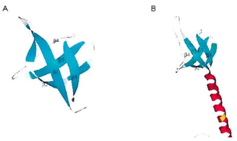
Two studies using NMR [24, 61] on solutions of HW-1 IN C-terminal domain showed homo-dimeric structures with each subunit composed of a five-stranded 3
-barrel that is topoiogically very similar to structures of SH3 domains, which occur in many signal transduction proteins [11, 46, 60, 62, 63]. These two solution structures agree well with each other (Fig. 1.6). The structure of the C-terminal domain monomer resolved by X-ray diffraction is also similar to the structures resolved by NMR. The two C-terminal subunits in the dimeric structure are related to each other by a 90° rotation relative to their two-fold axis.
the core domain plus the C-terminal domain [12, 29, 30]. The core domains in those two domain structures are almost identical to the structure of the isolated core domain and the two-fold symmetrical dimer interface is also similar. Superimposition of the catalytic core of these two-domain structures resuits in a proposed structure of the full-length IN dimer, which might be biologically relevant [13] and will be fùrther discussed in this thesis. Residues 271—28$ in the C-terminal domain are either not clearly resolved or deleted. The hinge region connecting the core and the C-terminal domain is different among HIV-1, Simian immunodeficiency virus (SIV) and Rous sarcoma virus (RSV) IN, which probably reflects flexibility in this region. Moloney murine leukemia virus (M-MULV) IN contains a sequence insertion ofunknown function in this region [64].
The interactions at the dimer interface are predominantly hydrophobic and localize to 3-strands 2, 3, and 4 , with the two triple-stranded antiparallel 3-sheets, one from each subunit, oriented antiparallel to each other. One surface of the dimer is a saddle-shaped groove with dimensions ofapproximately 24 x 23 x 12
À
in cross section, which could accommodate a duplex DNA molecule [24]. This groove contains basic residues favorably positioned to contact DNA. Lys264, which has been shown from mutational data to be involved in DNA binding, protrudes from this surface. The diversity and hydrophobic character of the protein—protein interactions forming dimer of the C-terminal domains from HW-1, RSV [30], and SP/ [29] suggest that they are weak and nonspecific [12].A B
FIGURE 1.6. Ribbon drawings of C-terminal domain structures shown in monomeric form. Coordinates from PDB file 1MV determined by NMR was used to generate (A). Coordinates from PDB file 1EX4 determined by X-ray diffraction was used to generate (B).
1.1.4 Integrase-DNA interaction
There are currently no IN structures available with substrate DNA docked in the active site. However, available IN structures and biochemical data provide considerable information about IN-DNA interaction. This section provides a review of the information available to date, which will be used as a basis for interpretation of the results presented in this thesis.
In the preintegration complex, IN must make specific interactions with viral DNA sequences, and afler transporting the preintegration complex into the nucleus, IN must also interact with the target DNA for the integration reaction to occur. It is clear that IN can distinguish between viral DNA ends and other oligonucleotides [65-6$] since IN protein requires both the subterminal and the distal position of its viral DNA recognition
sequence for efficient cleavage to occur; however ultraviolet (UV) cross-linking studies [55, 69], filter binding assays [37, 70], Southwestern blots [56, 59], and electrophoretic mobility shift assays [51] show that IN binds to substrate DNAs with affinities similar to those of nonsubstrate DNAs in vitro. In other words, although the 3’ processing and strand transfer activities of retroviral IN are sequence-dependent on both the distal and proximal sequences [67, 71-75], the binding of iN to DNA seems nonspecific. [55, 76, 77]. Experiments suggest that specificity in catalysis is achieved by nucleotides both distal and proximal to the conserved CA [74, 75] and metal cofactor t77-80]. for example, experiments showed that a stable complex of IN and viral DNA is formed in the presence of Mn2 and the IN-viral DNA complex is resistant to challenge by an excess of competitor DNA [78]. A comparative study shows that each iN from M-MUL’vÇ human T-cell leukemia virus (HTLV)-l, HTLV-2 and HIV-1 required specific terminal LTR sequences for optimal catalysis of 3’-processing reactions, while strand transfer and disintegration reactions do not. Furthermore, in the 3’-processing reaction, sequence specificity for each IN was traced to the three nucleotides proximal to the cons erved CA [81] in the presence of metal Mn2. Another study, by in vitro selection and specific photocrosslinking in the presence of Mg2, identified that distal positions in the LTR termini interact with the C-terminal domain of IN, providing evidence for the role of that domain in stabilization of viral DNA binding, while the terminal LTR interaction is mapped to the disordered loop of the iN core domain, specifically residues Q148 and Y143 t82]. Integrases need to fray viral LTR double-stranded DNA ends for 3’-processing to proceed since adding nucleotides to the 3’-end of the LTR sequence severely reduces 3’ processing while increased cleavage by IN was detected when the
nucleotides 3’ to the CA-3’ dinucleotide were present as single-stranded DNA [66]. Further evidence cornes from a nucleotide analog substitution study, in which substitutions increasing the hydrogen bonding between the ‘plus’ and the ‘minus’ strand decreases 3’ processing activity, while those which reduce or disrupt base pairing in the conserved CA dinucleotide increase activity [76, 79, 83-87]. This requirernent for base-pair disruption may account for the inability of iN to use intemal sites on DNA molecules as viral att (attachment) sites. Binding of IN to U5 LTR DNA is tighter, exhibiting a prolonged half-life in the presence of Mn2 cations compared to Mg2. The preference observed for Mn2 in standard in vitro integration assays can be attributed entirely to the augmentation in the DNAbinding affinity ofthe IN [77].
The core dornain contains the active site and is the only part of the IN protein capable of independent nucleotidyl transfer. This dornain interacts with viral LTR ends [43, 67, 71, 72, 76, 79, 82, 88-90]. for example, cross-linking data have demonstrated that conserved Lys156 and Lys159 residues are involved in binding ofthe adenosine ofthe conserved CA [71]. Furthermore, three active site residues ofthe DD35E motif in the core must contact both the viral LTR end and the target DNA for the integration reaction to occur. Cross-linking experiments also suggest that the adjacent conserved cytosine and the 5’ dinucleotide on the noncleaved strand also make contact with regions of the core domain, in and around the flexible ioop [$2, $8]. These data support a clear role in viral DNA end binding by the core domain. However, IN is known to function as a multimer (see section 1.1.5), and it remains to be determined which specific DNA contacts are in cis or trans with respect to the active site.
The N-terminal domain is in close proxirnity to target DNA 5’ to the site of integration as shown by crosslinking data [88]. By constmcting chimeras between HW-1
and Visna virus iNs, it is suggested that the N-terminus of IN does flot contribute to viral DNA specificity and is flot involved in determining substrate specificity for 3’-processing and strand transfer activities. Thus, this function must reside in the central region or C-terminus of IN [47]. Furthermore, the first 26 residues of RSV and HIV-1 IN, which include the first two histidines in the HHCC motif, are flot required for DNA binding [9, 76].
Experiments showed that the C-terminal domain of IN interacts with bases distal to the terminal bases of the LTRs [71, 82, 88]; this interaction may play an important role in stabilization of viral DNA binding t82]. This may help to explain several results in which mutations in the C-terminal domain affect the 3’-processing activity [57, 91], which does not require the binding of target DNA. Structurally, it is suggested that a strip of positively charged amino acids from both monomers extending from each active site of the dimer to the C-terminal domain of the other monomer may act as dimeric platform for binding each viral DNA end. This strip potentially may stabilize the viral att site DNA for 3’-processing and strand transfer. This putative DNA binding site involves residues from both monomers: the core from monomer A with the C-terminal domain from monomer B in the dimer implying that a viral end cleaved in the active site of one monomer is stabilized by residues from the C-terminus of the other monomer This is consistent with in vitro complementation experiments [11, 39]. Previously, the C-terminal domain has been presumed to be involved in target DNA binding and this IN-target DNA interaction has been presumed to be nonspecific [57, 91], which is suggested by target sites ofintegration known to exhibit very little sequence specificity [92, 93]. Mutation of conserved lysine-264 has a dramatic effect on the nonspecific DNA binding activity of the
isolated C-terminal fragment, as judged by ultraviolet (LTV) cross-linking, and by 3’-processing by full-length N containing a K264E mutation [24, 57]. Structural modeling based on NMR C-terminal structure, discussed in section 1.1.3, illustrates that the dimensions of the saddle-shaped groove consisting of amino acids 220-270 in the dimer are appropriate for DNA binding. In this model, amino acids 220-270 in the dimer can readiÏy bind to the major groove of DNA, where the side-chains of Ser230, Pro261, Lys25$, and Lys264 interact with the sugar-phosphate backbone, and the side chain of Arg231 interacts with the bases [91]. Some work has suggested a role for Arg-262 and Leu-234 in DNA binding [91]. Mutagenic analysis shows that Ser-230 and Arg-263 are involved in enzymatic activity and DNA binding [91]. Although the C-terminal domain has been implicated in binding of target DNA, certain work from chimeric N proteins shows that the core domain plays a more important role in target binding than the N- or C-terminal domains [47, 58]. Activity assays show that these chimeras exhibit the target site preference of the core domain, but flot ftanking domains. Some cross-linking experiments have crosslinked target DNA to portions of both the core and C-terminal domains, as well as a region of the N-terminal domain [94]. The exact function of the C-terminal domain is stiil not quite clear, but available experimental data suggest the idea of a complex network of DNA binding rather than a model in which individual domains are unilaterally responsible for binding to viral or target DNA [42]. Transposases and retroviral INs share a structurally related catalytic domain. They are members of the larger superfamily ofpolynucleotidyl transferases. Homology modeling based Tn5-DNA complex structure is likely to give us insights about N-DNA interactions.
1.1.5 Multimeric organization ofHIV-1 IN
Many evidences show that IN functions as a multirner. However, there is conflicting evidence with respect to the number of units composing the active multimer and with respect to the actual dimerization interfaces. Clearly, identification of the multirnerization interfaces must take into account the surfaces involved in DNA binding, and vice-versa. In the absence of structural data of multimers with bound DNA, our study set out to explore these potential binding surfaces using biochernical rnethods. The following section describes the information available to date, which will be used as a complement for interpretation of the resuits presented in this thesis.
Biochemical studies have revealed that multimerization determinants reside in the core domain, as well as in the N-terminal and C-terminal regions of HIV- 1 IN [95]. Deletion mutants ofHIV-1 IN that lack either the N-terminal or C-terminal domain have no 3’-processing or strand transfer activity [34]. However, if an N-terminally truncated IN is mixed with a C-terminally truncated IN, 3’-processing and strand transfer activities can be restored [34] [11]. Further evidences for multirnerization corne from mutagenesis and deletion studies which show that full-length N can multirnerize to form both dirners and tetramers in solution [45, 52, 96,
971.
Furthermore, the N-terminal domain of IN can function in trans but flot cis to the core domain [32, 9$], while the C-terminal dornain can function in cis [34] or trans [11, 34, 43, $9].The above results also suggest that the core domain contributes the active site enzymatic activity in partnership with an N-terminal domain from a different monomer of iN.cornes frorn the fact that purified recombinant INs can exist in a dynarnic equilibrium including monorners, dirners, tetrarners, and even higher order oligomers [50, 96, 99-101], and any species can be an active form of IN except the monorner [96]. The stoichiometry of retroviral IN complex is flot known in vitro.
Several structural studies show that a tetramer (dimer-of-dimers) or an octamer of IN wouldbe necessary to carry out concerted ifitegration ofboth LTRs on target DNA [13, 30, 43]. for example, the overail structure of an IN tetrarner formed by crystal lattice contacts frorn the N-terminal and core two-dornain structure ti 3]is structurally similar to a related bacterial transposase Tn5 dirner cornplex with its DNA substrate, which can be considered as evidence supporting the 51V tetrarneric model. Furthermore, this tetrarner model exhibits positively charged channels suitable for DNA binding [13]. A recent study that used iN complexes present in nuclear extracts from human cells suggested that the minimal cellular IN cornplex is a homotetramer, implying that at least an octamer of iN is required to carry out concerted integration of both LTR ends into target DNA [102]. Within this tetramer, it will be only one ofthe two active sites in each dimer that would be actually involved in the chemical reactions.
1.2 Purpose and Scope of the Study 1.2.1 Research objectives
The integration process is an obligate part of the retroviral life cycle. Retroviral iN is both essential and sufficient to catalyze this integration reaction. The overail aim of this study is to gain a finer understanding of the biochernistry of the integration reaction. The specific research objectives ofthis dissertation include:
The expression, purification and characterization of the previously obtained insertional mutants ofHIV-1 IN.
• To gain insights into the role ofpreviously predicted unstructured loops. • To map functionally tolerant region of insertions ofHIV-1 IN.
• To gain insights into IN-DNA interaction.
• To compare available tetramer models with our experimental data to see how they agree with our new data and with existing biochemical data.
• b define domain boundaries, which might be useful to express minimized IN constructs for crystallization studies.
1.2.2 Significance of the study
The development of effective inhibitors of HP! replication targeted to HP! reverse transcriptase and HIV protease has demonstrated the potential effectiveness of antiviral therapy for the treatment of ATDS, which benefits from the foundation of basic knowledge in understanding the mechanism of retroviral reverse transcription and the structure of the protease. Drugs targeted to HP! IN would be a valuable complement to reverse transcriptase and protease inhibitors. However, the lack of detailed structural information about IN/substrate interactions has so far hindered the search for strong and selective IN inhibitors. Although the structures of ail three domains of IN have been individually determined, their spatial arrangement in the active complex with DNA substrate is unknown. The studies I present herein using a linker-scanning approach will provide a better insight into the functioning of this enzyme. Resuits from this study will therefore provide valuable information for those concemed with the design of effective inhibitors of the retroviral IN.
Chapter H
Compreliensive Linker-scanning Analysis of
tlie 111V-1 Integrase Protein
2.1 Context of the work relative to resuits obtained previously
The work described in Chapter 2 was begun prior to registering as a M.Sc. student in the Bio-informatics program at Université de Montréal. Specifically, Sections 2.2.2: Mutagenesis: In vitro transposon-based linker insertion into pLNSD+Iis; 2.2.3: Selection of clones with insertions within the HW-11N coding region; and 2.2.4: Generating the IN frame 19-codon insertion, were performed by myseif while I was a research associate in the laboratory of Prof. C. Jonsson, currently at the Southern Research Institute, Department of Biochemistry and Molecular Biology, Birmingham AL. As this work is not yet published, it is described herein in full, with the approval of Prof Jonsson. Sections 2.2.5: Expression and purification of mutant and wild-type IN; 2.2.6: Substrate preparation; and 2.2.7: In vitro integration and disintegration assays, were initiated prior to registering as a M.Sc. student and were completed during the course of this degree. Section 2.2.8: Structural model of HIV-1 IN monomer were initiated and completed during the course of this degree.
The work presented in Chapter 2 has been submitted as part of a collaborative manuscript entitled: Comprehensive Linker-Scanning Analysis of the MuLV RNase H, MuLV and HIV-1 Integrase Proteins, Author: Jennifer Puglia1, Tan Wang2, Christine
Smith-Snyder’, Marie Cote’, Michael Scier’, Joelle Pelletier4, Sinu John3, Colleen B. Jonsson2 and Monica J. Roth’
‘Department of Biochemistry, Robert Wood Johnson Medical School, University of Medicine and Dentistry ofNew Jersey 675 Hoes Lane Piscataway, NI 08854.
2
Department of Biochemistry and Molecular Biology, Southem Research Institute, 2000 9th Ave S, Birmingham, AL 35205.
3Graduate Program in Biochemistry and Molecular Genetics, University of Alabama at Birmingham, Birmingham, AL 35294.
4Département de Chimie, Faculté des Arts et Sciences, et Département de Biochimie, Faculté de Médecine, Université de Montréal, C.P. 6128, Succursale Centre-Ville, Montréal, Québec H3C 317, Canada
2.2 Introduction
Various methods have been developed for the comprehensive analysis of a gene by construction of a saturating or near-saturating library of mutants t103-105]. These studies have defined domain boundaries, provided functional maps, and insights into previously predicted unstructured loops [106] [103-105, 107-109]. In this study, the method ofinsertional functional mapping is applied to the HW-1 IN (iN) protein.
The integration of retroviral particles is a complex process. Preintegrative complexes (PICs) lias been purified and characterized from infected cells [110-122]. Despite extensive study, the assembly of this complex is flot well understood. These efforts have been assisted by structural studies ofrelated retroviral iN subdomains [12, 13, 16, 19, 26, 29, 30, 123, 124]. However, to date, neither a structure of a complete
retroviral IN protein nor one of a subdomain in complex with DNA has been obtained. The linker-insertion genetic footprint provides a means to identify non-essential regions within proteins capable of withstanding insertions. Ibis method relies on retroviral-mediated insertion of a specific DNA sequence at various, random locations within target DNA. The insertion of a large DNA fragment is followed by excision of part of - but flot ail — the inserted sequence, leaving a shorter (57 base pairs), specific DNA sequence within the target gene. Upon expression, the inserted fragment is translated within the target protein, resulting in disruption of the native sequence. Disruption of function indicates that the area of insertion does not tolerate structural disruption, for any of a variety of reasons: the disrupted area may be directly required for function, may be required for oligomerization or may be essential to the folding of the target protein.
In this study, the HIV-1 IN gene was subjected to Tn5-based random insertion mutagenesis. Individual constructs, selected from the library, were assayed for the effects on in vitro iN enzymatic activity assays. The observed activities of the resulting iN mutants provide insights into the possible roles of the various parts of the HW- 1 iN protein. Using this approach, three regions that are functionally tolerant of insertions were identified within iN. These regions correlate with domain and protein junctions. 2.3 Experimental Procedures
2.3.1 Materia]s
Oligonuleotide PCR primers and oligonuleotide substrates were synthesized by Integrated DNA Technologies (Coralville, Iowa). Restriction enzymes, Taq polymerase, 14 DNA ligase and kinase were purchased from New England Biolabs (Beverly, Mass.). The EZ: TN In Frame Linker Insertion Kit was purchased from EPICENTRE (Madison,
WI). Ail chemicals were purchased from Sigma.
Bacteriai Strain and Plasmid: Plasmid pINSDHis containing HIV-1 IN was obtained from the ADS Research and Reference Reagent Program. It was used to construct HW- lIN mutants and was propagated in Escherichia cou strain DH5Œ. E cou
strain BL21 (DE3) was used to express wild type HW-l IN and its mutants.
2.3.2 Mutagenesis: In vitro transposon-based linker insertion into pINSDHis The EZ: IN In Frame Linker Insertion Kit was used to insert a nucleotide linker at random into the target piasmid pINSDHis, as described in FIGURE 2.1. The reaction mixture contained 1 jil EZ::IN lOx reaction buffer, 0.4 ig pINSD±Iis piasmid DNA, 1tl (0.1pmol/il) EZ::TN<Not/KAN-3>Transposon, liii EZ::TN Transposase in a final volume of 10tl. The reaction mixture was incubated for 2 hours at 37 oc and the reaction was stopped by adding 1 111 EZ::TN 10x Stop Solution. 1 tl — 10 al of the insertion reaction mixture was used to transform MAX Efficiency E. cou DH5Œ Competent Ceils from Invitrogen (Carlsbad, CA) and piated and selected on kanamycin-containing plates by ovemight growth at 37°C. Each individual colony was picked and transferred into 96 well culture blocks from Qiagen (Valencia, CA) with imi LB containing 50tg!mi kanamycin in each well. Afler ovemight growth at 37 oc with shaking, 50% glycerol was added to each well to bring the final concentration of glycerol to 10% and the blocks were stored at —80 °C.
2.3.3 Selection of clones with insertions within the 111V-lIN coding region The <Not IIKAN-3>Transposon shouid be randomly inserted into the target DNA plasmid pINSD±Iis. A PCR strategy was employed to determine whether the transposon insertion sites were within the HW-1 IN coding region and their distribution.
The pinsdBsreen primer (5’-CGG GCT TTG TTA GCA GCC GG -3’) and pinsdFscreen primer (5’-GGT GCC GCG CGG CAG CC -3’) were used to amplify the iN and encoding sequence from 301 to 320 and 335 to 351 of the pETl5b plasmid, which is parent plasmid of p1NSD. The PCR reactions contain 1xPCR buffer of ExpandTM long Template PCR system from Boebringer Mamiheim (Gmbh, Germany), 2.25mM MgSO4, 0.2mIVI dNTP, 2 iM primers, 2 units Taq polymerase and traces of the glycerol stocks stored at-80 OC .The reactions were carried out in 96 well PCR plates from Robbins Scientific Corporation (Suuyval, CA). PCR conditions were as follow: 94 °C for 4 minutes, followed by 35 cycles of: denaturing at 94 oc for 30 seconds, annealing at 69 °C for 30 seconds followed by an extension step at 72 oc for 2minutes and 15 seconds. This cycle was followed by a final extension period at 72 oc for 4 minutes, which was followed by a hold at 4 °C. Afler PER, the samples were loaded onto a 1.5 % agarose gel and examined to determine which of the clones are positive for linker insertion within the IN gene.
2.3.4 Generatïng the IN frame 19-codon insertion
The clones with a transposon insertion site within the IN coding region were individually digested with NotI according to manufacturer’s recommendations. Purification of the linearized DNA from the 1100 bp kanamycin—containing fragment was carried out by agarose gel electrophoresis. Religation of the linearized DNA by T4 DNA Ligase regenerated a single Not I restriction site and created the 57 nucleotide (19 codon) insertion into any of the three reading frames (Fig. 2.1). The religated DNA was transformed into E. cou DH5Œ cells and selection was done on Kanamycin-containing plates. DNA sequencing of transposon insertion clones was performed with the ABI
® TM ®
PRISM BigDye Pnmer v3.0 Cycle Sequencing Ready Reaction Kit with AmphTaq DNA Polymerase, FS. (Foster City, CA) to determine the site of the 19-codon insertion. The integrated sequence analysis software VectorNTl from InforMax Inc. (Frederick, MD) was used to analyze the sequence data.
2.3.5 Expression and purification of mutant and wild-type IN
Wild-type HIV- 1 IN and insertion mutants were expressed in E. cou BL2 1 (DE3) celis in 50 ml ofmedium and purified as hexahistidine-tagged fusion proteins under denaturing conditions as described previously [83]. 50 ml cultures purification yielded approximately 2 mg of 90—95% homogenous protein (Fig. 2.3). The protein fraction refolded at a concentration of 5 mg/ml exhibited greatest enzymatic activity. HPI-1 iN precipitated upon addition of Buffer C (0.2 M NaC1). The precipitated protein was re-suspended in Buffer D (0.5 M NaC1) to a final concentration of 1 mg!ml.
2.3.6 Substrate preparation for in vitro activity assays
Oligonucleotides were purified on 20% denaturing polyacrylamide gels, 32P-labeled at the 5’ end with 14 polynucleotide kinase, and hybridized to complementary strands as previously described (Jonsson et al., 1993). Unincorporated radioactivity was removed from labeled integration and disintegration substrates with G-25 or G-50 Quick Spin columns (Boehringer, Manriheim, iN).
2.3.7 In vitro integration and disintegration assays
Strand transfer and disintegration reactions were performed as described previously [83]. Reaction products were separated on 20% polyacrylamide denaturing gel and subjected to autoradiography or Phosphorimager screens (Molecular Dynamics). Products were quantified with ImageQuant software (Molecular Dynamics).
2.3.8 Structural model of 111V-1 IN monomer. The structural mode! of the HW- 1 IN monomer was constructed from a combination of two X-ray crystal structures, represented by PDB files lk6y and lex4. The “A” molecule of lk6y was superimposed onto the “A” molecule of 1e4 using the program O. The lky structure is comprised of residues 1-46, 56-139, and 149-210; and lex4 is comprised of residues 56-141 and 145-270. Thus, the superpositioning consisted of overlaying the CŒ atoms of ail common core residues (RMSD-0.83 angstroms). Where the mode! contained disordered regions (residues 47-55 and 142-144, inclusively), poiyalanine segments containing the correct number of amino acids were created and moved into the appropriate linking positions in the mode!. The Ala residues were then changed to the proper residues, and the regions were subjected to ieast squares minimization.
2.4 RESULTS
fig. 2.1 outiines the series of steps undertaken to generate the linker-insertion iibrary within the HW-1 IN construct pINSDHis. Briefly, the Tn5 mutagenesis system resuits in the random insertion of the transposon encoding the kanamycin resistance gene throughout the plasmid. To make the screening process high throughput, each individual coiony was picked and transferred into 96 weiis culture blocks. km total, 1056 colonies were picked into 96 well culture blocks to screen mutants with a transposon inserted into the N coding region by PCR. Any insertion within the IN will be amplified to a 2.1 kb product while an insertion within the vector sequence will amplified to a 0.9kb product. Each clone with a transposon inserted into the IN coding region was digested with NotI and religated to create the 57 nucleotide (19 codon) insertion. The amino acid sequence of the HW- 1 IN encoded by the target DNA pINSD is conserved
on both sides of the 19-codon insertion. These constructs with 19 amino acid insertions were transformed into E. cou and selected using the ampicillin-resistance gene (t3-lactamase) that is present on the original cloning vector. Selected insertion mutant proteins were expressed and purified. Strands transfer activity and disintegration activity were performed on these purified insertion mutants to evaluate the effect of insertions in different sites. The resuits obtained in each step ofthe procedure are described below. 2.4.1 Selection of clones with insertions within the HIV-1IN coding region by PCR
To determine the transposon insertion sites, PCR was used to screen the clones that have a transposon inserted into the HW-1 iN coding region. As shown in Fig. 2.2, insertion within IN coding region resulted in a 2100 bp PCR product; otherwise a 910 bp PCR product was obtained. 0f 1056 colonies (11 x 96 well plates) screened, 111 of them were positive for having one insertion within the IN gene. The 57-base pair insertion generated by the Tn5 In-Frame Linker Insertion scanning system resulted in insertions into all three reading frames of the original clone. Nine of the 57 nucleotides are the result of a 9 bp sequence duplication immediately ftanking the transposon insertion site. The amino acid sequence of the protein encoded by the target DNA is conserved on both sides of the 19 codon insertion. Sequencing from the library (Table 1) showed that insertions were distributed throughout the whole IN encoding region. The insertion sites, however, were not randomly distributed, with clustering of insertions within the C-terminal domain. This could indicate a preference for specific structure within the plasmid DNA by the transposase enzyme since a large number of duplicate isolates were identified within the population examined. The composition of 19-amino acids inserted
during the transposition as well as the sequences encoding the NotI restriction site and two 1 9-bp inverted repeat mosaic end sequences, which are recognized by Tn5 transposase. Depending on the reading-frame, the insertions will encode
XCLLYTSCGRKMCTRD(S/R)XX, LSLVHILRPQDVYKRQXXX or
HIV-1 IN pINSD + NotI NotI
4
KantEZ: TN transposon
4
Transposase
Transform
and
selection ofE.coh for Kan® (2x1
Q3colonies)
PCR to identify
the individuel
clones with insertions
are within
IN encoding region.
li
Digest with NotI, religateto generate 19 codon insertion, transformE.coh.
4
Expression mutated protein, assay for disintegration
and
strandtrans fer activity
FIGURE 2.1. Generation of the HIV-1 insertional library. The three steps required to generate the insertional library within the construct are outlined. The region of IN gene encoding the Integrase (IN) protein subjected to TN5 insertional mutagenesis
and
transposon Tn5 DNA, which contained the kanamycin resistance gene between its short 19 basepair Mosaic End (ME) Tn5 transposase recognition sequences,
are
shown. Restriction sites Not I flanked by the ME also are shown.2JOObp
9lObp
M
N coding region. A 2100-bp band indicates the presence of one single insertion within the IN coding region. A 91 O-bp product indicates the presence of an insertion within the vector pINSD but outside of the IN coding region.
2.4.2 Insertion Sites of HIV-l Individual mutants
The final insertion library for HW-1 N was characterized by analyzing individual isolates. Isolates of the final library were subjected to sequencing analysis. 0f the 111 insertions, 2 were within the N-terminal domain, 35 were within the catalytic core and 74 within the C-terminal domain. Afler eliminating the duplicate clones, where insertions are in the same position with same insertion sequence, 55 clones have unique insertion positions and correct sequence (summarized in Fig. 2.6. and Table 1). Each mutant N isolate was transformed into E. cou BL21 (DE3) and each mutant integrase was purified and further characterized by in vitro enzymatic analysis of individual clones.
2.4.3 Expression and purification of the wild type and mutant IN
Fifty-five insertional mutant proteins were expressed, purified and assessed for strand transfer and disintegration activity (Table 1 and Fig. 2.6). Mutants with a variety of activity levels were identified in each domain, the N-terminal, core and C-terminal.
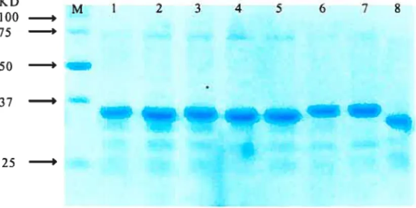
E. cou BL21 (DE3) ceils were used to express wild type IN or the insertion mutant INs by inducing with WTG. Following the induction period, total protein was extracted under denaturing conditions. Solubilized protein can be isolated by nickel affinity chromatography. This one-step affinity purification yielded approximately 2-3 mg of 90—95% homogenous protein from 50 ml of cells, and is well suited for manipulating multiple purifications in parallel. As can be observed following migration ofthe samples on 4-12% SDS polyacrylamide gel electrophoresis (PAGE) (Fig. 2.3), HW-1 IN insertion mutants with the extra 19 amino acids migrated a littie slower than wild type IN. Column fractions containing the most enriched INs were pooled and diluted to ascertain the optimal concentration for refolding as measured by the activity of the HW- 1 IN enzyme. The protein fraction refolded at a concentration of 5 mg/ml exhibited greatest enzymatic activity. HW-l iN precipitated upon addition of Buffer C (0.2 M NaC1). The precipitated protein was resuspended in Buffer D (0.5 M NaC1) to a final concentration of 1 mg!ml.
Table 1: Summary ofHIV-1 IN insertions 27L S127L ‘355 C D55C P58G D64_C 173_L 183I E 96_T T115D D116N T1 25_V 1&12 8A 13iW 113 5K FJ144P S147Q 0149V Vi 65_R 0167 Q 1U69E E 170_H K173T T174A Ml 7 SA A196G 1200_V 72010 LSLVHI LRPQDVYKRQDFN PVSCTHLAAARCVQETDFN LSLVHI LRPQDVYKRQQVD CLLYTSCGRKMCTRDRQVD VSCTHLAAARCVQETDCSP SVSCTHLAAARCVQETELD LSLVHILRPQDVYKRQKVI TVSCTHLAAARCVQETGGY LSLVHILRPQDVYKRQGQE CLLYTSCGRKMCTRDRVHT TVSCTHLAAARCVQETDTD AVSCTHLAAARCVQETGTT CLLYTSCGRKMCTRDRVKA LSLVHILRPQDVYKRQACW CLLYTSCGRKMCTRDRGI LSLVHI LRPQDVYKRQPYN CLLYTSCGRKMCTRDS PQS AVSCTHLAAARCVQETGQG LSLVHI LRPQDVYKRQGQV CLLYTS CGRKMCTRDRVRD CLLYTSCGRKMCTP]3RDQA PVS CTHLAAARCVQETEAE LSLVHI LRPQDVYKRQHLK CLLYTSCGRKMCTRDSLKT LSLVHI LRPQDVYKRQVQM CLLYTS CGRKMCTRDRYSA CLLYTS CGRKMCTRDRERI AVSCTHLAAARCVQETGIV A2 05_T 02071 E2 121 R2 2 BD S230R A239K K240L W243_K G245_E G247A A2 48_V V2501 V2501 1251 Q 025 3N S2 550 V259_V 1268_R R2 69_D G272K K2 73Q 0279 C V281A R2840 Q2 85_D E287D 0288 VSCTHLAAARCVQETDI lA LSLVHILRPQDVYKRQATD SVS CTHLAAARCVQETAKE AVSCTHLMARCVQETDYR S CLLYTSCGRKNCTRDRDS TVSCTHLAAARCVQETGPA PVS CTHLAAARCVQETAAK NCLLYTSCGRKMCTRDSLW CLLYTSCGRKMCTRDSWG LSLVHI LRPQDVYKRQGEG CLLYTSCGRKMCTRDSEGA CLLYTSCGRC.ICTRDRAW LSLVHILRPQDVYKRQAVV PVS CTHLAAARCVQETWI LSLVHI LRPQDVYKRQI QD CLLYTSCGRKMCTRDRDNS CLLYTSCGRKMCTRIDSIKV SCLLYTSCGRKMCT1flRI I AVSCTHLAAARCVQETVIR CLLYTSCGRKMCTRDRDYG PVSCTHLAAARCVQETDGK LSLVHI LRPQDVYKRQGDD CLLYTSCGRKMCTRDSDCV PVS CTHLAAARCVQETASR AVS CTHLAAARCVQETGRQ AVS CTHLAAARCVQETEDE CLLYTSCGRKMCTRDRDED
Positions are based on the protein sequence of wild-type HW-1 N. An arrow marks the insertion between the two amino acids indicated. Ail isolates are independent insertions. 2Sequence of the 19 amino acid insertion. 3Disintegration assay 4Strand-transfer assay 5Activity is based on WT IN activity. Symbols: 0%;
± O to 5%;
if
6 to 35%;tif
36 to 75%,ff
76 to 100%Insertion Inserted amino acid Insertion Inserted amino acid
)osition1 sequence2 DS35 ST4’5 position1 sequence2 DS3’5 ST4’5
tt li-t ttt ttt ttt + tt -t-+ t I-+ t + + t t + + tt tt ttt t t tt t t ttt tt tt tt ttt tt t-t t-t ttt ttt ttt ttt ttt -tt ttt ttt ttt ttt ttt ttt ttt ttt t-t ttt t-t t-t t-t tt t-t -t-t t-t-t t-t-t ttt ttt
75
50 —‘ —
-,
—
e s
ii
25 —‘
FIGURE 2.3. Purification of wild type HW-1 iN and insertion mutant INs as observed by 4—12% SDS-PAGE. M indicates the molecular weight markers. Lanes l-7, various insertion mutant INs. Lane 8, wiid type IN.
2.4.4 In vitro Analysis of Individual HW- 1 IN Mutants
HW-l wild type iN and its mutants were assayed for strand transfer and disintegration activities with LTR-specific substrates. Strand transfer activities were detected by the use of a precleaved duplex DNA substrate. Disintegration activities were tested by a Y-shaped substrate that resembies an integration intermediate. Oligonucleotides used in the synthesis of the substrates in these experiments are listed in Fig.2.4B.
N-terminal domain mutants. The HW-1 N-terminal domain is made of a three-heiix bundie structure. Two of the insertions at N27L retained full disintegration activity however their integration activity was barely detectable. Notably, these two mutants were inserted into same position, but with different amino acids sequences. Insertions D55C and P58G fali into the hinge region between HHCC domain and core domain. In the two-domain crystal structure, which was crystaliized in a tetrameric form, this connecting region (residues 47—55) is disordered in ail four monomers [125]. These two insertions have retained high disintegration activity and have moderate to full
integration activity. The D55/C56/S57 sequence is proposed to be involved in close proximity with the HIV LTR positions 1-4, based on a structural tetramer model [126].
Core Mutants. In the core domain, insertions D64_C, 173_L, Y83_L, E96_T, T125_V A12$A and W13 1W had completely diminished or very Iow disintegration and integration activity. These insertions are located within elements of secondary structure of the highly compact core consisting of a five-strand sheet together with six helices. These insertions most likely will disrupt the secondary structure. Insertions T115D and D116_N also completely diminished both disintegration and integration activity. Two of the insertions are located in the loop between $4 and cr2, and hence, would flot be expected to disrupt the packing of the core domain. Because D116 is part of the catalytic triad, these two insertions most likely disrupt the conformation of the catalytic triad. A group of insertions - 1135_K,
N144P, S 147_Q and G149 V — had different levels of disintegration activity, from trace or weak to moderate disintegration activity, and are ail located near the loop between $5 and a4. This agrees with a mutagenesis study on the Gly residues at 140 and 149 impaired catalysis of HIV-1 IN. Another group of insertions (V165_R, D167Q, A169 E and E170 H, K173 T and T174A), which retained weak or trace or no disintegration activity, are located in the loop between a 4 and a 5. These two loop regions correspond to an extended loop (residues 137-156) and a flanking region (residues 161-173), which are protected from proteolysis upon metal binding [127]. Insertions A196G 1200V, V201 D, A205 T, D2071 and E212 are distributed within o6. The activities of these insertion mutants range from low disintegration activity to low to moderate integration activity as one moves toward the C-terminai end of the lieux. 0f considerable interest, insertion E212L,
maintained nearly full disintegration and integration activity.
C-Terminal domain. The C-terminal domain bas been suggested to be
involved in target DNA binding as mutations in this domain can abolish nonspecific DNA binding [128, 129]. Insertion R228 D is located at the end ofj3l strand in the C-terminal domain, and S230_R is located in the hairpin connecting t31 and f32. Neither of these two mutants had strand transfer activity and both had greatly decreased disintegration activity. Since the C-terminal 1N can tolerate large deletions and can stiil have considerable level disintegration activity [33, 130], this implies that the effect of these two mutants may flot simply be in disruption of folding of the C-terminal domain.
38
A B
32p 5 E CAGT 3’ HIV-1 (U5)
GTCA A 5’ A 5’ ACTGCTAGAGATTtCCACAT processing 6 5’ ATGTGGAAAATCTCTAGGGCTGCAGGTCGAC C 5’ CAGCAACGCAAGCHG F CA 3’ 3• GTCA D 5’ GTCGACCTGCAGCCCAAGCTTGCGHGCTG A E 5’ AHGGAAAATCTCTAGCAGT
Disinteraon Strand transfer
F 5’ AHGGAAAATCTCTAGCA
c c “>1-c
32p 5’ 1—CAGT 3 GTCA D
FIGURE 2.4.(A) Schematic representation of the enzymatic activities catalyzed by the retroviral fl’1 in vitro: 3’-processing; strand transfer; disintegration. Strand transfer substrates are prepared by hybridizing the 32P-labeled F strand with the A strand. The substrate is identical to the 3’-processing substrate, except for the absence of two terminal nucleotides proximal to the CA dinucleotide. Strand transfer activity generates products both larger and smaller than the substrate, since integration occurs at random sites along the phosphate backbone of the target substrate DNA. The disintegration substrate represents a hypothetical strand transfer intermediate. The substrate is prepared by hybridizing the 32P-labeled C strand with the A, B, and strands. Disintegration reaction resuits injoining ofthe 3’-OH end ofthe C strand to the B strand, resulting in formation of the 30-nucleotide product. (B) Nucleotide sequences of HW-1 LIR substrates used in the assays.
c.
1 2345678910 D. 1 2 34567891011 Disintegration Products 30 nt I nteg ration products Substrate Substrate l9nt l6ntFIGURE 2.5 (A) Integration activities ofthe mutant iNs. (B) Disintegration activities of the mutant INs. Enzymatic activities of the mutant INs were assayed as described under Experimental Procedures using HW-1 U5 LTR-derived substrates (Fig. 2.4 3). Enzymatic activity is shown for integration (Panel A, lanes 1-10); and disintegration (Panel B, lanes 1—11) Assays were done in 15 d reaction volumes with a final substrate concentration of 0.07 pmol/ml. Reaction with substrate in the absence of protein is represented in lane 1. In A and 3, reactions were incubated at 37 oc for 60 min and terminated with proteinase K treatment for 30 min at 37°C.
al a2 a3
HI V-1 IN FLO GlO KAQEEIEKTHSNWRÀNASD FNLP PWAIŒIVAS C DKC Q LKQEAI
T
?fuT82
T83 a1 100HI V-1 IN HQQVD C Z P QIWQLD C71!L E QKVILVAWWÀ ZQflEAEVI PAETQQ TAYF
ioial
84ff
a2 a3flT?
T
T!’415oHI V-1 IN L LKLAQRWPVK1VffDNQ SNFTSflVKAA CWWAQIKQE FOI PYNP Q Z Q 0V 151 a4
TT
a5 fa6 T200HI V-1 IN lE ZNI’IICE LflCI I QQVPD Q AEHLKTAVQMAVFIIU’WKRKGQI QQYZÀQERI
HI V-1 INL IA! QTIS QKQ ITRI QNFLS
51T
PVWTCQP LI25fl134faB5
T? fftss
HIV-IIN IQONSO IKVVPPPRAKIIPDYQKQMAQDD CVAZRQDED
Q
Disintegntion activity 0%O-5%
• 6-35%
D
Strandtnnsferactivity 36-75%>76%
FIGURE 2.6. Positions of each insertion (indicated by arrow) and their activity (using
different color scheme) relative to disintegration (circle) and strand transfer activity (square) are shown for the amino acid sequence of 11W-l N protein. Numbering from
the N-terminus of HIV-1 N. Known structural elements of HW-1 N, determined by
crystallography of recombinant HW-1 N [125, 131], are also shown (bold horizontal unes) above the respective homologous segments. Their PDB accession numbers are
FIGURE 2.7. A three-dimensional structural model of the HW-1 monomer. The location of the insertional mutations and their subsequent effects on disintegration and strand transfer activity are shown using the color scheme corresponding to Fig.2.6. The large spheres denote disintegration activity and the widened colored linear portions denote strand transfer activity.
Insertions afier amino acids 239 (mutants A239 K, K240 L, W243_K, and G245E in f32; V250 I, 1251
Q
in f33, D253_N and S255_D in ioop between 3 and f34, V259V in f35) ail lost strand transfer activity while exhibiting full or slightly decreased disintegration activity. This agrees with the observation that the C-terminal deletion of 25 or 45 amino acids resuits in complete loss of integration and 3’-processing activities [33] and also agrees with the observation that the C-terminal deletion mutants (1-24$, 55-248, 1-206) exhibit higher or same level activities in disintegration [130]. Mutant G247_A was an exception as it retained full integration activity and disintegration activity. lnterestingly, the insertions in t32 and t33, which are right before and after 247 had no integration activity and were decreased in disintegration activity. Insertions afler 126$ and before Q2$4 had similar levels of activity in disintegration and retain moderate activity compared to wild type N. This agrees with the observation that deietion of 15 amino acids from end of the C-terminal domain retain similar or slightly reduced 3 ‘-processing activities and decreased integration activities in the presence of Mn2 (but not Mg2 )[33]. Insertions after R284 retain full activity both in the disintegration and integration reactions. This again agrees with the observation that C-terminai deletion of 5 amino acids exhibited wild type levels of 3’-processing activity and partial activity ofintegration in the presence ofMn2 (but flot Mg2[33].In summary, three regions retained full integration activity in this in vitro
study of HIV-1 IN. These correspond to the linge region connecting the N-terminal and the core domains, within the helix connecting the core and C-terminal domains, and the extreme C-terminus of the N.


![Table 1: Summary ofHIV-1 IN insertions 27L S127L ‘355 C D55C P58G D64_C 173_L 183I E 96_T T115D D116N T1 25_V 1&12 8A 13iW 113 5K FJ144P S147Q 0149V Vi 65_R 0167 Q 1U69E E 170_H K173T T174A Ml 7 SA A196G 1200_V 72010 LSLVHI LRPQDVYKRQDFNPVSCTHLAAARCVQETDFNLSLVHI LRPQDVYKRQQVDCLLYTSCGRKMCTRDRQVDVSCTHLAAARCVQETDCSPSVSCTHLAAARCVQETELDLSLVHILRPQDVYKRQKVITVSCTHLAAARCVQETGGYLSLVHILRPQDVYKRQGQECLLYTSCGRKMCTRDRVHT TVSCTHLAAARCVQETDTDAVSCTHLAAARCVQETGTTCLLYTSCGRKMCTRDRVKALSLVHILRPQDVYKRQACWCLLYTSCGRKMCTRDRGILSLVHI LRPQDVYKRQPYNCLLYTSCGRKMCTRDS PQSAVSCTHLAAARCVQETGQGLSLVHI LRPQDVYKRQGQVCLLYTS CGRKMCTRDRVRDCLLYTSCGRKMCTP]3RDQAPVS CTHLAAARCVQETEAELSLVHI LRPQDVYKRQHLKCLLYTSCGRKMCTRDSLKTLSLVHI LRPQDVYKRQVQMCLLYTS CGRKMCTRDRYSACLLYTS CGRKMCTRDRERIAVSCTHLAAARCVQETGIV A2 05_T02071E2 121 R2 2 BDS230RA239KK240LW243_KG245_EG247AA2 48_VV2501V25011251Q025 3NS2 550V259_V1268_RR2 69_DG272KK2 73Q0279 CV281AR2840Q2 85_DE287D0288 VSCTHLAAARCVQETDI lALSLVHILRPQDVYKRQATDSVS CTHLAAARCVQETAKE AVSCTHLMARCVQETDYRS CLLYTSCGRKNCTRDRDSTVSCTHLAAARCVQETGPAPVS CTHLAAARCVQETAAKNCLLYTSCGRKMCTRDSLWCLLYTSCGRKMCTRDSWGLSLVHI LRPQDVYKRQGEGCLLYTSCGRKMCTRDSEGACLLYTSCGRC.ICTRDRAWLSLVHILRPQDVYKRQAVVPVS CTHLAAARCVQETWILSLVHI LRPQDVYKRQI QDCLLYTSCGRKMCTRDRDNSCLLYTSCGRKMCTRIDSIKVSCLLYTSCGRKMCT1flRI IAVSCTHLAAARCVQETVIRCLLYTSCGRKMCTRDRDYGPVSCTHLAAARCVQETDGKLSLVHI LRPQDVYKRQGDDCLLYTSCGRKMCTRDSDCVPVS CTHLAAARCVQETASRAVS CTHLAAARCVQETGRQAVS CTHLAAARCVQETEDECLLYTSCGRKMCTRDRDED](https://thumb-eu.123doks.com/thumbv2/123doknet/2051906.5449/48.918.161.827.139.693/lrpqdvykrqdfnpvscthlaaarcvqetdfnlslvhi-lrpqdvykrqqvdcllytscgrkmctrdrqvdvscthlaaarcvqetdcspsvscthlaaarcvqeteldlslvhilrpqdvykrqkvitvscthlaaarcvqetggylslvhilrpqdvykrqgqecllytscgrkmctrdrvht-tvscthlaaarcvqetdtdavscthlaaarcvqetgttcllytscgrkmctrdrvkalslvhilrpqdv.webp)