HAL Id: tel-00790839
https://tel.archives-ouvertes.fr/tel-00790839
Submitted on 21 Feb 2013
HAL is a multi-disciplinary open access archive for the deposit and dissemination of sci-entific research documents, whether they are pub-lished or not. The documents may come from teaching and research institutions in France or abroad, or from public or private research centers.
L’archive ouverte pluridisciplinaire HAL, est destinée au dépôt et à la diffusion de documents scientifiques de niveau recherche, publiés ou non, émanant des établissements d’enseignement et de recherche français ou étrangers, des laboratoires publics ou privés.
The study of susceptibility and resistance of HIV
integrases to integrase strand transfer inhibitors and the
development of novel single domain antibody targeting
HIV integrase
Xiaoju Ni
To cite this version:
Xiaoju Ni. The study of susceptibility and resistance of HIV integrases to integrase strand transfer in-hibitors and the development of novel single domain antibody targeting HIV integrase. Biotechnology. École normale supérieure de Cachan - ENS Cachan, 2011. English. �tel-00790839�
THESE DE DOCTORAT DE
L’ECOLE NORMALE SUPERIEURE DE CACHAN
Présentée par
Mademoiselle Xiaoju NI
Pour obtenir le grade de
DOCTEUR DE L’ECOLE NORMALE SUPERIEURE DE CACHAN
Sujet de la thèse:
The study of susceptibility and resistance of HIV integrases to integrase strand transfer inhibitors and the development of novel single domain antibody
targeting HIV integrase
Thèse présentée et soutenue publiquement le 30 septembre 2011 Devant le jury composé de :
Dr. Xu-Guang XI Président du Jury Dr. Vincent PARISSI Rapporteur Dr. Karen MOREAU Rapporteur Dr. Olivier DELELIS Examinateur
Dr. Jean-François MOUSCADET Co-Directeur de thèse PI. Kai-Cheng QIAN Co-Directeur de thèse
Laboratoire de Biologie et Pharmacologie Appliquée (CNRS UMR 8113)
REMERCIEMENTS
J’ai effectuée ce travail de thèse dans l’équipe Interactions fonctionnelles des retrovirus au Laboratoire de Biologie et Pharmacologie Appliquées, UMR 8113 du CNRS. Je voudrais d’abord remercier Christian Auclair et Jean-François Mouscadet, qui ont assurée successivement la direction du laboratoire au cours de ces années, pour m’avoir accueillie au sein de leur unité de recherche et m’avoir permis de travailler dans un environnement scientifique aussi exceptionnel.
Je suis très sensible à l’honneur que m’ont fait les membres du jury de thèse en acceptant de lire et d’évaluer ce travail. Je les remercie d’avoir apporté un intérêt à mon travail. Ce mémoire a beaucoup bénéficié des commentaires qu’ils ont tous effectués.
Je tiens également à exprimer toute ma gratitude à Jean-François Mouscadet pour son accueil chaleureux au sein de son équipe. Je tenais aussi à le remercier pour les conditions de travail idéales dans lesquelles s’est déroulée ma thèse.
Je tiens également à remercier tous les membres passés et présents de cette équipe qui m’ont soutenu tout au long de cette thèse. Merci à Olivier Delelis, Stéphanie Bury-Moné, Osamuede Osemwota, Gladys Mbemba, Gwenola Manic, Frédéric Subra, Sylvain Thierry, Aurélie Maurin-Marlin.
Je tiens, d’autre part à remercier le Professeur KaiCheng Qian de l’East China Normal University, directeur de Shanghai transfusion institute, qui m’a accueillie au sein de son labo en Chine. Je le remercie pour son aide lorsque j’ai rencontrée des problèmes administratifs en Chine et surtout pour toutes les discussions concernant mon avenir professionnel.
Je remercie également tous les membres du LBPA pour les temps inoubliables que j’ai passées en votre compagnie. En particulier, je souhaite remercier à Marie-Alix Poul pour tous les aides concernant mes travails d’anticorps. Merci également à mes collègues au LBPA : Luba Tchertanov, Hervé Leh, Françoise Simon, Eric Deprez, Françoise Chaminade, Safwat Abdel-Azeim, Janine, Anne-Marie, Martine, Marie-Christine, Yingying Chen, Xiaoqian Xu, Li Na, Huan Wang, Jiayao Li, Ahmad Khodr, Victoria Fairweather, Bianca Sclavi, Claude Nogues, Anne Olliver, Philippe Fossé,… Merci pour toutes les discussions scientifiques ainsi que pour les bons moments que nous avons pu
partager. Merci à tous pour m’avoir installée dans une ambiance amicale.
Je souhaite remercier particulièrement à Serge Benichou, Jerome Bouchet de Institut Cochin qui nous avez aidé sur les manipulations d’anticorps. Un grand merci également à Daniel Baty, Patrick Chames, Julie Matz à Marseille pour leurs conseils précieux et patiences.
Un grand merci également à Charlotte Charpentier, Diane Descamps d’Hôpital Bichat-Claude Bernard, et Anne-Geneviève Marcelin, Vincent Calvez d’Hôpital Pitié-Salpêtrière.
Je souhaite aussi remercier le programme de coopération entre l’GENS (Groupes des Ecoles Normales Supérieurs) et l’ECNU (East China Normal University), qui m’offre une formation complète et véritablement professionnelle. Merci à Madame Bogdana Neuville, Christine Rose et Brigitte Vitale à l’ENSC pour leur aide pour résoudre les problèmes administratifs et dans la vie quotidienne en France. Mes grands mercis également à Madame Yun-Hua Qian, Monsieur Hai-Sheng Li à l’ECNU pour leur aide concernant les affaires administratives en Chine.
Enfin, merci à tous mes amis en France qui me tiennent compagnie pendant certain périodes longues ou courts, Ting Wu, Hua Ren, Ya-Xin Pen, Zhong-wei Tang, Jian Chen, Hua Yi, Zhe Sun, Min Zhang, Yao Wang, Tong Bu, Sosuke Mizusaki, Jakub kallas..., pour leur humour, leur amitié, leurs conseils et soutiens de chaque instant lors des moments difficiles et les bon moments passés ensemble.
Mon grand et même le plus important merci est pour César Rodríguez qui est apparu dans ma dernière année de thèse, et fait cet année beaucoup plus jolie et plus facile à passer. Merci pour me rappeler de temps en temps que ‘la vie est belle’.
Mes principaux remerciements s’adressent finalement à mes parents. Je vous remercie pour votre confiance que vous m’avez témoignée durant ce long parcours d’études. Merci de m’avoir donné la vie assez belle, de m’avoir aidée à croire en moi pendant toutes ces années d’études.
RESUME
Ce mémoire de thèse présente mes travaux sur la détermination de la susceptibilité et de la résistance des intégrases (INs) du virus de l’immunodéficience humaine (VIH) aux inhibiteurs de transfert de brins de l'IN (INSTIs) ainsi que le développement de fragments d’anticorps simple-chaîne (sdAbs) ciblant l’IN du VIH.
Tout d’abord, car les études antérieures ont suggéré que les variations significatives de l’IN de souche CRF02_AG pourrait avoir des effets consécutifs sur l'interaction entre l'inhibiteur et l’IN, la susceptibilité de l’IN de souche CRF02_AG du VIH-1 aux dernières INSTIs a été déterminée. Accord avec l'étude in silico, nous avons mis en évidence que l’activité de 3’-processing et de transfert de brin des INs de souche B et de souche CRF02_AG sont comparables. La susceptibilité des INs recombinantes de souche CRF02_AG aux INSTIs utilisés (Raltégravir-RAL, Elevitégravir-EVG et L-731, 988) est similaire à celle de l’IN de souche B, malgré les variations naturelles qui se produisent dans les INs de souche CRF02_AG. Le polymorphisme de l’IN de CRF02_AG n’a pas d’effet significatif sur la susceptibilité aux INSTIs.
Dans un second temps, la résistance de l’IN du VIH-2 au RAL, l’unique INSTI approuvé, a été confirmée in vitro avec des enzymes mutées portant des mutations de résistance. Les mutations aux positions 155 et 148 jouent un rôle similaire pour les VIH-1 et VIH-2, en rendant l'IN résistante au RAL. La mutation G140S confère peu de résistance, mais compense le défaut catalytique dû à la mutation Q148R. À l'inverse, Y143C seule ne confère pas de résistance au RAL excepté si la mutation E92Q est également présente. De plus, l'introduction de la mutation Y143C dans le mutant résistant N155H baisse le niveau de résistance de l’enzyme contenant la mutation N155H, ce qui pourrait expliquer l'absence de détection de ces deux mutations ensemble dans un seul génome.
Enfin, des anti-VIH sdAbs avec nombreuses propriétés intéressantes ont été sélectionnés pour développer des agents antirétroviraux. Après la sélection de sdAb ciblant l’IN du VIH, nous avons obtenu des qui sdAbs qui reconnaissent spécifiquement une vaste gamme d’INs in vitro, y compris le mutant G140S/Q148R résistant aux INSTIs. Néanmoins, l'activité inhibitrice des sdAbs n'a pas été observée. Les sdAbs ciblant l’IN du VIH peuvent être utilisés pour d'autres applications, telles que des réactifs ciblant des nanocapteurs. À l'avenir, en raison des avantages uniques des sdAbs, le développement de sdAbs anti-IN du VIH qui bloquent la réplication du VIH reste attractive pour l'obtenir des inhibiteurs efficaces de l’IN.
ABSTRACT
This thesis presents the determination of susceptibility and resistance of HIV integrases (INs) to IN strand transfer inhibitors (INSTIs) and the development of single domain antibody (sdAb) targeting HIV IN.
Firstly, the susceptibility of HIV-1 subtype CRF02_AG INs to the latest INSTIs was determined, since previous studies suggested that the significant variations of CRF02_AG IN may have consequential effects on the interaction between the inhibitor and IN. Consistent with in silico study, we found that 3’-processing and strand transfer activity of both HIV-1 subtype B IN and subtype CRF02_AG IN are comparable. The susceptibility
of recombinant CRF02_AG INs to employed INSTIs (Raltegravir-RAL,
Elevitegravir-EVG and L-731, 988) is similar to that of HIV-1 B IN. Hence, the polymorphism of CRF02_AG IN cannot significantly effect on the susceptibility to INSTIs.
Secondly, the resistance of HIV-2 IN to RAL, the unique approved INSTI, has been confirmed in vitro with mutated enzymes harboring resistance mutations. Mutations at positions 155 and 148 played a similar role in HIV-1 and HIV-2, rendering the IN resistant to RAL. The G140S mutation conferred little resistance, but compensated for the catalytic defect due to the Q148R mutation. Conversely, Y143C alone did not confer resistance to RAL unless E92Q is also present. Furthermore, the introduction of the Y143C mutation into the N155H resistant background decreased the resistance level of enzyme containing the N155H mutation, possibly accounting for the lack of detection of these two mutations together in a single genome.
Finally, anti-HIV IN sdAb that is endowed with many attractive properties was selected for developing antiretroviral agents. After the selections, we have obtained some sdAbs that specifically recognize a broad range of INs in vitro, including INSTI-resistance mutant G140S/Q148R. However, the inhibition activity of anti-HIV IN sdAbs has not been observed yet. Anti-HIV IN sdAbs can be applied for other application, such as targeting reagents for nanosensor. In future, development of anti-HIV IN sdAbs which are able to block HIV replication remains attractive for obtaining efficient inhibitor of IN.
TABLE OF MATERIALS
Remerciements ……….….………...………..…….1 Resume ……….………...………..…..3 Abstract ……….……….………….………..…..4 Table of materials ………..………...……..…..…5 Abbreviations ………...….7 Introduction.……….……….…….…….9I. Human immunodeficiency virus (HIV) ……….…..…..…10
I.1. Identification of HIV ...……….….… 10
I.2. Taxonomy of the retroviruses family ………..……….……. 11
I.3. Origins of HIV……….….………….…. 12
I.4. Basics of HIV replication ………...……… 14
I.5. Genomic organization and structure of HIV……….………...………...15
I.5.1. Genomic organization …….………..……….……….… 18
I.5.2. Structure of HIV………..………...………..19
I.6. HIV molecular epidemiology and mechanism of genomic diversity ………...24
I.6.1. HIV molecular epidemiology and circulating recombinant forms ……..…24
I.6.2. Mechanisms of HIV genomic diversity ………...…………....…27
I.6.3. Virus evolution and selection of drug resistance………....….…...29
II. HIV integrase and integration………...………..….31
II.1. Structure of integrase………….……….………..……..………31
II.2. Functions of integrase……….………...……….……35
II.2.1. Catalytic activities……….………...….…..35
II.2.2. Noncatalytic activities ……….………..….36
II.2.3. Kinetics of integrase ……….………..38
II.3. Preintegration complex ………..……….38
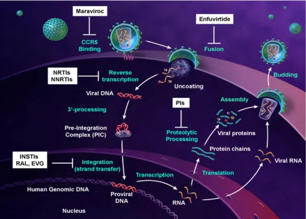
III. Anti HIV therapy………...42
III.1. Overview of current anti-HIV therapy ………...…42
III.2. Antiretroviral drugs ………...………..……....…43
III.2.1. Overview of present antiretroviral drugs………...43
III.2.2. Nucleoside and nucleotide reverse transcriptase inhibitors (NRTIs) … 45 III.2.3. Non-nucleoside reverse transcriptase inhibitors (NNRTIs)………….48
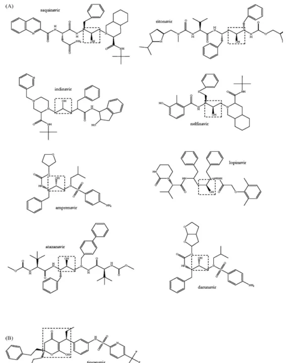
III.2.4. Protease inhibitors (PIs) ………...…50
III.2.5. Entry/Fusion inhibitors………..…....52
III.2.6. Integrase inhibitors (INIs)………..……..….…54
III.2.6.1. Development of integrase strand transfer inhibitors (INSTIs) 55 III.2.6.2. Resistance to INSTIs ………...58
III.2.6.3. Mechanism of INSTIs inhibition and resistance ………...…. 60
III.3. The strategy for HIV vaccine ...………..………..……....…62
III.4. Current challenges of antiretroviral therapy……….……….……...…..…..…64
Motivation and objective ……….………..…...…..67
Materials and methods ………...……….…….. 70
I. Purification of IN and in vitro activity tests ………..………..71
I.1. Cloning of IN……….……….71
I.2. Site-directed mutagenesis . ………. 71
I.3. Expression of HIV IN ………..……...72
I.4. Purification of HIV IN……….……...73
I.5. In vitro IN enzymatic activity test………...…. 74
I.5.1. Activity test using radio-marked oligonucleotides……….……..74
I.5.2. Activity test using static fluorescence anisotropy……….….. 76
Results ………...………...78
I. Determining the susceptibility of latest integrase strand transfer inhibitors on HIV-1 subtype CRF02_AG in vitro……….……….…….79
I.1. Introduction……….……...79
I.2. Manuscript………80
I.3. Conclusion………..….106
II. In vitro catalytic properties and resistance to raltegravir of HIV-2 integrase clinic isolates and site-direct mutants……….………...……..108
II.1. Introduction………..………..108
II.2. Manuscript………..…………...…110
II.3. Supplementary results………121
II.4. Conclusion ………..………...125
III. Development of anti-HIV-1 integrase camelid simple-domain antibody………….….….128
III.1. General introduction of single domain antibody ……….………..….130
III.1.1. VH and VHH sequence differences………..……….…131
III.1.2. Structure of VHH ………..……133
III.1.3. Unique features of sdAb ………...……….…...133
III.1.4. Biotechnological application of sdA………..135
III.2. Development of camelid single-domain antibody targeting HIV-1 integrase…... 137
III.2.1. Introduction………...137
III.2.2. Materials and methods……….…..…139
III.2.3. Results………..…….…….…156
III.2.4. Discussion……….……...….….…175
General conclusions ………..………178
ABBREVIATIONS
AIDS: Acquired immunodeficiency syndrome ART: Antiretroviral therapy
AZT: Zidovudine
BAF: Barrier-toautointegration factor bNAbs: Broadly neutralizing Abs CA: Capsid
CCD: Catalytic core domain
CCR5: C-C Chemokine Receptor Type 5 CDK9: Cyclin-dependent kinase 9
CDR: Complementarity determining region
cPPT-CTS : central polypurine tract-central termination sequence CRF: Circulating recombinant form
CTD: Carboxy-terminal domain
CXCR4: C-X-C chemokine receptor type 4 CYCT1: Cyclin T1
CypA: Peptidyl-prolyl isomerase cyclophilin A DC: Dendritic cell
DKA: Diketoacid moiety
dNTP: deoxynucleoside triphosphate DTT: Dithiothreitol
EED: Embryonic ectoderm development ELISA: Enzyme-linked immunosorbent assay EVG: Elvitegravir
FI: Fusion inhibitor FV: Foamy virus
HAART: Highly active antiretroviral therapy HCAb: Heavy-chain antibody
HIV: Human immunodeficiency virus HMGA1: High mobility group protein A1 HR: Heptat repeat
HSP60: Heat-shock protein 60
HTLV-III: Human T-Cell leukemia virus III IBD: Integrase binding domain
IN: Integrase
INI: Integrase inhibitor
INI1/hSNF5: Integrase interactor 1 Inr: Initiator
INSTI: Integrase strand transfer inhibitor
LEDGF/p75: Human lens epithelium-derived growth factor LTR: Long Terminal Repeat
MA: Matrix
MiRNA: micro-RNA
MOI: Multiplicity of infection Nabs: Neutralizing Abs
NC: Nucleocapsid Nef: Negative effector NF-B: Nuclear factor-B
NLS: Nuclear localisation signal
NNRTI: Non-nucleoside reverse transcriptase inhibitor NPC : Nucleoprotein complex
NRTI: Nucleoside reverse transcriptase inhibitor NTD: Amino-terminal domain
N-TEF: Negative transcription elongation factor ODN: Oligonucleotides
PCR: Polymerase chain reaction PI: Protease inhibitor
PIC: Pre-integration complex PPT: Polypurine tract
3’-P: 3’-processing
P-TEFb: Positive transcription elongation factor b RAL: Raltegravir
Rev: Regulator of virion gene expression RNAPII: Polymerase II
RT: Reverse transcriptase
RTC: Reverse transcription complex scFv: single-chain Fv
sdAb: Single-domain antibody
SIV: Simian immunodeficiency viruses SPRi: Plasmon resonance imagery ST: Strand transfer
TAM: Thymidine associated mutations TAR: Trans-activation response element Tat: Transcriptional transactivator Th: T helper
TRIM5 : Ttripartite motif protein 5 TRN-SR2 : Transportin SR-2
TSG101: Tumour-susceptibility gene 101 URF: Unique recombinant form
VH: Variable domain
VHH: Variable domain of heavy chain of heavy chain antibody Vif: Viral infectivity factor
VLP: Virus-like particles Vpr: Viral protein r Vpu: Viral protein u
I. Human immunodeficiency virus (HIV)
Nearly 30 years after the first reported cases of the acquired immunodeficiency syndrome (AIDS) and the discovery of the etiologic agent, effective control of the AIDS pandemic which is arguably the most serious infectious disease remains elusive. It is estimated that 33.3 million people worldwide are living with HIV in 2009, and 2.6 million people newly infected with HIV in that year alone.
The origin of this challenge is the molecular pathogenesis of HIV, a virus that has evolved a number of mechanisms to escape from host immune control. Among the most notable strategies are the virus’ direct targeting of the CD4+ molecule expressed by the key T lymphocyte in human immune system; the high rate of mutation which enable the virus to evade the host immune system (mutational escape); the highly glycosylation of the external glycoprotein, which protects neutralization epitopes; and integration into the human genome, which implies that cells which are not killed are permanently infected.
I.1. Identification of HIV
AIDS was first recognized in the United States in 1981, following an increase in the incidence of usually rare opportunistic infections such as the pneumonia caused by Pneumocystis carinii in homosexual men which were caused by a general immune deficiency. A dramatic reduction of CD4+ lymphocyte cells in peripheral blood was the common characteristic of all the patients.
In 1983 HIV was isolated by the team of L.Montagnier of Pasteur Institute at Paris (Barré-Sinoussi et al., 1983). The activity of reverse transcription was identified from a sample of patient with lymphoadenopathy. The presence of a retrovirus initially named as lymphoadenopathy associated virus (LAV) was confirmed by the visualization using electronic microscopy. Simultaneously, some American teams identified two virus considered as the pathogenic agent responsible of AIDS, human T-Cell leukemia virus III (HTLV-III) and AIDS associated-virus (Gallo et al., 1984; Levy et al., 1984 ). In 1985, the scientific community concluded that these 3 viruses are identical. This virus was nominated as HIV-1 in 1986 by the discovery of the second similar virus with slightly different genome structures, isolated from two West-African patients with AIDS and then named as HIV-2. By its genomic sequences and its proteins, this virus is different from the HIV-1 isolated from U.S.A., Europe and Central Africa. It differs also from simian immunodeficiency virus (SIV), but displays an antigenic relationship with the latter
virus, at the level of its external envelope protein (Clavel et al., 1986).
I.2. Taxonomy of the retrovirus family
The reference of classification is from the International committee of taxonomy of viruses (ICTV, http://www.ictvonline.org/virusTaxonomy.asp). The affiliation of one family is defined by common taxonomic denominators: genetic information as well as the replicative properties. Certain structure elements, as the presence of envelope and pathogenesis are equally counted in some cases.
Retroviruses are further divided into seven groups defined by evolutionary relatedness, each with the taxonomic rank of genus. Five of these groups represent retroviruses with oncogenic potential (formerly referred to as oncoviruses), and the other two groups are the lentiviruses and the spumaviruses (Table 1).
Retroviruses can be broadly divided into two categories, simple and complex, distinguishable by the organization of their genomes (Murphy et al., 1994). All oncogenic members except the human T-cell leukemia virus-bovine leukemia virus (HTLV-BLV) genus are simple retroviruses. HTLV-BLV and the lentiviruses and spumaviruses are complex.
HIV-1 and HIV-2 are both characterized by extensive genetic diversity. HIV-1 is phylogenetically divided into three groups, major (M), outlier (O), and nonmajor and nonoutlier (N), with the M group further split into 9 subtypes and 15 circulating recombinant forms. Today, group M has a near global distribution, whereas groups N and O are restricted to individuals of West African origin. HIV-2 is also most common in individuals from West Africa and is composed of seven subtypes. Despite its initial association with homosexual men, it is clear that HIV-1 and HIV-2 are now primarily transmitted by heterosexual intercourse and from mother to infant.
Subfamily Genus name Type species name
00.061.1. Orthoretrovirinae
00.061.1.01. Betaretrovirus 00.061.1.01.001. Mouse mammary tumor virus 00.061.1.02. Gammaretrovirus 00.061.1.02.001. Murine leukemia virus
00.061.1.03. Alpharetrovirus 00.061.1.03.001. Avian leukosis virus 00.061.1.05. Deltaretrovirus 00.061.1.05.001. Bovine leukemia virus
00.061.1.06. Lentivirus 00.061.1.06.001. Human immunodeficiency virus 1 00.061.1.08. Epsilonretrovirus 00.061.1.08.001. Walleye dermal sarcoma virus 00.061.2. Spumaretrovirinae 00.061.2.07. Spumavirus 00.061.2.07.001. Simian foamy virus
Table 1: Composition of retroviridae family. (ICTVdB - The Universal Virus Database, version 3. http://www.ncbi.nlm.nih.gov/ICTVdb/ICTVdB/)
I.3. Origins of HIV
The key to understanding the origin of HIV was the discovery that closely related viruses, the simian immunodeficiency viruses (SIVs), were present in a wide variety of African primates. Collectively, HIV and SIV comprise the primate lentiviruses, and SIVs have been isolated in more than 20 African primate species.
The evolutionary history of HIV-1 and HIV-2 has been reconstructed in great detail by inferring phylogenetic trees of the primate lentiviruses. It was discovered that the two human viruses are related to different SIVs and therefore have different evolutionary origins (Figure 1). Specifically, HIV-1 is most closely related to SIVcpz, which is found in some sub-species of chimpanzee that inhabit parts of equatorial Western and Central Africa, respectively (Gao et al., 1999; Santiago et al., 2002 ). This HIV-1 progenitor probably was passed from chimpanzees to human hunters through blood borne transmission.
Phylogenetic analysis of HIV-1 and related viruses from non human primates suggests that three independent transmission events early in the 20th century spawned three HIV-1 groups: M, N and O. Although strains related to the M and N groups have been found in chimpanzees, recent evidence suggests that group O HIV-1 may have originated in gorillas, in which the closest relatives of this group have been identified (Taylor et al., 2008). It is speculated that the virus then spread among humans along the Congo River into Kinshasa, Zaire, where the earliest documented case of HIV-1 infection (with group M strain) in humans has been traced to a blood sample from 1959 (Zhu et al., 1998).
high prevalence in sooty mangabey monkeys. Sooty mangabeys are most frequent in the regions of West Africa where HIV-2 is likely to have emerged.
Molecular phylogenies also show that there have been many cross-species transmissions to humans, because there is a mixing of HIV and SIV lineages. Although the vagaries of sampling make it difficult to determine exactly how many cross-species transfers have occurred, three jumps from chimpanzees to humans are considered to explain the current diversity of HIV-1, such that groups M, N and O each have an independent origin (Gao et al., 1999).
Figure 1: Evolutionary history of the HIV-1 and HIV-2.
Because both the HIV-1 and HIV-2 lineages (red branches) fall within the SIVs that are isolated from other primates, they represent independent cross-species transmission events. HIV-1 groups M, N and O represent separate transfers from chimpanzees (SIVcpz), again because there is a mixing of the HIV-1 and SIV lineages. Similarly, HIV-2 seems to have been transferred from sooty mangabey monkeys (SIVsm). Only some subtypes of HIV-1 and HIV-2 are shown for clarity (Rambaut et al., 2004).
I.4. Basics of HIV replication
HIV enters the body through the exchange of bodily fluids, and it infects mainly T helper (Th) cells, macrophages and, to some extent, microglial cells and dendritic cells (DCs). This tropism is determined at the level of viral entry by the use of CD4 as a primary receptor and the use of co-receptors which are strain and target specific (Figure 2). HIV R5 strains use C-C chemokine receptor type 5 (CCR5) as their co-receptor and can, therefore, enter macrophages, DCs and T cells, whereas X4 strains of HIV use C-X-C chemokine receptor type 4 (CXCR4) as a co-receptor and can infect T cells only (Doms and Trono, 2000). Early in infection, only R5 viruses can be detected in infected individuals. With time, X4 viruses come to predominate, which hastens the demise of Th cells, the hallmark of AIDS.
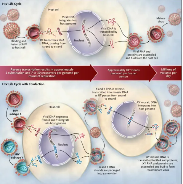
Upon membrane fusion, the viral core, containing the viral capsid (CA) and nucleocapsid (NC) along with the viral genome, reverse transcriptase (RT), integrase (IN), protease (PR), the viral accessory proteins Vif, Nef and Vpr, and regulatory proteins is released into the cytoplasm. Collectively called reverse transcription complex (RTC), this assembly traffics along the microtube network from the cell periphery toward the centrosome. Within the RTC, RT converts the viral RNA genome into a double-stranded viral complementary DNA, usually occurring upon entry into the host cell.
Reverse transcription and viral core disassembly are concomitant, tightly coupled processes (Zhang et al., 2000). More recent results indicate that viral core disassembly might occur later near the centrosome (Lehmann-Che et al., 2005) or the nuclear envelope (Arhel et al., 2007), likely depending on the virus and/or the cell metabolism. Uncoating might be the rate-limiting step that determines nuclear import of the viral genome (Lewinski et al., 2006 ).
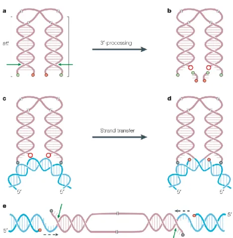
Reverse transcription process is quite complex and requires different catalytic activities. The process is initiated from the 3’ end of a primer tRNA3Lys partially hybridized to the primer binding site (PBS) which is located near the 5’ end of the viral genome. It results in the synthesis of a short stretch of minus, single-stranded DNA, which then relocates and hybridizes to the repeat sequence at the 3’ end of the RNA genome. After this strand transfer, the synthesis of the first DNA strand can continue. The synthesis of the first DNA strand requires multiple enzymatic activities: RNA-primed RNA-dependent DNA polymerization (RDDP), DNA-primed RDDP, strand-transfer, and RNaseH activity. The RNaseH which is embedded in the RT enzyme hydrolyzes the copied RNA template prior to strand-transfer. The synthesis of the second DNA strand uses RNA fragments still hybridized to the first DNA strand, like the polypurine tract (PPT), as primers, because of the RNaseH resistant conformation of those RNA/DNA duplexes. The second DNA strand synthesis also requires a strand transfer reaction. While reverse transcription proceeds, the RTC undergoes a progressive remodeling to become a pre-integration complex (PIC) as the viral nucleoprotein complex competent for proviral integration.
By undergoing an active phase of nuclear import through the nuclear pore, HIV does not require disassembly of the nuclear membrane during cell division, which is unlike to the other retroviruses. This property has been ascribed to the presence of karyophilic elements present in viral nucleoprotein complexes (NPCs) (Bukrinsky et al., 1992; Suzuki and Craigie, 2007). However, the role of different compenents of NPC in nuclear import remains controversial. Human lens epithelium-derived growth factor (LEDGF/p75) which contains the basic and classical nucleusnuclear localization signal (NLS) consistent stretch
146RRGRKRKAEKQ156 is involved in tethering the viral PIC to chromatin (Maertens et al., 2004b; Maertens et al., 2003; Meehan and Poeschla, 2010; Vanegas et al., 2005). IN residues 161 to173 had been thought as a transferable karyophilic signal (Bouyac-Bertoia et al., 2001); however, further studies suggested that no NLS is present at this location (Dvorin et al., 2002; Limón et al., 2002) . Moreover, it was shown that a three-stranded central DNA flap which is a structure present in neo-synthesized viral DNA, specified by the central polypurine tract-central termination sequence (cPPT-CTS), acts as a cis-acting determinant of HIV-1 genome nuclear import (Arhel et al., 2007; Rivière et al., 2010; Zennou et al., 2000). These elements may mediate nuclear import directly or via the recruitment of the host's proteins. Indeed, several cellular proteins have been found to influence HIV-1 infection during nuclear import, such as the karyopherin α2 Rch1 (Gallay et al., 1996); importin 7, an import receptor for ribosomal proteins and histone H1 (Fassati et al., 2003); the transportin SR-2 (TRN-SR2) as a binding partner of HIV-1 IN (Brass et al., 2008; Christ et al., 2008); or the nucleoporins Nup98 (Ebina et al., 2004), Nup358/RANBP2, and Nup153 (Brass et al., 2008; König et al., 2008). By interacting directly with the NPC via the binding of PIC-associated IN to the C-terminal domain of Nup153, HIV-1 sub-viral particles gain access to the nucleus (Woodward et al., 2009).
In vivo, integration is a rare event. The integrated viral genome represents only 5 to 10% of the total viral DNA content of an infected cell (Brussel and Sonigo, 2004). Both the extra linear molecules and circular molecules, containing 1 or 2 long terminal repeats (LTR), represent the most abundant forms of the viral DNA genome. Only the linear form cDNA can integrate into the host genome, yielding the LTR flanked provirus. The linear double-stranded cDNAs have IN and chromatin-remodelling complexes at their termini (Miller et al., 1997b). Broadly sketched, lentiviruses favor integrating in active transcription units without favoring the promoter regions in particular. HIV integration is also strongly disfavored in centromeric heterochromatin, which is transcriptionally repressed and a poor substrate for viral transcription (Poeschla, 2008). The full-length linear cDNA decays rapidly if not integrated (estimated half life of 1 to 2 days) (Zhou et al., 2005), while 1- and 2-LTR circles accumulate in the nucleus at later times of infection. These circular forms, which are considered dead-end products of aborted integration events, can also lead to viral gene expression (Gelderblom et al., 2008).
Once integrated in the host genome, the provirus behaves like any human gene or so Pol II-transcribed genes. At this point, the 5’ LTR which contains enhancer and promoter sequences with binding sites for several transcription factors, behaves like any eukaryotic promoter; and the 3’ LTR acts as the polyadenylation and termination site (Peterlin and
Trono, 2003). Moving upstream from the transcription start site, the initiator (Inr), the TATA Box and three SP1-binding sites are found. These elements position RNA polymerase II (RNAPII) at the correct site for initiating transcription. Further upstream is the enhancer, which binds nuclear factor-B (NF-B) and nuclear factor of activated T cells (NFAT), as well as members of the E-twenty six (ETS) family of transcription factors (Jones and Peterlin, 1994). These activators ensure that the virus replicates at a high level in activated T cells and differentiated macrophages.
Additionally, the presence of a strong regulatory element located 3’ to Inr is the most unusual feature of the LTR. This RNA structure, which is found at the 5’ end of all viral transcripts, is known as the trans-activation response element (TAR) and it binds the transcriptional transactivator (Tat) (Taube et al., 1999). In the absence of Tat, HIV transcription begins, but elongation is inefficient. Tat and its cellular cofactor, positive transcription elongation factor b (P-TEFb), cooperate to bind TAR with high affinity, allowing RNAPII to produce full-length viral transcripts. P-TEFb contains two components: cyclin T1 (CYCT1) and cyclin-dependent kinase 9 (CDK9). Once recruited to the nascent HIV RNA, CDK9 phosphorylates the carboxy-terminal domain of RNAPII and negative transcription elongation factor (N-TEF), thereby allowing efficient elongation (Price, 2000). The ability of Tat to recruit P-TEFb through an RNA sequence is unique among transcriptional activators, and it renders HIV replication particularly sensitive to inhibition by compounds that target CDK9. In contrast, Tat is inefficient at recruiting RNAPII, and it requires a strong basal promoter for optimal effects (Jones and Peterlin, 1994). NF-B, which also recruits P-TEFb, can substitute partially for Tat (Barboric et al., 2001). Furthermore, Tat and P-TEFb affect the ability of HIV to establish latency and affect the transcription of MHC class II genes.
Viral proteins are synthesized from more than 30 mRNA species, which are all derived from the same primary transcript. These transcripts that encode the late viral proteins, which are mainly structural and enzymatic components of the virion and factors that fine-tune infectivity, are singly spliced or unspliced, while those encoding the most of early regulatory proteins are fully spliced.
The timely production of viral gene products requires the regulator of virion gene expression (Rev), which transports slightly spliced and unspliced genomic transcripts from the nucleus to the cytoplasm. To function optimally, this protein needs to reach threshold levels (Pomerantz, 1992), the splicing of genomic transcripts must be slow and an active Crm1/RanGTP complex must be present in the cell. For HIV and other lentiviruses, the only really fundamental post-integration peculiarity in the nucleus is a necessary
mechanism to bypass the cells splicing checkpoint for the viral genome RNA and some coding mRNAs.
After their translation, viral structural and enzymatic proteins travel to the plasma membrane, where immature virions assemble in cholesterol-rich lipid rafts (Nguyen and Hildreth, 2000). The carboxy-terminus of Gag, p6, is ubiquitylated, and it recruits components of multivesicular bodies, such as tumour-susceptibility gene 101 (TSG101) and vacuolar protein sorting 4 (VPS4), which facilitate the release of progeny virions from the cell. Processing of Gag and Gag-Pol yields mature HIV particles. Some accessory viral proteins such as Vpr and Nef, as well as cellular components, e.g., MHC class I and II molecules, and some CD proteins, are incorporated into virions (Esser, 2001). The envelope glycoprotein is an essential viral component that allows the infection of new cells. The high cholesterol content of the virion, which is a consequence of its budding through rafts, is crucial for this process as well.
I.5. Genomic organization and structure of HIV
I.5.1. Genomic organization
The genome of HIV composes a double copy of positive-sense, single stranded RNA of about 9 kilo nucleotides and encodes 15 distinct proteins (Figure 3). The two RNA molecules are linked by their 5’ extremity and classic proprieties of eukaryotic mRNA: a 5’ cap (m7GpppGm) and a poly (A) tail at 3’ extremity. The RNA is flanked by repeated R sequence and U5 region at 5’ and U3 region at 3’. Reverse transcription produces a duplication of region U5 and U3 when RNA is transformed into DNA double strands. The new extremity called long terminal repeat (LTR) contains some essential elements for viral replication (e.g., promoter region at 5’ U3 and poly-adenylation site at 3’ U3) and sites for integration (sequence ACGT at 5’ and CAGT at 3’).
From the point of genetic view, HIV is a complex virus which codes for the characteristic retroviral genes (gag, pol and env) but also for regulatory proteins. The gene
gag (group antigen) codes for polyprotein Pr55gag. This precursor is digested by viral protease to release different virion elements. The viral protease, integrase, and reverse transcriptase are always expressed within the context of a Gag-Pol fusion protein. The Gag-Pol precursor (p160) is generated by a ribosomal frame shifting event, which is triggered by a specific cis-acting RNA motif (Dulude et al., 2002). When ribosomes encounter this motif, in approximate 5% of the cases, they shift to the pol reading frame (at
position -1 according to gag reading frame) without interrupting translation.
I.5.2. Structure of HIV
The viral particles exist as spheres of approximately 80 to120 nm in diameter with a spherical morphology (Figure 4). The cone-shaped core, which contains 2 copies of genomic RNA, reverse transcriptase, integrase, protease, and etc, is surrounded by lipid matrix containing key surface antigens and glycoproteins. Those proteins that are derived from the gag (group-specific antigen), pol (polymerase) and env (envelope) genes are classical structural and enzymatic factors that are required by all retroviruses.
A
B
Figure 3: Genomic organization of HIV-1 and HIV-2 (A) and a summary of the functions of 9 genes encoding 15 proteins of HIV provirus (B) (Greene and Peterlin,
Figure 4: Photo and schematic structure of viral particle.
http://www.ictvdb.org/WIntkey/Images/dg_HIV.jpg
Viral envelope (Env)
The envelope (Env) spikes on HIV-1 define the viral tropism, mediate the fusion process and are the prime target of the humoral response. Three gp120 subunits comprise the ‘head’ of Env and three gp41 subunits comprise the ‘stalk’ and other membrane-associated elements.
The gp120 moiety has five hypervariable regions, designated V1 through V5 loop. V3 loop is not involved in CD4 binding, but is rather an important determinant of the preferential tropism of HIV-1 for either T lymphoid cell lines or primary macrophages (Hwang et al., 1991 ). Sequences within the V3 loop interact with the HIV chemokine co-receptors CXCR4 and CCR5, which partially determine the susceptibility of cell types to given viral strains (Deng et al., 1996). The V3 loop is also the principal target for neutralizing Abs (Abs) that block HIV-1 infectivity.
Structural proteins
Gag precursor protein, also called p55, is expressed from the unspliced viral mRNA. The membrane-associated Gag polyprotein recruits two copies of the viral genomic RNA along with other viral and cellular proteins that triggers the budding of the viral particle from the surface of an infected cell. After budding, p55 is cleaved by the virally encoded protease during the process of viral maturation into four smaller proteins designated matrix (MA, p17), capsid (CA, p24), nucleocapsid (NC, p7) and p6. MA is a 132 amino acid protein and
post-translationally myristylated at the N-terminus. MA and CA play critical roles in both late and early stages of the viral replication cycle. NC contains two successive zinc fingers, promotes viral RNA dimerization and encapsidation and activates annealing of the primer tRNA to the initiation site of reverse transcription (Morellet et al., 1992). The p6 protein regulates the final abscission step of nascent virions from the cell membrane by the action of two late assembly (L-) domains. The 52 amino acid peptide of p6 protein binds at least to two cellular budding factors (Tsg101 and ALIX), and mediates the incorporation of the HIV-1 accessory protein Vpr into viral particles (Votteler et al., 2011).
Nonstructural proteins
During viral maturation, the virally encoded protease cleaves Gag-Pol precursor, releases the Pol polypeptide away from Gag and further digests it to separate the protease, reverse transcriptase, and integrase.
Protease (Pro)
HIV protease activity is not required for virus production and release per se, but is essential for viral maturation leading to infectious viral particles. It generates mature infectious virus particles through cleavage of the viral Gag and Gag-Pol precursor proteins.
HIV protease is in the family of aspartic proteases (with an aspartic acid in the active site at position 25) and is a symmetrically assembled homodimer consisting of two identical subunits of 99 amino acids (Navia et al., 1989; Wlodawer et al., 1989). The centre of the enzyme is formed by the substrate-binding cleft, which interacts with the different substrate cleavage site sequences in the Gag and Gag-Pol proteins. Dimerization of Gag-Pol precursor proteins is required by the activation of viral protease which is embedded in the Gag-Pol protein. Despite its critical function in viral maturation and infectivity, HIV protease shows great plasticity and polymorphisms which are observed at one-third of the 99 amino acids (Rhee et al., 2003).
HIV-1 protease recognizes the asymmetric shape of the peptide substrates, rather than a particular amino acid sequence of the cleavage site (Prabu-Jeyabalan et al., 2002). The peptide substrates have a secondary structure, yielding a substrate ‘envelope’ which fits within the protease substrate-binding region. However, a few differences between the cleavage site substrates in which amino acid side chains protrude out of the ‘envelope’. These amino acid side chains protruding out of the ‘envelope’, which makes small differences between cleavage sites, contribute to the highly ordered and regulated process in which all individual cleavages occur at different rates. The ordered cleavage appears to
be regulated by the amino acid sequence near the actual protease cleavage site.
Reverse transcriptase (RT)
The reverse transcriptase is an asymmetric heterodimer comprising a p66 subunit (560 amino acids) and a p51 subunit (440 amino acids) (Kohlstaedt et al., 1992). Both subunits are encoded by the same sequence in the viral genome. The DNA- and RNA-dependent DNA polymerase activity is catalyzed by the p66 subunit, which is responsible for the critical step of conversion of the minus stranded RNA viral genome into double stranded DNA. RNase H consists of the last (carboxy terminal) 120 amino acids of the p66 subunit, which correspond to the p15 fragment cleaved from the p66 subunit by the viral protease to generate the p51 subunit. RNase H activity removes the original RNA template from the first DNA strand, allowing synthesis of the complementary strand of DNA.
Like other polynucleotide polymerases, the overall three-dimensional structure of the p66 subunit is often compared to a right hand, with a fingers (amino acids 1-85 and 118-155), a palm (amino acids 86-117 and 156-237) and a thumb (amino acids 238-318) domain (Steitz, 1999). The palm domain contains the polymerase active site with its three aspartic acids (110, 185 and 186) and the YMDD characteristic motif. The incoming dNTP binds between the palm and the finger subdomains and the ribose and base make important contacts with residues including L74, Y115, M184 and Q151. The catalytic pocket is formed by the fingers folding down into the palm domain (Huang et al., 1998).
Next to the catalytic domain, the p66 subunit also contains the RNaseH domain (amino acids 427-560), linked by the connection domain (amino acids 319-426). The connection domain is also involved in interactions with the nucleic acid and the p51 subunit.
A two-metal model has been proposed for the phosphoryl transfer reaction of polymerases including HIV RT (Steitz, 1999). In the RT active site, two Mg2+ ions are coordinated by the catalytic triad of D110, D185 and D186 that, along with residues including K65, interact with triphosphate and 3’-terminus of the primer (Huang et al., 1998).
Structural studies support an induced fit model where proper base pairing by the incoming dNTP results in formation of a closed polymerase, primer/template, and dNTP complex with the incoming dNTP appropriately aligned to be attacked by the 3’ OH at the terminus of the elongating primer strand (Doublie et al., 1998). Kinetic studies showed a corresponding rate limiting step that might reflect this conformation change. The dependence of the rate-limiting step on correct dNTP binding and base-pairing forms the basis for the fidelity of polymerization (Joyce and Benkovic, 2004).
whereas the p51 subunit is rather compact, and seems to play a structural role, devoid of catalytic activity, with the three aspartic acids buried inside.
Integrase (IN)
The IN protein mediates the insertion of the HIV proviral DNA into the genomic DNA of an infected cell. The structure and function of IN will be described in more detail in part II. HIV integrase and integration.
Regulatory proteins and accessory proteins
HIV encodes two regulatory proteins: the transcriptional transactivator (Tat) and the regulator of virion gene expression (Rev), and four so-called accessory proteins: the ill-named ‘negative effector’ (Nef), viral infectivity factor (Vif), and the viral proteins r (Vpr), u (Vpu) for HIV-1 and u (Vpx) for HIV-2.
Rev is a 13 kDa sequence-specific RNA binding protein. Produced from fully spliced
mRNAs, Rev regulates the transition from the early to the late phases of viral gene expression by allowing the export of incompletely spliced and unspliced viral mRNA species from the nucleus to the cytoplasm (Kim et al., 1989).
Tat is capable to interact with a couple of cellular proteins. Tat binds to a short-stem
loop structure, known as the transactivation response (TAR) element that is located at the 5’ terminus of HIV RNAs. Tat binding occurs in conjunction with cellular proteins that contribute to the effects of Tat. The binding of Tat to TAR activates transcription from the HIV LTR at least 1000 fold (Feng and Holland, 1988; Kao et al., 1987).
Nef has been shown to have multiple activities, including the down regulation of the cell
surface expression of CD4, the perturbation of T cell activation, and the stimulation of HIV infectivity. Tat and Nef are not only crucial for high levels of HIV replication, but also have important roles in promoting viral immune evasion.
Vpr is a multifunctional accessory protein. It plays a role in the ability of HIV to infect
non dividing cells by facilitating the nuclear localization of the PIC (Heinzinger et al., 1994 ). Other critical functions include transcriptional coactivation of viral and host genes, induction of cell-cycle arrest, both direct and indirect contributions to T-cell dysfunction, and regulation of nuclear factor kappa B (NF-κB) activity (Kogan and Rappaport, 2011).
Vpu of 16 kDa is unique to HIV-1. There is no similar gene of Vpu in HIV-2 or SIV.
HIV-1 Vpu is an oligomeric, type I integral membrane phosphoprotein. Two distinct biological activities, via distinct molecular mechanisms, have been attributed to Vpu: enhancement of viral particle release from the plasma membrane of infected cells and the
specific degradation of the HIV-1 receptor molecule CD4 in the endoplasmic reticulum (ER) (Subbramanian and Cohen, 1994; Willey et al., 1992 ). Vpu can efficiently antagonize the restriction of releasing enveloped viruses from infected cells which is imposed by Tetherin (BST-2/CD317), an interferon-inducible antiviral host protein (Neil et al., 2008; Van Damme et al., 2008). Vpx of 12 kDa was found only in HIV-2/SIVagm. Vpx function in relation to Vpr is not fully elucidated.
Vif is a 23 kDa polypeptide that is essential for the replication of HIV in peripheral
blood lymphocytes, macrophages, and certain cell lines (Strebel et al., 1987). Vif inhibits the antiviral activity of the cellular protein APOBEC3G from entering the virion during budding from a host cell by targeting it for ubiquitination and proteasomal degradation (Lecossier et al., 2003; Sheehy et al., 2002; Yu et al., 2003).
Cellular proteins in viral structure
Certain cellular compositions are packed during virus assembly. This encapsidations are due to the interaction of viral components and cellular proteins. For example, the peptidyl-prolyl isomerase cyclophilin A (CypA), binding a proline-rich loop on the surface of HIV-1 CA, is involved in assembly. This interaction increases HIV-1 infectivity in humans but promotes an anti-HIV-1 restriction activity in non-human primates (Sokolskaja
and Luban, 2006). CypA is a ubiquitous cytoplasmic protein that catalyzes
the cis/transisomerization of peptidyl-prolyl bonds. Via isomerization of a peptide bond in CA conformation (Bosco et al., 2002), CypA modulates HIV-1 virion core detection by a class of innate pattern recognition molecule tripartite motif protein 5α (TRIM5α) (Stremlau et al., 2004 ). It is speculated that the rapid turnover of TRIM5α and presumably TRIM5α-virus complexes by proteasomes leads to an early block to infection, before the virus has had the opportunity to reverse transcribe. How exactly TRIM5α renders the virus uninfectious remains unclear (Towers, 2007).
During budding, viral particle is enveloped with the help of lipid bilayer of infected cell. It is also possible to find some cellular proteins on the surface of virus. Among them, the antigen of major histocompatibility complex (MHC) can be numerous and as important as the viral glycoproteins (Arthur et al., 1992).
I.6. HIV molecular epidemiology and mechanism of genomic diversity
HIV-1 subtypes, also called clades, are phylogenetically linked strains of HIV-1 that are approximately the same genetic distance from one another; in some cases, subtypes are also linked geographically or epidemiologically. Genetic variation within a subtype can be 15 to 20%, whereas variation between subtypes is usually 25 to 35%. Group M is the predominant circulating HIV-1 group which has a near global distribution. It has been divided into subtypes, denoted with letters, and sub-subtypes, denoted with numerals: subtypes and sub-subtypes A1, A2, A3, A4, B, C, D, F1, F2, G, H, J, and K.
Over the past decade, advances in full-genome sequencing of HIV have led to the identification of circulating and unique recombinant forms (CRFs and URFs, respectively). These are the result of recombination between subtypes within a dually infected person, from whom the recombinant forms are then passed to other people. The recombinant progeny are classified as circulating recombinant forms if they are identified in three or more people with no direct epidemiologic linkage; otherwise they are described as unique recombinant forms (Taylor et al., 2008). Among the 40 identified CRFs, the most described are CRF01_AE playing an important role in epidemic in Southeast Asia. CRF02_AG is responsible for the epidemic in West Africa, and more and more frequently found in other countries (Machado et al., 2009; Malet et al., 2008c; Njai et al., 2006; Ye. J. et al., 2011).
The global distribution of subtypes and CRFs reflects the complexity of the molecular epidemiology of HIV-1 (Figure 5). In 2004-2007, subtype C accounted for nearly half (48%) of all global infections, followed by subtypes A (12%) and B (11%), CRF02_AG (8%), CRF01_AE (5%), subtype G (5%) and D (2%). Subtypes F, H, J and K together cause less than 1% of infections worldwide. Other CRFs and URFs are responsible for 4% of global infections respectively, rendering the combined total of worldwide CRFs to 16% and all recombinants (CRFs along with URFs) to 20% (Hemelaar et al., 2011). Analysis of the global distribution of HIV-1 demonstrated a broadly stable distribution of HIV-1 subtypes worldwide with a notable increase in the proportion of CRFs (Hemelaar et al., 2011). CRF02_AG isolates showed a slightly but not significantly higher replication advantage compared with subtype A or G isolates. This advantage in replicative fitness of CRF02_AG may contribute to its dominant spread over A and G subtypes in West and West Central Africa (Njai et al., 2006). Recombination that may contribute to rescue high-fitness HIV-1 variants is a common feature among retroviruses and, as well, between HIV-1 strains, particularly clade A (Spira et al., 2003).
From 1985, it was demonstrated that the genetic variability of HIV is very important. Some further studies showed that it does not exist two identical viral strains. And in one
individual the virus presents in the form of a polymorphic viral population with a multitude of different genomes called ‘quasi species’. In a recently infected patient, the circulating viruses are genetically homogeneous. But the viral population evolutes over time with a global rate of change (estimated at 1% each year for gene env, 0.5% for gene gag). A complex mixture of variants progressively appears and evolutes in different and independent way in different tissues and cells over time. The initial view that the virus is classifiable into distinct subtypes or clades now needs to reflect the reality of a dynamic genetic evolutionary process, through which new HIV-1 strains are constantly emerging (Taylor et al., 2008).
A
B
Figure 5: Global distribution of HIV.
(A) Global distribution of HIV-1 subtypes and recombinant forms (Taylor et al., 2008). (B) Distribution of all HIV-2 sequences. Note that this map only includes sequences for which the sampling country is known. Subtype distributions represent the frequency in the HIV Database and not the population. (http://www.hiv.lanl.gov/components/sequence/HIV/geo/geo.comp).
I.6.2. Mechanisms of HIV genomic diversity
Mathematical models that predicted the eradication of virus from a patient within 2 to 3 years failed to adequately take into account two key aspects of HIV biology: viral reservoirs and evolution (Perelson et al., 1997). HIV has several intrinsic mechanisms, e.g., frequent random mutation and genetic recombination, which ensure rapid viral evolution, in response to both host immune activity and antiretroviral therapy (ART).
Frequent random mutation
The reverse transcriptase of HIV lacks exonucleolytic proofreading activity which confirms that the DNA transcript produced is an accurate copy of the RNA template, and confers a mutation rate of approximately 3.4×10⁻⁵ mutations per base pair per replication cycle. Since the HIV genome is an estimated 105 base pairs in length, and HIV infection is characterized by a very high replication rate, with the production of 1 to 10 billion new virus particles per day in an untreated infected individual, millions of viral variants are produced within any infected person in a single day, and a mutant at each nucleotide position in the viral genome is produced every day (Béthune, 2010; Perelson et al., 1996).
Genetic recombination
The recombination phenomenon is possible due to the capacity of reverse transcriptase to jump from one RNA molecule to another one, creating a recombinant DNA (Figure 6). Recombinogenic template switching is an even more common occurrence with an estimated three recombination events occurring per genome per replication cycle during HIV-1 DNA synthesis thereby exceeding the introduction of base substitution errors rate per replication. The discovery that most infected cells harbor two or more different proviruses, and the evidence for dual infection, set the stage for recombination to have a central role in generating HIV diversity (Figure 6). Recombination is an inherent property of retroviral DNA synthesis, and hence the vast majority of HIV-1 DNAs are biochemical recombinants (An and Telesnitsky, 2002).
Most mutations lead to the production of defective virions, certain of them confers a great power of adaptation permitting virus to escape from its host immune system. Only the viruses best adapted could be selected and multiply in organism. The ART also confers a selection in viral population during viral replication, which favors the most resistant variants to that drug under treatment. Moreover, it has been suggested that the genetic diversity of HIV may, at least under certain conditions, be promoted by the host DNA deaminase APOBEC3G that is known to induce G-to-A mutations in HIV genome (Mulder
et al., 2008).
Figure 6: Evolution of diversity in HIV-1 during the viral life cycle and creation of unique recombinant forms in the context of coinfection with two subtypes (Taylor et
I.6.3. Virus evolution and selection of drug resistance
The capacity of evolving rapidly is a key biological property of HIV-1 which results in extensive genetic diversity, and facilitates the emergence of drug resistance. Within a chronically infected patient without effective treatment, continuous high level replication, the error-prone nature of HIV reverse transcriptase, and recombination events lead to the generation of massive numbers of genetically distinct viral variants, referred to as a viral quasispecies. Within the viral quasispecies, wild-type is the most fit and common individual sequence. However, the total number of HIV variants is orders of magnitude higher than the number of wild-type viruses present in a patient. The entire viral quasispecies are subject to selection and evolution. Response to different selection pressures is allowed by its genetic flexibility. For example, a mutant that is less fit may demonstrate an increased fitness under the selection pressure imposed by antiretroviral drug and would be considered as a drug-resistant variant. The presence of antiretroviral drugs, particularly at suboptimal concentrations in certain celluar compartments, exerts a positive selection pressure for the expansion of drug-resistant viral isolates (Kepler and Perelson, 1998). Moreover, recombination events can accelerate the emergence of multidrug-resistant HIV isolates (Figure 7).
The evolutionary potential of HIV was severely underestimated by those proposing highly active antiretroviral therapy (HAART) as a cure for AIDS. This occurred for two reasons. First, though the evolution of drug resistance in single drug therapy had been documented, it was thought that the new triple-drug combination therapy would prove too much difficulty to evolve a solution for escape, because mutations were believed to occur singly and to accumulate in an ordered fashion (Molla et al., 1996). However, this ignored recombination, which allows the virus to accumulate and exchange drug resistant mutations in a nonlinear fashion, leading to rapid evolution of drug-resistance mutants, even between different reservoirs. Second, early studies from patients on HAART concluded that the virus was not undergoing evolution in those with ‘undetectable’ levels of viral load. In fact, the data from later studies clearly indicated that the virus was evolving (Rambaut et al., 2004).
Figure 7: Multiple drug resistance induced by recombination.
Hypothetical example shows how recombination will be an important mechanism to generate drug resistance in HIV. Two different HIV strains that are resistant to drug A (in red) and drug B (in blue) recombine to produce a new strain resistant to both drugs (Rambaut et al., 2004)
II. HIV integrase and integration
II.1. Structure of integrase
Mechanistically and structurally, integrase (IN) belongs to a diverse family of polynucleotidyl transferases (Dyda et al., 1994b), which notably includes RNaseH and the transposases from Tn5 and eukaryotic mobile element Mos1. In each newly formed virion, approximately 50 to 100 copies of the IN enzyme are packaged. IN is a 32 kDa protein that comprises three structurally independent domains: the amino-terminal domain (NTD), the catalytic core domain (CCD) and the carboxy-terminal domain (CTD) (Figure 8).
Amino-terminal domain (NTD)
The amino-terminal domain encompasses residues 1 to 49. It has a non-conventional HHCC zinc finger motif consisting of 4 α-helices arranged as a three-helix bundle which is common to all retroviral integrases. The HHCC motif formed by His12, His16, Cys40 and Cys43, with the last 43 to 49 amino acids disordered. Binding of one Zn2+ to the HHCC motif promotes IN multimerization which is a key process in integration: more specifically, the N-terminal tail and the first half of helix α3 mediate dimer interface through hydrophobic amino acids F1, L2, I5, P29, L31, V32 and hydrophilic Q35 (Eijkelenboom et al., 1999; Eijkelenboom et al., 2000; Lee et al., 1997)
Figure 8: Structural domains of HIV-1 integrase (Pommier et al., 2005). CCD
Catalytic core domain (CCD)
The catalytic core domain or central domain (CCD) (amino acids 50 to 212) is structurally similar to other retroviral integrases and to RNase H (Dyda et al., 1994a). This family of polynucleotide transferases contains a canonical three-amino acid D, D(35)E motif corresponding to D64, D116 and E152 in HIV IN and involved in DNA substrate recognition. Mutation of any of these residues abolishes IN enzymatic activities and viral replication. These residues D64, D116 and E152 coordinate presumably two divalent metal ions (Mg2+ or Mn2+) in complex with the viral and host DNA (Chiu and Davies, 2004). The core domain has a mixed α/β structure, with five β-sheets and six α-helices (Dyda et al., 1994a). The active site residues D64, D116 and E152 are located in different structural elements: β-sheet (β1), coil and helix (α4), respectively.
Another structural feature of the catalytic core domain is the 10 amino acid disordered ‘flexible loop’ (residues G140 to G149) which is probably stabilized by DNA binding. Conformational changes in flexible loop are required for 3’-processing and strand-transfer reactions. Those two glycines potentially act as hinges for the overall movement of the loop that may serve as a clamp for the binding of the viral DNA ends to the catalytic site of IN. Binding of the viral DNA extremities mainly via the residues Q148, K156 and K159. Q148 has been shown to bind selectively to the penultimate cytosine at the 5’ end of the viral DNA (Johnson et al., 2006). Moreover, Q148 is also a key residue for IN catalytic activity and resistance to raltegravir and elvitegravir (Delelis et al., 2009a; Sichtig et al., 2009a).
The CCD also contains other functional domains and residues such as an RNase H-fold (Rice and Baker, 2001); the K186R187K188 multimerization motif at the dimmer/dimer interface (Wang et al., 2001); and several important residues involved in the chemical bond and hydrophobic contacts with LEDGF/p75 (Cherepanov et al., 2002; Poeschla, 2008).
Carboxy terminal domain (CTD)
The C-terminal domain (amino acids 213 to 288) is the least conserved among retroviral integrases but has an overall SH3 fold (Chiu and Davies, 2004). CTD binds DNA non-specifically and helps to stabilize the IN/DNA complex. Mutagenesis data support a role for the CTD in proper folding of the IN protein (Moreau et al., 2003).
Integrase functions in a multimeric form, at least a tetramer. Dimers associated with either end of the viral DNA molecule. Pairs of dimers bring together the two ends of the viral DNA and form a tetramer, stable synaptic complex for concerted integration. Viral DNA is processed within these complexes which go on to capture the target DNA and
integrate the viral DNA ends (Li and Craigie, 2005 ; Li et al., 2006).
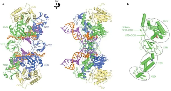
Overall structure of IN and model for target DNA binding
Despite its acute importance for antiretroviral drug discovery and decades of rigorous research, the complete structure of HIV IN, either as a separate protein or in the context of the functional intasome defined as the minimal functional complex involving viral DNA and IN, is lacking, due to poor solubility and interdomain flexibility problems. However, several structures of isolated domains or of two consecutive domains have been reported (Mouscadet et al., 2010a). Recently a crystal structure of full-length IN from the prototype foamy virus (PFV), another lentivirus, in complex with its cognate DNA has recently been reported (Figure 9) (Hare et al., 2010a). In this complex, the retroviral intasome consists of an IN tetramer tightly associated with a pair of viral DNA ends. The inner subunits of the tetramer are responsible for all contacts involved in tetramerization and viral DNA binding. The CCDs of the outer subunits seem to provide supporting function. Within the PFV intasome, the CCD-CTD linker adopts an extended conformation for most of its length, tracking parallel to the NTD-CCD linker from the same subunit. Each CTD makes contact with the phosphodiester backbone of both viral DNA molecules, essentially crosslinking the structure. The overall conformation of the assembled intasome is well constrained.
The active sites of the inner IN subunits, engaged with the 3’ termini of the viral DNA, are located deep within the dimmer/dimer interface. Therefore, the only mode of host chromosomal DNA (target DNA) binding that would not require marked rearrangement of the intasome or severe DNA bending is along the cleft between IN dimers (Figure 10). Note that the CTD, juxtaposed to the target DNA in this model, is known to possess sequence non-specific DNA binding activity (Hare et al., 2010a).