HAL Id: tel-01827378
https://tel.archives-ouvertes.fr/tel-01827378
Submitted on 2 Jul 2018
HAL is a multi-disciplinary open access archive for the deposit and dissemination of sci-entific research documents, whether they are pub-lished or not. The documents may come from teaching and research institutions in France or abroad, or from public or private research centers.
L’archive ouverte pluridisciplinaire HAL, est destinée au dépôt et à la diffusion de documents scientifiques de niveau recherche, publiés ou non, émanant des établissements d’enseignement et de recherche français ou étrangers, des laboratoires publics ou privés.
Structures and functions of the C-Terminal domain of
HIV-1 integration
Oyindamola Oladosu
To cite this version:
Oyindamola Oladosu. Structures and functions of the C-Terminal domain of HIV-1 integration. Im-munology. Université de Strasbourg, 2017. English. �NNT : 2017STRAJ025�. �tel-01827378�
UNIVERSITÉ DE STRASBOURG
ÉCOLE DOCTORALE DES SCIENCES DE LA VIE ET DE LA SANTÉ
[ UMR 7104 ]
THÈSE
présentée par :[ Oyindamola OLADOSU ]
soutenue le : 16 Mai 2017
pour obtenir le grade de : Docteur de l’université de Strasbourg Discipline/ Spécialité : Biophysique et Biologie Structurale
Structures et Fonctions du Domaine C-Terminal de L’Intégrase du VIH-1
THÈSE dirigée par :M. RUFF Marc Directeur de Recherche, université de Strasbourg
RAPPORTEURS :
Mme. ANDREOLA Marie-Line Directeur de Recherche, université de Bordeaux M. BOUAZIZ Serge Directeur de Recherche, université de Paris Descartes
AUTRES MEMBRES DU JURY :
Mme. BERNACCHI Serena Chargé de Recherche, université de Strasbourg M. NEGRONI Matteo Directeur de Recherche, université de Strasbourg M. PARISSI Vincent Directeur de Recherche, université de Bordeaux
For Toyin and Toyin You made me everything I am today I hope I have made you proud
Acknowledgements
I would like to start by thanking God for bringing me this far and making all of this possible. I am nothing without Him.
A big thank you to all of the members of my jury: Dr. Marie-Line Andreola, Dr. Serge Bouaziz, Dr. Serena Bernacchi, Dr. Matteo Negroni and Dr. Vincent Parissi, for taking the time to read and evaluate my work.
I am forever indebted to my thesis supervisor Marc Ruff for allowing me to join his team and giving me the opportunity to work on this project. Marc, you are such a brilliant scientist and it was a huge privilege to learn from you. Thank you for all your support and for being an overall super awesome boss.
I thank the PhD program through the LABEX and SIDACTION for funding my thesis.
I would like to especially thank Raphael Recht and Bruno Kieffer for countless hours spent helping me with NMR and discussing about my project. I would also like to thank Yves Nomine for helping me with CD experiments, and for the helpful discussions about my results. I would like to thank the Molecular Biology platform, especially Paola Rossolillo for producing so many expression vectors used for my projects.
To all the past and present members of the Ruff team: BenOlas (Benoit and Nicolas), Julien (JuJu), Karine and Sylvia (just realized I don’t have nicknames for you guys) and Eduardo. I am grateful for the opportunity to have worked with you over the past 3.5 years. You guys taught me pretty much everything in the lab. Thank you so very much for all the scientific discussions, for always answering my stupid questions, and for always being there to lend a helping hand. You are all super brilliant, and each one of you contributed immensely to my time in the lab. I am also thankful for all your contributions towards the completion of this manuscript.
A special shout out to my friends Chantal (ChanChan), Francesca and Lorraine, for all their support and always being there to hear me rant and listen to my “business ideas” during lunch. I’m especially thankful that you guys help me with everything from taxes to hospital visits to making phone calls. Arielle, thanks for being the president of the “fat ass club”. It was nice having another ‘sista’ at the IGBMC. Thanks to Fabrice and Hubert for all your
help, from talking about my project during lunch, to helping me move apartments, you guys were always there, and I really appreciate it.
Dr. Nicaise Ndembi, thank you for introducing me to HIV research and allowing me to work in your lab at IHVN. That experience helped shape my scientific interests and pushed me to do this PhD.
Sam, I could write a whole book about what an incredible friend you have been to me over the past few years. I can’t believe I was fronting on you at first. I came to Strasbourg for a PhD and I also got a sister in the process. You have been there for me every step of way and I am forever thankful to you.
To my mamas Aunt Deola and Mama Hart, you are both so inspiring. I’m blessed to have you in my life.
To my friends: Ona, Kunle, Wole, Brendan, Bolaji, Abby, Faith, Gloria, thank you for always being a phone call away and for keeping me grounded.
Bimpe and Deoti, the best sisters in the world. You girls inspire me so much and push me to be the best version of myself. Thank you for everything.
Finally, to my husband Valentine, thank you for all your support. None of this would have been possible without you. I’m blessed to be on this journey with you!
Abstract en Français
L’Integrase du VIH est une ADN recombinase catalysant deux réactions qui permettent l'intégration de l'ADN viral dans l'ADN hôte. L’intégrase du VIH comprend 3 domaines : N-terminal impliqué dans la réaction de « 3' processing » et le transfert de brin, le domaine catalytique contenant le site actif et le domaine C-terminal liant l'ADN non-spécifiquement (CTD). Des recherches récentes mettent en évidence l'importance du CTD dans la liaison avec d'autres protéines virales comme la transcriptase inverse.
L'intégration du génome viral dans le génome de l'hôte nécessite un ciblage dans des régions spécifiques de la chromatine. Des facteurs de ciblage tels que BET ou LEDGF ont été découverts comme jouant un rôle dans la sélection des sites d'intégration. Ces facteurs reconnaissent des modifications spécifiques des histones. Cependant, les processus d'association avec le nucléosome et l'intégration dans la chromatine ne sont pas pleinement compris. L’IN-CTD de l’intégrase possède un domaine « SH3 like » avec une structure en tonneau beta. Cette structure est compatible avec la superfamille « Royal Domain » qui reconnaît les histones modifiées. Nos collaborateurs (Vincent Parissi, Bordeaux) ont montré que la queue N-terminale mono-methylé de l’histone H4 (H4K20Me1) interagit spécifiquement avec le CTD.
Une caractéristique du VIH est sa grande variabilité génétique. Cette souplesse, qui permet une diversité antigénique importante est centrale pour l'adaptation virale à la réponse immunitaire et pour échapper aux antiviraux. L'inconvénient de la variation génétique est la perte de la fonctionnalité des protéines nécessaire pour l'infectivité virale. La façon dont l'équilibre est maintenu entre ces exigences divergentes est essentielle pour notre compréhension de l'évolution virale. L'examen de ces questions peut fournir de nouvelles perspectives sur la biologie du VIH et sur l'identification de cibles pour les antirétroviraux. Les chimères inter-groupes entre le groupe M (sous-type A2) et le groupe O ont fourni la preuve qu'un motif N222K240N254K273 dans le groupe M, remplacé par le motif
K222Q240K254Q273 dans le groupe O, est important pour l'intégration (Matteo Negroni, IBMC).
Nous sommes intéressés par les différences structurales entre le CTD de ces différents groupes et sous-types.
Le but de ma thèse était de comprendre les rôles et l'importance du domaine C-terminal de l’intégrase dans deux contextes : l'intégration dans la chromatine et la coévolution, avec l'objectif de comprendre le rôle de la multimerisation dans la fonction de l’intégrase.
La première partie de mon projet a porté sur l'élucidation du rôle du CTD dans les interactions avec les histones en comprenant les bases structurales de l'interaction entre le CTD et H4K20me1. La procédure globale impliquait des études biochimiques, fonctionnelles et structurales.
En utilisant la thermophorèse, j’ai montré que l’IN-CTD se lie préférentiellement au peptide H4K20Me1 avec un Kd de 0,8 µM. Ces résultats ont été confirmés à l'aide de la NOESY-RMN où un changement du signal NOE sur le peptide indiquait la formation d’un complexe CTD/H4K20Me1.
Les spectres HSQC 15N ont montré que les résidus 271-288 étaient désordonnés. En outre, les résultats de RMN ont indiqué que la protéine est plus désordonnée dans 150mM NaCl comparativement à 1M NaCl et moins oligomérisée à pH 8 par rapport à pH 7. En utilisant le CTD marqué isotopiquement, j'ai attribué avec succès 75% des résidus et calculé des structures RMN montrant une topologie semblable aux structures publiées. J'ai observé des interactions dépendant du pH avec H4K20Me1. Lors de la liaison du peptide à pH 8, les spectres HSQC suggèrent un changement de conformation conduisant à une oligomérisation plus importante. En quantifiant les changements de déplacements chimiques observés pendant le titrage peptidique, j'ai identifié les résidus affectés à pH 7 et à pH 8.
En utilisant la cristallographie, j'ai obtenu des structures du CTD à pH 6.5, 7 et 7.5. En utilisant la structure à pH 7 (1.5 Å), j'ai cartographié les résidus dont les déplacements chimiques ont été perturbés par la liaison au peptide et observé qu'ils étaient situés dans la partie N et C terminale. Plusieurs de ces résidus sont hydrophobe suggérant qu'ils forment un patch hydrophobe pour la fixation de H4K20Me1. Nos expériences de docking suggèrent que la lysine méthylée peut interagir avec ce patch.
Sur la base de ces données, je propose que la liaison de l’IN-CTD au peptide se produit via le patch hydrophobe, induisant des changements de conformation (oligomères), et provoquant des changements dans l'exposition au solvant de certains résidus suggérant que la propension à la multimerisation du CTD joue un rôle dans la fonction de l’IN.
Le but de la deuxième partie de mon projet était de résoudre la structure du CTD sauvage et chimères afin de comprendre l'importance du motif N222K240N254K273 pour la fonction de l’IN
et son rôle dans la coévolution. À cette fin, j'ai purifié des constructions CTD 220-270 sauvage ainsi que des chimères inter groupe. J'ai obtenu des cristaux et résolu les structures de la protéine sauvage et des mutants sélectionnés.
J'ai obtenu une structure à 1.7 Å du CTD A2, qui montre trois surfaces de contact possibles pour la dimérisation. La structure du mutant K240Q/N254K (2 Å) était similaire à la structure du sauvage, sauf dans les boucles et les régions flexibles. Les résultats suggèrent que le motif N222K240N254K273 non conservé joue un rôle dans la formation d'oligomères d'ordre supérieur.
En particulier, K240/N254 sont importants pour la dimérisation. Comme l’IN doit oligomériser afin d'être actif, ce motif est important la fonction de l’IN.
Globalement, les résultats de mon projet indiquent que l'IN-CTD joue un rôle important, en contribuant à la formation de multimères d'ordre supérieur importants pour la fonction de l’IN. Le projet souligne l'importance d'une approche de biologie structurale intégrée pour répondre aux questions biologiques. Les résultats obtenus dans ce projet sont importants pour comprendre les relations structure/fonction de l'intégrase pendant le cycle de vie du VIH-1. Les méthodes développées au cours de ce projet peuvent être utilisées pour cribler de nouveaux inhibiteurs de conformation du l’IN du VIH-1.
Abstract in English
HIV Integrase is a DNA recombinase that catalyzes two endonucleolytic reactions that allow the viral DNA integration into host DNA for replication and subsequent viral protein production. HIV Integrase consists of 3 structural and functional domains: The N-terminal zinc domain involved in 3’ processing and strand transfer, the catalytic core domain which contains the active site, and the C-terminal domain that binds DNA non-specifically. Recent research highlights the importance of the CTD in binding with other viral proteins such as Reverse Transcriptase.
The integration of the viral genome into the host’s genome requires targeting into specific regions of the chromatin. Cellular targeting factors such as BET or LEDGF/p75 have been discovered to play a role in the integration site selection because they recognize and bind specific histone modifications. However, the process of nucleosome association and chromatin integration is not yet fully understood. The C-terminal domain of HIV Integrase possess a SH3 like domain made up of approximately 60 amino acids that displays a β-barrel fold. This SH3 fold is consistent with the Royal Domain superfamily that recognize post translationally modified histone tails. Our collaborators (Vincent Parissi, Bordeaux) show that the monomethylated N-terminal tail of H4K20Me1 interacts specifically with the C-terminal Domain of HIV integrase.
A main feature of the human immunodeficiency virus (HIV) is its great variability, reflecting a remarkable genetic flexibility. This flexibility, which allows antigenic variation, is central for viral adaptation to the immune response mounted by the host and to escape antiviral treatments. The drawback of genetic variation can be the loss of proteins and nucleic acids functionality, necessary to ensure viral infectivity. How the balance is kept between these two divergent requirements is central for our understanding of viral evolution. Addressing these issues can provide new insights into HIV biology and on the identification of potential targets for antiviral strategies.
Inter-group chimeric constructs between group M (subtype A2) and group O provided evidence that a non-conserved motif, N222K240N254K273 in group M, replaced by
K222Q240K254Q273 in group O, is important for integration (Matteo Negroni, IBMC). We are
interested in the structural differences between the CTD of these different groups and subtypes.
The aim of my thesis was to understand the roles and importance of the C-terminal domain of HIV-1 Integrase in two contexts: chromatin integration, and co-evolution, with the overall purpose of understanding the role of multimerization in IN function.
The first part of my project focused on elucidating the role of the IN-CTD in histone interactions, by understanding the structural basis of the interaction between IN-CTD and mono-methylated N-terminal tail of histone H4K20 (H4K20me1). The overall procedure involved biochemical, in-vitro functional and structural studies.
Using Microscale Thermophoresis, my results show that the CTD preferentially binds to the fluorescent mono-methylated H4K20 peptide with a Kd of 0.8µM. These results were
confirmed using NOESY-NMR, where a change in NOE signal on the peptide indicated complex formation between the CTD and H4K20Me1.
15N HSQC spectra showed that the residues 271-288 of the CTD were disordered.
Additionally, NMR results indicated that the protein is more disordered in 150mM NaCl, compared to 1M NaCl and less oligomerized at pH 8, compared to pH 7. Using isotopically labeled CTD, I successfully assigned 75% of the protein residues, and calculated NMR structures, whose overall topology was similar to published structures. I also observed pH dependent interactions with H4K20Me1. Upon binding of the peptide at pH 8, the HSQC spectra suggest a change to a more oligomerized protein conformation. By quantifying the chemical shift changes observed during peptide titration, I was able to identify the residues affected by peptide binding at pH 7 and pH 8.
Using X-ray crystallography, I obtained high-resolution structures of the isolated IN-CTD at pH 6.5, 7 and 7.5. Using the crystal structure of the IN-CTD at pH 7 (1.5 Å), I mapped the residues whose chemical shifts were perturbed by peptide binding, and observed that they were mostly located on the N and C terminus of the IN-CTD. Interestingly, several of these residues are hydrophobic in nature, suggesting that they form a hydrophobic patch, which serves as the binding surface for H4K20Me1. Our preliminary docking experiments suggest that the methylated lysine is in close proximity and may interact with this patch.
Based on this data, I propose that binding on IN-CTD to the peptide occurs via the hydrophobic patch, inducing conformational changes (oligomerization), and causing changes
in solvent exposure of some residues. This suggests that the propensity for multimerization of the IN-CTD plays a role in IN function.
The goal of the second part of my project was to solve the structure of IN-CTD from WT and chimeric constructs in order to understand the importance of the motif N222K240N254K273 for
IN function and its role in co-evolution. To this end, I purified HIS-IN-CTD 220-270 from WT and inter-group chimeric constructs. I obtained crystals and solved the structure of selected WT and mutant proteins.
I obtained a high-resolution structure (1.7Å) of the Group A2 WT IN-CTD, which presented three possible surface contacts for dimerization. The structure of the K240Q/N254K mutant (2Å) was similar in structure to the WT, except in loops and flexible regions. Results suggest that the non-conserved N222K240N254K273 motif plays a role in the formation of higher order
oligomers in the IN-CTD. In particular, K240/N254 are important for dimerization. As IN needs to be oligomerized in order to be active, this motif is important for IN function
Overall, results from my project indicate that the IN-CTD plays an important role, by contributing to the formation of higher order multimers that are important for IN functionality. Project highlights the importance of an integrated structural biology approach to answering biological questions. Insights gained from this project are important for understanding structure/function relationships of Integrase during the HIV-1 life cycle. The methods developed (NMR and X-ray) during this project can be used to screen for new conformational inhibitors for HIV-IN.
Table of Contents
TABLE OF ABBREVIATIONS ... 1 TABLE OF FIGURES ... 4 CHAPTER 1 - INTRODUCTION ... 6Origin and Epidemiology of HIV ... 6
HIV Phylogeny ... 8
HIV Structure ... 10
HIV Genome Description ... 10
HIV life cycle ... 12
Binding ... 12
Fusion ... 12
Reverse Transcription ... 12
Integration ... 12
Transcription and Translation ... 13
Assembly and Budding ... 14
Maturation ... 14
HIV Inhibitors ... 15
HIV Integrase ... 17
Functions of HIV Integrase ... 17
HIV Integrase domains ... 19
The role of LEDGF in HIV Integration ... 23
Important factors for Integration Site Selectivity ... 25
Proposed role of HIV-1 Integrase CTD in Chromatin Association ... 27
Genome diversity in HIV ... 28
HIV-1 Co-Evolution ... 30
Proposed Importance of CTD in Co-Evolution ... 30
Project Objectives ... 30
CHAPTER 2 – HISTONE ASSOCIATION STUDIES ... 32
MATERIALS AND METHODS ... 32
DNA cloning ... 32
Protein Production ... 38
Peptide Production and Quantification ... 44
Biochemical Studies ... 45
Structural Studies ... 48
RESULTS ... 60
Purification Results ... 60
Biochemical Results ... 65
Preparation of Isotopically labeled protein (Gateway construct) ... 69
Results from NMR experiments ... 73
NMR structure ... 100
Results from Crystallization experiments ... 101
DISCUSSION ... 114
Protein Production and Purification ... 114
Biochemical Studies ... 114
Structures of protein only ... 115
Interactions with peptide (15N HSQC data) ... 115
CHAPTER 3 – CO-EVOLUTION STUDIES ... 118
MATERIALS AND METHODS ... 118
DNA cloning ... 118 Protein Production ... 118 Crystallization ... 119 RESULTS ... 120 Purification Results ... 120 Crystallization ... 124 Discussions ... 133 Appendix ... 137 Résumé en Français ... 140 Bibliography ... 145
1
TABLE OF ABBREVIATIONS
AAIDS Acquired Immune Deficiency Syndrome B
βME β-mercaptoethanol
BET Bromodomain and Exter terminal domain C
CA Capsid
CCD Catayltic Core domain
CCR5 Chemokine (C-C motif) receptor 5 CDC Center for Diseases Control CDK9 Cyclin dependent kinase 9 CD4 Cluster of Differentiation 4 CTD C-terminal Domain
CRFs Circulating Recombinant Forms CRM1 Chromosome region maintenance 1 CXCR4 Chemokine (C-X-C motif) receptor 4 D
D2O Deuteriom Oxide
dsDNA double stranded Deoxyribonucleic Acid dNTPs deoxynucleotides
E
Env envelope
F
FDA Food and Drug Administration FIV Feline Immunodeficiency Virus G
Gag group specific antigen GST Glutathione S- Transferase H
H4K20Me1 monomethylated H4K20 peptide HAART Highly Active Anti-Retroviral Therapy HIV Human Immunodeficiency Virus I
IBD Integrase binding domain
IN Integrase
IPTG IsoPropyl β-D-1-ThioGalactopyranoside K
2
Kd Dissociation Constant L
LEDGF Lens Epithelium Derived Growth Factor M
mRNA messenger RNA
MA Matrix
MHz megahertz
MLV Murine Leukemia Virus MST Microscae thermophoresis MVV Maedi-Visna Virus
MWCO Molecular Weight Cut Off N
Nef Negative Regulatory Factor
NC Nucleocapsid
NMR Nuclear Magnetic Resonance
NNRTIs Non-nucleoside reverse transcriptase inhibitors NRTIs Nucleoside reverse transcriptase inhibitors NOEs Nuclear Overhauser Effects
P
P24 protein 24
P3C Protease 3C precision PCR Polymerase Chain Reaction PIC Pre-integration complex PFV Prototype foamy virus
Pol Polymerase
Ppm parts per million
PR- Protease
R
RNAse1 Ribonuclease 1
RNA Ribonucleic Acid
RT Reverse Transcriptase
RTC Reverse transcriptase complex S
SH3 Src Homology 3
SIV Simian Immunodeficiency Virus STC Strand transfer complex
SDS-PAGE Sodium Dodecyl Sulfate Polyacrylamide (SDS-PAGE) Electrophoresis T
TAR transactivation response protein Tat transactivator of transcription TCC target capture complex
TE Tris-EDTA
3
U
U3 Unique sequence element at the 3’end of the viral RNA U5 Unique sequence element at the 5’end of the viral RNA URFs Unique Recombinant forms
V
Vif Viral infectivity factor Vpr Viral Protein R
Vpu Viral Protein U W
4
TABLE OF FIGURES
FIGURE 1:WORLD MAP SHOWING HIV PREVALENCE BY REGION. ... 6
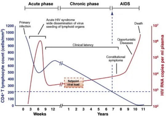
FIGURE 2:TYPICAL COURSE OF HIV PROGRESSION. ... 7
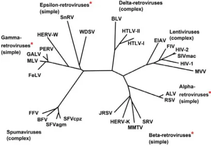
FIGURE 3:MEMBERS OF THE RETROVIRIDAE FAMILY ... 8
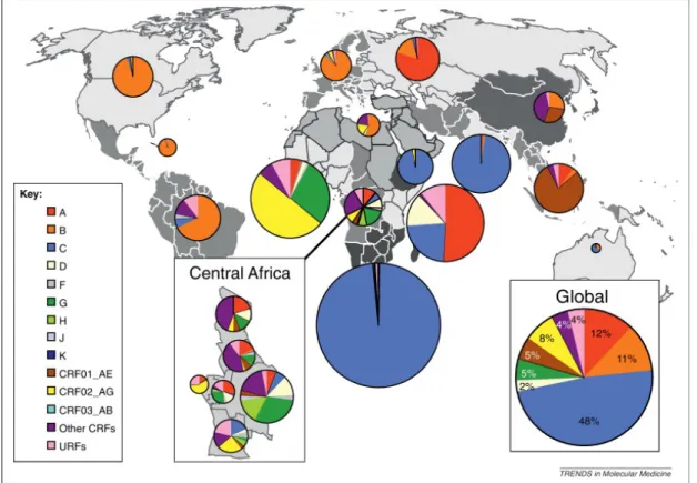
FIGURE 4:WORLD MAP SHOWING GLOBAL DISTRIBUTION OF HIV GROUPS AND SUBTYPES. ... 9
FIGURE 5:CROSS SECTION OF MATURE HIV-1 VIRION ... 11
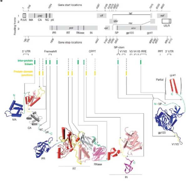
FIGURE 6:ORGANIZATION OF HIV GENOME. ... 11
FIGURE 7:THE LIFE CYCLE OF HIV-1 ... 14
FIGURE 8:TIMELINE OF HIV-1 INHIBITORS APPROVED BY THE FDA ... 16
FIGURE 9:DOMAIN ORGANIZATION OF HIV-1INTEGRASE ... 17
FIGURE 10:SCHEMATIC REPRESENTATION OF THE 3’ PROCESSING AND STRAND TRANSFER ACTIVITIES ... 19
FIGURE 11:SUPERPOSITION HIGHLIGHTING IN FLEXIBILITY ... 21
FIGURE 12:STRUCTURE OF THE STC INTASOME ... 23
FIGURE 13:MODEL FOR IN/LEDGF INTERACTION IN DNAINTEGRATION ... 25
FIGURE 14:MODEL OF HIV-1INTEGRATION AT THE NUCLEAR PORE COMPLEX ... 26
FIGURE 15:MEMBERS OF THE ROYAL DOMAIN SUPERFAMILY ... 28
FIGURE 16:GATEWAY CLONING STRATEGY ... 33
FIGURE 17:THEORY OF AFFINITY CHROMATOGRAPHY ... 42
FIGURE 18:THEORY OF SIZE EXCLUSION CHROMATOGRAPHY ... 43
FIGURE 19:1D SPECTRUM OF THE TRP/H4K20ME1 PEPTIDE MIXTURE ... 44
FIGURE 20:SCHEMATIC REPRESENTATION OF MST ... 46
FIGURE 21:1D SPECTRUM OF WELL-FOLDED HEN EGG WHITE LYSOZYME ... 49
FIGURE 22:TRANSFER OF MAGNETIZATION DURING 15NHSQC ... 51
FIGURE 23:RED ARROW INDICATE TRANSFER OF MAGNETIZATION FOR 13CHSQC ... 51
FIGURE 24:TRANSFER OF MAGNETIZATION FOR HNCA. ... 52
FIGURE 25:TRANSFER OF MAGNETIZATION FOR CBCACONH. ... 53
FIGURE 26:TRANSFER OF MAGNETIZATION FOR HNCO. ... 53
FIGURE 27:TRANSFER OF MAGNETIZATION FOR HNCACB. ... 54
FIGURE 28:TRANSFER OF MAGNETIZATION FOR 15NNOES. ... 55
FIGURE 29:VAPOR DIFFUSION CRYSTALLIZATION PHASE DIAGRAM ... 57
FIGURE 30: VAPOR DIFFUSION METHODS. ... 58
FIGURE 31: 2L PURIFICATION OF GST-IN-CTD ... 61
FIGURE 32:6LPURIFICATION OF HIS-IN-CTD(GATEWAY CONSTRUCT) ... 62
FIGURE 33:2LPURIFICATION OF HIS-IN-CTD220-270 AT PH7 ... 63
FIGURE 34:2LPURIFICATION OF HIS-IN-CTD220-270 AT PH8. ... 64
FIGURE 35:2LPURIFICATION OF DOUBLE LABELLED HIS-IN-CTD220-270 AT PH8. ... 65
FIGURE 36:DATA FROM MST EXPERIMENTS. ... 66
FIGURE 37:DATA FROM TRNOESY EXPERIMENTS. ... 67
FIGURE 38:1D SLICES EXTRACTED AT 0.8 PPM FROM SEVERAL TRNOESY EXPERIMENTS: ... 68
FIGURE 39:DATA FROM WATERLOGSY SPECTRA ... 69
FIGURE 40:SUPERPOSITION OF 1D SPECTRA ... 70
FIGURE 41:SUPERPOSITION OF 1D SPECTRA OBTAINED FOR ALL SAMPLES AT 298K. ... 71
FIGURE 42:SUPERPOSITION OF 1D SPECTRA OBTAINED FOR ALL SAMPLES AT 280K. ... 72
FIGURE 43:SUPERPOSITION OF 15NHSQC PERFORMED AT VARIOUS TEMPERATURES ... 73
FIGURE 44: 15NHSQC OF HIS-INCTD220-288 IN 25MMHEPES PH7,2MM ΒME,1MNACL. ... 74
FIGURE 45:15NHSQC OF HIS-IN-CTD220-288 IN 25MMHEPES PH7,2MM ΒME,500MMNACL. ... 75
FIGURE 46:PEPTIDE INTERACTIONS WITH OLD CONSTRUCT. ... 76
FIGURE 47:HSQC SPECTRA OF NEW CONSTRUCTS. ... 77
FIGURE 48:HSQC SPECTRA COMPARING PET15B AND GATEWAY CONSTRUCTS ... 77
FIGURE 49:PROTEIN STABILTY AT VARYING TEMPERATURES. ... 78
FIGURE 50:PEPTIDE INTERACTIONS IN 1MNACL ... 79
FIGURE 51:15NHSQC OF HIS-IN-CTD220-270 ... 80
FIGURE 52:INTERACTION WITH 1MM PEPTIDE AT PH7 IN 1MNACL: ... 80
FIGURE 53:INTERACTION WITH 2MM PEPTIDE AT PH7 IN 1MNACL. ... 81
FIGURE 54:HIS-CTD-220-270 IN 150MMNACL AT PH7 ... 82
FIGURE 55:COMPARISON OF HIS-CTD-220-270 IN DIFFERENT SALT CONCENTRATION. ... 83
FIGURE 56:INTERACTION WITH 1MM PEPTIDE AT PH7 IN 150MMNACL ... 84
5
FIGURE 58:HIS-IN-CTD220-270 AT PH8 ... 85
FIGURE 59:COMPARISON OF SPECTRA AT PH7 AND PH8. ... 86
FIGURE 60:INTERACTION WITH 1MM PEPTIDE AT PH8 IN 150MMNACL ... 87
FIGURE 61:INTERACTION WITH 2MM PEPTIDE AT PH8 IN 150MMNACL ... 88
FIGURE 62:COMPARISON OF COMPLEX AT PH8 AND PROTEIN ONLY AT PH7. ... 88
FIGURE 63:CONTROL EXPERIMENTS WITH WATER. ... 89
FIGURE 64:EFFECTS OF GLYCINE ON SPECTRA QUALITY. ... 90
FIGURE 65:PROTEIN ASSIGNMENT. ... 91
FIGURE 66:PEAK VOLUMES OF ASSIGNED RESIDUES. ... 92
FIGURE 67:DIHEDRAL ANGLE PREDICTION. ... 93
FIGURE 68:SECONDARY STRUCTURE CHART. ... 93
FIGURE 69:ASSIGNMENT RESULTS AT PH8. ... 94
FIGURE 70:RESIDUES AFFECTED BY 1MM PEPTIDE AT PH8. ... 95
FIGURE 71:RESIDUES AFFECTED BY 2MM PEPTIDE AT PH8. ... 95
FIGURE 72:ASSIGNMENT RESULTS AT PH7. ... 96
FIGURE 73:RESIDUES AFFECTED BY 1MM PEPTIDE AT PH7. ... 97
FIGURE 74:GRAPHICAL REPRESENTATION OF CHEMICAL SHIFT DIFFERENCES AT PH8 ... 98
FIGURE 75:GRAPHICAL REPRESENTATION OF CHEMICAL SHIFT DIFFERENCES AT PH7 ... 98
FIGURE 76:SUPERPOSITION OF CD SPECTRA. ... 99
FIGURE 77:NMR STRUCTURES OF HIS-IN-CTD220-270. ... 100
FIGURE 78:INITIAL CRYSTALLIZATION HITS FOR GST-IN-CTD FROM THE ROBOT. ... 101
FIGURE 79:GST-IN-CTD CRYSTALLIZATION ATTEMPTS. ... 101
FIGURE 80:CRYSTALS OBTAINED AFTER OPTIMIZATION AT DIFFERENT PH ... 102
FIGURE 81:HIS-IN-CTD220-270 MONOMER. ... 104
FIGURE 82:HEXAHISTIDINE TAG BOUND TO NI2+ ION FORM CRYSTAL PACKING CONTACTS. ... 105
FIGURE 83:IN-CTDINTERFACE 1. ... 105
FIGURE 84:IN-CTDINTERFACE 2. ... 106
FIGURE 85:IN-CTDINTERFACE 3 ... 106
FIGURE 86:COMPARISON OF STRUCTURES AT DIFFERENT PH. ... 107
FIGURE 87:CRYSTALS OBTAINED AT EACH PH WITH PEPTIDE AT 1:10 RATIO ... 109
FIGURE 88:CRYSTALS OBTAINED AT 24°C AND 27°C WITH PEPTIDE AND DNA ... 109
FIGURE 89:X-RAY DIFFRACTION PATTERN FOR CRYSTALS WITH DNA ... 110
FIGURE 90:RESIDUES THAT DISAPPEAR IN THE PRESENCE OF PEPTIDE. ... 111
FIGURE 91:RESIDUES SHIFTING IN THE PRESENCE OF PEPTIDE. ... 112
FIGURE 92:FIRST ROUND OF DOCKING RUN. ... 112
FIGURE 93:DOCKING MODEL WITH BEST-WEIGHTED SCORE FROM DOCKRUN. ... 113
FIGURE 94:1LPURIFICATION OF A2HIS-IN-CTD ... 120
FIGURE 95:1LPURIFICATION OF N222K/K240QHIS-IN-CTD. ... 121
FIGURE 96:1LPURIFICATION OF K240Q/N254KHIS-IN-CTD. ... 122
FIGURE 97:1LPURIFICATION OF OHIS-IN-CTD. ... 123
FIGURE 98:CRYSTALS OF A2CTD OBTAINED AT 20°C ... 124
FIGURE 99:CARTOON REPRESENTATION OF A2IN-CTD MONOMER ... 126
FIGURE 100:A2IN-CTDINTERFACE 1 ... 127
FIGURE 101:A2IN-CTDINTERFACE 2 ... 127
FIGURE 102:A2IN-CTDINTERFACE 3. ... 128
FIGURE 103:CRYSTALS OF K240Q/N254KCTD OBTAINED AT 20°C ... 129
FIGURE 104:K240Q/N254KINTERFACE 1. ... 131
FIGURE 105:K240Q/N254KINTERFACE 2 ... 132
6
CHAPTER 1 - INTRODUCTION
Origin and Epidemiology of HIV
Human Immunodeficiency Virus (HIV) is the causative agent of AIDS (Acquired Immune Deficiency Syndrome). According to the WHO, by the end of 2015, there were 36.7 million people living with HIV, with the highest population of 25.5 million in Africa. Recent advancements in HIV testing and treatments have led to an overall decline in new infections with 18.2 million people living with HIV on anti-retroviral therapy, and 1.1 million AIDS-related deaths in 2015 (W.H.O 2016). However, with 2.1 million new infections in 2015 (approximately 5700 new infections daily), the lack of a preventive vaccine or cure, and high HIV drug resistance rates, HIV remains a global public health threat.
Figure 1: World map showing HIV prevalence by region.
AIDS was recognized as a disease in 1981 (Sharp and Hahn 2011), and in 1983 (Barre-Sinoussi, Chermann et al. 1983), HIV-1 was determined to be the cause of AIDS. HIV can be transmitted through three transmission routes (Shaw and Hunter 2012): mucosal transmission through body fluids such as sperm and vaginal fluid during unprotected sex, parenteral transmission through blood transfusion and needle sharing, and mother to child transmission
7
in utero, during childbirth or breastfeeding. With 80% of adults acquiring HIV through mucosal surfaces, it is considered to be a sexually transmitted virus (Cohen, Shaw et al. 2011).
HIV infection occurs in 3 stages if untreated (Coffin and Swanstrom 2013, AIDS.gov 2015, AIDSinfo 2016). The acute infection stage develops within 2-4 weeks of infection, with some people showing flu-like symptoms such as fever, headache, sore throat and body rash. HIV replicates and spreads rapidly in this stage, depleting CD4 levels, and compromising the immune system. The process of seroconversion occurs, where the body develops antibodies against HIV, which are detectable for HIV test. In the chronic infection stage or the asymptomatic stage, the virus goes into latency and continues to multiply at low levels, without showing symptoms in some people. It can take up to 10 years before the disease progresses to the final stage of symptomatic infection. Symptoms at this stage include chronic diarrhea, persistent cough and weight loss. By this stage, the immune system is completely weakened, with a CD4 count of less than 200 cells/m3, making the patient prone to opportunistic infections such as tuberculosis, malaria and cancers. Without treatment, the lifespan of people living with AIDS is about 3 years. Three types of tests can detect HIV infection: antibody tests that detect antibodies against HIV in the blood, usually with test kits, a combination test that detects viral p24 antigens and antibodies, and a nucleic acid test that detects viral load levels in the blood (CDC 2016).
8
There is currently no cure for HIV, and the best treatment is prevention. For people living with HIV, HAART (Highly Active Antiretroviral Treatment), a combination therapy using multiple classes of antiretroviral regimens that target different steps of the HIV life cycle is used to treat HIV. HAART therapy offers several benefits including viral load reduction, disease progression delay and opportunistic infection prevention. Additionally, HAART reduces side effects by reducing drug dosage, and decreases the emergence of drug resistant viruses by reducing the ability of the virus to adapt and mutate. There are currently six classes of antiretrovirals on the market.
HIV Phylogeny
HIV emerged in the late 19th/early 20th century from non-human primates through cross-species zoonosis of Simian Immunodeficiency Virus (Hahn, Shaw et al. 2000, Sharp and Hahn 2011). HIV belongs to the Lentivirus genus in the Retroviridae family. Lentiviruses are characterized by long incubation periods, and are typically enveloped viruses. Retroviruses contain a positive single stranded RNA genome, and contain the reverse transcriptase protein, which allow them to transcribe their single stranded RNA into double stranded DNA once inside the host cell. Other lentiviruses include SIV, Feline Immunodeficiency Virus (FIV), and Maedi-Visna Virus (MVV) (Clapham and McKnight 2002). HIV is also an enveloped virus, budding off from the host cell enveloped by fragment of the host cell membrane.
9
There are two classes of HIV: HIV-1 and HIV-2. HIV-1 is classified into 4 groups – M, N, O, and P, while HIV-2 is classified into groups A-H. Group M is the most widely spread group, responsible for approximately 33 million infections worldwide (Hemelaar 2012). Group M is further sub-divided into subtypes A, B, C, D, F, G, H, J, K. There are also circulating recombinant forms (CRFs) and unique recombinant forms (URFs) of HIV-1. Subtype B is most common subtype in Group M. Group O infections are found predominantly in Western-Central Africa. HIV Group M and N originated from SIVcpz in the chimpanzee Pan troglodytes troglodytes in West–Central Africa (Gao, Bailes et al. 1999). HIV Group O and P originated from SIVgor in Western lowland gorillas in Cameroon (Van Heuverswyn, Li et al. 2006, Plantier, Leoz et al. 2009). HIV-2 originated from SIVsm in sooty mangabey monkeys (Cercocebus atys) in West Africa (Wertheim and Worobey 2009).
Figure 4: World map showing global distribution of HIV groups and subtypes. J. Hemelaar, Trends in Molecular Medicine, 2012
10
HIV Structure
The HIV virion is about 120nm in diameter with a spherical shape. The lipid bilayer, derived from the host cell, contains host membrane proteins including antigens, major histocompatibility complex, and ubiquitin (Arthur, Bess et al. 1992). On the surface, the envelope consists of trimers of glycoprotein gp120, attached to gp41 via the transmembrane protein(Ganser-Pornillos, Yeager et al. 2008). On the inside, the matrix (MA) shell lines the inner membrane and maintains the structural integrity of the virus. There is a fullerene cone capsid (CA) in the center, and a nucleocapsid (NC) that contains the viral genome - two single unspliced positive RNA strands of approximately 9.2kb in size, tightly bound to viral proteins essential for the life cycle such as reverse transcriptase (RT), integrase (IN). Lysine tRNA is bound to the viral RNA, and acts as a reverse transcription primer. Protease (PR) is also found within the nucleocapsid. Some accessory proteins like Nef, Vif and Vpr are packaged in the virus, while other accessory proteins like Rev, Tat and Vpu are not packaged in the virus.
HIV Genome Description
The HIV genome contains nine genes (Frankel and Young 1998, Watts, Dang et al. 2009). Three main genes: Gag, Pol and Env encode for structural proteins, viral enzymes and envelope proteins respectively. Gag encodes for core structural proteins including matrix, capsid and nucleocapsid. Pol encodes for viral enzymes including reverse transcriptase, integrase and protease. Env encodes for envelope protein gp160, which is spliced to gp120 and gp41. Tat and Rev encode for regulatory proteins involved in transcription activation and RNA splicing and export. Vif, Vpr, Vpu and Nef encode for accessory proteins involved in the synthesis regulation, viral RNA processing and other functions.
11
Figure 5: Cross section of Mature HIV-1 virion Structural proteins are shown in blue, viral enzymes in
magenta, accessory proteins in green and viral RNA in yellow. Host proteins and tRNA are shown in purple. David S. Goodsell, http://hive.scripps.edu/resources.html
12
HIV life cycle
The life cycle of HIV can be broken down into 7 main steps namely binding, fusion, reverse transcription, integration, transcription and translation, assembly and budding. Each of these steps is described briefly below.
Binding
HIV-1 primarily targets T-helper lymphocytes, monocytes, macrophages and dendritic cells that contain the CD4+ receptor and a co-receptor such as CCR5 and CXCR4 (Clapham and McKnight 2002, Liu, Bartesaghi et al. 2008). The spikes on the HIV envelope contain trimers of glycoprotein gp120 non-covalently bound to gp41 (Rizzuto, Wyatt et al. 1998).
Fusion
Upon binding to CD4 cells, a conformational change is induced in gp120, exposing a binding site for the co-receptor. Binding to the co-receptor induces further structural changes in gp120, exposing the hydrophobic fusion peptide on gp41 and causing insertion of the fusion peptide into the cell membrane and subsequent fusion of the viral and host cell membranes (Kwong, Wyatt et al. 1998, Moscoso, Sun et al. 2011, Munro, Gorman et al. 2014).
Reverse Transcription
After viral entry, the capsid shell is partially dissolved, allowing for the formation of the reverse transcriptase complex (RTC) (Fassati and Goff 2001, Forshey, von Schwedler et al. 2002, Peng, Muranyi et al. 2014). This complex consists of viral proteins including reverse transcriptase, capsid, integrase as well as host proteins. During reverse transcription, reverse transcriptase catalyzes the formation of double stranded DNA using the single strand RNA of the virus as a template. This double stranded DNA is rich in uracil to prevent auto-integration (Yan, O'Day et al. 2011). Viral proteins such as nucleocapsid have been shown to improve reverse transcription by improving the binding of primer tRNA Lys 3 to viral RNA (Barat, Lullien et al. 1989). Furthermore, Vif has also been shown to contribute to reverse transcription by increasing the polymerization rate (Cancio, Spadari et al. 2004).
Integration
Following the synthesis of double stranded DNA; the RTC is transformed into the Pre-Integration complex (PIC). The HIV-1 PIC consists of cellular proteins such as LEDGF (Raghavendra, Shkriabai et al. 2010), viral proteins such as nucleocapsid, matrix, Vpr and
13
reverse transcriptase, and integrase, the core viral protein in the PIC (Matreyek and Engelman 2013). More recently, capsid has also been shown to be in the PIC (Hulme, Kelley et al. 2015). Integrase is responsible for catalyzing the two main interactions (3’ processing and strand transfer) that allow for DNA integration. While in the cytoplasm, integrase catalyzes a 3’ processing reaction (Miller, Farnet et al. 1997). The PIC travels along microtubules to reach the nuclear envelope, and crosses the nuclear pore complex to reach the genome of non-dividing cells. Vpr promotes nuclear localization of viral DNA during nuclear import (Heinzinger, Bukinsky et al. 1994). Following the active transport, integrase catalyzes the strand transfer reaction, allowing for integration of the viral DNA into the host DNA. Host machinery repairs any gaps, and the viral DNA is replicated along with host DNA (Craigie and Bushman 2012). Post integration, the virus can go into latency, establishing reservoirs that make HIV incurable so far (Dahabieh, Battivelli et al. 2015).
Transcription and Translation
Transcription can occur pre- and post- integration. Pre-integration transcription involves the production of viral regulatory proteins such as Tat, Rev and Nef that interact with cellular proteins to regulate viral transcription and translation (Sloan and Wainberg 2011). Tat initiates post integration transcription from the U3 promoter in the upstream LTR by binding to the viral transactivation response protein (TAR) (Bieniasz, Grdina et al. 1998). This leads to the recruitment the cyclin dependent kinase (CDK9) and cyclin T1 to the TAR. CyclinT1 binds to Tat, increasing its affinity and specificity for RNA. CDK9 mediated phosphorylation of the C-terminal domain of RNA polymerase II stimulates transcription elongation (Fujinaga, Cujec et al. 1998, Isel and Karn 1999, Zhou and Rana 2002).
Newly synthesized unspliced or partially spliced viral mRNAs are exported via Rev or Gag mediated pathways. In the Rev mediated pathway, Rev binds to Rev Response Element (RRE) and recruits cellular nuclear export factors such as chromosome region maintenance 1 (CRM1) and RanGTP to export mRNAs to the cytoplasm through the nuclear pore for the synthesis of Gag and Gag-Pol precursors (Jain and Belasco 2001, Rausch and Grice 2015). Env precursor (gp160) is synthesized on the rough endoplasmic reticulum (Checkley, Luttge et al. 2011). In the Gag mediated pathway, interaction between the nuclear export signal of matrixGag and CRM1 results in mRNA export (Parent 2011). Rev, Gag, CRM1 and RanGTP are re-imported into the nucleus.
14
Assembly and Budding
Nascent virions are assembled at the plasma membrane, containing viral RNA, Gag and Gag-Pol, viral proteins such as Vif, Nef, Vpr, and host proteins (Freed 2015) . Following synthesis, Env (gp120 and gp41) trimers are exported to the plasma membrane by secretory pathway. CD4 proteins are also synthesized in the endoplasmic reticulum. Vpu interacts with CD4 to promote Env packaging by inducing CD4 degradation (Le Noury, Mosebi et al. 2015). N-terminal myristoylation of Gag contributes to the membrane association (Morikawa, Hockley et al. 2000). Interactions between proline-rich motifs in Gag and cellular class E vacuolar protein sorting (VPS) proteins cause the virion to be pinched off the plasma membrane (Ren and Hurley 2011). Tetherin, a cellular protein that restricts viral budding, is inhibited by Vpu (McNatt, Zang et al. 2013).
Maturation
About 2400 copies of Gag bud to form an immature particle with 2 copies of unspliced viral genome (Carlson, Briggs et al. 2008). During maturation, non-infectious immature virions are converted to infectious virions by the proteolysis of precursor proteins by protease (Pettit, Moody et al. 1994). Gag and Gag-Pol precursor are processed to produce structural proteins (Matrix, Capsid, Nucleocapsid), and enzymes (Protease, Integrase and Reverse Transcriptase) (Pettit, Moody et al. 1994, Briggs, Riches et al. 2009).
Figure 7: The life cycle of HIV-1. Inserts show pictorial representations of the virus at each step. Adapted from
Engelman and Cherapanov, Nature Reviews, Microbiology, 2012, and David S. Goodsell http://hive.scripps.edu/resources.html
15
HIV Inhibitors
There are currently six classes of inhibitors approved by the Food and Drug Administration (FDA) for the treatment of HIV infections (Cihlar and Fordyce 2016, F.D.A 2016). Due to the high rates of drug resistance of HIV to inhibitors, it is important that new inhibitors are designed frequently. There is an emerging class of HIV inhibitors known as maturation inhibitors that inhibit the protease mediated Gag cleavage process (Wang, Lu et al. 2015). HAART (Highly Active Anti Retroviral Therapy), a combination of 3 or more different classes of antiretrovirals is used to improve efficacy and reduce drug resistance rates.
Entry Inhibitors: Maraviroc (Pfizer) is a CCR5 antagonist approved for HIV treatment by the FDA in 2007. It functions by binding to CCR5, preventing the binding of gp120 to the co-receptor (Henrich and Kuritzkes 2013).
Fusion Inhibitors: Enfuvirtide (Roche) was approved by the FDA in 2003 and was the first fusion inhibitor on the market. It works as a peptide mimetic, locking a conformation of gp41, and preventing the structural changes that allow for membrane fusion between host and viral cells (Greenberg and Cammack 2004).
Reverse Transcriptase Inhibitors: RT inhibitors were the first class of inhibitors to be approved by the FDA. They are divided into two sub-classes: nucleoside reverse transcriptase inhibitors (NRTIs) and non-nucleoside reverse transcriptase inhibitors (NNRTIs). They both function by inhibiting the reverse transcription process. NRTIs are nucleoside analogs that compete with dNTPs and inhibit reverse transcriptase when incorporated in the nascent viral DNA chain. NNRTIS bind to reverse transcriptase and act as allosteric inhibitors, preventing conformational changes necessary for activity (Sluis-Cremer, Wainberg et al. 2015). Azidothymidine (AZT), a NNRTI, was the first antiviral drug approved for HIV treatment in 1987 (Fischl, Richman et al. 1987). Since then, there have been 13 NRTIs approved by the FDA. There are currently 6 FDA approved NNRTIs on the market
Integrase Inhibitors: Raltegravir (Merck) was the first FDA approved integrase inhibitor on the market in 2007 (Hicks and Gulick 2009). Dolutegravir (GlaxoSmithkline) and Elvitegravir (Gilead) were approved in 2013 and 2014 respectively. Mg2+ ions are necessary for catalytic activity of integrase. These inhibitors prevent the strand transfer process by acting as competitive inhibitors, interfering with Mg2+ binding, and preventing the integration of viral DNA into host DNA. Other types of integrase inhibitors, which act as allosteric
16
inhibitors, binding to the IN/LEDGF complex, and stabilizing IN dimers, are currently under development (Engelman, Kessl et al. 2013, Le Rouzic, Bonnard et al. 2013).
Protease Inhibitors: There are currently 9 protease inhibitors approved by the FDA on the market. Protease inhibitors prevent the formation of mature infectious viruses by binding to the active site of protease and inhibiting the cleavage of HIV protein pre-cursors.(Eron 2000)
The FDA approved Saquinavir (Roche), the first protease inhibitor in 1995.
Combination Inhibitors: Recent developments in HAART include the usage of a fixed dose
combination of inhibitors from the same class or different classes in one pill, often taken once a day. The first of these inhibitors on the market was Atripla (Bristol-Meyers Squibb, Gilead), a combination of NRTIs and NNRTIs, approved for use in 2006. There are currently six approved combination inhibitors on the market. These inhibitors improve adherence and lower hospitalization risk in patients (Sax, Meyers et al. 2012).
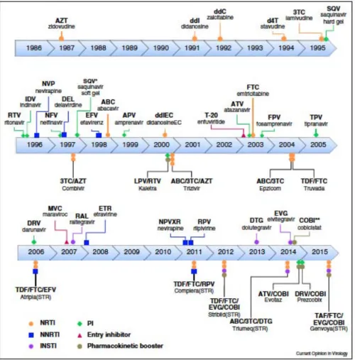
Figure 8: Timeline of HIV-1 inhibitors approved by the FDA. Cihlar and Fordyce, Current Opinions in
Virology, 2016
17
HIV Integrase
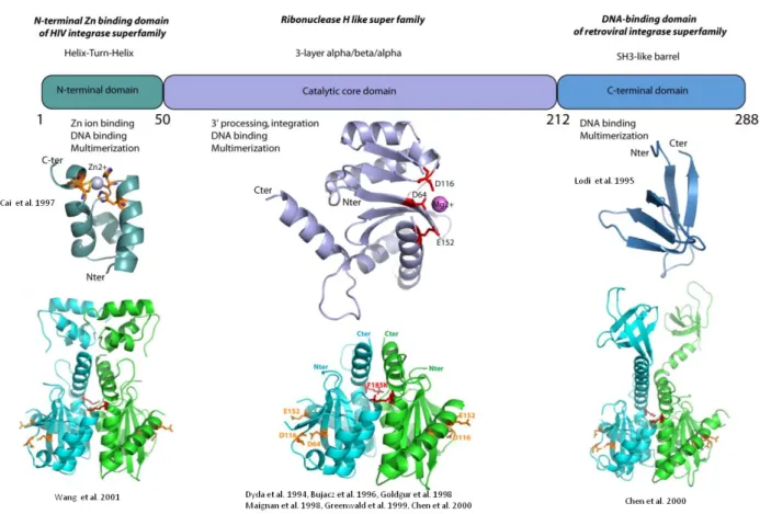
Integrase is a 288-amino acid (32kDa) protein that is encoded by the end of the pol gene. It is produced as part of the Gag-Pol polyprotein, after which it is cleaved by protease mediated cleavage. It consists of 3 structural and functional domains: the N-terminal zinc finger domain (1-49), which is responsible for protein multimerization, the Catalytic core domain (50-212), which contains the active site of the protein, and the C-terminal domain (213-288) that binds DNA non-specifically.
Figure 9: Domain Organization of HIV-1 Integrase showing structures of individual domains
Functions of HIV Integrase
Two reactions are necessary for the covalent integration of viral DNA into host DNA. The full-length integrase is essential for catalyzing the 3’ processing and strand transfer reactions (Bushman, Fujiwara et al. 1990). No energy co-factor is required for either reaction to occur. In addition to its primary roles in integration, integrase may play a role in other steps of the replication cycle, as mutations in integrase affect other steps beyond integration. For example, viruses with mutated integrase display lower levels of reverse transcription and viral particles
18
with abnormal morphology (Engelman, Englund et al. 1995). Moreover, the use of IN/LEDGF allosteric inhibitors suggests a role for integrase in viral maturation (Le Rouzic, Bonnard et al. 2013)
3’ processing
The pre-integrated viral DNA exists as a linear double stranded DNA fragment. In the cytoplasm, integrase binds to a long terminal repeat sequence on the 5’ and 3’ end, and catalyzes an endonucleolytic cleavage, removing a GT di-nucleotide corresponding to the U5 and U3 end of the viral DNA (Engelman, Mizuuchi et al. 1991). The reaction is a trans-esterification where the phosphodiester bond on the viral DNA is broken by nucleophilic attack. This reaction exposes 3’-OH groups, forming a target capture complex (TCC) that will be covalently joined to target DNA in the strand transfer reaction (Lesbats, Engelman et al. 2016). Divalent cations such as Mn2+ and Mg2+ are essential for both 3’ processing and strand transfer reactions, with Mg2+ being the preferred cation due to better cleavage specificity than Mn2+, with water as the nucleophilic agent (Engelman, Englund et al. 1995).
Strand transfer
Sequentially, the cleaved DNA is inserted into the target DNA during the strand transfer reaction. A nucleophilic attack on the target DNA is catalyzed by integrase, using the Asp and Glu residues of the D, D-35, E motif of the catalytic core domain motif to coordinate two divalent metal ions, thereby activating the 3’-OH groups from the viral DNA to attack the phosphodiester bond in the target DNA (Bushman, Engelman et al. 1993). The exposed 3’-OH groups from the viral DNA are joined to the phosphate ends of the target DNA, allowing integration of the viral DNA and forming the strand transfer complex (STC) (Lesbats, Engelman et al. 2016). Cellular repair proteins such as DNA polymerases and DNA ligases repair both ends (Yoder and Bushman 2000). Integrated viral DNA is then replicated along with cellular DNA.
19
Figure 10: Schematic representation of the 3’ processing and strand transfer activities . Lesbats et al; Chemical Reviews, 2016
Disintegration
Disintegration is a process that is considered to be the opposite of the strand transfer reaction, whereby integrase reverses the DNA cleavage and ligation reaction. This reaction can be catalyzed by catalytic core domain only or with truncated versions of integrase (IN1-212 or IN52-288) (Chow, Vincent et al. 1992). This reaction has only been observed in vitro and there is currently no experimental evidence for this process in vivo.
HIV Integrase domains
N-terminal Domain
The N-terminal domain (1-49) contains an HHCC (H12, H16, C40 et C43) motif that resembles a zinc finger, and acts as a zinc-binding domain, with zinc being required for the folding of the isolated N-terminal domain (Bushman, Engelman et al. 1993). Additionally, zinc promotes the multimerization of integrase, which is needed in order to be catalytically active (Zheng, Jenkins et al. 1996). The N-terminal domain of integrase exists as a dimer in solution, with each monomer consisting of four helices (Cai, Zheng et al. 1997) . In the upper
20
part of the monomer, the HHCC motif coordinates the zinc ion, while a hydrophobic core formed by helices 1, 2 and 3 stabilizes the lower part.
Catalytic Core Domain
The catalytic core domain (50-212) contains the active site of integrase, and contains a D, D-35, E catalytic triad (D64, D116, E152) that is essential for the catalytic activity of the protein (Bushman, Engelman et al. 1993). D64 and D116 coordinate the binding of metallic cofactor (Mg2+ or Mn2+), which is required for activity. Mutating any of these residues eliminates enzyme activity. This motif is conserved among all retroviral and retrotransposon integrase proteins (Dyda, Hickman et al. 1994). This domain also contains amino acids that are essential for interactions with viral DNA: Q148, K156 and K159, Y143. Mutating K156 and K159 eliminates the specific interactions with DNA (Jenkins, Esposito et al. 1997). A F185K mutation improves solubility of the catalytic core domain for structural studies (Jenkins, Hickman et al. 1995). The catalytic core domain has a topology that resembles that of ribonuclease H, consisting of a central five-strand β sheet and six helices (from α1 to α6). There is also a disordered loop from residues 141-153. Mutations in the loop G149A and G140A/G149A eliminates catalytic activity in the catalytic core domain by stiffening the loop without interfering with DNA binding (Greenwald, Le et al. 1999).
C-terminal Domain
The C-terminal domain (213 to 288) is the least conserved domain and has been shown to bind viral DNA and non-specific DNA. Triple mutants of the C-terminal domain of HIV-2 integrase (R262D, R263V, K264E) are defective in DNA binding (Lutzke, Vink et al. 1994). Specifically, K264E mutants show reduced binding to DNA. R263K mutants confer low-level resistance to Dolutegravir (Quashie, Mesplède et al. 2012). Additionally, these triple mutants are unable to cleave viral DNA. The structure of the C-terminal domain consists of five β-strands that form 2 anti-parallel β-sheets, with one helical turn between the fourth and fifth β strand (Eijkelenboom, Puras Lutzke et al. 1995, Lodi, Ernst et al. 1995). The fold of the C-terminal domain is similar to the SH3 domains (Src-Homology 3), which display a β-barrel fold consisting of 5 or 6 anti-parallel β-strands. SH3 domains have been shown to be present in proteins involved in protein-protein interactions important for intracellular signaling pathways and DNA binding (Weng, Rickles et al. 1995).
21
Structure of two domains
Chen et al. solved a two-domain structure of the CCD and CTD (52-288) with C56S, W131D, F139D, F185K, and C280S mutations in order to improve solubility (Chen, Krucinski et al. 2000). Wang et al. solved the structure of the NTD and the CCD (1-212) with W131D, F139D and F185K (Wang, Ling et al. 2001). The IN52-288 crystal structure forms a symmetric dimer where each monomer of the CCD is linked to the CTD by residues 195-220 in helix α6. Similarly, the IN1-212 exists as a dimer, with the interface being mediated by the side chains of R20, K34, Q209, T206 and E212. Unlike the structure of IN1-212 that is compact, the structure of IN52-288 is extended, with the CCD being globular, and the CTD extending away from the catalytic core.
Intrinsic flexibility of Integrase
In order to carry out its diverse functions, Integrase needs to adopt several conformations that allow it to interact with multiple partners in multiple steps of the virus life cycle. Thus, it is a highly flexible protein, whose conformation changes as a function of partner protein, in relation to biological functions being performed. A superposition of the catalytic core domain of structure of integrase from various viruses shows variability between domain organization between viral species (Maillot, Lévy et al. 2013).
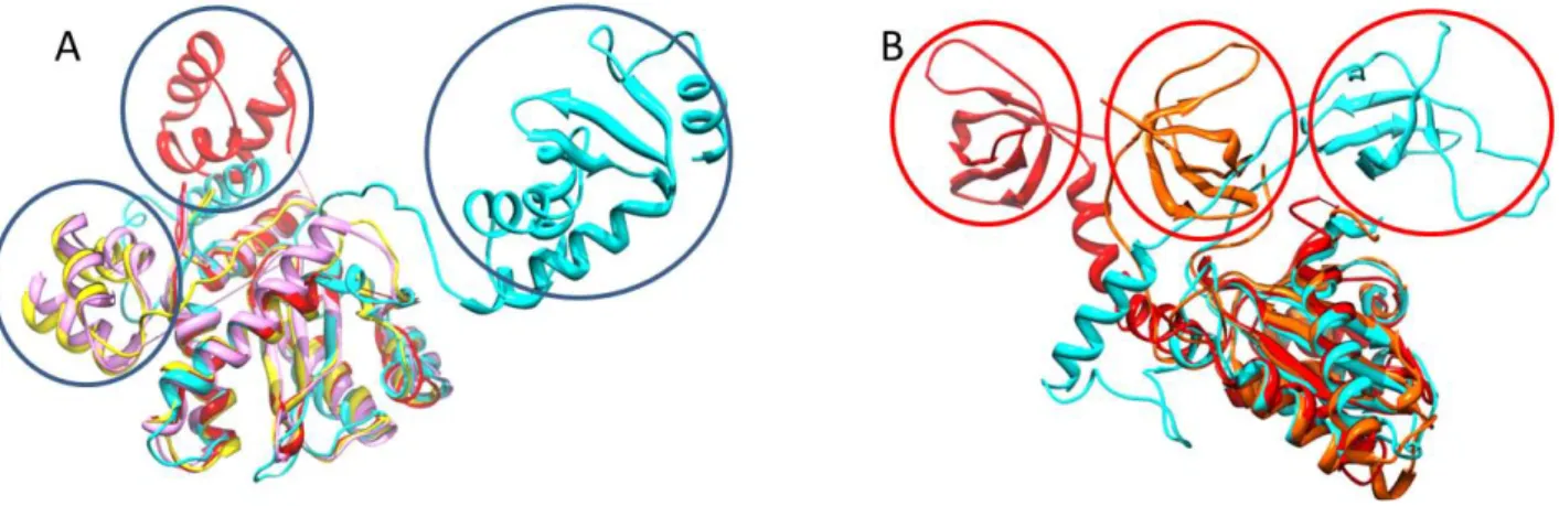
Figure 11: Superposition highlighting IN flexibility. A: Overlay of IN catalytic core domain of MMV (pink),
PFV (cyan), HIV-2 (yellow) and HIV-1 (red). N-terminal domains are highlighted in blue. B: Overlay of IN catalytic core domain of RSV (orange), HIV-1 (red) and PFV (cyan). C-terminal domains are highlighted in red. Maillot et al; PLOS One, 2013
22
HIV-1 Intasome structure
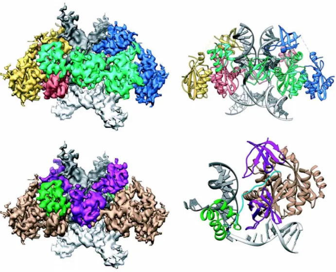
Recent advancements in cryoEM have made it possible to solve the structure of the full-length strand transfer complex, with integrase in complex with viral DNA and target DNA (intasome) (Passos, Li et al. 2017). Fusing the DNA binding protein Sso7d to the N-terminal of integrase improved solubility without interfering with activity in vivo, and Sso7d-IN was used to assemble intasomes for cryo-EM studies. The HIV intasome consists of a dimer of dimers with four IN protomers (one NTD, one CCD, one CTD each) arranged with two-fold symmetry around the target and viral DNA. A newly identified residue (K46), and previously identified residues (K156, K159 and K160) were confirmed to play roles in viral DNA binding, sequence specificity and catalysis (Jenkins, Esposito et al. 1997, Chen, Weber et al. 2006, Krishnan, Li et al. 2010). Additionally, R231 interacts strongly with target DNA and viral DNA (Serrao, Krishnan et al. 2014). The presence of the host co-factors such as the Integrase binding domain of LEDGF results in the formation of higher order assemblies, where the CTDs are reorganized to engage viral DNA, highlighting the structural flexibility of integrase. Notably, in the CTD, residues L242, I257, and V259 are involved in the formation of CTD-CTD interface. Other residues such as K14, E35, K240, K244 and R269 were also important for higher order oligomers. Mutating residues in the CTD affected strand transfer reactions and virus replication. This data suggests the importance of higher order oligomers for integrase function.
23
Figure 12: Structure of the STC intasome. Top left: Cryo-EM reconstruction of the STC, showing IN
protomers: inner protomers (green and red), outer protomers (yellow and blue), viral DNA (dark grey) and target DNA (light grey). Top right: Atomic model derived from the cryo-EM density. Bottom left: Segmented cryo-EM density showing IN domains: NTD (green), CCD (beige) NTD-CCD linker (blue) CTD (purple), viral DNA (dark grey), target DNA (light grey) Bottom right: Asymmetric subunit of the atomic model, using the same color scheme as in the bottom left. Passos et al, Science, 2017
The role of LEDGF in HIV Integration
Several host factors have been shown to be involved in the integration process. One of the most widely studied co-factors is Lens Epithelium Derived Growth Factor (LEDGF/p75). LEDGF/p75 is a 64kDa transcriptional co-activator that belongs to the hepatome derived growth factor (HDGF) related protein (HRP) family (Baid, Upadhyay et al. 2013). It is made up of 550 amino acids and contains two small structural domains. The N-terminal PWWP domain (1-91), along with the nuclear localization signal (178-197) (Maertens, Cherepanov et al. 2004) and a pair of AT-hook motifs (178-197) are responsible for the association of LEDGF to the chromatin (Llano, Vanegas et al. 2006, Turlure, Maertens et al. 2006, Shun, Botbol et al. 2008). The C-terminal (347-429) Integrase Binding Domain (IBD) mediates the
24
IN/LEDGF interaction (Cherepanov, Devroe et al. 2004, Llano, Saenz et al. 2006). LEDGF/p75 was shown to stimulate the catalytic activities of integrase in vitro (Cherepanov, Maertens et al. 2003). HIV-1 infectivity of RNAi LEDGF knockdown cells is severely depleted, due a lack of IN-chromatin association (Llano, Vanegas et al. 2004). The IN/LEDGF interaction has been shown to be specific to lentiviruses only (Busschots, Vercammen et al. 2005, Cherepanov 2007).
The LEDGF-IBD is composed of four long α-helices (α1, α2, α4 and α5) as revealed by NMR spectroscopy (Cherepanov, Sun et al. 2005). A structure of the IN-CCD (F185K)– LEDGF IBD complex has been solved (Cherepanov, Ambrosio et al. 2005). Residues I365, F406 and V408 were shown to be involved in the interaction with IN. D336N is essential for the interaction with IN, as well as for the enzymatic activity of LEDGF. The IN-CCD contains residues involved in the LEDGF interaction (V165, R166, Q168, L172 and K173), and residues directly interacting with LEDGF (A128, A129, W131 and W132). The IN-NTD is important for high affinity IN/LEDGF interactions (Maertens, Cherepanov et al. 2003). LEDGF has been implicated in the stabilization of the functional tetramer of IN (Cherepanov, Maertens et al. 2003), as well as in 3’ processing and strand transfer (Michel, Crucifix et al. 2009). Furthermore, LEDGF protects integrase from proteasomal degradation (Llano, Delgado et al. 2004). Overall, the model for the role of LEDGF in integration postulates that the N-terminal region of LEDGF binds to host chromatin at active transcription sites, and interacts with the PIC through its IBD, allowing to PIC to integrate into transcriptionally active sites, while also stimulating strand transfer (Ciuffi, Llano et al. 2005).
The Ruff team solved the structure of the full-length integrase in complex with LEDGF, and DNA by cryoEM, and proposed a binding mechanism for the IN/LEDGF/DNA complex (Michel, Crucifix et al. 2009). In this model, the IN/LEDGF complex contains 4 integrase and 2 LEDGF molecules, supporting the evidence that IN tetramer is the basic functional unit for integration.
25
Figure 13: Model for IN/LEDGF interaction in DNA Integration 15Å distance between viral DNA (yellow) and target DNA (red). Red arrows indicate conformational changes in viral DNA for integration to occur. Michel et al; The EMBO Journal, 2009
Important factors for Integration Site Selectivity
HIV-1 and other retroviruses preferentially integrate into transcriptionally active sites (Schröder, Shinn et al. 2002, Mitchell, Beitzel et al. 2004). However, HIV-1 can infect both dividing and non-dividing cells (Lewis, Hensel et al. 1992). The ability of HIV-1 and other retroviruses to infect non-dividing cells in the G0 stage makes non-dividing macrophages an important reservoir for HIV-1 in people living with HIV.
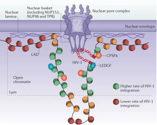
HIV enters the nuclear envelope through the nuclear pore complex (NPC) (Bukrinsky, Sharova et al. 1992). Capsid (CA) has been shown to mediate this entry by interacting with NPC components such as Polyadenylation Specificity Factor 6 (CPSF6) (Price, Fletcher et al. 2012, Sowd, Serrao et al. 2016) and Nucleoporins (NUPs) (Ocwieja, Brady et al. 2011, Matreyek, Yücel et al. 2013). The nuclear entry route is the first determinant factor for HIV-1 integration. HIV-1 PIC has been shown to integrate into areas of euchromatin closer to the
26
nuclear periphery (Di Primio, Quercioli et al. 2013), and HIV-1 integration targets are closely associated with the nuclear pore complex (Marini, Kertesz-Farkas et al. 2015), suggesting that HIV-1 integrates into the chromatin regions it encounters immediately after nuclear translocation.
Figure 14: Model of HIV-1 Integration at the Nuclear Pore Complex . Lusic and Silicano. Nature Reviews Microbiology, 2016
Additionally, the IN/LEDGF interaction has been shown to be important for targeting transcriptionally active sites, and recognizing transcription associated histone modifications. LEDGF interacts with DNA and tri-methylated H3K36 histones via its PWWP domain (Pradeepa, Sutherland et al. 2012, Eidahl, Crowe et al. 2013, van Nuland, van Schaik et al. 2013), allowing integrase to target actively transcribed genes in this histone mark. HIV integration in LEDGF mutants is decreased, and shifted away from transcriptionally active sites (Ciuffi, Llano et al. 2005, Shun, Raghavendra et al. 2007). In contrast to HIV-1, Gamma retroviruses like MLV use the Bromodomain and Extra terminal domain (BET) proteins for
27
chromatin targeting and integration near transcription start sites (De Rijck, de Kogel et al. 2013, Gupta, Maetzig et al. 2013, Sharma, Larue et al. 2013)
Furthermore, there is evidence that severely kinked DNA, such as nucleosomal DNA is preferred for integration. Structures of the PFV intasome suggest that there are interactions between the C-terminal domain of the intasome and H2A/H2B nucleosome (Maskell, Renault et al. 2015). PFV intasomes are able to catalyze integration into mononucleosomes. Studies performed using DNA mini-circles to mimic curved nucleosomal DNA show HIV-1 integrase preferentially targets curved DNA, compared to linear DNA (Pasi, Mornico et al. 2016). In addition to nucleosomal target, physical properties of target DNA have been shown to be important for integration, where factors such as the energy required to fit the DNA into the CCD, and DNA wrapping around a nucleosome determine integration sites (Naughtin, Haftek-Terreau et al. 2015). Moreover, intasome architecture and compactness of the
chromatin surrounding the nucleosome target are determinants for integration site selectivity (Benleulmi, Matysiak et al. 2015). While PFV and MLV integrate into dense and stable nucleosomes, HIV-1 preferentially integrates into sites with low nucleosome occupancy.
Proposed role of HIV-1 Integrase CTD in Chromatin Association
While cellular co-factors such as LEDGF and BET have been shown to be important for nucleosome targeting, these co-factors are not the only determinants for cellular integration. It is evident that additional interactions between the intasome and nucleosomal DNA need to be further elucidated in order to fully understand HIV-1 integration.
According to recent work done by our collaborators (Vincent Parissi, Bordeaux), another important factor for HIV-1 nucleosomal binding and chromatin integration is histone tails. N-terminal histone tails have been shown to be important for IN interactions with mononucleosomes in vivo. Using histone peptide array, it was shown that there is a specific interaction between HIV-1 integrase and the monomethylated N-terminal tail of histone H4. Structurally, the C-terminal domain displays a β-barrel fold, similar to the SH3 fold. This SH3 fold is consistent with members of the Royal Domain superfamily that includes chromo, Tudor, MBP, PWWP and Agenet domains (Chen, Nott et al. 2011). The structures of all members of this superfamily consist of anti-parallel β-strands that form a β-barrel with an aromatic cage located in the groove of the barrel. Members of this superfamily are involved in
28
recognizing and binding arginine or lysine-methylated ligands. The aromatic cage serves as the binding site for these proteins by interacting with the methylated side chain using electrostatic and hydrophobic contacts. Specifically, Tudor, Chromo, PWWP and MBT domains bind to methylated histone tails (Kim, Daniel et al. 2006, Yap and Zhou 2010).
Figure 15: Members of the Royal Domain Superfamily. Chen et al; Nature Reviews, Molecular Cell Biology,
2011
Although the CTD of HIV-1 Integrase most closely resembles the Tudor domains, it lacks the aromatic cage consistent with Tudor domains. However, the CTD of HIV-1 Integrase
probably functions as a chromodomain in vivo to mediate integrase/chromatin associations. Indeed, using the far dot blot assay, our collaborators show that the CTD is responsible for the interactions between HIV-1 integrase and histone H4 mono-methylated at lysine 20 (H4K20). Additionally, structures of the HIV-1 and PFV integrase (Maskell, Renault et al. 2015, Passos, Li et al. 2017) suggest that contacts between the CTD of the intasomes and target DNA are essential for integration. In the HIV-1 intasome, Residue R231 has been shown to be important for binding to target DNA. The intasome structure also suggests that the CTD is involved in higher order oligomerization that is essential for integrase function.
Genome diversity in HIV
HIV-1 reverse transcriptase lacks a proofreading activity, leading to high mutation rates of about9.3 × 10−5 mutations per base pair in plasma virus (Cuevas, Geller et al. 2015). There is